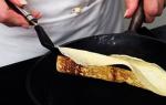
Lizard symptoms in humans. What is foot and mouth disease and why is it dangerous? Pharmacological agents for the treatment of foot and mouth disease - photo gallery
Foot and mouth disease is a dangerous, acute, highly contagious viral disease many species of animals, characterized by fever, salivation, aphthous-erosive lesions of the mucous membrane of the tongue and oral cavity, skin of the nasal mirror, limbs, mammary glands, myocarditis and myositis with high mortality of young animals in the first days of life. FMD can also be transmitted to humans from animals.
History reference. For the first time foot and mouth disease was described and pointed out its contagiousness in Italy in 1546. D. Fracastoro. In the 17th and 18th centuries foot-and-mouth disease, which often proceeded malignantly and was often confused with rinderpest of those cattle, was established in many countries of Europe. FMD has been known in Russia since the first half of the 19th century. In 1897, Loeffler and Frosch discovered the causative agent of the disease, the filter virus.
Currently, foot and mouth disease is found in many countries in Europe, Asia, Africa and South America. The last outbreak of foot-and-mouth disease in Russia was recorded in 2005 in the Amur Region, Khabarovsk and Primorsky Territories as a result of its introduction through the territory of Mongolia from China, where there was an epizootic of foot-and-mouth disease of the Asia-1 type.
Economic damage from foot and mouth disease consists of losses as a result of the death of mainly young animals (calves, piglets, lambs), a decrease in the milk productivity of cows by 50-75%, a decrease in the live weight of sick animals and abortions. Especially huge losses are carried out by quarantine measures. So, type O foot-and-mouth disease in Taiwan in 1997, where there were more than 6 thousand foot-and-mouth disease foci and more than 4 million were destroyed. pigs, brought a total economic loss of about 10 billion US dollars. In the UK, a similar FMD epizootic in 2001, when 2030 FMD foci were registered and more than 4 million heads of animals were destroyed, the damage amounted to about 12 billion dollars. In Russia in 2005, due to FMD foci of the Asia-1 type in the Amur Region, Khabarovsk and Primorsky Territories, direct economic damage amounted to more than 45 million rubles.
The causative agent of the disease The virus belongs to the Picornaviridae family, genus Aphthovirus. The viral particle consists of ribonucleic acid (RNA) surrounded by a peripheral protein capsid of 32 capsomeres. In the body of a sick animal, the virus is in the highest concentration in the epithelium of the walls of aphthae and in the lymph in the first 24-48 hours of illness. It can, but in much lower concentrations, be found in saliva, blood, urine and feces. Moreover, the virus from aphtha becomes contagious already at a dilution of 1:100-200 million, and from feces only at a dilution of 1:100. 7 types of the virus are known: O, A, C, Asia-1, CAT-1, CAT-2, CAT-3 and many of their variants. Now in the world they are predominantly distributed types O, A, Asia-1 and CAT-2. Animals that have been ill with one type of foot-and-mouth disease may become ill again if they are infected with another type of virus.
FMD virus is relatively resistant to environmental factors. On the surface of objects contaminated with the secretions of animals with foot-and-mouth disease, the virus remains 150, in manure - up to 168, in slurry - up to 40, in sewage- up to 103 days. In the blood of cattle and pigs (after quick freezing of meat carcasses), the virus remains active for 40 days. In chilled milk it lasts up to 47 days, in fresh milk at a temperature of 37 degrees it dies after 12 hours, while at the same time in sour milk and when cooking cheese, the virus quickly dies. The virus retains the coat of animals and clothing of people for 28-40 days. With properly conducted biothermal manure neutralization, the virus dies in 10-15 days. Heating up to 60 degrees kills the virus in 15 minutes, and at 80-100 degrees it is destroyed almost instantly. The best disinfectant to kill FMDV is to use 2-3% hot sodium hydroxide solution and 1% formaldehyde solution.
epidemiological data. Foot-and-mouth disease affects all types of artiodactyl animals. Most susceptible to FMD cattle, then descending pigs, sheep, goats and deer, less sensitive buffaloes, camels. Cases of FMD infection of dogs and cats through the milk of cows with FMD are described. Birds and horses are not susceptible to FMD. Of the laboratory animals, guinea pigs, rabbits, and mice suffer from foot and mouth disease.
Animals of any age are ill with foot and mouth disease, however, young animals under the age of 2-3 months are more easily infected and more severely ill.
The source of the infectious agent is infected, sick animals, as well as animals - convalescents, which can be virus carriers for a long time.
The main ways of spreading infection. The FMD virus is transmitted mainly by alimentary and aerogenic routes. The FMD pathogen is introduced into the farms when sick or recovered animals are admitted to the farm by private household plots, peasant farms; in contact with sick (recovered) animals, including wild ones; when grazing, watering, hauling; with infected feed, water, as well as when using milk from sick animals; when importing products of slaughter of sick (recovered) animals (use of untreated kitchen waste in feed); spread with the wind (small particles of feed, affected tissue, saliva, dust, etc.), with infected (contaminated) care items; clothing and footwear for caregivers, vehicles.
Pathogenesis. The virus, which enters the body of an animal through the digestive tract or external integuments, penetrates through the mucous membranes into the epithelial cells, where it is fixed and multiplied. The reproduction of the virus causes a response of the body in the form of a serous inflammatory process. There is the formation of one or two primary aphthae, which are usually not noticed by the owners of the animals. The general condition of the animal in this phase of the disease usually does not change. After 24 hours, in most animals, the disease enters the second phase. From the places of primary localization, the virus, after reproduction, enters the bloodstream, and then to all organs and tissues. Generalization of the process causes an acute febrile reaction in the animal.
The protective agents available in the animal's body neutralize the virus in the blood in most organs and tissues, but due to the fact that the epidermis is relatively poorly supplied with blood, and hence with antibodies, the virus begins to multiply in it. As a result of intensive reproduction of the virus in the epidermis, multiple secondary aphthae appear in the oral cavity, in the region of the interhoof gap and the corolla of the limbs, on the skin of the udder teats (generalized process). FMDV can replicate in the heart and skeletal muscles.
The course and symptoms of the disease. In all animals under natural conditions, foot and mouth disease usually proceeds acutely. In adult animals, an abortive course of foot and mouth disease is sometimes observed, accompanied by a short-term increase in body temperature and a rapidly onset recovery. In adult animals, FMD usually proceeds benignly. It is customary to distinguish between typical and atypical form(malignant, abortive and latent) foot and mouth disease.
In cattle the incubation (hidden) period is 1-3 days, but can be from 12 hours to 7 days, reaching in some cases up to 14-21 days.
In a benign course, we first note a decrease in appetite, lethargy of chewing gum, increased salivation. Then, in a sick animal, an increase in body temperature to 40.5-41.5 degrees is observed, the pulse quickens, the state of the animal becomes depressed, there is a refusal to feed and the absence of chewing gum. By the second and third days of illness, on the inner surface of the upper and lower lips, on the toothless edge of the lower jaw, on the tongue and buccal mucosa, aphthae. Almost simultaneously, some animals form aphthae in the area of the interhoof gap and on the skin of the udder. With foot and mouth disease, all four limbs are often affected, but there are cases when only two front or two rear limbs are affected.
At the beginning of the disease, aphthae are the size of a millet grain, then they merge and increase to the size of a pea or walnut. After 12-24 hours, the walls of the afts break, leaving behind fresh erosion. At this point, the body temperature of the animal drops to normal. When examining a sick animal, we note profuse salivation, in the corners of the mouth a foamy mass and a characteristic smacking are formed. The resulting erosions heal in 6-8 days, but in the case when the process is complicated by a secondary infection, the healing of erosions is delayed up to 2-3 weeks.
On the extremities, foot-and-mouth disease begins with painful and hot swellings appearing on the skin of the corolla and crumbs, in the region of the interhoof gap, due to which the animal develops lameness. If all limbs are affected, then such animals lie down and can be raised with great difficulty. In the future, aphthae appear at the site of the resulting swelling, which soon burst with the release of the contents in the exudate aphthae. With timely, properly started treatment and keeping animals on dry litter, erosion heals within 5-8 days. If aphthous lesions are extensive, then the animal develops phlegmon of the corolla, deep purulent pododermatitis, purulent arthritis, up to the subsidence of the horn shoe.
In lactating cows, the formation of aphthae of various sizes is often observed on the skin of the udder and teats. After opening the aphthae, erosion remains. The inflammatory process tends to spread to the top of the nipple and to the mucous membrane of the nipple canal. All these inflammatory processes lead to dysfunction of the affected quarter of the udder, which is manifested by a change in the composition of the milk, the milk becomes slimy, acquires an acidic reaction and a bitter taste. As a result of blockage of the teat canal with fibrinous, casein plugs and scabs, leading to difficulty in the exit of milk, mastitis develops in cows. In lactating cows, milk production is reduced to 75%. Milk productivity, with timely and correctly started treatment, is restored in cows slowly, sometimes it takes several months. In some adult animals, we note a disorder of function digestive tract accompanied by diarrhea.
In calves under 2 months of age, foot-and-mouth disease usually occurs in an aphthous form, but with symptoms of acute gastroenteritis. If the necessary remedial measures, the disease will end in the death of calves. In some calves, as a result of a complication of secondary microflora, foot and mouth disease is complicated by bronchopneumonia.
With a benign course, foot and mouth disease continues for 8-10 days. If complications join foot and mouth disease, then the disease stretches for 25 or more days.
Complications in animals with foot-and-mouth disease arise as a result of the attachment of pathogens of secondary infections to the foot-and-mouth disease process: streptococci, staphylococci, as a result of which purulent pododermatitis, corolla phlegmon, endometritis, mastitis, nephritis, bronchopneumonia, etc. appear in sick animals. Complications mainly develop in animals with weak body resistance. The FMD virus itself can lead to the development of disorders in the activity of the cardiovascular system due to myocardial dystrophy and metabolic disorders.
There are cases when foot and mouth disease can have a malignant course. In the malignant form of foot-and-mouth disease, when the disease initially proceeds with symptoms typical of foot-and-mouth disease, on the 8-12th day (in the recovery stage), the animal suddenly begins sharp deterioration the state of the animal. In the animal, we note weakness, depression, the pulse quickens to 120-140 beats per minute, the animal refuses to feed, chewing gum stops. We note paralysis in some animals hind limbs. Death in a malignant form of the disease occurs from cardiac arrest.
In some cases, foot and mouth disease acquires a malignant course from the very beginning of the disease, when the body temperature rises by 0.5-1 degrees, against the background of weak aphthous lesions with simultaneous damage to the udder. The disease proceeds with loss of appetite, severe depression and disorder of cardiac activity. Mortality in animals is 20-50%.
Pigs. In pigs, the incubation (hidden) period is most often 24-48 hours, but sometimes it is delayed up to 8 days. Among pigs, the disease is acute, with high mortality of young animals. The disease is characterized by fever, depression and decreased appetite. In pigs, mainly the limbs are affected, lameness appears, in some pigs we note the fall of the hooves. Aphthae appear on the patch, mammary glands, occasionally in the oral cavity. After the rupture of the aphthae, erosion remains. In adult pigs, the disease lasts 8-25 days. In piglets, foot-and-mouth disease occurs in a septic form, leading to the death of 60-100% of animals already in the first days of the disease. In severe cases of the disease, hemorrhages occur in the mucous membranes of the digestive tract, lungs and kidneys, under the serous membranes.
Sheep the incubation (hidden) period lasts 2-3 days. The disease is less acute than in cattle. We rarely note such a symptom characteristic of foot-and-mouth disease as salivation. Aphthae small, open early and in the absence of complications quickly heal. In connection with the defeat of the limbs (aphthae in the area of the interhoof gap and corolla, as well as pododermatitis), lameness occurs in sheep. With the mass distribution of foot-and-mouth disease in the flock, in some sheep we reveal a characteristic symptom complex: aphthous-erosive changes on the lips, tongue, gums, toothless edge of the upper jaw, on the limbs and udder, increased body temperature (up to 41.5 degrees), decreased appetite, periodic cessation of chewing gum, depression. It often happens that foot and mouth disease in sheep in flocks occurs with weak signs or these signs are completely absent, as a result, foot and mouth disease remains undiagnosed. Sheep with such a development of foot and mouth infection can remain virus carriers for several months, acting as a hidden source of the virus. In lambs, foot and mouth disease often occurs in the form of septicemia and is accompanied by a large case.
Goats the incubation (hidden) period lasts from 2 to 8 days. It is less acute than in cattle. In the first days of the disease, we note fever, depression, loss of appetite, damage to the mouth and limbs, which leads to lameness. The mouth of sick goats is closed, we note the gnashing of teeth. Abundant salivation is absent. Often the udder is damaged. Recovery usually occurs within 10-14 days. Goats are more resistant to foot-and-mouth disease, but sometimes they also have a malignant course.
Deer with foot and mouth disease, we note diarrhea, damage to the mucous membranes of the oral cavity and extremities, which are often complicated by necrobacteriosis. In the absence of complications, deer recovery occurs in 10-12 days.
pathological changes. We find aphthae and erosion in the oral cavity, sometimes on the mucous membrane of the esophagus and in the proventriculus in ruminants. In piglets, lambs and calves, at autopsy, we find hemorrhagic inflammation of the intestine, degenerative changes in the heart muscle (tiger heart).
In the malignant form of the course of foot and mouth disease in the heart muscle, we find severe lesions. The cardiac muscle becomes pale and flabby, grayish-reddish spots and caseous degenerate gray-white foci of various sizes appear on the cut. Under the epicardium and endocardium we find hemorrhages. In the liver, kidneys and skeletal muscles - degenerative changes.
Diagnosis as with all infectious diseases is put complex taking into account epizootological data, clinical signs of the disease, pathoanatomical changes and mandatory results laboratory research.
For laboratory research send walls and contents of fresh aphthae (lymph), blood samples at the time of fever in animals, The lymph nodes head area, esophageal-pharyngeal mucus and blood serum samples (not earlier than 14 days after the onset of clinical signs). Selected materials are placed in closed sterile vials, frozen or transported in a preservative liquid (equal volumes of neutral glycerol and 0.8% solution sodium chloride) in thermal containers with ice (refrigerant) with strict observance of safety precautions (safety of working with microorganisms of 1-2 pathogenicity (danger) groups. Sanitary and epidemiological rules SP 1.3. 1285-03).
Materials for research sent by courier with a cover letter with a detailed description of the epizootic situation in the farm (settlement).
Laboratory diagnostics FMDV is based on the isolation and identification of FMDV from samples of pathological material (walls and contents of aphthae, lymph, lymph nodes, meat samples, meat products and feed), detection of the pathogen antigen and viral RNA, establishing the primary structure of the BP 1 gene of FMDV and identifying post-infectious antibodies (research in the RSK, ELISA, PCR, RMN, virus isolation).
differential diagnosis. When making a diagnosis of foot and mouth disease, veterinarians should exclude feed stomatitis and, vesicular stomatitis kr.r.sk.,. In sheep and goats, we exclude contagious pustular stomatitis and.
Treatment. Given that the success of treating animals with foot and mouth disease largely depends on strict adherence to the rules of feeding and keeping, we provide peace to sick animals in order not to overstrain the heart. Premises for sick animals should be clean, with enough bedding material and have a constant supply fresh air. In order to combat dehydration, sick animals should receive plenty of clean cool water in agricultural enterprises, household plots, peasant farms. The diet should contain soft digestible feed (grass, flour talker, good silage in winter, soft hay). The mouth is washed clean water with the addition of 2% acetic acid, you can use a 0.1% solution of potassium permanganate, a 0.5% solution of furatsilina. In case of severe damage to the oral mucosa, we use an ointment (anesthesin 2.5 g; novocaine 2.5 g; blue vitriol 5 g; fish oil 20 g; Vaseline 70 g). This ointment accelerates the healing of erosions and, having an analgesic effect, allows animals to take food.
The limbs are cleaned of dirt and every 1-2 days the hooves, the corolla, the skin of the arch of the interhoof gap are lubricated with tar in half with fish oil. For the same purpose, animals are passed through disinfectant barriers with sawdust, which are impregnated with tar, or through baths with a 5% formalin solution.
In case of severe damage to the limbs (phlegmon of the crumb, corolla, interdigital fiber), the inflamed areas are smeared with tincture of iodine. We clean the hooves, remove dead tissues, ulcers and wounds, cauterize with potassium permanganate powder in half with streptocide and apply a protective bandage or use shoes made of tarpaulin and other dense material. In the event that foot and mouth disease in an animal is complicated by sepsis as a result of a secondary infection, a 0.5% solution of novocaine is administered intravenously at the rate of 0.5 ml per 1 kg of animal weight. Antibiotic therapy is used, incl. modern antibiotics cephalosporin series. To prevent udder aphthous lesions in cows, milkmaids must keep their hands clean and follow the rules for milking cows. With aphthous lesions on the udder, trypoflavin-novocaine ointment is used (trypoflavin 1g, novocaine 4g, vaseline 100g), synthomycin emulsion or 15% propolis ointment on vaseline.
In case of severe foot and mouth disease and cardiac disorders, the use of a mixture is recommended: valerian tincture 10 ml, lily of the valley tincture 15 ml, potassium bromide 6 g, distilled water 400 ml; inside at one time.
In malignant forms of foot-and-mouth disease, sick cows must be injected daily through a tube or with a bottle of 20-30 liters of flour mash. Weakened animals are not bad to give honey 100-200g or drink back with the addition of 200-400g of sugar.
From specific means, cited blood of convalescents or serum is used for animals at the rate of 2 ml per 1 kg of weight. These tools are effective if applied before the generalization of the process. FROM therapeutic purpose FMD immunolactone can also be used.
Immunity and specific prophylaxis. In newly vaccinated animals, immunity is formed by 21 days. Virus-neutralizing antibodies in the blood serum appear 5-7 days after infection, reaching a maximum after 3-4 weeks and can persist for about a year. Collostral antibodies in calves persist for 3-5 months.
For prophylactic immunization of animals, depending on the epizootic situation, inactivated mono-, bi- and polyvalent vaccines are used according to certain schemes. A stable level of post-vaccination antibodies that protect adult animals from FMD is maintained for 6 months.
Control and prevention measures. FMD well-being in the Russian Federation has been achieved through the implementation of a program that includes monitoring the epizootic situation, controlling the movement of animals and products of animal origin, taking measures to prevent the introduction of FMD virus into livestock farms, observing the “closed-type enterprises” regime, and conducting preventive vaccination of animals in areas of high risk of introduction and spread of the disease, the elimination of sick or all animals in the foci of infection, strict adherence to the system of quarantine measures. In the event of an outbreak of foot-and-mouth disease, by the decision of the Governor of the region on the farm, settlement quarantine is imposed. Quarantine and restrictive measures are being taken in accordance with the instructions "On measures to prevent and eliminate the disease of animals with foot-and-mouth disease", approved by the Main Directorate of Veterinary Medicine of the USSR Ministry of Agriculture dated March 15, 1985.
Preventive vaccination of animals is carried out in threatened areas with subsequent control of the immune background. Specific prevention of foot-and-mouth disease is carried out using inactivated vaccines produced by FGU "ARRIAH" and FGPU "Shchelkovo Biokombinat".
In Vladimir region for the purpose of all kinds of emergencies for foot and mouth disease, a 30-kilometer buffer zone has been created around FGU VNIIZHZ, which includes Sobinsky, Suzdalsky districts and the city of Vladimir, where vaccination of the entire livestock of the kr.r.sk. and small cattle incl. and those in the private sector. The veterinarians of these regions constantly monitor the intensity of immunity by conducting monitoring studies of blood samples at the FGU "ARRIAH".
Foot and mouth disease (Latin - Aphtae epizooticae; English - Foot-and-Mouth disease) is an acute, highly contagious viral disease of domestic and wild artiodactyl animals, characterized by fever and aphthous lesions of the mucous membrane of the oral cavity, hairless areas of the scalp, udder, corolla , interhoof gap and accompanied by impaired movement; in young animals - damage to the myocardium and skeletal muscles (see color insert). Sometimes a person is sick with foot and mouth disease.
Historical information, distribution, degree of danger and damage. Foot and mouth disease has been known to mankind for over 400 years. animal disease associated with profuse salivation, was repeatedly noted in a number of European countries in the 17th-19th centuries.
The FMD virus, the first of the causative agents of viral animal diseases, was discovered in 1897 by German scientists Lefler and Frosch. At the beginning of the XX century. French, German and English scientists established the multiplicity of types of the pathogen, which was of great practical importance in the development of means for diagnosing and preventing the disease.
FMD is registered in many countries of the world. According to the OIE, every year 55...70 countries become unaffected by FMD. Information about the disease of animals with foot-and-mouth disease in Russia began to appear in the literature from the middle of the 19th century. In the XIX-XX centuries. foot-and-mouth disease in Russia was recorded periodically as an epizootic, covering large areas of the country. Since 1989, Russia has been free of foot and mouth disease, but periodically the pathogen is brought into our territory from unfavorable, in particular neighboring, countries. Thanks to the developed strategy, the disease can be eliminated in the primary foci.
AT modern conditions people practically do not get sick with foot-and-mouth disease.
Foot and mouth disease can cause great economic damage even today. For example, during an epizootic of foot-and-mouth disease in pigs in Taiwan in 1997, the total economic damage amounted to about 10 billion US dollars. With the modern integration of European countries, the occurrence of FMD in them has led to serious economic and social problems. With an epizootic of foot-and-mouth disease type O in the UK in 2001, over 1000 foot-and-mouth disease foci arose within 6 weeks and the total economic damage amounted to more than 20 billion US dollars.
The causative agent of the disease. The causative agent of foot-and-mouth disease is a very small RNA-containing virus belonging to the genus of rhinoviruses of the Picornaviridae family. The virus has a complex antigenic composition: there are 7 serological types (O, A, C, CAT-1, CAT-2, CAT-3, Asia-1). Each type has a certain number of options (subtypes): type A has 32 options, O - 13, C - 5, CAT-1 - 7, CAT-2 - 3, CAT-3 - 4, Asia-1 - 2. In the world type O causes disease in 38% of cases, A - in 33%, C - in 26%. On the territory of our country, during the years of the epizootic, mainly foot and mouth disease types A (76.4%) and O (19.2%) were recorded. However, in recent years, FMD type O has become prevalent, as in the rest of the world. The types and variants of the virus differ immunologically: each of them can cause disease in an animal immune to other types and variants of the virus.
The virus reproduces well in the cell culture of epithelial tissues of susceptible animals with the manifestation of CPD. It has a high virulence: lymph from aphtha in a dilution of 1: 106 causes foot and mouth disease in
Receptive animals. Passaging of the virus is carried out on laboratory animals (guinea pigs, mice, rabbits). In animals, the virus induces the formation of antibodies specific for each serotype of the pathogen. Therefore, serological tests are used to differentiate FMD virus serotypes and variants.
The causative agent of foot-and-mouth disease in terms of resistance to chemical disinfectants refers to stable (group 2). The virus is resistant to ether, chloroform, carbon tetrachloride, is not inactivated by 1% phenol solution, 75% ethyl alcohol, withstands the action of lysol and toluene in concentrations that are detrimental to a number of other viruses and bacteria. The stability of the virus is significantly increased if it is contained in the torn off walls of aphthae. On mountain pastures, it may persist until the next pasture season; in sewage in the cold season survives up to 103 days, in summer - 21 days, in autumn - 49 days. On the coat of animals, the virus persists for up to 50 days, on clothes - up to 100, in rooms - up to 70, in feed - up to 30 ... 150, soil - up to 40 ... 150, in fresh milk (4 ° C) - up to 15, in sausages - up to 56 days. In salted and smoked products - up to 50 days. In quick-frozen meat (below -20 °C), the virus can persist for years. In a 50% solution of glycerol in phosphate buffer (pH 7.2) at 4-8 °C, the virus-containing material remains infective for 40 days. This preservative is used when sending materials to the laboratory.
The most effective in carrying out disinfection measures for foot and mouth disease are 1 ... 2% hot solutions of sodium and potassium hydroxide, which have a detrimental effect on the foot and mouth disease virus in the first 10 ... 30 minutes, 2% formalin - 10 minutes, Virkon C 1 : 200, 1% iodine, 4% hydrogen peroxide solution.
Epizootology. The highest contagiousness of the disease, long-term carrier of the virus in the body of animals and its long-term preservation in the external environment, wide range susceptible domestic and wild animals, the multiplicity of types and subtypes of the virus - all these factors ensure the stability of the pathogen, its preservation in nature and the reproduction of the epizootological process (Table 5.1).
5.1. The main epizootological data for foot and mouth disease

A characteristic feature of foot-and-mouth disease is its almost absolute specificity for artiodactyls. Wild artiodactyls (buffaloes, saigas, etc.) can perform the function natural reservoir viruses of various types.
The amount and virulence of the virus varies in various stages illness and various kinds animals.
The distribution of foot and mouth disease largely depends on economic and economic ties, methods of animal husbandry, animal population density, the degree of population migration, conditions for harvesting, storage and processing of products and raw materials of animal origin. The risk of introducing a pathogen associated with the importation of animals has also long been recognized.
The following factors can be considered as an epizootological feature of foot and mouth disease: diverse ways of transmission of the infectious agent and a very short incubation period, as a result of which the circulation of the virus among animals is accelerated and new sources of the infectious agent quickly appear; a large number of naturally susceptible domestic and wild animal species; selection a large number a virus highly resistant to various environmental factors; the presence of several immunological types and many variants of viruses.
Pathogenesis. The primary reproduction of the virus occurs in the mucous membrane of the nasopharynx, lymph nodes of the head, neck and tonsils 18 hours after infection. In places of introduction of the virus, primary aphthae are formed. From here, the virus enters the blood through the lymphatic pathways and then to the organs of the lymphoid-macrophage system, where there are optimal conditions for its abundant accumulation and the formation of a focus of infection. Clinically, this phase of the disease is manifested by an increase in body temperature, the rapid formation of secondary or generalized aphthae and exanthema on areas of the skin not covered with hair (nasal mirror, nasal openings, udder skin, sometimes the scrotum and horn roots), on mucous membranes (oral cavity, esophagus, scar, vagina) and on the skin around the
Pyt (corolla, interhoof gap, crumbs). This usually occurs 48 hours after infection.
Possessing myotropic properties, the FMD virus is also fixed in the fibers of the heart and skeletal muscles, causing various functional disorders heart and tissue defects.
With low virulence and a low infectious dose of the virus, as well as the resistance of the animal, the development of the infection may stop or proceed latently. Latent infection is especially common in sheep.
Current and clinical manifestation. Clinical signs of the disease depend on the individual sensitivity of the animal to the FMDV, its physiological state and the degree of virulence of the pathogen. The most characteristic signs of the disease are expressed in adult cattle. In other animals (lambs, piglets, calves) they may be less typical. Possible benign and malignant manifestation of foot and mouth disease.
With foot-and-mouth disease in cattle, the incubation period averages 1 ... 3 days, less often up to 21 days. The course of the disease is acute. With a benign course of foot and mouth disease, at first the animal's appetite worsens, chewing gum slows down, and salivation increases. Then the body temperature rises to 40.5...41.5°C. Animals eat little or do not take food at all, are depressed, they have a rapid pulse and breathing, lack of chewing gum, and milk yield is sharply reduced. During this period, the mucous membrane is dry, hot, hyperemic. On the 2nd ... 3rd day after the onset of fever on the mucous membrane of the oral cavity (on the upper and lower lip, toothless edge of the lower jaw), on the tongue, on the wings of the nose, sometimes aphthae (vesicles) appear on the nasal mirror, filled at first with a clear, then turbid liquid. When the process is generalized, characteristic aphthous lesions are formed on the teats of the udder, on the skin of the corolla, in the interhoof gap, on the crumbs of the hooves, sometimes at the base of the horns. After 12 ... 36 hours, the aphthae open, the lymph contained in them mixes with saliva and is released from the oral cavity; note profuse salivation, a kind of smacking, foamy mass in the corners of the mouth. In place of bursting aphthae, painful erosions with jagged edges, healing after 6-8 days. If the process is complicated by a secondary infection, healing occurs after 2-3 weeks. Body temperature drops to normal when aphthae appears. In complicated cases, recovery is slow. The udder is more commonly affected in lactating cows. Aphthae appear on the nipples various shapes and magnitude. In place of the opened bubbles, erosions form, milking is difficult. Milk is slimy, with a bitter taste. Sometimes patients have mastitis, endometritis and diarrhea. Milk yields are reduced by 20...75% and after recovery they are not fully restored. In newborn calves, aphthae are not formed, gastroenteritis is typical. The duration of the disease with a benign course is 8 ... 10 days, with complications up to 25 days. At malignant course calves often die from foot and mouth disease. The disease first proceeds with typical features. On the 7th ... 10th day after the onset of the disease, the animal's condition deteriorates sharply; noted a secondary increase in body temperature, depression, rapid pulse- up to 120 ... 140 beats per minute, muscle tremors and convulsions. The animal dies of paralysis of the heart.
Sheep have an incubation period of 1...6 days. The limbs are mostly affected. Redness, swelling and soreness appear on the skin of the corolla and in the interhoof gaps. Later on these
In some places, aphthae arise, which are torn, and in their place, foci of erosion are formed, which leads to lameness. In ewes with a disease, lactation often stops, aphthae or small foci of erosion are found on the udder. In the oral cavity, aphthae are rarely formed, but salivation does not occur. With the appearance of foot-and-mouth disease during the lambing period, a massive death of newborn lambs from damage to the heart muscle is observed. Often, foot and mouth disease in sheep occurs in a latent form, without pronounced symptoms of the disease.
Goats have an incubation period of 2-8 days. The disease proceeds more typically, more often with the formation of small aphthae and foci of erosion, the mucous membrane of the oral cavity, the skin of the extremities and less often the udder are affected. Salivation is expressed weakly. When the limbs are affected, goats are very lame. Foci of erosion heal slowly, general state animals worsens, oppression sets in, appetite disappears, body temperature rises to 41 ° C and above. Sick animals lie down more often. Comes from the mouth bad smell have diarrhea or constipation. With a benign course, the animals recover in 10-14 days. Goats are seriously ill, there is a high mortality rate.
With foot-and-mouth disease of pigs, the incubation period lasts 2 ... 14 days. FMD occurs relatively easily in gilts. In adult pigs, especially boars and lactating sows, the disease is more severe. Suckling piglets are acutely and seriously ill. In pigs with foot-and-mouth disease, oppression, a decrease in appetite are observed, and during the formation of aphthae - an increase in temperature to 41 ... 42 "C.
The most common clinical sign of foot and mouth disease in pigs is aphthae and erosion on the corolla, hoof crumbs, in the interhoof gap, which is accompanied by lameness and often falling of the hooves. Aphthae and erosion are also formed on the patch, and in suckling sows - on the udder. When the udder is affected, lactation often stops in sows, many suckling pigs under them die. The oral mucosa is rarely affected. foot-and-mouth disease in pigs develops rapidly, clinical symptoms in a sick animal, they can disappear within 10 days, but with a large concentration of livestock, the spread of infection can be prolonged.
In suckling pigs, foot and mouth disease is characterized by the appearance of many aphthae, which develop not only on the mucous membrane of the oral cavity and patch, but also on the skin almost over the entire surface of the body. Often, foot and mouth disease in young animals occurs without the formation of aphthae in the form of a general septic process, sometimes there are signs of acute gastroenteritis, in most cases, piglets die in the first 2-3 days of illness.
FMD is also diagnosed in wild animals. Repeated mass disease of foot-and-mouth disease in saigas in Kazakhstan, as well as in Russia in the Lower Volga region and the North Caucasus are described. FMD has been reported in reindeer, goitered gazelle, roe deer, wild goats, moose, deer, argali, wild boars, bears, etc.
Cases of foot-and-mouth disease were also noted in zoos among reindeer and spotted deer, fallow deer, ibexes, tours, gayals, deer, European moufflons, marals, yaks, buffaloes, bison, bison, bison and other wild animals. In some cases, they recorded a malignant course of foot-and-mouth disease, accompanied by death, especially of young animals. Wild animals can remain asymptomatic carriers of the pathogen and be a source of its spread in natural conditions.
pathological signs. At autopsy of the corpses of dead animals, exanthema, aphthae, and erosion characteristic of foot and mouth disease are found on the mucous membrane of the oral cavity, often the esophagus and pancreas. In young farm animals different types(calves, piglets, lambs) changes are characterized by hemorrhagic inflammation of the mucous membrane gastrointestinal tract, inherent acute gastroenteritis. In the malignant course of foot and mouth disease, the main changes are noted in the heart muscle. The myocardium is flabby, has a gray-dirty-yellowish, whitish color or striping ("tiger heart"); under the epi- and endocardium - hemorrhages. The same changes are found in the skeletal muscles. The liver is enlarged, regenerated.
Diagnosis and differential diagnosis. Timely diagnosis of foot-and-mouth disease, determination of the type and variant of the virus have importance for rapid localization and elimination of infection during the first outbreak of the disease, as well as preventing its further spread.
The diagnosis is made on the basis of epizootological data, clinical signs of the disease, pathological changes and laboratory results.
From the epizootological data in the diagnosis, the following are taken into account: 1) the range of susceptible animals - artiodactyls; 2) the degree of distribution and the speed of coverage - within 10 ... 15 days most of the animals of the farm fall ill; 3) economic relations of the enterprise with farms that are unfavorable for foot-and-mouth disease in a given district, region, republic; 4) absence expressed connection diseases with seasonality and natural and climatic conditions; 5) data of previous vaccination and disease of animals with foot-and-mouth disease.
Suspicion of foot and mouth disease is caused by any disease of susceptible animals, characterized by the appearance of a vesicular rash in the oral cavity, on the limbs and udder, increased salivation, smacking, difficulty in eating and chewing food, and when examining the oral cavity, the detection of aphthae and erosions. In addition, attention is paid to lameness, aphthae on the corolla and in the interhoof gap, sometimes the fall of the horn shoe, aphthae on the nipples and the pain of the latter during milking and sucking (with a pronounced defensive reflex). During the period of the threat of foot and mouth disease, it is necessary to pay attention to the depressed state of the animal, decreased appetite and milk secretion, increased body temperature, etc.
Laboratory diagnosis of foot and mouth disease is shown in Figure 5.1.
Material to be studied: from cattle, the walls of mature, unruptured aphthae are taken from the tongue, from pigs - from the patch or udder, from small cattle - from the toothless edge of the lower jaw, the skin of the interhoof gap or corolla; blood at the time of the temperature reaction; from the corpses of young animals - the lymph nodes of the head and pharyngeal ring, the pancreas, the muscle of the heart. To study for virus carriers, scrapings from the mucous membrane of the pharynx or esophagus are taken with a probe.
Collection, preservation and shipment of materials for laboratory diagnosis of foot and mouth disease is carried out in accordance with the current instructions and guidelines.
ELISA and PCR are currently widely used as an express diagnostic method.

Rice. 5.1. FMD virus isolation and identification scheme
According to the results of laboratory tests, the farm is considered unfavorable for FMD in any of the following cases: 1) positive results (with a score of at least three crosses) in the RSK in the study of pathological material and determining the type of virus, taking into account clinical and epizootological data; 2) development guinea pigs(24-72 hours after the introduction of the material) primary aphthae, and then a generalized process, accompanied by the appearance of secondary aphthae on the tongue and the plantar surface of the forelegs; 3) the development of paresis and paralysis in three mice (4 ... 5 days of age) after infection with their pathological material, and then their death under normal physiological state three control animals.
Retrospective diagnostics in order to determine the type and variant of the FMD virus that caused the disease in the past is based on the identification of antibodies in RDP, RID, NRIF, and the reaction of seroprotection in mice in ROP in cell culture.
In the differential diagnosis of foot and mouth disease, it is necessary to exclude viral vesicular stomatitis, viral diarrhea, malignant catarrhal fever, rinderpest, smallpox, necrobacteriosis, infectious rhinotracheitis, contagious ecthyma, catarrhal fever of sheep, vesicular exanthema of pigs, stomatitis, traumatic diseases, poisoning with certain substances. Diseases with vesicular syndrome exclude bioassay (Table 5.2).
5.2. Differential Diagnosis diseases of pigs with vesicular syndrome

Immunity, specific prophylaxis. Animals that have been ill acquire immunity to the type and variant of the virus that caused the disease. The duration of immunity in cattle is 8...12 months, in pigs 8...10, in sheep about 18 months. Colostral immunity is well expressed, but calves that have not received colostrum do not have serum antibodies. In calves, passive protection lasts up to 3 months.
Inactivated vaccines with preventive purpose, as well as for the forced processing of animals in disadvantaged and threatened areas for foot and mouth disease (Table 5.3).
Prevention. The multiplicity of FMD pathogen types, the diverse transmission mechanism and the wide range of susceptible animals present major challenges in FMD control.
The system of anti-FMD measures in our country is based on scientifically substantiated forecasting of the epizootic situation and provides for the zonal principle of their implementation. Priority in the system are general veterinary and sanitary measures to prevent the introduction of foot-and-mouth disease virus, and in areas of permanent threat and in areas of high risk of occurrence and spread of foot-and-mouth disease, along with them, vaccination is provided.
Treatment. Treatment is carried out only in countries where foot-and-mouth disease has a significant prevalence. In the event of a primary foot-and-mouth disease on the territory of Russia, treatment of sick animals is not carried out.
AT initial stage disease, serotherapy using hyperimmune serum or blood (serum) of convalescents is effective. In order to reduce morbidity and mortality among animals and prevent the development of complications, patients are improved conditions of detention, often watered, prescribed dietary feed (grass, flour mash), symptomatic treatment in the form of disinfectant solutions or ointments for the treatment of affected mucous membranes and skin. Glucose is administered intravenously, antibiotics and cardiac agents are used.
Control measures. When foot-and-mouth disease occurs, measures to eliminate it are determined by the epizootic situation, geographical conditions, the method of animal husbandry, the level of development of the country, etc. Taking this into account, measures to combat foot-and-mouth disease in different countries can be divided into four areas.
1. A radical method of fighting FMD (the so-called stamping out) consists in the immediate slaughter of all patients, suspected of the disease and suspected of infecting susceptible animals and refusing to vaccinate. This method used in developed prosperous countries with the initial appearance of the disease. This method can make it possible to completely eradicate FMD in the primary focus.
2. Refusal of prophylactic immunization of animals, and in case of foot and mouth disease, slaughter (destruction) of animals in the outbreak and forced vaccination around the source of infection.
3. Systematic prophylactic immunization of susceptible animals in threatened areas. If foot-and-mouth disease occurs, slaughter (destruction) of patients and ring vaccination around the focus of infection (successfully used in our country).
4. An integrated method of combating foot-and-mouth disease consists in combining the method of slaughtering diseased and suspicious animals with active immunization of susceptible livestock while simultaneously
5.3. Characteristics of vaccines used for prophylactic and forced vaccination of farm animals against foot-and-mouth disease


Rice. 5.2. Measures to eliminate foot and mouth disease
Carrying out sanitary and quarantine measures. The integrated method is used in areas previously unfavorable for foot and mouth disease, in border areas, especially when there is a threat of introducing foot and mouth disease, in areas of operation of institutions and enterprises engaged in the manufacture of anti-foot-and-mouth disease biological preparations. In the event of foot-and-mouth disease, sick and suspicious animals are isolated or killed. The unfavorable zone is quarantined, all animals in the threatened zone are immunized. This method should be considered the most effective, since the activities are aimed at all links of the epizootic chain.
Measures to eliminate foot and mouth disease in a disadvantaged area are shown in Figure 5.2.
When organizing events, one should distinguish between an epizootic focus, a disadvantaged point and a zone threatened by foot and mouth disease.
Quarantine is removed 21 days after the recovery of the last sick animal at this point.
Restrictions after the lifting of quarantine are quite strict. Prohibited: export and import of animals within 1 year after the removal of quarantine; use of pastures and livestock tracts for 1 year. Inoculated animals can be administered 21 days after vaccination. Animals that have recovered from illness within 3 months after the removal of quarantine can be sent for slaughter only to a meat processing plant in this region. Non-sick but vaccinated animals can be sent for slaughter 21 days after vaccination. Animal and vegetable products that have been in contact with FMDV are used locally.
Measures to protect people from infection with foot-and-mouth disease. FMD is very rare in humans. Infection occurs when caring for sick animals; people with a weakened body or children are more likely to get sick when drinking raw milk from sick cows. The prognosis is often favorable. Recovery occurs in 10 ... 15 days.
Personal prevention in disadvantaged areas is reduced to the prohibition of the consumption of raw meat, milk and dairy products. Boiling or pasteurization of milk is required. Caution is required when caring for sick animals (washing and disinfection of hands, overalls - apron, gloves, boots).
Control questions and tasks. 1. What are the antigenic differences of the FMD pathogen that must be taken into account in anti-FMD work? 2. What are the features of the epizootic process in foot and mouth disease? 3. What are the sources and reservoirs of the FMD virus, how it is transmitted and how the disease spreads? 4. How is FMD diagnosed and from what diseases should it be differentiated? 5. List the complex of general and specific measures for the prevention and elimination of foot-and-mouth disease in animals of different species. 6. What anti-foot and mouth disease measures are carried out in the epizootic focus, disadvantaged point and threatened zone?
The causative agent of the disease is a virus
FMDV is transmitted from animal to animal and from animal to human. A sick person is a carrier of the virus, but cannot transmit the virus to another person. Due to the underformed immune system, children are more susceptible to the lizard virus than adults.
The virus that causes foot-and-mouth disease is extremely pathogenic: when it enters the body, the virus is not just in it, it necessarily causes the disease. At the same time, the virus is quite dermatotropic - it likes to be localized on the skin, so if a disease occurs, it can be seen on the skin.
The causative agent of foot-and-mouth disease is quite resistant to low temperatures and drying. However, exposure to ultraviolet rays is detrimental to him. For foot-and-mouth disease, high temperatures are also fatal: at a temperature of 60 ° C, the virus dies. Ordinary disinfectants are also capable of killing the pangolin virus, the virus is very sensitive to them.
An epidemic of foot-and-mouth disease is periodically observed in various animals. In animals, the disease manifests itself in the form of ulcers in the region of various mucous membranes: the nasopharynx, nasal cavity, mouth, tongue, and sores on the lips are possible. Also, sores containing the virus can be seen on the animal's skin: in the gaps between the hooves, on the udder of cows, near the horns, etc.
When animals start to get sick, there is a high probability of illness and people who work with these animals. People who care for animals, slaughter them, milk them and treat them can become infected with FMDV from animals through the air. Therefore, all people who work with artiodactyl animals must wear and change masks during work. A person can become infected with the virus even before he knows that the animal is sick: the incubation period (the period from the virus entering the body until the first symptoms appear) in animals is 2-4 days.
The FMD vaccine is only for animals and is constantly being improved. There is no such vaccine for humans due to the short duration of the disease and successful recovery.
Ways of disease transmission

You can become infected with the virus through the raw milk of sick animals and dairy products prepared from it. dairy products
Often, artiodactyl animals can develop foot and mouth disease - a disease that can become a real epidemic among animals. A person is infected with a virus from an animal. There are several ways the virus can be transmitted from animal to person.
- Through raw milk of sick animals and dairy and sour-milk products prepared from it (sour cream, kefir, fermented baked milk, cottage cheese, cheese).
- By airborne droplets for people who constantly work and are in contact with animals, if there are foot-and-mouth disease among the animals.
- The virus can be transmitted to humans through objects that have come into contact with the secretions of a sick animal - buckets, bowls, rags that wipe the animal, etc.
- Sometimes a person can get sick when eating the meat of a diseased animal, but this situation possible if you eat raw meat or meat that is not fully thermally processed, as well as meat with blood.
Symptoms of the disease

The disease begins with a sharp rise in temperature, chills appear, as well as muscle and headaches.
FMD enters a person through scratches, microcracks and damage to the skin, mucous membranes of the mouth or nose. At the site of entry of the virus into the body, a primary lesion occurs - a vial in which the virus multiplies and accumulates. Then the virus enters the blood. It is at this stage of the disease that a person has pronounced symptoms, one of them is intoxication. Then the virus from the blood again passes into the mucous membranes and onto the skin. It can be seen on the mucous membranes of the mouth and nose, or on the skin of the feet and hands.
The most important indicator of foot-and-mouth disease is symptoms and a biological test on animals (isolation of the virus in humans from vesicles and infection of laboratory animals with it). The incubation period in humans lasts from 2 to 4 days (sometimes it can reach 12 days).
- The disease begins with a sharp rise in temperature: up to 39-40°C.
- Chills appear, and then headaches and muscle pains.
- By the end of the first or on the second day, a person feels a burning sensation in the oral cavity, he has a strong salivation.
- The conjunctiva of the eyes begins to turn red, the person feels some pain when urinating, diarrhea may occur.
- There are swelling of the mucous membranes of the cheeks, lips, tongue, palate. After a few days, small blisters can be seen on the edema, which are filled with a clear liquid. After a certain amount of time, the bubbles change their color - the liquid in them becomes cloudy.
- Bubbles on the edema of the mucous membranes disappear, in their place erosions form, which often merge and form a fairly large lesion.
- Regional lymph nodes are enlarged, with their palpation a person feels some soreness.
- In humans, salivation increases (the amount of saliva secreted can reach 2-4 liters).
- Foot and mouth disease is characterized by blisters that are found between the fingers and toes of a person, near the nails.
Foot and mouth disease in humans goes away within a week, but sometimes the disease can drag on for months (in case of weakened immunity). Foot and mouth disease is a disease that affects children more and is more severe in them than in adults. Gastrointestinal disorders are added to the main symptoms in children, vomiting, nausea, and inflammation of the gastric mucosa are possible.
Treatment of the disease

Treatment should begin with antipyretic drugs.
Diagnosis of foot and mouth disease is carried out after the first symptoms of the disease appear in a person. When the diagnosis is confirmed, a person begins to be treated: for successful treatment apply means traditional medicine and traditional medicines. The mandatory combination of these two methods allows a person to quickly get rid of the disease and transfer it easily. It is the combination of methods of traditional and traditional medicine that prevents the disease from developing and stretching for months.
FMD treatment begins with antipyretic drugs, namely drugs, because standard teas are not enough here. In no case should you wipe a sick person with vinegar - he will receive additional skin burns.
All rashes must be smeared with oxolinic, tebrofen or florenal ointment. These ointments will kill the virus and prevent it from spreading further throughout the body.
Nutrition of a sick person should be fractional: 5-6 times a day. At the same time, food is taken in a liquid and semi-liquid state: it is quite difficult for people with swelling of the mouth, lips, tongue and cheeks to consume liquid food. At the same time, traditional medicine advises to avoid spicy food so that it does not injure the gastric mucosa.
Folk remedies

As a folk remedy, you can use a decoction of calendula
- Treatment of foot and mouth disease with traditional medicine begins with rinsing the mouth with a decoction of chamomile. For this, 2 tsp. dry chamomile is poured with a glass of boiling water, allowed to cool until room temperature and rinse the mouth 3-5 times a day.
- Bubbles on the skin can be lubricated with low-fat sour cream. However, if the bubbles have opened, and their contents have come out, then nothing can be lubricated. It is best to moisten a piece of bandage in warm water and wipe the opened vial, and then apply a dry piece of bandage to that place. This is necessary so that erosion does not spread over the skin.
- As a folk remedy, you can also use a decoction of calendula: 1 tbsp. l. dry calendula inflorescences are poured with a glass of boiling water and infused for an hour. A piece of gauze is moistened in a decoction, squeezed out and the unopened bubbles are gently wiped. You can wipe not only the bubbles on the skin, but also on the lips and in the nasal cavity.
- Rinsing the mouth with warm water is another folk method fight against foot-and-mouth disease. In this case, the water temperature should reach 60 ° C, but not be hot. Such a rinse should be carried out 4-5 times a day.
- In connection with the intoxication of the body, a person should drink plenty of fluids. Due to the high temperature, a person loses not only liquid, but also salts, which are vital for the body. Be sure to drink salty drink or mineral water. For a glass of warm boiled water you need to add 1/4 tsp. regular salt. Drink at least 4 glasses of this solution per day to restore balance in the body. In total, a sick person needs to drink at least 2 liters of clean water per day.
Foot-and-mouth disease is the most dangerous viral disease that is extremely difficult to tolerate. Most often diagnosed in domestic animals, however, in the event of favorable conditions, the causative agent of foot and mouth disease can affect humans. The disease is widespread in regions where people are actively engaged in agriculture.
General information
FMD provokes an acute zoonotic infection, which is transmitted by the fecal-oral route. In this case, the mucous membranes and skin in the brush area. All this is accompanied by fever and intoxication.
The disease has been known to science for more than 400 years, although its causative agent was discovered only in 1897 by Lefleur and Frotem.
They noticed similar symptoms in both animals and humans and became interested in them. In both cases, there were:
- profuse salivation;
- aphthae, ulcers in the oral cavity;
- damage to the myocardium, skeletal muscles;
- in animals, lesions of areas of the body devoid of hair were also observed.
Note!The causative agent of the disease is considered to be a virus from the genus of aphthoviruses of the picornovirus family. It belongs to the group of highly resistant, as it can withstand drying, disinfection. It can live on pastures in mountainous regions for 12 months, in drains, on clothes or on wool - 3 months, in sausages - 3 months, indoors - 2 months, in milk at a temperature of about 15C degrees - 2 weeks, in frozen foods - for years, posing a great danger to others.
It is possible to destroy the pathogen of foot and mouth disease only in solutions or when heated. He does not tolerate:
- hot 1% solution of Na and K, if placed in them for half an hour;
- 2% formalin solution if placed there for 10 minutes;
- 10% peroxide solution.
In addition, the virus is unstable to ultraviolet rays. Thanks to modern medicine and preventive measures people have become much less likely to get sick with foot-and-mouth disease, meanwhile, for animal husbandry, outbreaks of the disease are still detrimental to this day. That is why all identified sick individuals are destroyed in order to avoid new infections. In a matter of hours, the virus can infect hundreds of livestock.
Causes and ways of infection with foot and mouth disease
 You can catch FMD only from animals, contacting them or eating food contaminated with the virus.. At risk are adults and children who regularly take care of cattle, sheep, pigs, goats, dogs, cats, horses, poultry, or get along with them.
Important! The carriers of the disease are also rodents, ticks, flies, camels.
Mechanism of infection - contact, fecal-oral or contact-household. In other words, you can catch the disease by eating fresh dairy products, contacting aphthae of sick animals or infected objects. These can be: bedding, drinking bowls, water, manure, feed and even clothes of people caring for animals.
Note!Animals can become infected both by airborne droplets and through open wounds. At the same time, the foot-and-mouth disease virus is not transmitted from person to person. It is also known that adults are more resistant to it than children, in whose diet dairy products play a huge role.
Clinical picture and symptoms of the disease
Gates of infection are mucous membranes and damaged human skin. Moreover, initially the pathogen enters the oral cavity and / or upper Airways. Here it multiplies, as evidenced by the appearance of bubbles in this area of the body. The incubation period lasts 3-8 days, after which the virus is directly in the blood.
Important!During incubation period the disease may not manifest itself. It all depends on the individual characteristics of the organism.
As soon as the virus enters the bloodstream, there are:

The first rashes that appear on the human body last about 7 days, after which small wounds form in their place, which subsequently merge into one affected area. Often they are localized in the language of the patient, preventing him from eating and talking. At the same time, the lips swell, also becoming covered with ulcers and crusts.
After the appearance of secondary rashes (on the hands), the temperature persists for about 3 to 5 days, and then slowly decreases. From this moment, recovery begins, which in some cases can stretch for 2 weeks.
Important!After the transfer of foot-and-mouth disease, a person develops immunity to it.
Doctors distinguish 3 forms of the course of the disease, including:
- damage to the skin;
- damage to the mucous membranes of the oral cavity;
- mucocutaneous injury.
AT medical practice there were also erased forms of the disease, in which foot and mouth disease manifested itself as a general malaise, single aphthae on the skin or in the oral cavity.
Diagnostics
The typical picture of the disease simplifies the diagnosis. Meanwhile, the situation is complicated by the presence of erased forms of the disease and the similarity of symptoms with signs of other diseases. In particular, foot and mouth disease can be confused with:
- herpetic;
- erythema;
- for medicinal products.
To exclude them, the doctor not only examines the patient, but also collects an anamnesis (he is interested in contact with sick animals, drinking raw milk, living in an area where outbreaks of foot and mouth disease in animals have been recorded). Additionally, laboratory tests can be carried out, namely:
- biological - blood tests, saliva, feces;
- serological tests that allow you to track the reaction of the interaction between the antigen and the antibody. In other words, such studies can detect the presence of antibodies to the pathogen in the blood.
Treatment
People who have been diagnosed with foot and mouth disease are subject to mandatory hospitalization for a period of at least 2 weeks. As in the case with for the most part viral diseases, specific treatment foot-and-mouth disease is not provided. During this period, doctors simply try in every possible way to alleviate the patient's condition. To do this, he:

Despite the insidiousness of the disease, the prognosis for its treatment in most cases is favorable.. As a rule, recovery occurs within 2 to 3 weeks, and all symptoms disappear almost without a trace for a person. Serious complications and consequences for his health in case of timely treatment no doctor.
Important!Small children and newborns tolerate foot and mouth disease much worse than adults. Lack of proper medical care in this case, it is fraught with a fatal outcome, although in practice this happens extremely rarely.
FMD prevention
 The best prevention of FMD infection is considered to be strict adherence to sanitary standards and rules of personal hygiene.. At the same time, a special sanitary and epidemiological service is engaged in identifying the disease in animals, after which quarantine is introduced in the region, and the infected individuals themselves are destroyed.
After that, all the premises where the animals were kept, as well as their care products, including the working clothes of their owners, are treated with disinfectant solutions.
Note! In order to prevent an epidemic of foot-and-mouth disease, vaccination should be carried out regularly.
Compliance with safety instructions when working with animals is also of great importance in prevention. According to them, it is necessary to take care of them only in special clothes, and after contact, wash your hands with soap and water.
Foot and mouth disease is a disease that causes serious damage to livestock farms, but remains harmless to humans, provided that elementary rules personal hygiene. To neglect them, even in the case of transferring the disease earlier, doctors do not recommend. The duration of immunity to foot-and-mouth disease has not yet been established.
In the regions of Russia with developed animal husbandry, even in our age of high technology, outbreaks of zoonoses periodically occur - diseases transmitted from animals to humans. They inflict not only severe harm health, but also huge damage to the country's economy. One of these diseases is foot-and-mouth disease - a dangerous acute viral infection, characterized by lesions in humans of the mucous membrane of the mouth and nose, the skin of the periungual bed and interdigital folds.
Features of the virus
The causative agent of foot-and-mouth disease is a rather small (diameter does not exceed 30 nm) RNA-containing virus belonging to the family Picornaviridae and the genus Aphthoviruses. Science knows its seven types: O, A, C, SAT-1, SAT-2, SAT-3, Asia-1. Moreover, if the animal was ill with one of these types, then after recovery it can easily become infected with any other. In addition, immunity after the disease is maintained for a period not exceeding eighteen months.
The FMD virus can be in the external environment for quite a long time. So, on the surface of objects, it retains its ability to infect animals and humans for 150 days, and remains active in manure for 168 days. On the wool of mammals and clothing of people, the virus remains unchanged from 28 to 40 days.
In dairy products, the timing of the persistence of the virus varies somewhat. For example, in milk, whose temperature is maintained at 37 degrees, the pathogen remains active for twelve hours, but in a chilled product, this period increases to 47 days. But in dairy products and when making cheese, the virus dies quickly.
Low temperatures do not destroy the pathogen, and in frozen carcasses it lives for about 40 days. And heating, on the contrary, quickly inactivates the pathogen. So, at 60 degrees, the virus dies after 15 minutes, and a temperature of 80-100 degrees kills it almost instantly.
For disinfection of premises and care items, 2-3% hot solution of caustic soda or potassium, 1-2% formaldehyde solution and 20% solution of freshly slaked lime are best suited. These agents inactivate the virus within ten minutes.
Some veterinary information
All artiodactyl animals are susceptible to foot and mouth disease - cattle, pigs, small cattle, deer. Buffaloes and camels are less susceptible. If dogs and cats drink infected milk, they too can easily become infected. Of the laboratory animals, sea animals, rabbits and mice are susceptible.
It should be noted that birds and horses do not get sick with foot and mouth disease. But birds, like rodents, take an active part in the spread of infection.
 FMD can infect all susceptible animals, regardless of sex and age. But young animals get sick much more severely than adults, and among calves, piglets and lambs, deaths are not uncommon.
In our time, outbreaks of the disease are recorded in Europe, Asia, Africa and South America. In Russia, the FMD epidemic was last recorded in 2015 in the Amur Region, Khabarovsk and Primorsky Territories.
Healthy animals become infected alimentary and aerogenic. The virus can be introduced into the farm in various ways:
- with sick animals.
- With contaminated food and water.
- With infected care items.
- Attendants - cattlemen, milkmaids, veterinary workers - can bring the pathogen on clothes and shoes.
- On the wheels of cars entering the farm.

Animals on the farm eat feed and inhale dust containing the infectious agent. And, getting into the body through the mucous membranes and damaged skin, the virus penetrates the epithelium and begins to actively multiply. At the same time, primary aphthae are formed in the interhoof gap or in the mouth - no more than two pieces. The next day, the virus spreads through the bloodstream throughout the body. Because of this, the animal has an increase in body temperature, refusal to feed, and a depressed state.
The immune system of the sick person destroys the pathogen in almost the entire body. The exception is the cells of the mucous membrane and skin, which are poorly supplied with blood and, accordingly, with antibodies. In them, the virus multiplies perfectly, as a result of which a lot of aphthae form in the mouth, in the interhoof gap, on the rim of the hoof and on the skin of the udder teats, which cause great anxiety to the animal.
 In artiodactyls weak immunity, as well as in young animals, damage to the skeletal and cardiac muscles is possible.
In adult animals, foot and mouth disease occurs in two forms:
- Typical.
- Atypical, which, in turn, is divided into malignant, abortive and latent.
In adult cows and sheep, the symptoms of the disease are almost the same - a short-term increase in temperature, the formation of aphthae, erosion, salivation. The defeat of the udder is accompanied by the development of mastitis. After 8-10 days, recovery occurs. In young animals, foot and mouth disease is accompanied by signs of gastroenteritis or bronchopneumonia. If help is not provided in time, the young will die.
 In adult pigs, the oral cavity is rarely affected, aphthae are located on the limbs, patch and mammary glands. The duration of the disease is from eight to twenty-five days. In piglets, a septic form of foot and mouth disease is observed, in which 60–100% of young animals die in the first days of the disease.
In adult goats clinical picture less pronounced than in cows. The oral cavity, limbs and udder are affected. There is no salivation, but you can hear the grinding of teeth. Usually after two weeks the animals recover.
In adult deer, aphthae appear on the mucous membrane of the mouth and skin of the extremities. In addition, there is diarrhea. If the disease is not complicated by concomitant microflora, then after 14 days the animals recover.
During treatment, it is necessary to create optimal conditions for patients - the room must be clean and well ventilated, and dry bedding is required. It is best to use bran mash as feed. Patients are given complete rest. All ulcers are treated with a 0.1% solution of potassium permanganate or a 0.5% solution of furacilin. For hooves, baths are made with a 5% formalin solution.

If the disease has a severe course or is complicated by a concomitant infection, then treatment takes more time and effort. Special drugs are used to support the immune system, cardiovascular system. And for the treatment of hooves with signs of necrosis, novocaine blockades and antibiotics are used.
For the prevention of foot-and-mouth disease on the farm, they carry out routine vaccination artiodactyl animals, do not allow the introduction of the virus into the territory with feed and water. It is mandatory to install sanitary passes and quarantine newly imported animals.
FMD and medicine
Foot and mouth disease is a disease that is transmitted from a sick animal not only to a healthy fellow, but also to a person. There are several options for contracting this viral infection:

Most often, the virus enters the human body during the consumption of contaminated products. Therefore, unpasteurized milk must be boiled, and meat obtained from artiodactyl animals must be cooked longer.

The mechanism of development and symptoms of the disease
The virus enters the human body through mucous membranes and injured skin. At the point of penetration, a bubble is formed, filled with a cloudy liquid - aphtha. It is here that during the entire incubation period, which lasts an average of two to six days, the virus multiplies. After this time, the bubble bursts, and the pathogen with the blood flow penetrates into the epithelial cells of the oral mucosa. In addition, the foot and mouth disease virus in humans is also localized in the skin around the nails and between the fingers and toes.
Numerous aphthae filled with cloudy exudate appear in the mouth and on the surface of the skin. After a few days, they burst, ulcers form, which gradually unite, occupying large areas.

For foot and mouth disease, the symptoms are quite characteristic, so it is difficult to confuse it with other diseases. In humans, the disease is always acute. At the beginning of the pathology, a person feels weak, the body temperature rises to 40 degrees, headache attacks torment, appetite disappears.
With the appearance of aft, the symptoms become more. There is a burning sensation in the mouth, the formation of bubbles filled with liquid, pain when chewing food, increased salivation. After aphthae appear on the body, hyperthermia persists for about 5-6 days. And then the temperature starts to decrease gradually. This indicates the beginning of the healing process, which can last about 14 days.
At the same time, the disease is quite easily tolerated by adults. Unlike children, who have much more rashes and pain is much stronger. If the infection of the child occurred when eating milk, then the clinical picture will resemble poisoning. In this case, abdominal pain, nausea, vomiting, diarrhea are observed.

Small children are very sick. Unfortunately, their prognosis may be poor. There are cases of death of children as a result of infection with the foot-and-mouth disease virus.
Establishing diagnosis
Foot and mouth disease is a disease whose specific symptoms make it possible to establish a diagnosis fairly quickly. But sometimes you can observe less pronounced Clinical signs. In this case foot and mouth disease is quite easy to confuse with other diseases:
- Chicken pox.
- Herpetic stomatitis.
- Erythema.
 Sometimes aphthous skin lesions viral infection similar to a rash resulting from an allergy.
For staging accurate diagnosis the attending physician collects the most complete information about the patient. First of all, the doctor is interested in the place of residence of the victim, namely the well-being of the area for foot and mouth disease. This is followed by questions about whether the person had contact with sick animals, and also whether he ate raw milk. After collecting an anamnesis, the doctor sends the patient to undergo laboratory tests. The patient must be tested - blood, saliva and feces. And in the laboratory they will conduct a serological diagnosis, which will determine the presence of antibodies in the blood.
Only after collecting anamnesis and conducting all the necessary laboratory tests, the doctor will be able to make a final diagnosis.
Giving help
Having determined the disease, the doctor sends the patient to the hospital for two weeks, where he is provided with specialized care. Since there are no specific drugs for the treatment of foot and mouth disease, the patient is prescribed drugs that alleviate his condition and a special diet.
Due to the fact that foot and mouth disease affects the mucous membrane of the oral cavity, eating is accompanied by severe pain. Therefore, the patient is organized frequent meals small portions and plenty of fluids. Food must be liquid and warm. If the pain is too strong, then they resort to feeding through a tube.

Treatment is reduced to the treatment of the affected mucosa and skin. To do this, use hydrogen peroxide, a solution of silver nitrate, potassium permanganate or rivanol. After washing the wounds, various topical preparations are applied to them. Most often, oxolinic, florenal, interferon and other antiviral ointments are used for these purposes.
Damaged areas are treated with antiseptic sprays. These drugs destroy pathogenic microflora without giving it a chance to develop.
Often prescribed to take antiviral drugs in the form of tablets and capsules. They help the body cope with the pathogen and help to alleviate the general condition.

Due to the large number of prescribed drugs, a person may develop an allergy. To avoid this, the patient is prescribed medicines with antihistamine properties.
Except specific symptoms, FMD is also characterized by clinical signs common to most infections - fever, soreness and aches in muscles and joints. To alleviate the condition, the patient is prescribed antipyretic and analgesic drugs.
If a secondary microflora joins the viral process, then it is imperative to use antibiotics that destroy microbes that are the causative agents of various diseases.
To increase the body's resistance, the patient is prescribed various vitamins and, if necessary, immunostimulants.
In addition to medicines, physiotherapy is often used in a hospital setting. These include, for example, laser treatment. Before using it, the doctor applies a solution of methylene blue to the affected areas. After that, a laser beam enters the work, which acts on the damage for five minutes, contributing to tissue regeneration.

For the treatment of mucous membranes, not only a laser is used, but also aerosol therapy. Wherein medicinal substances, in the form of the smallest particles, penetrate deeply into the affected tissues, where they exert their therapeutic effect.
Preventive actions
The main point in the prevention of foot and mouth disease is the observance of the rules of personal hygiene. If an epidemic outbreak occurs on the farm, then all sick animals are isolated. They are cared for by farm workers only in overalls. After leaving the premises in which sick animals are kept, the working uniform must be removed, and hands must be washed thoroughly. The passage from the isolator is carried out only through the disinfection barrier.

It is strictly forbidden to eat milk from animals suspicious of the disease, without exposing it to heat treatment. Products from sick artiodactyls are destroyed.
Meat is always better to thoroughly boil and fry. After cooking, all utensils, as well as hands, are recommended to be thoroughly washed with detergents.
After contact with any animals, regardless of their state of health, it is necessary to thoroughly wash your hands with soap and water.
Foot and mouth disease is a disease that occurs in humans almost always in acute form and ends with recovery. And although it is not so difficult to treat this infection, it is much easier to prevent its occurrence by observing necessary rules personal hygiene. In addition, having noticed the first clinical signs of the disease, you do not need to engage in self-diagnosis. It is better to consult a doctor immediately qualified help without waiting for complications to develop.