Foot-and-mouth disease in humans consequences. What is foot and mouth disease: causes, symptoms, diagnosis and treatment. General information about the disease
The content of the article
foot and mouth disease(synonyms of the disease: disease of ril-hoofed animals, pruritus) is an acute infectious disease from the group of viral zoonoses, characteristic mainly of artiodactyls, transmitted to humans by contact or alimentary means, characterized by fever, salivation, papulo-vesicular-aphthous lesions of the mucous membranes and skin.FMD Historical Data
FMD was first described in animals in 1546 by S. Fracastorius, and in humans in 1764 p. M. Sagar. In 1834 p. Hertwig et al. themselves proved the possibility of FMD infection by drinking raw milk from sick animals.Etiology of foot and mouth disease
The causative agent of foot-and-mouth disease, Dermaphilus pecoris, belongs to the genus Rhinovirus, family Picornaviridae. This is one of the smallest viruses, contains RNA. It is characterized by high virulence, pronounced dermatotropism, variability antigenic structure. 8 serological types of foot-and-mouth disease virus have been identified. In our country, viruses of types O and A are most often detected. The virus is quite stable in the external environment, it tolerates freezing and drying well, it remains in manure for a long time, raw foods and??raw materials obtained from sick animals. On the clothes of people caring for them, the virus persists for up to three weeks, on the fur of animals - about a month. It quickly dies when boiled and under the influence of sunlight. It is detrimental to alkali solutions, formalin.FMD epidemiology
The main source of infection for humans are artiodactyls - cattle, less often pigs, sheep, goats. The disease is especially severe in young animals. Sick animals excrete the pathogen with saliva, milk, feces, urine.Deer, camels are susceptible to foot-and-mouth disease, horses, dogs, cats, and rodents rarely get sick. Some birds do not get sick themselves, but they secrete a virus through the intestines that has entered with cbrm. Animals become infected on common pastures, in cowsheds, stables.
The main route of human infection is alimentary - through raw milk and dairy products, less often through meat. The infection can be transmitted through infected objects, forage, bedding, troughs, manure, wool, sometimes by airborne droplets. In the milk and saliva of animals, the virus appears during the incubation period and ceases to be excreted after the 10-12th day of illness. In some recovered animals, a virus carrier is possible, which sometimes lasts for a year.
Adults are unfavorable to foot-and-mouth disease, children are more likely to get sick. The incidence of foot-and-mouth disease can be occupational in nature. Workers of livestock farms (primarily milkmaids), meat-packing plants and veterinarians can become infected by direct contact during milking and caring for sick animals, if the contents of aphthae are brought by hand to the mucous membranes of the eyes, nose, mouth, and damaged areas of the skin. The virus is not transmitted from person to person. Immunity is type-specific, associated with the presence of virus-neutralizing antibodies.
In our country, FMD epizootics were in 1952-1953 pp. and 1965-1966 pp.
For animals with foot-and-mouth disease, a characteristic rash of vesicles (vesicles), which later turn into ulcers, on the mucous membrane of the mouth, nose, tongue, lips, gums, in the inter-pitched crevices (rill-hoofed disease).
Pathogenesis and pathomorphology of foot-and-mouth disease
The entrance gate of infection is the oral mucosa, damaged skin. At the site of initial entry, the virus multiplies in epithelial cells of the mucous membrane or skin, which causes inflammatory response with the formation of specific bubbles. After the virus penetrates from the primary vesicles into the blood, it disseminates and forms secondary aphthae on the mucous membrane of the lips, tongue, nose, conjunctiva, vagina, and urethra. With the development of secondary aft, the virus disappears from the blood. The pathomorphology of foot-and-mouth disease in humans has not been studied enough due to the fact that fatal cases are very rare. In biopsies of the affected areas of the skin and mucous membranes of the digestive tract, foci of necrosis are detected. Described purulent-necrotic changes in the larynx, trachea, urethra.FMD clinic
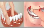
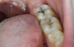
The incubation period lasts 2-12 days, with an average of 4 days. The onset is usually acute, with chills, fever, headache, and muscle pain.On the 2-3rd day of illness, a burning sensation in the mouth, soreness during chewing, salivation, redness of the eyes, sometimes vomiting and diarrhea, heartburn during urination are added. IN oral cavity and against the background of hyperemic and edematous mucous membrane of the lips, tongue, palate, inner surface cheeks reveal small, 2-4 mm in diameter oval vesicles filled with light, transparent, and then cloudy yellow contents. Bubbles sometimes densely cover the mucous membrane of the oral cavity, including the gums and palate. After a day or two, the bubbles burst, areas covered with erosions and ulcers form, signs appear general stomatitis. It is difficult for the patient to swallow and speak through swelling of the tongue, increased salivation(hypersalivation). Characteristic squinting outside the patient for the need to wipe the streams of saliva, suffering facial expression, irritability. In the case of a significant rash, the ulcers merge with each other. The regional lymph nodes swell (increase) and become painful. After opening the bubbles, the body temperature decreases.
Aphthae- vesicles that have turned into erosions can occur on the mucous membrane of the nose, vagina, urethra, conjunctiva with corresponding symptoms. Often a characteristic rash appears around the mouth, back, on the skin of the hands, feet. Typical for foot and mouth disease is the localization of the rash in the region of the terminal phalanges of the fingers, near the nails, between the fingers of the upper and lower extremities, accompanied by a burning sensation, crawling, itching. Due to the defeat of the terminal phalanges of the fingers, the nails may come off. In patients with a severe course of the disease, a maculopapular, sometimes hemorrhagic rash may appear on the skin of the trunk, neck, and extremities. The rash period lasts for 5-7 days, sometimes several weeks.
The illness lasts 6-10 days. Healing of aphthae on the skin and mucous membrane of the lips, mouth, tongue occurs on the 4-6th day of illness without scarring.
Some patients have lingering course disease (up to several months) with repeated vesicular rashes at a temperature and satisfactory general condition.
With uncomplicated foot and mouth disease, the internal organs are not affected, sometimes the liver and spleen can be enlarged; symptoms became arterial hypotension and bradycardia, leukopenia, eosinophilia.
Complications are usually associated with secondary bacterial flora - pneumonia, rarely meningitis, sepsis, myocarditis.
The prognosis is favorable. At severe course in weakened children, the prognosis can be serious.
FMD diagnosis
Supporting symptoms clinical diagnostics foot and mouth disease e acute onset of the disease, characteristic vesicles and erosion on the conjunctiva, mucous membranes of the lips, oral cavity, nose, urethra, vagina, severe salivation, rash on the skin of the fingers of the upper and lower extremities, especially near the nails and in the interdigital folds, around the mouth, back . Significant assistance in the diagnosis of the disease can provide epidemiological data, epizootological situation.Specific diagnosis of foot and mouth disease
The virus can be isolated from blood, saliva, aphthous elements, faeces of patients, in kidney cell cultures of pigs, calves, Syrian hamster. Serological studies carried out in the dynamics of the disease using RIGA, RSK, as well as RN.The best and most convenient diagnostic test is the biological sample. The contents of the vesicles-aft from patients are rubbed into the "pads" of the paws of guinea pigs. In the presence of foot-and-mouth disease virus, characteristic vesicular-aphthous elements appear at the site of its injection after 24-36 hours, and after 1-3 days secondary aphthae develop on the surface of the tongue.
Differential diagnosis of foot and mouth disease
FMD is often differentiated from aphthous stomatitis. In patients aphthous stomatitis ulcers are deep, have the correct round shape, and the bottom is covered with a whitish coating. There is no eosinophilia characteristic of foot and mouth disease.Herpetic disease is characterized by vesicles more often on the edges of the lips, wings of the nose. Erosions after opening the bubbles are deep and do not heal for a long time.
For chickenpox characterized by thin-walled vesicles surrounded by a narrow zone of hyperemia. They are formed on any places of the skin and scalp, but they are not on the feet and hands. Unlike chicken pox, foot-and-mouth disease does NOT spill onto the scalp and does not have a characteristic location on the limbs.
FMD treatment
Mandatory hospitalization and isolation of patients until they disappear acute manifestations but not less than 14 days from the onset of the disease. For today antiviral treatment patients with foot and mouth disease have not been developed, and antibiotic therapy is ineffective. The patient first of all needs careful care of the affected mucous membranes and an appropriate diet. Sometimes the patient is given food through a tube. special attention requires oral hygiene. Use frequent irrigation with a 3% solution of hydrogen peroxide, 1% solution of potassium permanganate, infusions of chamomile or sage, 0.25% solution of novocaine. Eyes are washed with 2% solution boric acid, instill 30% sodium sulfacyl solution, apply hyoxysone ointment, which is anti-inflammatory, relieves itching and pain. In case of severe disease, use cardiovascular agents, detoxification treatment, painkillers and antihistamines.FMD prevention
Disease prevention is sanitary and veterinary measures: isolation, quarantine, sometimes forced slaughter of sick animals. Conduct active immunization of animals with the threat of infection. Disinfection in the foci is mandatory, manure is disinfected by the biothermal method. Personnel caring for animals with foot-and-mouth disease must strictly observe all measures of individual protection.In endemic foci, pasteurization and boiling of milk are mandatory. The meat of sick or suspicious animals for foot-and-mouth disease can be used after appropriate heat treatment in agreement with the sanitary and veterinary service.
Foot and mouth disease is an acute, highly contagious disease of artiodactyls, manifested by fever, vesicular lesions of the mucous membranes of the mouth, corolla and udder skin, in young animals - damage to the myocardium and skeletal muscles. FMD is registered in many countries of the world.
IN vivo Domestic and wild artiodactyls are susceptible to FMDV. Dogs and cats can become infected and asymptomatic. A person rarely becomes infected by drinking uncontaminated milk from sick animals.
Exciter characteristic. The RNA-containing virus belongs to the family Picomaviridae, genus Aphtovirus. The genome is represented by a single single stranded linear plus RNA. Virus virions are particles of cubic symmetry with a diameter of 22-30 nm, devoid of a supercapsid shell.
Resistance to physical and chemical influences. FMD virus is resistant to ether, chloroform, freon. It is rapidly inactivated in an environment with a pH of 6.0 and below. Most stable at pH 7.0-7.5. Bleach, creolin, cresol, phenol kill the virus only after a few hours. Alkali solutions (2%) inactivate it in 10 minutes. The virus is resistant to environmental factors; aphthous lymph containing the virus is inactivated at a temperature of 31 ° C for 24 hours; in milk at a temperature of 66 to 78 ° C, the virus dies after 1 minute. Low temperatures it is preserved; at minus 40 - minus 70 °С retains biological properties some years. IN sewage the virus survives up to 103 days. A good preservative is a 50% solution of glycerol in phosphate buffer; in it, at 4-8 ° C, the virus is stored for 40 days. The best disinfectants are 2- or 3% hot solutions of sodium bicarbonate and 1% formaldehyde solution.
Antigenic structure. The main structural proteins of FMDV are VP1, VP2, VP3 and VP4. The VP1 protein is located on the surface and causes the induction of virus-neutralizing antibodies that protect the animal from the virulent virus.
The virus-containing suspension contains infectious and non-infectious viral particles: 140S - complete virions; 75S - capsids without RNA; 12S-14S - protein subunits and Via antigen, which is found in infected cells but is not integral part virion. All of these components have antigenic properties, but only 140S and 75S particles are immunogenic. Only 140S particles (complete virions) are infectious.
Antigenic variability. Currently, 7 antigenic types of foot-and-mouth disease virus are known: A, O, C, Sat-1, Sat-2, Sat-3 and Asia-1. Within the main types, there are variants, or subtypes, that differ from each other. Type A has 32 options, type O - 11 options, type C - 5, type Sat-1 - 7 options, type Sat-2 - 3 options, type Sat-3 - 4 options and type Asia-1 - 2 options. Antigenic types and variants established in CSCs also differ immunologically. Animals that have been ill acquire pronounced immunity to the homologous virus. Therefore, for specific prevention foot and mouth disease for each type of virus should be a vaccine.
Antigenic activity. In naturally susceptible animals, the virus induces the production of virus-neutralizing, complement-fixing, and precipitating antibodies.
Virus cultivation. The virus is cultivated on naturally susceptible and laboratory animals: newborn mice and rabbits, 60-day-old hamsters, and adult guinea pigs. It multiplies well in the culture of kidney cells of susceptible animals, in the culture of explants of the epithelium of the epithelium of the tongue of cattle and in some transplanted cell lines (VNK-21, SPEV, etc.) with a pronounced cytopathic effect.
experimental infection. It is easily reproduced by applying a virus-containing material to the scarified surface of the mucous membrane of the tongue, gums of cattle, sheep and pigs (in the patch), as well as by subcutaneous inoculation of the virus in newborn mice or rabbits and intradermal injection of the material into the plantar surface of the hind legs guinea pigs.
Hemagglutinating properties. The virus does not have them.
Clinical signs. The incubation period lasts 1-3 days, sometimes up to 7-10 days. The most characteristic sign this disease in animals - vesicular lesions of the mucous membranes of the mouth, skin of the corolla and udder. In cattle and pigs, foot and mouth disease is acute, in adult animals, as a rule, it is benign. The disease spreads very quickly. Initially, a deterioration in appetite, increased salivation, and an increase in body temperature (up to 40.5-41.5 ° C) are noted. On the 2-3rd day, aphthae appear on the inner surface of the lips and on the tongue. In some animals, aphthae are formed in the region of the interhoof gap and on the udder. Limb disease is accompanied by lameness. A day later, the aphthae are torn and erosion is formed. After 2-3 weeks, the erosions heal and the animals recover. In pigs, sheep and goats, lesions are observed more often on the limbs and less often on the mucous membranes of the mouth. Quite often the udder is affected. In young animals, foot and mouth disease usually proceeds malignantly (death - 80% or more), as a rule, there is no aphthae.
Pathological changes. At autopsy of dead young animals, hemorrhagic inflammation of the intestine and degenerative changes in the muscles of the heart ("tiger" heart), similar changes are found in skeletal muscles.
Virus localization. From sick animals, the virus can be detected already during the incubation period from milk, semen, saliva (4-7 days before clinical signs). The largest amount of virus is contained in the epithelium and fluid of vesicles (up to 10 8 ID/g). Excrements and secrets of sick animals are infectious for more than 10 days. The virus is also excreted with exhaled air. The disease may be accompanied by a long virus carrier. About 50% of cattle can shed the virus within 8 months, and some - up to two years. In pigs, persistent carriage of the virus has not been established. In herds of buffalo, the infection has been maintained for many years by virus carriers and animals with undercurrent infections.
source of infection are sick animals and virus carriers. The epizootological role of wild artiodactyls is very significant. The virus is highly contagious, so the disease spreads rapidly among susceptible animals. An important role in the spread of foot and mouth disease is played by products and raw materials of animal origin, as well as care items, manure and feed contaminated with the secretions of sick livestock. Animals immune to foot-and-mouth disease (dogs, cats, horses and birds) can also be carriers of the infection.
Diagnostics. FMD is diagnosed on the basis of epidemiological data (high contagiousness and selective damage only to artiodactyls), clinical signs (vesicular lesions of the mucous membranes of the mouth, skin, limbs and udder), pathological and anatomical changes (with the death of young animals - damage to the intestines and heart muscles) and results laboratory research.
Diagnose foot and mouth disease clinical signs quite easily, but it is important for the farm doctor to know what type of virus is causing the disease in order to administer the appropriate vaccine. The type of virus is determined in the laboratory.
Taking and preparing material. For laboratory studies, at least 5 g of the wall and contents of aphthae are taken from 2-3 sick animals on the mucous membrane of the tongue (in cattle), on the patch (in pigs), on the skin of the corolla and interdigital gap (in cattle and small cattle, pigs, camels, etc.). In the absence of aphthae, the blood of animals is taken at the time of the temperature reaction, from the corpses of young animals of all kinds - the lymph nodes of the head and pharyngeal ring, the pancreas and the heart muscle. To study for virus carriers, esophageal-pharyngeal mucus is taken (with a special probe).
The material must be obtained in such a way as to prevent the removal of the virus outside the dysfunctional focus and laboratory, and to protect personnel working with infectious material. To do this: a) the veterinarian of the farm must have certain skills in taking material from sick animals; b) it is necessary to prepare everything for the selection of material - tweezers, scissors, napkins, thick-walled vials, adhesive plaster, rubber stoppers, 50% sterile glycerin solution in isotonic sodium chloride solution, thermos with a cooling mixture, disinfectant - 2% NaOH solution or 1% solution of acetic or lactic acid; overalls - dressing gowns, overalls, scarves or hats, masks, rubber boots, gloves, etc. Everything you need is placed in a container and they go to a dysfunctional hearth, where, before entering the room with sick animals, they change clothes; c) after taking material from sick animals, tools, mask, gloves are immersed in a disinfectant solution; outer surface bottles and a thermos are treated with disinfectant. In the sanitary inspection room, they take off all their clothes and take a shower.
In the nasal cavity of humans, the foot-and-mouth disease virus survives up to 7 days, therefore, during this time after visiting a dysfunctional farm, contact with healthy artiodactyl animals is undesirable.
Samples of the material without signs of decomposition are placed in vials with screw or ground stoppers and frozen, and in the absence of freezing conditions, they are filled with a preserving liquid (50% sterile glycerol solution in isotonic NaCl solution). Labels are attached to the bottles indicating the type of animal, the name of the material, its quantity, the date of selection and the address of the sender. The vials are placed in an impenetrable metal container, sealed and placed in a thermos with ice, which is also sealed. A cover letter signed by the doctor is attached to the material, which indicates: the date of taking the material, from what type of animals and what material was taken, the epizootic situation for foot-and-mouth disease on the farm, the name of the doctor. The material is sent by courier. To work with the FMD virus in the laboratory, a separate room is allocated (a box with a pre-box), where there should be necessary equipment and materials for diagnostic work(preparation of material, setting up RSC, bioassays, etc.). When working in the box, they completely change their overalls and shoes, put on rubber gloves and a mask. After work, nothing that has not been neutralized can not be taken out of the box. The dishes and tools are boiled, overalls are immersed in a container for autoclaving; tables, floors, walls are treated with a disinfectant solution, followed by irradiation with UV rays.
The laboratory keeps a strict record of the incoming material and its consumption with an accuracy of 1 mg. The material received by the laboratory is stored until the study and during the period of use in a locked and sealed refrigerator. Upon completion of the work, an act is drawn up for the destruction of the material remaining from the study and animals after the bioassay.
Laboratory tests for foot-and-mouth disease include: detection and identification of the antigen of the foot-and-mouth disease virus in the RSC (determination of its type and variant affiliation); detection and titration of antibodies to foot-and-mouth disease virus in recovered animals (convalescents) in the reaction of radial immunodiffusion (RRID) and indirect immunofluorescence reaction (IRIF).
Detection and identification of FMD virus antigen using RSK. Reaction components: test antigens from epizootic virus strains from diseased animals; sera of guinea pigs hyperimmunized with standard type and variant strains of foot-and-mouth disease virus (biofactory production); control antigens - from typical and variant strains of foot-and-mouth disease virus (biofactory production); complement - fresh or dry normal guinea pig serum; biofactory hemolysin; sheep erythrocytes - in the form of a 2% suspension in saline; 0.85% solution chemically pure chloride sodium in distilled water; a set of specific sera and antigens to other viruses that cause vesicular lesions.
When examining material from pigs in RSK, specific antigens and sera to the swine vesicular disease virus are included.
RSK is placed in various volumes: in a total volume of 1 ml - take 0.2 ml of each component, in a total volume of 0.5 ml - take 0.1 ml of each component or by the micromethod - a total volume of 0.125 ml, while each component is 0.025 ml .
Preparation of FMD virus antigen. The walls of aphthae from sick animals are washed from the preservative liquid with physiological saline solution pH 7.4-7.6, dried with filter paper, weighed, crushed and carefully ground in a porcelain mortar with sterile broken neutral glass until a homogeneous mass is obtained, to which is added double with respect to mass of aft the amount of physiological solution (pH 7.4-7.6), i.e. for 1 g of aft - 2 ml of solution. The resulting 33% suspension is extracted at room temperature for 2 hours, frozen at minus 10-20 °C for 5-18 hours. After thawing, centrifuge for 15-30 minutes at 3000-5000 min -1 . The supernatant is inactivated at 58°C for 40 min. After inactivation, if flakes remain in the liquid, it is centrifuged again for 10-15 min at 3000 min -1 and then used as an antigen in the CSC.
Stages of setting RSC.
1. Titration of hemolysin. It is carried out upon receipt of a new series according to the generally accepted method. In the main experiment, hemolysin is taken at a 4-fold concentration of its limiting titer (working dilution).
2. Cooking hemolytic system(heme systems). To do this, hemolysin is mixed in a working dilution with an equal amount of a 2% suspension of sheep erythrocytes.
3. Complement titration. It is carried out in the hemolytic system on the day of setting up the main experiment according to the generally accepted method. For the main experiment of RSK, complement is taken with an excess of 1% of its titer in the heme system. Correctly taken working dose of complement is an indispensable condition normal flow reactions, which ensures the reliability of the results.
4. Preparation of working dilution of type-specific sera. In the main experiment to determine the type of foot-and-mouth disease virus, serum is used in a double titer (from the limiting titer), for example, if the limiting serum titer is 1: 40, then the working titer will be 1: 20.
5. Preparation of working dilution of type-specific antigens. Antigens are also used in double titers, for example. if the limiting titer is 1:6, then the working titer will be 1:3.
6. The test antigen in the reaction is examined whole (33% suspension) and in dilutions of 1:2, 1:4 and 1:8.
7. Statement of the main experiment to determine the type of foot-and-mouth disease virus. Simultaneously with the main experience put the control of all specific FMD antigens and sera according to the scheme. The components are poured in the following order: 1) specific sera in a working titer of 0.2 ml for each serum - one row of test tubes vertically; 2) specific antigens in a working titer of 0.2 ml - in the first seven horizontal rows, one row for each antigen; 3) test antigen in dilutions of 0.2 ml - for each dilution one row of test tubes horizontally; 4) saline 0.2 ml each - in the last horizontal row (sera control) instead of the antigen and in the last vertical row (antigen control) instead of sera; 5) a complement of 0.2 ml and a working dilution - in all test tubes of the main experiment. The tubes are shaken gently and placed in water bath for 20 min at 37-38 °C; 6) pour 0.4 ml of the hemolytic system into all test tubes. The test tubes are shaken again and placed in a portable bath for 30 minutes at 37-38 °C.
The reaction is recorded 5-10 minutes after the water bath, and final result receive after 10-12 hours. The degree of hemolysis delay is evaluated in crosses: (++++) - 100% hemolysis delay; (+++) - 75%; (++) - 50%; (+) - 25% hemolysis delay; (-) - complete hemolysis.
If the tested antigen is homologous to specific antibodies, then there will be a delay in hemolysis and the reaction will be positive; if homologous antibodies are absent, the reaction is negative and complete hemolysis is observed.
In case of production necessity, after determining the type of FMDV, its subtype (option) is established. To do this, put RSK according to the same method, but use variant sera and variant antigens of the established type. Moreover, variant sera are used in the limiting titer, and antigens in doubled.
The antigen (tested) refers to the variant, with the serum of which it gives a positive reaction in higher dilutions.
When the viral material delivered from the farm is not enough for research in the RSC, it is bred in cell culture or on 3-6-day-old suckling mice, or on adult guinea pigs. Permission from higher authorities is required to carry out such work. veterinary authorities and the presence in the diagnostic laboratory of conditions of a strict sanitary regime. In mice, the suspension under study is injected subcutaneously in the back at a dose of 0.1-0.2 ml, in guinea pigs - intradermally into the pads of both hind limbs at a dose of 0.2-0.5 ml. Animals are observed for 5-7 days.
In the event of the death of mice, an antigen for CSC is prepared from their carcasses. In guinea pigs, in positive cases, aphthae form on the legs; aft walls and their contents are used in RSC. If necessary, 2-3 "blind" passages are carried out. A sample of the test material is considered negative if no degeneration of cells and death of white mice is noted in the third passage, and when examining the suspensions obtained from them, the FMD virus antigen is not detected in the CSC.
Retrospective diagnosis. The material for testing for the presence of antibodies to the FMD virus is the blood serum of animals suspected of having FMD or other vesicular diseases. Blood sera should be taken no earlier than 7 days after the appearance of signs of vesicular disease in animals. 5-10 serum samples from each animal should be sent for research. age group. If the results of the primary study are doubtful, it is necessary to take blood again from the same animals after 7-10 days.
The serum obtained by the conventional method is preserved with antibiotics (500 IU/ml of penicillin and streptomycin) or frozen at minus 20 °C. At least 5 ml of serum in a thermos with ice is sent to the study from each animal.
In the laboratory, serum is examined using a radial immunodiffusion test (RRID) and an indirect immunofluorescence test (IRIF).
RRID. The essence of the reaction is the formation of a zone of specific precipitation of viral antigens by antibodies included in the agar gel. RRID is type-specific.
To set up the reaction, melted 2% agar is mixed with an equal volume of test serum heated to 50-55 ° C in dilutions of 1:5, 1:10, 1:20, etc. up to 1: 320 and applied 1 ml per slide. Wells (4-7.7 mm in diameter) are cut out in frozen agar, which are filled with reference type antigens. The slides are then placed in a humid chamber at 37°C. The results are taken into account after 6-7 hours and finally after 18 hours.
A positive reaction is characterized by the formation of a precipitation ring in the form of an opalescent zone around the well with an antigen homologous to the pathogen that caused the disease.
Antibodies found in the test serum sample are assigned to the serotype with which they tested positive. Their titer is considered the maximum dilution of the test serum, with which a positive reaction is observed.
After the animal has been ill, antibody titers usually exceed 1: 160.
NRIF. This reaction is based on the fact that the presence of antibodies in the blood serum of recovered animals reveals a specific luminescence (antigen + antibody complex), and when using sera from vaccinated animals, the luminescence of the complex is not observed.
The setting technique is as follows. On the preparation from the culture of cells BHK-21, PEC, PES, infected with foot-and-mouth disease virus of any type, apply the test serum at a dilution of 1: 10 and 1: 20; incubated in a humid chamber at 37 °C for 30 minutes; wash off unbound antibodies; air dry and stain with a mixture of working dilutions of fluorescent anti-species serum and bovine albumin labeled with rhodamine; incubated in a humid chamber at 37 °C for 30 minutes; launder; dried and viewed under a fluorescent microscope (x40 objective, x4 or x5 eyepiece).
A positive reaction is characterized by a green or emerald green glow of the cell cytoplasm.
The reaction is accompanied by setting appropriate controls.
The diagnostic result is considered positive if a specific luminescence is detected in at least one of 5-10 sera sent from this farm.
To determine the level of antibodies detected in this way in the test serum, it is titrated. To do this, the test serum is diluted from 1:40 to 1:1280, and a known infected preparation is treated with each dilution, as described above. The titer of post-infection antibodies in serum is judged by its limiting dilution, which is capable of producing a positive NRIF. The presence of a specific luminescence in preparations treated with the test serum in dilutions of 1:10, 1:20 and 1:40 indicates that the serum was obtained during the period of acute illness of the animal with foot and mouth disease, i.e., about 7 days have passed since its illness , and the presence of a specific glow in dilutions of 1: 80 and above indicates that the serum was taken from a convalescent animal.
The results of the study on foot and mouth disease are drawn up in the form of a protocol, which indicates the date of the study, the name of the farm, material, brief epizootological data, etc., and the name of the components used in the study, the characteristics of the controls are required.
It should be noted that many other methods have been developed for the indication and typing of FMDV, such as PCR, RNHA, ELISA, the method of cross-immunity, etc.; for the detection and typing of antibodies - PH, RNHA, sulfur protection reaction on sucking mice, etc.
Differential Diagnosis. It is necessary to exclude other diseases of animals with vesicular syndrome, such as VD, RTI, vesicular stomatitis, in pigs - vesicular disease, vesicular exanthema, in sheep - catarrhal fever.
Immunity and specific prophylaxis. The duration of immunity in animals with FMD is 8-12 months, in pigs - 10-12, in sheep - 18 months. With very tense immunity, there may be some resistance to infection with a heterologous type of virus. With foot and mouth disease, tissue and humoral immunities. Humoral immunity factors are of primary importance in protecting animals from disease. For the specific prevention of foot-and-mouth disease, apply inactivated vaccines. found in our country wide application the following 3 vaccines: lapinized aluminum hydroxide saponformol vaccine, which is prepared from a virus reproduced in the body of newborn rabbits; aluminum hydroxide saponformol vaccine from a virus cultivated in the surviving tissue of the mucous membrane of the tongue, not immune to foot-and-mouth disease in cattle (Frenkel method); and a vaccine from a virus obtained in a suspension culture of BNK-21/13 cells. For pigs, an emulsified vaccine made from lapinized virus is used.
Immunity after vaccination in adult animals lasts 4-6 months. After revaccination, immunity is more intense and prolonged.
Young animals born from immune animals passively receive antibodies from colostrum. Antibodies in calves persist for 5 months, although passive protection lasts up to 3-4 months.
Inactivated vaccines can be mono - or polyvalent, i.e. contain antigens of one or many types and variants of the virus. Live vaccines against FMD have not been developed. Research is being conducted on the development and use of synthetic vaccines, as well as molecular vaccines obtained using genetic engineering methods.
Foot-and-mouth disease is an acute zoonotic infection that is transmitted from diseased artiodactyls to humans. The infection is accompanied by fever, as well as with the appearance of small blisters on the mucous membrane of the mouth and near the oral surface, and in addition, they form at the nail bed.
Due to the characteristic clinical picture foot-and-mouth disease in humans and animals has been known since ancient times. However, the fact that the infection is able to pass from a sick artiodactyl animal to a person was established only at the end of the 18th century. Leffler in 1897 was able to prove that the FMD pathogen is able to penetrate through the pores of a bacterial filter.
FMD is caused by an RNA virus. It is characterized by high virulence, and it also resembles the epithelial structures of the mucous membranes and skin. FMD virus is persistent. He is steadfast in various factors the external environment, namely freezing and drying, retains its viability in animals on wool for up to about 4 weeks and up to 3.5 weeks on clothes. The death of this virus can be achieved by heating, UV irradiation, as well as exposure to disinfectants. These include alkalis 2%, formalin 1% and ethylene oxide 1%.
As already mentioned, the source and reservoir of foot-and-mouth disease are ungulates, both domestic and wild. Some types of rodents can also be distributors, but it is worth noting that they do not have a significant impact on its distribution. As for birds, they cannot get sick with foot-and-mouth disease, but during migration they can carry it.
Foot-and-mouth disease can be attributed to occupational disease. It is mainly distributed in countryside, and those people who work in agricultural livestock enterprises, meat processing plants that slaughter livestock and process animal raw materials.
Foot and mouth disease can also occur in children. Children get the virus after eating contaminated dairy products.
Causes of FMD
The cause of foot-and-mouth disease in humans is poor personal hygiene. The main source of the disease, as already mentioned, are animals (cattle, pigs, sheep, goats, very rarely - dogs, cats, horses, poultry). Rodents, flies and ticks play a secondary role in the distribution. The mechanism of infection is fecal-oral (through food), contact and household contact, i.e. either through direct contact with aphthae of sick animals, or through contact with infected objects.
In addition, products such as milk and meat obtained from animals that suffer from foot and mouth disease can become a source of the disease. The infection cannot be transmitted from person to person.
Despite the above factors of infection, children can also be infected with FMD. In the case of children, infection occurs through digestive tract. Foot-and-mouth disease can enter a child's body through contaminated milk and dairy products.
FMD symptoms

The incubation period of the disease is on average 3 to 4 days, but the disease can increase from 10 to 14 days or, conversely, be reduced to two days. The disease process begins with a strong chill, while the temperature rises to 40 degrees. To these symptoms are added, appetite decreases, observed muscle pain and especially strong in the lumbar region. On the first day of the course of the disease, the infected person feels a burning sensation and dryness in the mouth, as well as strong salivation. After some time, small bubbles begin to pour out on the mucous membrane of the mouth, from about 1 to 3 mm. Their largest accumulation is located at the tip and along the edges of the tongue, as well as on the gums, mucous membrane of the cheeks and lips. The liquid that fills the formed bubbles is transparent, it gradually becomes cloudy, the diameter of the bubbles increases, while forming erosion.
If foot-and-mouth disease has struck children, then they complain of difficulty swallowing, as well as pain when talking and chewing. Because of this, they become irritable and subsequently refuse to eat at all. Often salivation can be so strong that saliva can flow out in a stream. Eruptions of bubbles can also be on the mucous membranes of the nose, conjunctiva, stomach. In addition to the mucous membranes, rashes can also be on the skin of the face, forearms, hands, feet and legs, they are especially common between the fingers. Provided that the disease proceeds without complications, the fever lasts from 3 to 6 days. After this, a period of convalescence begins, which is accompanied by fast healing all ulcers. Total time The course of the disease is about 2 weeks.
There are cases when the duration of the disease is from 1 to 1.5 years, with periodic rashes.
If the virus penetrates through the gastrointestinal tract, then stomatitis may not appear. In this case, the whole disease proceeds according to the type acute gastroenteritis. Symptoms of intoxication appear, fever, abdominal pain, nausea, and liquid stool. Usually this form of the course of the disease is observed in children who become infected through milk and dairy products.
In most cases, the disease ends with a complete recovery, while it does not leave any traces behind. But a completely different prognosis has a severely flowing foot and mouth disease, which affects children. early age. Some cases of the disease end in death.
Like many diseases, foot and mouth disease can cause complications. But this happens quite rarely. Complications may include pneumonia, purulent diseases skin.
FMD prevention
The basis of foot-and-mouth disease prevention is to prevent the spread of the foot-and-mouth disease pathogen, as well as constant control over the various movements of animals and food products that are obtained from animals. If sick animals are found, they must be immediately isolated from society. It is forbidden to take milk and meat from a sick animal.
FMD prevention also consists in observing personal precautions in the focus of the disease and various sanitary and veterinary measures. Mandatory heat treatment of milk, processing of components of living origin is required, from which butter and other products are made. Mandatory compliance with all safety measures when caring for and working with a sick animal. An important role is played by the spread of sanitary and educational work among the population.
If work is carried out where there are sick animals, a number of precautions must be observed. A worker who has wounds or abrasions on his hands and exposed parts of his body is not allowed to work with such animals. After completion of work with a sick animal, 1% chloramine is disinfected. A complete and thorough disinfection of the premises and equipment, and even manure is carried out.
FMD treatment

Patients with foot and mouth disease need hospitalization. There are no specialized medicines for the treatment of this disease. Treatment is carried out in a hospital. The main treatment measures are aimed at caring for the mucous membranes of the mouth and nose, as well as local treatment and general relief symptoms.
For a period of severe damage to the oral mucosa, the patient should eat semi-liquid food of moderate temperature, which is easily absorbed by the body. It should not include irritating components. These include horseradish, mustard, pepper, etc. If extensive lesions are observed, the nutrition process occurs parenterally or through a tube. For local treatment of foot and mouth disease, ointments are used, such as oxolinic, florenal and interferon ointments, as well as physiotherapeutic methods are used: laser and UV irradiation.
Antipyretics, analgesics, cardiovascular and desensitizing agents are usually prescribed according to indications. For general strengthening purposes, adaptogens are often prescribed. To treat and alleviate the symptoms of foot and mouth disease, people turn to traditional medicine recipes.
Expert editor: Mochalov Pavel Alexandrovich| MD general practitioner
Education: Moscow Medical Institute. I. M. Sechenov, specialty - "Medicine" in 1991, in 1993 " Occupational diseases", in 1996 "Therapy".
This disease is rarely seen by physicians, however, is well known to veterinarians. Since it is animals that are susceptible to it, as a rule, livestock. Much less often, foot-and-mouth disease affects deer, elk and camels. This viral infection is high fever and damage to the mucous membranes of the skin of the animal.
The disease is not safe for humans. The virus penetrates into all fluids of an infected individual (saliva, feces, blood, milk). In most cases, a person becomes infected with foot-and-mouth disease after eating contaminated milk or meat. It is also possible to get sick from prolonged close contact with an animal, for example, when caring for him during the period of illness. The human body is quite capable of coping with the pathogen, for an adult the risk of infection is low, however, the virus is dangerous for children. Their immune system is not strong enough to resist the disease.
You can only get infected from a sick animal. FMD is not transmitted from person to person. The virus is not afraid of freezing or drying. But sensitive to sunlight, heat treatment and disinfectants.
Symptoms
The period from infection to the first symptoms is 4 days. In isolated cases - from 2 to 14 days. The patient begins to have a fever, the temperature can reach 40 degrees. There are headaches and muscle pains, a burning sensation in the mouth, dry mucous membranes and profuse salivation.
The first bubbles can be found on the tongue, lips and gums. In the future, the rash affects the nose, eyes and skin, spreading throughout the body from the face to the feet. Erosion can also occur on the mucous membranes of the stomach. It is difficult for the patient to speak, swallow and chew, as a result - complete failure from food. In some cases, the virus penetrates the mucous membranes of the genitourinary system, which leads to pain during urination.
In the youngest children, the virus does not manifest itself in rashes, but causes symptoms of gastroenteritis. Namely:
- high body temperature,
- intoxication,
- stomach pain,
- nausea,
- vomiting and diarrhea.
As a rule, this form of the disease manifests itself when children become infected. younger age through milk and fermented milk products that have not undergone sufficient heat treatment.
Course of the disease
It takes about two weeks from the onset of symptoms to full recovery. Heat lasts 3 to 6 days. Another 7-10 days are needed for healing and disappearance of erosion. But in rare cases, the course of the disease can last up to one and a half years. Rashes appear periodically.
Diagnostics
To confirm the diagnosis, it is necessary to donate blood, saliva, feces, as well as ichor from the rashes to isolate the FMD virus from them. Such a study is carried out if a person hospitalized with a rash and fever has contacted a sick animal.
Treatment
There is no specific cure for FMD. A sparing diet is recommended, consisting of easily digestible liquid and semi-liquid foods, as well as plentiful drink. The oral cavity is treated with hydrogen peroxide, novocaine, lapis solution (2 or 5%) and potassium permanganate. Depending on the patient's condition, immunoglobulin may be administered. good effect has ultraviolet and laser irradiation.
Treatment of the disease at home is almost impossible. People infected with foot-and-mouth disease must be hospitalized. As a rule, the disease has no complications. However, a severe course in young children can be fatal.
Prevention
It is necessary to avoid places where in recent times there have been cases of infection of animals with foot-and-mouth disease. And also always give in to thorough heat treatment of milk and meat.
What is foot-and-mouth disease virus? How is the infection transmitted? What are the symptoms of the disease in animals and humans? What is foot-and-mouth disease? How is the disease treated? About all this will be discussed in our publication.
The causative agent of the disease
The causative agent of foot and mouth disease virus is a specific structure of ribonucleic acids, which belongs to the family of picornaviruses. The size of such infectious particles is about 30 nanometers. The microscopic structure consists of RNA surrounded by a protein coat. Once in the human or animal body, the virus infects the lymph. The development of infection occurs within 48 hours.
FMD virus is resistant to heat and cold in natural conditions. However, it instantly dies when exposed to temperatures above +80 ° C. Being in the feces of an animal that ended up in environment, the causative agent of infection passes into an inactive phase, maintaining vital activity for more than 100 days. The foot-and-mouth disease virus loses its ability to reproduce under the influence of ultraviolet rays, as well as disinfectants.
Mechanism of infection development
Having entered the body, the viral pathogen concentrates on the mucous membranes of the oral cavity and damaged areas of the skin. In places of penetration into the tissue, the infection accumulates in small vesicles. Here, an active infection is observed. Then the infection spreads through the bloodstream, attacking the tissues of organs and systems. Over time, intoxication of the body develops. Pathological ribonucleic structures settle in the epithelium of the oral cavity and nasopharynx, concentrate in the urethra.
At-risk groups

What categories of the population are in the group increased risk foot-and-mouth disease virus infection? Usually the development of the disease is observed among the personnel of livestock enterprises. As practice shows, most often a viral infection affects milkmaids, cattle drivers, people who work in slaughterhouses and meat processing plants. Sometimes, as a result of negligent attitude to work, the lizard virus in humans is observed among veterinarians and livestock specialists.
At the same time, not a single case of transmission of pathogens from one person to another has been recorded. This is due to the low susceptibility of a person to infection. Among other things, after recovery, people acquire short-term immunity, which lasts about a year.
Symptoms of foot and mouth disease in animals
Often, the infection affects young cattle. Immature animals do not have immunity against the virus and are more difficult to tolerate the disease. The development of the disease is characterized by the appearance of fever, which is accompanied by rashes on the limbs, mucous membranes of the mouth, tissues that are adjacent to the horns, and also on the skin of the udder.
FMD virus in animals attacks the body for 10-15 days. This is preceded by an incubation period that lasts 2-4 days. In most cases, livestock can be successfully cured. However, in severe cases of the disease, death occurs.
Symptoms in humans

What are the symptoms of FMDV in humans? Often already during the incubation period, which lasts about a week, infected person the first characteristics diseases. These include:
- chills;
- headache attacks;
- general malaise;
- pain in the muscles;
- temperature rise to +38 ... +39 ° С.
Then the foot-and-mouth disease virus in humans begins to progress. A few days later, a burning sensation and dryness in the oral cavity are added to the above symptoms. Photophobia is manifested, there are pain during urination.
Concerning external signs foot-and-mouth disease virus, the appearance of small whitish bubbles on the palate, lips, inner surface of the cheeks is noted. After about a day, such aphthae open, which leads to the formation of sores of a bright red hue. At this stage of the development of the disease, a gradual decrease in body temperature is observed. Despite this comparative relief, the general condition of the infected person worsens. Severe pain occurs when swallowing copious excretion saliva. Then the tissues of the tongue swell, the lips swell. Speech becomes slurred.
With absence adequate treatment foot-and-mouth disease virus, in humans, blistering formations move to the skin of the legs and arms. Here, aphthae heal much faster than on mucous membranes. Within 3-5 days, no trace remains of them.
The course of the disease in children has more. Nausea is often added to the above symptoms, frequent urges to vomiting, disorder digestive organs, change in stool structure, diarrhea.
Features of the transmission of infection

The infection can spread between animals and from livestock to humans. People affected by the virus are only carriers. However, they are not able to transmit the pathogen to another person. Children are the most susceptible to the virus. This is facilitated by the presence of a weak immune system.
How is FMDV transmitted? The infection is spread by contact. Infection occurs when the pathogen enters the body when caring for large cattle. The virus can concentrate on animal hair, be contained in pollution, feces.
Usually, people become infected by inhaling airborne dust. Sometimes infection occurs through contact dirty hands with mucous membranes of the mouth. The disease can also develop with the use of meat and milk of animals.
Treatment of foot and mouth disease in animals

The destruction of infection among occurs by isolating sick animals from the rest of the herd. The latter are contained in separate rooms. Destroy the viral pathogen by introducing into the body disinfecting sera containing substances such as convalescents, lactoglobulins, immunolactones.
During the recovery period, animals are offered an abundance clean water and nutritious food. The mucous membranes of the oral cavity are periodically treated with antiseptics. In order to eliminate ulcers on the surface of the skin, ointments with a healing effect are prescribed. Additionally, antibiotics and pain medications may be used.
In case of widespread infection in the herd, quarantine is introduced. When epidemics occur, sick cattle are destroyed. Animal carcasses are disposed of by burning in furnaces. Quarantine measures are terminated after 21 days have passed since the last case of infection was recorded.
Treatment of foot and mouth disease in humans

Therapy for infection with a viral pathogen requires the placement of an infected person in a hospital. Treatment involves regular disinfection of the oral cavity, healing of formed ulcers, the use of measures aimed at alleviating the general condition of the patient.
Infected people are offered easily digestible food with a semi-liquid consistency. Food must have room temperature and do not contain ingredients that can irritate mucous membranes. With extensive distribution of ulcerative manifestations, nutrition is provided to the patient through the introduction of food through a probe.
For the purpose of local treatment, exposure to the affected areas of the skin with laser and ultraviolet radiation. For the speedy healing of sores, tissue treatment with florenal, oxolinic or interferon ointment is prescribed.
To alleviate the suffering of the patient during treatment, painkillers, cardiovascular, antipyretic pharmacological drugs are used. If necessary, carry out activities aimed at removing toxins from the body. Vitamin complexes are prescribed to maintain immunity.
In most cases, foot and mouth disease does not pose a mortal danger to humans. The prognosis for such a viral infection is extremely favorable. Full recovery with the formation of appropriate immunity occurs in quite short period. The disease does not leave behind any consequences. cases lethal outcome occasionally observed only among newborns and toddlers.
Prevention

To prevent infection with the FMD virus allows, first of all, personal hygiene and compliance with sanitary standards. In order to prevent the disease, appropriate vaccination of animals is often carried out.
Of particular importance in terms of prevention is the implementation of instructions while working on farms, slaughterhouses, meat processing plants. According to the regulations, it is necessary to take care of livestock by wearing overalls, a protective mask, and gloves. After finishing work, it is important to wash your hands with soap and water.
In order not to once again expose yourself to the risk of infection with the virus, it is worth eating only proven, safe products of animal origin. Dishes in which meat or milk has been stored raw must be thoroughly cleaned using a detergent.
Last reported case of FMD epidemic
In October of this year, the foot-and-mouth disease virus was detected in Bashkiria. The state of emergency was introduced in the villages of Ermukhametovo and Urmekeyevo, which are located on the territory of the Tuymazinsky district. In the region, roads were blocked that lead to the marked settlements, and checkpoints were also established. Special units Disinfection stations were equipped to eliminate emergencies. Active measures have begun to disinfect farms.
During the elimination of the epidemic, the FMD virus was found in dairy products. The sale of the latter from hand to hand was banned. in the above settlements had to be exterminated. The rest of the animals in the surrounding regions were vaccinated. On this moment meat and milk are not sold to both the population and enterprises until the FMD virus in Bashkiria is completely eradicated.
Finally
As you can see, foot and mouth disease is a rather dangerous infection of a viral nature, which can cause significant losses to livestock farms. However, subject to the established instructions and personal hygiene, the disease does not pose a danger to humans. If the infection still manages to hit the body, forecasts for full recovery here are positive.