Causes and treatment of ulcerative stomatitis. General information and the cause of the disease. What is this symptom
Most often, the appearance of Vincent's stomatitis occurs in people with low level immunity, especially against the background chronic diseases. Necrotizing stomatitis is one of the varieties bacterial stomatitis with pronounced necrotizing ulcers affecting soft tissues and oral mucosa. To a greater extent, young people under the age of thirty are susceptible to this disease, however, this does not mean at all that ulcerative necrotic stomatitis does not manifest itself in other age categories.
isolated leak this disease happens extremely rarely, since in most cases it is a complication of long-term illnesses gastrointestinal tract or a logical continuation of untreated catarrhal stomatitis. In this ulcerative form of the disease, damage to the mucous membrane is inherent in the entire depth of its layer with the possible formation of ulcers on the lips, the inside of the cheeks, on the gums, soft and hard palate and on the tongue.
Causes
The most important and obvious reason for the development of any form of stomatitis is poor oral care. In the absence of proper sanitation and timely treatment of diseased teeth and gums, the risk of pathogenic spirochetes of Vincent and spindle-shaped bacteria getting into the resulting microcracks and damage to the mucosa increases. As a result of the symbiosis of these bacteria and their active reproduction, ulcers form in the mouth, and soft tissues undergo necrosis and decomposition.
Often the cause of the development of Vincent's gingivostomatitis is the complications of such serious illnesses such as agranulocytosis, infectious mononucleosis, leukemia, poisoning of the body by prolonged exposure to salts of heavy metals, immunodeficiencies. IN this case fertile ground for development ulcerative stomatitis are periodontal disease, gingivitis, periodontitis, dental trauma, untreated deep caries, tartar and even wisdom teeth.
In addition, frequent smoking, drinking too hot drinks and food, stress and depression contribute to the development of inflammatory processes. favorable conditions work.
Symptoms
One of the most unpleasant signs of ulcerative stomatitis, in addition to severe pain — horrible smell from the mouth, formed due to rotting of soft tissues. In some cases, ulcerative stomatitis is not limited to the oral mucosa and spreads deep inside, affecting many vital organs. In this case, the following symptoms appear on the face:
- a sharp increase in body temperature, chills, fever;
- loss of strength, apathy, aching muscles and joints;
- gums swell, become loose and red;
- severe pain when eating, drinking and when trying to speak, because of which the patient is more often silent and sometimes completely refuses to eat;
- Enlargement and soreness of the cervical and submandibular lymph nodes;
- migraine-type headaches;
- increased salivation;
- an increase in leukocytes in the blood according to the results of the analysis;
- the number of ulcers is constantly increasing, the older ones are covered with a gray film, when removed, a deep bleeding and non-healing wound is exposed;
- there is constant bleeding of the gums;
- in rare cases, if left untreated, necrosis can affect not only soft tissues, but also the alveolar process, as a result of which teeth can be severely damaged and even fall out;
- if treatment is neglected, pathogenic bacteria can go to the palatine tonsils, resulting in both adults and children can become Simanovsky-Vincent's angina.
Treatment
Bacterial causes of this disease involve treatment with antibiotics, anti-inflammatory and disinfectant drugs. The most commonly prescribed medications are:
- Rinsing and processing with a weak solution hydrogen peroxide, furatsilina or potassium permanganate;
- Solutions of trypsin, pancreatin, chemotripsin and other proteolytic enzymes to remove tissues prone to necrosis;
- Trichopolum, flagyl, metrogil, dioxidin or klion for a week;
- Fortifying agents, immunomodulators, vitamin-mineral complexes for raising immunity;
- Antibiotics: ampioks, gentamicin, lincomycin, penicillin, kanamycin;
- Antihistamines to relieve possible adverse reactions from antibiotics;
- Keratoplasty: metacil, solcoseryl, retinol acetate, aloe and kalanchoe juice, sea buckthorn oil.
How to treat sores on the tongue and soft tissues oral cavity, your attending physician will tell you in detail at the appointment in dentistry. Proper treatment contributes to the removal of the main symptoms within one to two days and the healing of ulcers within a week.
Prevention
The main rule is the observance of oral hygiene! If you're vigilant about your gums and teeth, brush them at least twice a day. medicinal pastes and visit your dentist regularly for check-ups, removal of plaque and tartar your chance of getting this disease is extremely low. And, of course, serious illness that lead to necrotizing stomatitis should not be left untreated. Eliminate the cause and complications will bypass you.
There are many pathologies of the oral mucosa, often their diagnosis and treatment become big problem even for experienced professionals. One of the exceptions is Vincent's ulcerative necrotic gingivostomatitis. It is enough to encounter it only once, then to confidently recognize this disease in any of its manifestations.
Ulcerative necrotic stomatitis - infection oral cavity caused by specific pathogens. It was very common during the First World War and was called "trench mouth" or "trench disease".
Video: stomatitis
Causes
Pathogen
The immediate cause of the disease is the symbiosis of opportunistic microorganisms - fusiform bacillus and Vincent's spirochete.
Normally, they are natural inhabitants of periodontal pockets, and are also found in abundance under hoods - gum folds over incompletely erupted lower third molars. Under adverse conditions, they multiply rapidly, acquiring pathogenic properties.
Provoking factors
Provoking factors should include such as:
- lack of high-quality hygienic care for the oral cavity;
- hypothermia of the body;
- prolonged stress;
- unbalanced diet, especially lack of vitamins;
- abundance of tartar;
- advanced form of catarrhal gingivitis;
- decrease in body resistance due to infectious and viral diseases;
- suppression of the activity of other microflora of the oral cavity with the irrational use of antibacterial drugs.
The mechanism of the development of the disease
Ulcerative necrotic stomatitis of Vincent begins acutely against the background of a significant decrease in the body's resistance and in the presence of adverse factors in the oral cavity (lack of hygienic care, abundant dental deposits, etc.).
The activated anaerobic symbiosis of the fusiform bacillus and Vincent's spirochete practically displaces most representatives of the microflora of the oral cavity.
There is a sharp soreness, swelling and bleeding of the gums, accompanied by the formation of abundant necrotic plaque. Multiple dense necrotic layers appear, which are very difficult to separate from the mucosa, and an ulcerated, hyperemic surface is exposed.
In the future, suppuration from pathological periodontal pockets is observed. All this is accompanied by general weakness and fever.
In case of inadequate treatment acute form disease can become chronic. Often there is chronic ulcerative stomatitis in children, especially of school age, who neglect oral care. It manifests itself in the form of swelling of the gums with small ulcerations in the region of some gingival papillae.
It is also possible to exacerbate chronic stomatitis. It usually occurs as a consequence of the transferred colds and also after severe stress.
Symptoms
Symptoms depend on the severity of the disease:
- Easy degree. The patient complains of pain and bleeding in the gums while eating, bad smell from the mouth, and increased salivation. An objective examination reveals diffuse swelling of the gums, the gingival papillae are edematous, bleed easily from touch and are very painful. The general condition of the body usually does not suffer.
- Average degree. The patient's complaints increase significantly. The contour of the gingival margin is distorted, covered with bleeding necrotic crusts of gray-green color with putrid fetid odor. Suppuration is observed from the periodontal pockets. Lymphatic nodes are enlarged, mobile and painful. The patient's condition worsens: general weakness, inactivity and apathy, the temperature rises to 38°C.
- Severe degree. The patient's condition is similar acute sepsis. Body temperature can change abruptly from an increase to 40°C to a decrease to 35°C, which indicates a significant decrease in the body's resistance. The patient is tormented by severe headaches, as well as pain in the oral cavity. Ulceration of the mucosa from the gums extends to the cheeks, palate and floor of the mouth. With significant progression, damage to the bone tissue of the jaws is possible.
Diagnostics
Diagnosis of ulcerative necrotic stomatitis does not cause difficulties. This takes into account the anamnesis, as well as an assessment of the general condition of the patient and the data of an objective examination.
At severe course diseases, as well as with a low effectiveness of the treatment, differential diagnostics should be carried out with such diseases and pathological conditions as:
- blood diseases accompanied by ulceration of the oral mucosa (leukemia, agranulocytosis);
- mercury poisoning;
- secondary syphilis;
- AIDS.
To clarify the diagnosis, a detailed blood test is performed, as well as the Wasserman reaction. Urine is tested for mercury content.
The most accurate answer is given by a study of a smear taken from the oral mucosa, the predominance of fusiform rods and spirochetes confirms the diagnosis of ulcerative necrotic stomatitis.
Treatment
Treatment of ulcerative necrotic stomatitis begins with anesthesia of the oral mucosa.
Preference should be given to application anesthesia, since when injections are carried out in conditions of extensive damage to the mucosa by anaerobic microflora, numerous complications are possible associated with their penetration into the deep tissues of the maxillofacial region.
Stages of treatment:
- Softening and removal of necrotic tissue.
- Antibacterial therapy.
- Restorative and desensitizing therapy.
Preparations
For treatment, the use of non-irritating medicines. So for pain relief it is better to choose more gently active solution anesthesin in glycerin than the more powerful 10% lidocaine hydrochloride solution.
To remove necrotic tissue, mechanical methods should not be used, because. this will cause additional injury to the ulcerated mucosa.
First you need to put gauze swabs moistened with solutions of proteolytic enzymes such as chymotrypsin, trypsin or chymopsin. They soften non-viable tissues without affecting intact ones.
The areas of inflamed mucosa cleared of necrotic crusts should be carefully treated with antimicrobial and antiseptic drugs that are active against anaerobic microflora.

Most preferably, the use of a combination of metronidazole (trichopolum) with a solution of chlorhexidine bigluconate. However, it is also possible to use solutions of hydrogen peroxide and potassium permanganate.
From general treatment The following medications should be given:
- desensitizing drugs (tavegil, suprastin or fenkarol);
- vitamins (large doses ascorbic acid with routine)
- with a severe degree of the disease - antibiotics a wide range actions.
After removing the severity of inflammation, it is necessary to carry out an extended sanitation of the oral cavity - removal of supra- and subgingival dental deposits, elimination of traumatic dentures, treatment of caries and its complications.
If possible, it is better to postpone the extraction of teeth until the signs of inflammation are completely removed.
To improve the healing of ulcerative surfaces, ointments, such as Solcoseryl, help well, but other keratoplastic agents can also be used ( oil solutions vitamins A and E, etc.).

Treatment is practically no different from that of adults. However, it should be noted that the child may be capricious, afraid and interfere with the quality treatment of the oral cavity.
Diet
Since, with necrotizing ulcerative gingivitis, the oral mucosa is extremely sensitive to even the lightest influences, food should not be irritating - warm, not rough, and also not spicy.
Requires protein and fortified diet. It is desirable to exclude viscous and sticky products that make it difficult to treat with antiseptics and hygienic care.
Forecast and prevention
In the absence of treatment, as well as in case of its insufficiency, the disease usually progresses.
An increasing area of the oral mucosa is involved in the inflammatory process, with the possibility of further damage to the skin of the face.
However, with the timely start of treatment, relief occurs after the first visit and the patient quickly recovers. In the future, such patients need regular monitoring at the dentist at least 2-3 times a year, and the next visit should be no later than two months after the end of treatment.
One of the most important measures for the prevention of necrotizing ulcerative gingivitis is careful oral care.
It is also important to timely professional removal of hard dental deposits and the elimination of other factors leading to injury to the mucosa. Emerging viral and infectious diseases should be avoided and promptly treated.
FAQ
Is it possible to treat folk remedies?
Ulcerative necrotic gingivitis does not come completely unexpectedly, it occurs where the body's resistance has already been significantly reduced and all conditions have been created in the oral cavity for the rapid progression of the disease.
Therapeutic dentistry. Textbook Evgeny Vlasovich Borovsky
11.3.2. Ulcerative necrotic stomatitis Vincent
Ulcerative necrotic stomatitis Vincent (stomatitis ulceronecroticans Vincenti) is an infectious disease caused by spindle-shaped sticks (Bacillus fusiformis) and Borellia vincentii.
Described under various names: ulcerative gingivitis and ulcerative stomatitis, ulcerative-membranous stomatitis, fusospirochetal stomatitis, Plaut-Vincent stomatitis, "trench mouth", ulcerative-membranous stomatitis, etc. modern classification the disease is called "Vincent's ulcerative necrotic stomatitis", or "Vincent's stomatitis". In the case of gum disease, the disease is defined as Vincent's gingivitis; with simultaneous damage to the gums and other parts of the mucous membrane - Vincent's stomatitis, and with the localization of the process in the area palatine tonsils- Simanovsky's angina - Plaut - Vincent.
Etiology. Ulcerative necrotic stomatitis of Vincent is caused by a symbiosis of the fusiform rod and Vincent's spirochete. Under normal conditions, these microorganisms are representatives of the resident microflora of the oral cavity and are detected in a small amount all people with teeth. They are found mainly in the gingival groove, periodontal pockets, carious cavities, crypts of the palatine tonsils. In an unsanitized oral cavity, with its poor hygienic condition, as well as with periodontitis, the number of fusobacteria and spirochetes increases sharply. The development of necrotizing ulcerative stomatitis Vincent is associated with sharp decline the body's resistance to infection, which can occur as a result of past viral diseases (acute respiratory infections, herpetic stomatitis, pneumonia, etc.). beriberi, stress, overwork, malnutrition. Ulcerative necrotic gingivitis often complicates the course of severe general diseases (leukemia, agranulocytosis, pneumonia, infectious mononucleosis). May occur as a complication of multiforme exudative erythema, erosive allergic stomatitis. Virulence of fusobacteria and spirochetes increases when specific and nonspecific defense mechanisms are violated in the body. Their number increases to such an extent that they become dominant compared to other microflora. The decrease in the overall resistance of the body negatively affects the resistance of the oral mucosa - it cannot play the role of a reliable barrier to the introduction of infection, and the violation of its integrity, which always occurs in an unsanitized oral cavity, due to the presence of local traumatic factors (sharp edges of teeth, prostheses, deposits of tartar, etc.) creates conditions for the introduction of fusobacteria and spirochetes. Therefore, Vincent's ulcerative necrotic stomatitis often develops in people with an unsanitized oral cavity and an unsatisfactory state of hygiene.
Despite the fact that cases of group morbidity with Vincent's stomatitis are known (in military units, schools, kindergartens), this disease is considered non-contagious, and such cases are explained by similar unfavorable living conditions (malnutrition, lack of vitamins, lack of hygienic measures for oral care, etc.).
Clinical picture. Affects predominantly individuals young age(17–30 years), mostly men.
The provoking factor in the development of Vincent's ulcerative necrotic stomatitis is often hypothermia, which explains the highest frequency of its occurrence in the autumn-spring period; The maximum cases of morbidity occur in the period from October to December.
Clinically, acute and chronic course of necrotizing ulcerative stomatitis of Vincent is distinguished, and according to severity - its mild, moderate and severe form.
The disease begins acutely with an increase in body temperature to 37.5-38 ° C. Regional The lymph nodes increase, thicken, become painful on palpation, retain mobility. Patients are worried about headache, general weakness, soreness of the oral mucosa, aggravated by eating, talking, bleeding gums, hypersalivation, putrid breath.
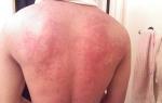
Catarrhal phenomena on the oral mucosa quickly turn into ulcers. The process most often begins on the gums, and then moves to other parts of the mucous membrane. Gums become swollen, hyperemic mi, sharply painful and bleed when touched. The epithelium of the gingival margin and interdental papillae becomes cloudy and then necrotic. As a result, the gingival margin looks as if cut off, with uneven notches, its surface is covered with an easily removable grayish-yellow coating. Subsequently, the affected gingival margin is not fully restored and remains deformed. Most often and to a greater extent, the area at the lower eighth teeth is affected, where necrosis with alveolar process quickly spreads to the buccal mucosa and the retromolar region, causing in some cases trismus and pain when swallowing. Ulcers on the mucous membrane of the cheeks can reach large sizes(up to 5–6 cm in diameter) and depth (Fig. 11. 1 5). Their edges are uneven, soft. The bottom is covered with a thick grayish-greenish necrotic coating, which has a putrid fetid odor, after the removal of which the bleeding bottom of the ulcer is exposed. There is no compaction in the area of the base and edges of the ulcer. The mucous membrane around the ulcer is edematous, hyperemic. If there are local traumatic factors in the oral cavity (roots of destroyed teeth, sharp edges of teeth, prostheses), other parts of the oral mucosa (hard, soft palate, arches, tonsils, tongue) may also be affected. Ulcers in the oral cavity can be single or multiple, various sizes and depth. When the ulcer is localized on the hard palate, necrosis of all layers of the mucous membrane develops quite quickly and the bone is exposed. Isolated pharyngeal lesion(Simanovsky's angina - Plaut - Vincent), as a rule, is unilateral, in the practice of a dentist it is rare. 2–3 weeks after the onset of necrotizing ulcerative stomatitis, the process usually resolves with complete epithelialization of the ulcerative surfaces.
Rice. 11.15. Acute ulcerative necrotic stomatitis of Vincent. An extensive ulcer of the buccal mucosa along the line of teeth closure in the area of large molars, covered with necrotic plaque.
In rare cases, when treatment is not carried out or it is ineffective, a chronic form of necrotizing ulcerative stomatitis of Vincent develops, in which general symptoms missing. Patients are concerned about constant bleeding and soreness of the gums, as well as bad breath. The clinical picture is blurred, the gum is stagnant-hyperemic, edematous, its ulcerated edge is often compacted, necrotic areas are located mainly in the interdental spaces and can be seen during a cursory examination. With careful examination and probing of the gingival margin, exposed bone tissue is determined. Affected areas are present only in some teeth. Lymph nodes (submandibular, submental) in patients with chronic ulcerative necrotic stomatitis of Vincent are compacted, slightly painful. With a disease duration of 4–8 months, the lymph nodes acquire a cartilage-like consistency.

Rice. 11.16. Histological picture in Vincent's ulcerative necrotic stomatitis. Area of tissue necrosis in the superficial layers (1), maturing granulation tissue (2), edema and small cell infiltration (3).
When pathohistological examination of areas of the ulcerated edges of the gums, two zones are revealed: superficial - necrotic and deep - inflammatory (Fig. 11.16). In the surface layers of the necrotic gum tissue, a rich and diverse microflora (cocci, bacilli, fusobacteria, spirochetes, etc.) is revealed. Fusobacteria and spirochetes sharply predominate in the deeper layers (Fig. 11.17). Deep layers connective tissue inflamed: they are edematous, the vessels are dilated. In the perivascular inflammatory infiltrate, shaped elements blood. In the same zone of inflammation inside intact tissue, only spirochetes that have penetrated between epithelial cells are found.
The cytological picture of scrapings from the ulcerative surfaces of the mucous membrane in patients with Vincent's ulcerative necrotic stomatitis corresponds to a nonspecific inflammatory process. At the beginning of the disease, an abundance of structureless masses is determined, a sharp predominance of neutrophils, mainly in a state of decay, and erythrocytes (due to severe bleeding). In the second period of the disease, when healing begins, along with decayed neutrophils, full-fledged phagocytic cells and many macrophages appear. During the period of epithelialization, layers of young epithelial cells appear, the number of fusobacteria and spirochetes decreases.

Rice. 11.17, Acute ulcerative necrotic stomatitis. Fusospirochetosis. Scraping from an ulcer. Fusobacteria and spirochetes (1), neutrophils in the stage of deep decay (2). Cytogram. x 700.
In the chronic course of Vincent's ulcerative necrotic stomatitis, the relative number of fusobacteria and spirochetes decreases and the number of cocci increases, but fusospirochetes still prevail.
Vincent's ulcerative necrotic stomatitis is diagnosed on the basis of characteristic clinical picture and detection of an abundance of spindle-shaped fusobacteria and spirochetes in scrapings from the surface of ulcers.
Differential Diagnosis. Ulcerative necrotic stomatitis of Vincent must be differentiated primarily from ulcerative lesions with blood diseases (leukemia, agranulocytosis, infectious mononucleosis). Along with clinical differences (pallor of the oral mucosa, the presence of hemorrhages, leukemic infiltrates, severe and prolonged bleeding of the gums in blood diseases), the detected changes in the peripheral blood in leukemia, agranulocytosis, and infectious mononucleosis are of decisive importance in this case.
Vincent's ulcerative necrotic stomatitis is differentiated from allergic stomatitis on the basis of anamnesis data, features of clinical manifestations and results of bacterioscopic examination.
An ulcerative necrotic process in the oral cavity, similar to Vincent's stomatitis, can occur with mercury intoxication. If contact with mercury is detected, a urine test for mercury content is performed.
It should be remembered that ulcerative-necrotic lesions of the oral mucosa can complicate the course specific infections(syphilis, HIV infection) or malignant tumors (cancer, sarcoma). To avoid diagnostic errors in these cases, you should carefully collect an anamnesis, take into account not only local, but also general clinical manifestations diseases and conduct the necessary laboratory tests (clinical blood test, Wasserman serological tests or for the detection of HIV infection, cytological, bacterioscopic, pathohistological, etc.).
Treatment. Volume medical measures with ulcerative necrotic stomatitis, Vincent is determined mainly by the severity of its course. Since the disease often occurs in young people, most of them practically healthy, local treatment is of decisive importance. The earlier and more thoroughly the treatment of the oral cavity is carried out, the faster the patient's condition improves. High-quality processing of the oral mucosa, removal of traumatic factors are decisive in the further course and outcome of the disease.
Local treatment consists in removing local traumatic factors, necrotic tissues, affecting the microflora and stimulating the processes of regeneration of the oral mucosa. Treatment of the oral cavity should begin with anesthesia. For this purpose, application and injection anesthesia with solutions of pyromecaine, trimecaine, lidocaine is used. After that delete everything mechanical stimuli: grind sharp edges of teeth and prostheses, remove tartar and plaque. Carious cavities should be treated with antiseptic solutions. The removal of decayed teeth should be postponed until the ulcers are epithelialized, since the removal of teeth in the infected oral cavity of a patient with ulcerative necrotic stomatitis is fraught with serious complications (alveolitis, periostitis, abscess, phlegmon). Purification of ulcerative surfaces from necrotic tissues is carried out using proteolytic enzymes: trypsin, chymotrypsin, lysoamidase, deoxyribonuclease.
The entire oral cavity is treated with warm solutions of antiseptics (0.5% hydrogen peroxide solution, 1% chloramine solution, 0.06% chlorhexidine solution, 0.5% aethonium solution, potassium permanganate solution 1:5000, etc.). Gingival pockets, the surface of the ulcer, interdental spaces and undercapillary spaces are best washed with a jet under pressure. On the first visit, the entire oral cavity should be treated. Further processing is carried out daily. At home, the patient is prescribed warm antiseptic rinses: 3% hydrogen peroxide solution (2 tablespoons per glass of water), 0.25% chloramine solution, potassium permanganate solution (1:5000), 0.06% chlorhexidine solution or other antiseptic drugs every 3-4 hours
With a mild course of ulcerative necrotic stomatitis, Vincent is enough local treatment. In more severe cases, it is necessary to general therapy. For the purpose of antimicrobial action, metronidazole (trichopolum, flagyl, klion) is administered orally at a dose of 0.25 g 2 times a day for 7-10 days. Metronidazole is also used for local treatment (a liquid aqueous slurry is applied to ulcerative surfaces after removal of necrotic tissue). In severe cases, broad-spectrum antibiotics are sometimes used (bicillin-3, 300,000 IU once every 3 days or 600,000 IU once every 6 days; erythromycin, oletethrin, oxytetracycline in daily dose 800,000-1,000,000 IU for 5-10 days). For any degree of severity, it is recommended to prescribe B vitamins, ascorutin (0.1 g orally 2 times a day, for 10-14 days).
With proper treatment, improvement in the condition of patients with Vincent's ulcerative necrotic stomatitis occurs within 24-48 hours, pain decreases or disappears, patients can eat, sleep. Edema and hyperemia of the oral mucosa decrease, epithelialization of ulcers begins, which, when mild degree disease and a satisfactory condition of the oral cavity is completed by the 3-6th day. In an unsanitized oral cavity, epithelialization of ulcerative surfaces proceeds more slowly. After the improvement of the general condition of the patient and the disappearance of acute inflammatory phenomena it is necessary to carry out a thorough sanitation of the oral cavity with the removal of tartar, teeth roots, to carry out treatment carious teeth, periodontal disease.
Relapses of necrotizing ulcerative stomatitis can occur if foci remain in the oral cavity chronic infection(periodontal pockets, hoods over incompletely erupted third molars), or traumatic factors (overhanging fillings, carious cavities, roots of destroyed teeth, tartar, low-quality prostheses, etc.). The reason for recurrence may be unsatisfactory hygienic condition of the oral cavity.
Treatment of symptomatic ulcerative necrotic stomatitis with blood diseases allergic conditions, mercury intoxication is mainly in the general treatment of the underlying disease that causes these changes.
Forecast. With timely and proper treatment, the prognosis is favorable. Epithelialization of ulcerative surfaces in an acute process occurs after 3-6 days, in a chronic process it is somewhat slower. In an unsanitized oral cavity in the presence of many traumatic factors and untimely or improper treatment omission (retraction) or deformation of the gums, resorption of the bone tissue of the alveolar process may occur. These changes contribute to the further progression of periodontitis.
Patients who have had Vincent's stomatitis are subject to active observation during the year. The first examination is carried out after 1-2 months, subsequent - after 6 months.
Prevention. It consists in observing oral hygiene, regular sanitation, full and timely treatment of infectious and other diseases that lead to a decrease in immunity.
From the book Skin and Venereal Diseases author Oleg Leonidovich IvanovChronic ulcerative-vegetative pyoderma Chronic ulcerative-vegetative pyoderma is a mixed strepto-staphylococcal chronic form of deep pyoderma.
From the book Dog Treatment: A Veterinarian's Handbook author Nika Germanovna Arkadieva-Berlin From the book Blue iodine - and the disease will go away author Nina Anatolyevna BashkirtsevaStomatitis Stomatitis is a variety of diseases of the oral mucosa. The cause of stomatitis can be various microtraumas - chemical, thermal or physical. The first is the effect of acids and alkalis, the second is the effect hot food
From the book Kidney Diseases and Bladder author Julia PopovaNecrotizing nephrosis Necrotic nephrosis or necronephrosis of the kidneys is an acute shockogenic, infectious or toxic lesion of the kidneys, accompanied by impaired blood supply, ischemia of the kidneys, followed by necrosis of the epithelium of the renal tubules and the development of acute
From the book Dentistry author D. N. Orlov21. Catarrhal stomatitis and ulcerative stomatitis Catarrhal stomatitis is the most common lesion of the oral mucosa; develops mainly in case of non-compliance with hygiene measures, lack of oral care, which leads to
From the book Therapist. Folk ways. author Nikolay Ivanovich MaznevStomatitis With insufficient dental care, soft deposits on the teeth turn into tartar, which causes inflammation of gingiva. Recipes * In case of stomatitis, rinse your mouth and throat with lime-colored infusion. Pour 1 glass cold water 1 st. l. lime color, insist
From the book Homeopathic Handbook author Sergei Alexandrovich NikitinStomatitis Stomatitis with salivation; gum sensation; bad breath - acidum
From book Sauerkraut- health and beauty recipes author Liniza Zhuvanovna ZhalpanovaUlcerative angina Ulcerative angina usually lasts 6-8 days. If the general condition of the body at the time of the disease is unsatisfactory, more time will be required to treat the ailment. The diagnosis is made after examining a swab taken from the patient's throat, in
From the book Hydrogen Peroxide for Your Illness author Liniza Zhuvanovna ZhalpanovaStomatitis Stomatitis is an inflammation of the oral mucosa. The cause of the disease is some infectious diseases - such as diphtheria, measles, syphilis, tuberculosis, etc.; diseases of the blood and skin: leukemia, anemia, lichen planus, etc. The patient feels a general
From the book Calendula, aloe and badan thick-leaved - healers for all diseases author Yu. N. NikolaevStomatitis Stomatitis is an inflammation of the oral mucosa. The cause of the disease is some infectious diseases - such as diphtheria, measles, syphilis, tuberculosis, blood and skin diseases: leukemia, anemia, lichen planus, etc. The patient feels a general
From the book Home Directory of Diseases author Ya. V. Vasilyeva (ed.) From book Apple vinegar, hydrogen peroxide, alcohol tinctures in the treatment and cleansing of the body author Yu. N. NikolaevUlcerative membranous angina It is characterized by yellow- white coating on the tonsils, sometimes on the inside of the cheeks and back wall throats. If it is removed, then ulcers are found under the plaque, an unpleasant odor is felt from the mouth. Body temperature rises to 38 °C. Generally
From book home doctor on the windowsill. From all diseases author Yulia Nikolaevna NikolaevaStomatitis Stomatitis is an inflammation of the oral mucosa. The cause of the disease is some infectious diseases, such as diphtheria, measles, syphilis, tuberculosis, etc.; blood and skin diseases - leukemia, anemia, lichen planus, etc. The patient complains of general
From the book Healing Soda author Nikolai Illarionovich DanikovStomatitis Stomatitis is an inflammation of the oral mucosa. The cause of stomatitis can be infectious diseases, such as pulmonary tuberculosis, diphtheria, syphilis, blood and skin diseases. With stomatitis, the patient feels weakness, drowsiness,
From the author's bookStomatitis In the treatment of stomatitis, products prepared from fresh or canned aloe juice are used. It can also be mixed with decoctions and infusions of other medicinal plants. Recipe 1 Aloe leaves are washed, peeled and chewed for 3-5
From the author's bookStomatitis Stomatitis - very unpleasant disease Everyone gets sick, especially children. At first, small sores in the mouth and white plaque cause simply uncomfortable sensations, but further, if no measures are taken and stomatitis is not cured at an early stage,
Ulcerative necrotic stomatitis of Vincent - an infectious alternative-inflammatory disease of the mucous membraneoral cavity, which occurs against the background of a decrease in rebody activity and adverse conditions in the oral cavity. Other names for the disease: ulcerative stomatitis, fusospirochetal stomatitis, Vincent's stomatitis, "Trench" mouth. One of the first to reveal the infectious naturedo disease French bacteriologist Vincent(1895).
The causative agents of the disease are fusobacteria{ Bad. fusiformis) and spirochetes(Borrelia vin- centi). Under the influence of a number of unfavorable local and general factors, the number of fusobacteria and spirochetes, which are normally saprophytes (resident flora) of the oral cavity, increases sharply, which leads to ulcerative-necrotic lesions of the gums and oral mucosa. Vincent's stomatitis often develops after stress, illness, hypothermia, surgical interventions, can complicate traumatic, trophic, cancerous ulcers; be a symptom of blood diseases, radiation injuries; intoxication with salts of heavy metals, HIV infection. Patients with Vincent's stomatitis have an unsatisfactory hygienic condition of the oral cavity, dental deposits, sharp edges of carious teeth, deep-set artificial crowns, pericoronitis of the third molars of the lower jaw, and inflammatory diseases of periodontal tissues.
Allocate mild, moderate and severe forms, as well as acute and chronic process. According to localization, Vincent's gingivitis, Vincent's gingivostomatitis (stomatitis) and Vincent's angina are distinguished. The severity of the signs of intoxication depends on the severity of the process: general weakness, headache, fever, aching joints and muscles, sleep and appetite disorders, and a tendency to faint.
On the mucous membrane of the oral cavity, on a hyperemic and edematous base, there is one or more ulcers covered with a gray coating. Most often, ulcers are localized in the retromolar space, on the mucous membrane of the cheeks along the line of closing of the teeth, on the lateral surfaces of the tongue. The gum, when involved in the pathological process, is hyperemic, edematous, bleeds when touched. Interdental papillae with signs of necrosis. Pungent putrid odor from the mouth. Possible trismus. Regional lymph nodes are enlarged and painful
DIAGNOSIS OF NUCLEAR-NECROTIC STOMATITIS OF VINCANT
|
Examination order |
Identified symptoms |
Pathogenetic substantiation of symptoms |
||||
|
Survey | ||||||
|
complaints | ||||||
|
Pain, burning of the mucous membrane |
Irritation of nerve endings by toxins, |
|||||
|
mouth, aggravated by taking pi- |
mediators of inflammation, chemical and me- |
|||||
|
cabbage soup, conversation |
chanic stimuli. Violation of the integrity of the oral mucosa, gums due to a hyperergic reaction with the release of substances leading to limited necrosis |
|||||
|
Weakness, feeling unwell |
General intoxication decay products of non- |
|||||
|
headache, fever |
crotic areas of the mucous membrane |
|||||
|
body, tendency to faint, sleep disturbance |
mouth and microorganism toxins |
|||||
|
Putrid odor from the mouth. Increased salivation. Limited mouth opening |
Activation anaerobic infection |
|||||
|
chronic |
Painful ulcers on the mucous membrane |
The process of alteration is joined by proli- |
||||
|
on the forehead with a dense white coating |
ferrative inflammation, in which there is a multiplication of connective tissue cells, mainly lymphocytes, plasma cells, fibroblasts. Then a cell-rich granulation tissue is formed |
|||||
|
Bleeding gums Putrid breath |
Expansion of capillaries and venules due to exposure to inflammatory mediators Activation of anaerobic flora |
|||||
|
Anamnesis the onset of the disease efficiencyearliertreatment |
Acute. The disease is not prone to self-healing. The onset of the disease can be associated with exposure common factors: ARVI, tonsillitis, influenza, viral diseases, hypothermia, malnutrition; local factors: poor hygiene mouth, multiple caries, peri-coronitis, chronic mechanical trauma, inflammatory processes in the periodontium No effect |
Decreased body resistance; microcirculation disorder Incorrect choice medicines. Late visit to the doctor |
||||
|
Age |
Mostly persons aged 17-30 years |
Unfavorable local factors contribute to the occurrence of the disease |
||||
|
Past and associated diseases |
SARS, tonsillitis, influenza, viral diseases, hypothermia |
Decreased resistance of the body and oral mucosa as a result of impaired microcirculation, nervous trophism, phagocytic activity of cellular elements |
||||
|
Material and householdconditions |
Malnutrition, violation of the regime of work and rest, overload |
Helps reduce immunity |
||||
|
Inspection visual inspection |
Facial skin is pale In some patients, facial asymmetry due to soft tissue edema Regional lymph nodes are enlarged, soft, slightly painful, not soldered to surrounding tissues |
The result of intoxication In the focus of inflammation, the outflow of blood and lymph is difficult, which causes their release into the tissue; edema develops Barrier response of lymphoid tissue to infection from lesions in the oral cavity |
||||
|
Oral examination hygienic condition dental conditionranks |
Abundant soft plaque, supragingival and subgingival calculus Sharp edges of teeth, fillings, multiple caries, deep-set crowns pericoronitis |
Tartar prevents the normal desquamation of the surface layers of the epithelium, closes the entrance to the gum pockets and thus creates favorable conditions for the development of anaerobic infections. Chronic trauma of the mucous membrane of the gums leads to a decrease in the resistance of the mucous membrane, favorable conditions arise for the activation of spirochetes and fusobacteria Promotes the accumulation of microflora (cocci, rods, leptotrichia, etc.) and the activation of spirochetes and fusobacteria |
||||
|
mucosal examination |
At the site of injury, one or more |
Serous inflammation occurs |
||||
|
shell cavity |
Most often, ulcers are localized on the |
isolation of biologically active substances |
||||
|
viscous membrane of the cheeks along the line of closure |
substances leading to limited |
|||||
|
teeth, on the lateral surfaces of the tongue, in re- |
necrosis of the oral mucosa. Vospa- |
|||||
|
tromolar area. Ulcers are soft |
litelny hyperemia in a zone of action |
|||||
|
jagged edges, thick necrotic on- |
pathogenic factor is limited to the focus |
|||||
|
years of grayish-green color, after the removal of which a loose, heavily bleeding bottom is visible. There is no compaction at the base and around the ulcer. The surrounding tissues are edematous, sharply hyperemic |
defeat |
|||||
|
Additional | ||||||
|
research methods | ||||||
|
general analysis blood |
Without changes. Shift formula to the left |
There is no intoxication. Elevated ESR |
||||
|
(leukocytosis, elevated ESR) |
The result of a shift in the protein fractions of the blood towards coarse proteins. Leukocytosis - the result of stimulation of leukopoiesis in an infectious disease |
|||||
|
cytological |
Picture of nonspecific inflammation. IN |
The presence of red blood cells is the result |
||||
|
method |
the onset of the disease are marked by a sharp |
bleeding of the ulcerative surface. |
||||
|
the predominance of neutrophils in the state |
Granulocytes carry out phagocytic |
|||||
|
decay, an increase in phagocytic neutrophils, lymphocytes and histiocytes. Later, along with decayed neutrophils, full-fledged phagocytic neutrophils and many histiocytes appear. When epithelization begins, there are layers of young epithelial cells |
new function |
|||||
|
bacterioscopic |
In the surface layers, an abundance of spirochetes |
Fusobacteria and spirochetes inhibit |
||||
|
sky method |
(Borrelia vincenti), spindle-shaped |
growth of other oral microflora and |
||||
|
check (Bact. fusiformis) and regular microflo- |
may represent an almost pure culture |
|||||
|
mouth cavity. In the deep layers there is an almost pure culture of fusobacteria and spirochetes. During the healing period of ulcers, the number of spirochetes and fusobacteria decreases | ||||||
|
serological |
Diagnosis of specific diseases | |||||
|
blood test | ||||||
|
Specialist consultations |
According to indications |
Leading role in the development of the disease |
||||
|
sheets |
has a decrease in the reactive forces of the body (immunity) - the result of previous or concomitant diseases |
|||||
DIFFERENTIAL DIAGNOSIS OF NUCLEAR-NECROTIC STOMATITIS OF VINCANT
|
Disease |
Are common Clinical signs |
Features |
||
|
Blood diseases | ||||
|
acute leukemia |
Young age of patients (up to 30 |
nic picture is determined by hemorrhagic |
||
|
years). Weakness, loss of appetite, |
ii, hyperplastic, anemic and in- |
|||
|
malaise. Pale skin |
ication syndromes. Hemorrhagic |
|||
|
covers. Ulcerative necrotic |
1rom manifests itself in the form of petechiae, ecchymo- |
|||
|
gingivostomatitis. Regional lim- |
hematomas on the skin and mucous membranes |
|||
|
phatic nodes are enlarged, painful |
and mouth. Severe bleeding in the mouth |
|||
|
nenn, soft to the touch, not soldered to the surrounding tissues |
s, hemorrhages on the mucous membrane along the line of teeth |
|||
|
chronic leukemiaagranulocytosis |
Weakness, fatigue, loss of appetite. Ulcerative-necrotic lesions of the oral mucosa. Regional lymph nodes are enlarged, painful, soft to the touch, not soldered to surrounding tissues Weakness, malaise. Paleness of the skin. Ulcerative-necrotic processes of the oral mucosa. Regional lymph nodes are enlarged, painful, soft to the touch, not soldered to surrounding tissues |
Spontaneous bleeding. Hyperplastic processes in the palate, gums, back of the tongue. Pain in intact teeth and jaws, in bones. Blood test: in the leukogram, the predominance of undifferentiated blood cells; the number of erythrocytes is 1-1.5 million in 1 mm 3. The total number of leukocytes at acute leukemia ranges from leukopenic figures to 200,000-300,000 in 1 mm 3 of blood or more. On bacterioscopic examination, the absence of pronounced fusospirochetosis It is determined in persons aged 30-60 years. Pain in bones, joints, neuralgic pain. Hemorrhages on the skin and mucous membranes. Post-extraction bleeding. Blood picture: at the beginning of the disease, slight leukocytosis, an increase in basophils, then the number of leukocytes, eosinophils, and basophils increases. The number of mature granulocytes drops sharply. Anemia progresses Absence inflammatory response tissue surrounding foci of necrosis. Blood test: leukopenia, neutropenia up to the complete disappearance of granulocytes. Anemia and thrombocytopenia. Absence of mature neutrophils. On bacterioscopic examination, the absence of pronounced fusospirochetosis | ||
|
Hard chancre (ulcerative form) |
Ulcer on the mucous membrane of the mouth. Regional lymph nodes are enlarged, painful, soft to the touch, not soldered to surrounding tissues |
Prolonged existence of a painless ulcer with dense edges and base. In scrapings from an ulcer, they find pale treponema. Regional lymph nodes are enlarged, compacted (scleradenitis). Wasserman's reaction is positive after 3 weeks. after hard chancre | ||
|
traumatic ulcer |
painful ulcer on the oral mucosa, pain when eating. Regional lymph nodes are enlarged, painful |
The ulcer is located at the site of chronic injury, can exist for a long time, its base is infiltrated. Bacterioscopic examination of the usual microflora of the oral cavity, accompanying nonspecific inflammation (cocci, rods, leptotrichia). Solitary fusobacteria and spirochetes. Putrid odor from the mouth is uncharacteristic. Elimination of the traumatic factor, as a rule, leads to healing of the ulcer in 5-6 days | ||
|
Decaying malignant tumor (cancer, sarcoma) |
An ulcer on the mucous membrane of the oral cavity can be located at the site of the traumatic factor (sharp edges of the teeth, etc.). Regional lymph nodes are enlarged, painful |
Mostly in older people. Long-term existence (up to several months) of the ulcer, lack of a tendency to heal after the injury has been eliminated, reduction in soreness, thickening of the edges and base. Lymph nodes are soldered to the surrounding tissues. A cancerous ulcer is not always associated with mechanical trauma. Cytologically, conglomerates of atypical cells with their characteristic cellular and nuclear polymorphism are determined. Bacterioscopic examination of the usual microflora of the oral cavity | ||
|
Trophic ulcer |
An ulcer on the oral mucosa, pain during eating of varying severity, possibly associated with traumatic factors |
An ulcer with a sluggish, prolonged course without a tendency to epithelization even after the elimination of the traumatic factor. It is observed in patients with cardiovascular and cardiopulmonary insufficiency II-III degree. The ulcer is covered with fibrinous plaque, slightly painful, the surrounding mucous membrane is slightly inflamed. Epithelialization is possible only in the treatment of a general disease | ||
TREATMENT OF NUCLEAR-NECROTIC STOMATITIS OF VINCANT
|
Stages |
Facilities |
Purpose of use |
Mechanism of action |
|
treatment |
treatment |
drugs |
drugs |
|
Treatment |
in the clinic | ||
|
Anesthesia | |||
|
injection |
1-2% lidocaine solution, |
For painless |
Blockade of sodium channels in the cell |
|
(with generalization |
4% solution of articaine, 0.5- |
removal of supragingival |
accurate nerve membrane, as a result |
|
bathroom process) |
1% mepivacaine solution |
tartar, |
whereby depolarization does not occur |
|
necrotic masses |
membrane and along the axon |
||
|
from the surface of ulcers and |
nerve impulse passes |
||
|
application |
1% dicaine solution, 10% |
Block sensitive windows |
|
|
lidocaine solution, 10% pyromecaine solution, xylonor spray |
chani nerve fibers |
||
|
cleansing |
proteolytic enzymes |
Lysis and removal of non- |
With local action, splitting |
|
necrotic |
you: trypsin, chymotrypsin, |
crotic masses |
yut necrotic tissue, |
|
surfaces |
chymopsin, lidase (as |
liquefy viscous secrets, exsu- |
|
|
applications for 10 min. to areas of necrosis) | |||
|
Antibacterial |
Antiseptics (rinses, |
Eliminate or donkey |
Hydrogen peroxide has an |
|
therapy |
irrigation, applications, ro- |
beat action sec- |
tiseptic action due to |
|
bath tubs). start |
rich microflora |
fission of atomic oxygen. |
|
|
to damaged |
Bactericidal action on micro- |
||
|
hydrogen peroxide and then |
mucous membrane |
roflora. Cleansing, deodorizing |
|
|
drugs are used in any |
rubbing action. Denaturation |
||
|
fight sequence: 1% |
squirrel bacterial cell. Lizo- |
||
|
hydrogen peroxide solution |
cym - enzyme preparation, yav- |
||
|
0.25% chloramine solution, |
lyatsya natural factor en- |
||
|
potassium permanganate solution 1:5000, 0.06% chlorhexidine solution, furacilin solution 1:1000, lysozyme (2 teaspoons per glass of water), calendula tincture (1 teaspoon per glass of water), ethacridine lactate solution 1 :1000 |
tibacterial protection |
|
Sanguiritrin 1% line- |
To fight anae- |
Has antimicrobial activity |
|
|
ment, 0.2% alcohol solution |
robust microflo- |
with respect to grampolo- |
|
|
creative, 1% water solution |
swarm, as well as for |
resident and gram-negative |
|
|
healing of ulcers |
bacteria. Acts on yeast-like fungi and Trichomonas. It also has anticholinesterase activity. |
||
|
Metronidazole in the form of app- |
To suppress pa- |
Suppresses 90% of anaerobic infections |
|
|
lications on lesions |
togenic microflo- |
tion, easily penetrates into bacterial |
|
|
cell, forms highly toxic substances that destroy DNA |
|||
|
elimination |
Treatment of carious polos |
Elimination of foci |
Elimination of the action of annoying |
|
traumatic |
steak concentrated |
infections |
factors and exclusion of patho- |
|
factors |
solutions of antiseptics, followed by the imposition of temporary fillings, grinding of the sharp edges of the teeth, removal of supragingival tartar |
gene influence of microflora |
|
|
Stimulation |
Solcoseryl, vitamins A E, tezana liniment, sodium mefenaminate, Kalanchoe juice, aloe |
Stimulation of reparative processes |
Improvement of metabolism and trophic tissues. Stimulation of regeneration processes |
|
processes | |||
|
regeneration | |||
|
(with a beginner- | |||
|
Xia epithelialization, |
Vitamins and their analogues: |
To speed up the epi- |
Complex compound of sodium |
|
the absence of necro- |
0.5-1% aqueous solution |
telization for pain |
salts of ascorbic acid. Day- |
|
tic raid) |
galascorbin |
shi ulcerative time- |
the effect of the drug is associated with the presence |
|
ascorbic acid (about 20%) and the astringent action of sodium gallate. Gallic acid and its salts to some extent have the properties of a vitamin R |
|||
|
Sanitation of the cavity |
Professional hygiene- | ||
|
mouth during |
on the mouth. Treatment | ||
|
recovery |
caries and its complications, removal of decayed teeth. Treatment of periodontal diseases. Prosthetics |
Ulcerative necrotic stomatitis Vincent(stomatitis ulceronecroticans Vincenti) is an infectious disease caused by the fusiform bacillus and Vincent's spirochete (Borrelia). In the world literature, it is described under the following names: ulcerative stomatitis, ulcerative-necrotic stomatitis, ulcerative-membranous stomatitis, fusospirochetal stomatitis, "trench mouth", angina Botkin - Simanovsky - Plaut - Vincent, etc.
When the gums are affected, the disease is defined as Vincent's gingivitis, with simultaneous damage to the gums and other parts of the mucous membrane - stomatitis, with involvement of the palatine tonsils - Vincent's angina.
What provokes Vincent's Ulcerative necrotic stomatitis:
The causative agents of necrotizing ulcerative gingivostomatitis Vincent belong to the resident flora of the oral cavity and are found in a small amount normally in all people with teeth, especially in the gingival groove. With poor care and unsanitized oral cavity, especially with periodontitis, their number increases dramatically.
Fusobacteria and Borrelia Vincent belong to opportunistic pathogens. The decisive role in the occurrence of the disease is played, as a rule, by a decrease in the body's resistance to infections. It occurs especially often with general cooling, due to a general illness, overwork, stress, malnutrition (for example, in wartime).
A predisposing factor is also a violation of the integrity of the mucous membrane, which creates conditions for the invasion of microorganisms. This happens with injuries, more often chronic, for example, with sharp edges of teeth, with difficult eruption of third molars. Breakthrough of the epithelial barrier also occurs in periodontitis. Ulcerative necrotic stomatitis of Vincent occurs more often with careless hygiene care behind the oral cavity against the background of previously existing inflammatory processes of the gums, with the deposition of supra- and subgingival calculus, which prevents the normal process of epithelium desquamation, irritates the gums and, by closing the entrance to periodontal pockets, creates favorable conditions for the development of anaerobic infection.
Ulcerative necrotic stomatitis of Vincent may occur as a complication viral infections(influenza, herpetic stomatitis), erosive allergic stomatitis, exudative erythema multiforme, some severe common diseases - leukemia, agranulocytosis, infectious mononucleosis, joins in case of poisoning with salts of heavy metals, scorbute. Cancer ulcers and syphilides in the mouth are sometimes also complicated by fusospirochetosis.
Symptoms of Ulcerative necrotic stomatitis Vincent:
By the nature of the flow diseases distinguish acute, subacute, chronic ulcerative necrotic stomatitis and relapse.
According to the severity of the course- light, medium and heavy forms.
At the onset of the disease there is weakness, headache, body temperature rises, aching joints. Disturbed by bleeding gums, burning sensation and dryness of the mucous membrane. This period can last from several hours to several days, depending on the form of the course of the disease.
As you progress stomatitis, general weakness increases, body temperature rises, headache intensifies, and working capacity decreases.
The pain in the oral cavity increases sharply from the slightest touch, the tongue is inactive during a conversation. Eating and brushing your teeth become almost impossible. Increased salivation, there is a strong putrid smell from the mouth. With the localization of the inflammatory process in the area of the wisdom tooth, there is a limited opening of the mouth (trismus).
More often ulceration of the mucous membrane begins with the gums, from areas where local irritating factors are present: tartar, decayed teeth, dental crowns that injure the gums. Gradually, ulceration spreads to neighboring areas of the mucous membrane.
With ulcerative necrotic stomatitis gums swollen, loosened, reddened, sharply painful, bleed from a light touch. Initially, necrosis affects the tops of the interdental papillae, and then spreads to the entire gum. Over time, the gum becomes covered with necrotic masses of white-gray, gray-brown or gray color.
For light form ulcerative necrotic stomatitis is characterized by a limited spread of the process. More often, only the tops of the interdental papillae in a certain group of teeth are necrotic. General well-being does not change significantly. Working ability, as a rule, is not broken.
In case of severe Vincent's stomatitis body temperature rises to 38.5-40°C. General well-being deteriorates sharply.
Ulceration extends to a significant area of the mucous membrane, the depth of the ulcer can reach muscle tissue, tendons, bones. With this course of the disease, osteomyelitis (melting of the bone) of the affected area of the jaw bone develops.
When spread ulcerative necrotic focus on the palate and tonsils, stomatitis is called Simanovsky-Plaut-Vincent's angina.
Acute ulcerative necrotic stomatitis at insufficient treatment may recur and become chronic. This transition is more often observed against the background of chronic somatic pathology, and also, with an unsanitized oral cavity.
Predominantly young people (17-30 years old), mostly men, are ill. The disease begins more often in autumn and spring, the maximum of new cases occurs in October - December.
There are pains in the oral cavity, especially when eating, bleeding gums, increased salivation, putrid breath, general weakness. The patient is usually pale due to severe intoxication. Regional lymph nodes are enlarged, compacted and painful on palpation, retain mobility.
The process, as a rule, begins on the gum and manifests itself in the form of foci of necrosis of the gingival margin and gingival papillae. Then necrosis can move to other parts of the mucosa. The region of the lower third molars suffers most and most of all, where necrosis quickly spreads to the buccal mucosa and the retromolar region, often causing trismus and pain when swallowing. In some cases, inflammation leads to a pronounced asymmetry of the face due to swelling of the surrounding tissues. In more severe cases, necrotic lesions occur on the lateral surfaces and back of the tongue, on the hard and soft palate. They have soft uneven edges, a thick, fetid, necrotic greyish-green coating, after the removal of which a loose, heavily bleeding bottom is visible. Surrounding tissues are edematous, sharply hyperemic. There are no seals at the base and around the ulcers.
On the hard palate, the process quickly leads to necrosis of all layers of the mucous membrane and exposure of the bone. Isolated lesions of the pharynx (Vincent's angina), as a rule, are unilateral and are rare in dentistry.
The general condition of the patient during an acute process, as a rule, worsens: the body temperature in the first 2–3 days rises to 37.5–38 ° C, but may remain normal, headache worries. bad dream, difficulty in eating, intoxication weaken the patient. There is a tendency to fainting. In the hemogram, pronounced changes may be absent, but often there is a slight leukocytosis, a shift of the formula to the left, moderate increase in ESR; in severe cases - toxigenic granularity of neutrophils.
The chronic form of this disease usually develops with careless treatment or its absence, but it can also occur initially, without a previous acute process.
Diagnosis of Ulcerative necrotic stomatitis Vincent:
Diagnosis Vincent's angina is put on the basis of the clinical picture and the detection of fusospirillary symbiosis.
Examination of biopsy specimens from the edges of ulcers reveals two zones: superficial - necrotic and deep - inflammatory. In the superficial layers of necrosis, the flora is abundant and varied (cocci, bacilli, fusobacteria, spirochetes, etc.), in the deeper layer adjacent to living tissues, fusospirochetes sharply predominate. The underlying tissues are in a state of acute inflammation. Inside living tissue, only spirochetes are found.
The cytological picture of scrapings from ulcers in Vincent's ulcerative necrotic stomatitis corresponds to a nonspecific inflammatory process.
Differential Diagnosis. The first step is to rule out HIV infection. In addition, Vincent's ulcerative necrotic stomatitis is differentiated from ulcerative lesions in blood diseases (leukemia, agranulocytosis, infectious mononucleosis), mercury poisoning, scurbut. In necrotic ulcers in these diseases, fusospirochetes are also found in large quantities. Rashes with secondary syphilis in the oral cavity can be secondarily complicated by Vincent's stomatitis. To avoid possible errors, in all cases ulcerative gingivitis and stomatitis, you should carefully collect an anamnesis, taking into account not only local, but also general clinical manifestations, do a general clinical blood test, an HIV test, a Wasserman reaction, and if contact with mercury is detected, a urine test for mercury content. As mentioned above, fusospirochetosis can complicate the course of other ulcers (eg, decaying cancer of the oral mucosa). Therefore, great importance in differential diagnosis acquires a cytological method of research.
Treatment of Ulcerative necrotic stomatitis Vincent:
An important condition successful treatment is a thorough sanitation of the oral cavity. After anesthesia, the decay of necrotic tissues and dental deposits are removed. Rapid cure is facilitated by the use of broad-spectrum antibiotics. Frequent (4-5 times a day) rinsing is necessary antiseptic solutions(0.05 - 0.1% chlorhexidine solution, 1 - 2% hydrogen peroxide solution). A good effect is achieved by using Trichopolum 0.5 g 2 times a day for 5 to 7 days. To suppress microbial sensitization, antihistamine therapy (fenkarol, tavegil or suprastin) is carried out. Vitamin C is also prescribed (up to 1.5 g per day). Locally, enzyme preparations are used for the lysis of necrotic plaque, and then keratoplastic ointments (solcoseryl, methyluracil). When the process is localized in the pharynx, an interferon solution is instilled. The prognosis for timely and adequate treatment is favorable. IN acute stage associated with difficult eruption of third molars, surgical manipulations are not recommended. With proper treatment, epithelialization occurs in an acute process after 3-6 days, in a chronic process it is somewhat slower. Severe cases ulcerative necrotic stomatitis of Vincent, especially recurrent, when treatment is carried out untimely or incorrectly, lead to irreversible changes: bone resorption, subsidence (retraction) of the gums, severe forms periodontitis. After treatment, the gingival papillae may disappear, conditions are created for food retention, progression of periodontitis. In other parts of the mucous membrane, except for the gums, tissues are usually restored during healing, only after deep and extensive ulcers do scars remain.
Treatment of symptomatic ulcerative necrotic stomatitis in blood diseases, scurbut, mercury poisoning consists mainly in the general effect on the body.
Persons who have had Vincent's stomatitis should be on dispensary observation at least 1 year, and the first examination is carried out after 1 - 2 months.
Forecast of angina and stomatitis Vincent favorable, although in some cases, in the absence of rational therapy, the disease is delayed and can last several months. Relapses are possible.