Muscles and fascia of the neck. A - sternohyoid muscle B - coracoid process of the scapula
Function: during contraction, it stretches the cervical fascia, lowers the hyoid bone.
Innervation: cervical loop, C I -C II.
Rice. 63. Places of origin and attachment of muscles on the hyoid bone: 1 - large horn of the hyoid bone; 2 - stylohyoid ligament; 3 - small horn of the hyoid bone; 4 - chin-hyoid muscle; 5 - the body of the hyoid bone; 6 - maxillofacial muscle; 7 - sternohyoid muscle; 8 - scapular-hyoid muscle; 9 - fibrous plate of the digastric muscle; 10 - stylohyoid muscle; 11 - thyroid muscle; 13 - middle constrictor of the pharynx; 14 - cartilaginous muscle.
2. Sternohyoid muscle(t. sternohyoideus) starts from the inner surface of the handle of the sternum, the sternal end of the clavicle, goes up; attached to the lower edge of the body of the hyoid bone (see Fig. 61).
Function: lowers the hyoid bone. Innervation: cervical loop, C I -C III.
3. Sternothyroid muscle(T. sternothyroideus) starts from the inner surface of the handle of the sternum and cartilage of the 1st rib; attached to the plate of the thyroid cartilage (see Fig. 61).
Function: pulls the thyroid cartilage, and with it the entire larynx down.
Innervation: cervical loop, C I -C III.
4. Thyrohyoid muscle(T. thyrohyoidus) starts from the plate of the thyroid cartilage; attached to the hyoid bone (see Fig. 61).
Function: lowers the hyoid bone, with a fixed hyoid bone raises the larynx. Innervation: cervical loop, C I -C III.
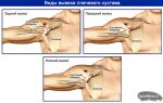
With fractures of the lower jaw, the function of each of the masticatory muscles is realized differently than normal, and depends on how the fracture line passes. So, if the fracture line passes through the neck of the lower jaw, then the superficial part of the masticatory muscle and the medial pterygoid muscle displace the lower jaw (without condylar processes) anteriorly and upwards.
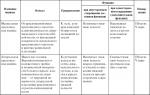
Table 10 Muscles involved in the movements of the lower jaw
Continuation of the table. 10

The end of the table. 10

Typical features of masticatory muscles
The surface layer of the masticatory muscle in brachycephaly and chameprosopic face shape is usually wide and low, muscle fibers diverge downward (Fig. 85); with dolichocephaly and leptoprosopic face shape, it is long and narrow, muscle fibers run parallel. The intermediate layer of this muscle in dolichocephaly and leptoprosopia protrudes more from under the posterior edge of the superficial layer than in brachycephaly and chameprosopia.
The temporal muscle with a dolichocephalic form of the skull is low and long, and with a brachycephalic it is high and short (see Fig. 85).
Both heads of the lateral pterygoid muscle with a brachycephalic skull shape are short and wide, with a narrow gap between them, with a dolichocephalic one they are long and narrow, with a wide gap between them (Fig. 86).
The medial pterygoid muscle with a dolichocephalic shape of the skull and a leptoprosopic face shape is long and narrow, and with brachycephaly and chameprosopia it is low and wide (Fig. 87).
The shape of the pterygoid and chewing muscles is determined by the shape of the mandibular branch and the infratemporal fossa, but at the same time it corresponds to the structure of the bone components of the temporomandibular joint. This relationship is especially clearly reflected in the external structure of the lateral pterygoid muscle. When opening the mouth (lowering the lower jaw) and moving the lower jaw forward in people with a brachycephalic skull, the head of the joint is shifted to the top of the flat articular tubercle, i.e. the articular path slightly deviates from the horizontal plane. This movement of the head of the jaw is provided by the lower head of the lateral pterygoid muscle, which lies almost horizontally. In the dolichocephalic form of the skull, the articular head slides down the steep and high slope of the articular tubercle rather than horizontally. This movement is provided by the lower head of the lateral pterygoid muscle, the beginning of which is lower on the high lateral plate of the pterygoid process, and the muscle pulls the head of the jaw down rather than forward.
It originates from the styloid process of the temporal bone.
Not far from the point of attachment, the muscle is perforated by the intermediate tendon of the digastric muscle.
Function:
Raises the hyoid bone and pulls it back.
3. Maxillofacial muscle (m. Mylohyoideus).
It starts on the inner surface of the lower jaw from the maxillo-hyoid line.
The posterior fibers are attached to the body of the hyoid bone, the anterior and middle fibers are connected to the same fibers of the opposite side, forming a tendon suture along the midline, which stretches from the middle of the chin to the hyoid bone.
Both maxillohyoid muscles are involved in the formation of the floor of the mouth and are called the diaphragm of the mouth (diaphragma oris).
Functions:
4. Geniohyoid muscle (m. Geniohyoideus).
It starts from the mental spine of the lower jaw.
Attached to the body of the hyoid bone.
Functions:
When the jaws are closed, the muscle raises the hyoid bone along with the larynx;
With a strengthened hyoid bone, it lowers the lower jaw (chewing, swallowing, speech).
Hyoid muscles:
1. Scapular-hyoid muscle (m. omohyoideus) - has two bellies: upper and lower, which are connected approximately at the middle of the length of the muscle by a tendon bridge.
The upper abdomen (venter superior) starts from the lower edge of the body of the hyoid bone outward from the attachment of the sternohyoid muscle, in the middle of the length of the muscle lies behind the sternocleidomastoid muscle, where it passes into the tendon bridge, which fuses with the sheath of the neurovascular bundle of the neck.
The lower abdomen (venter inferior) starts from the tendon bridge, is attached to the upper edge of the scapula.
Functions:
Pulls the vagina of the neurovascular bundle of the neck and prevents compression of blood vessels and nerves;
With a strengthened scapula, it pulls the hyoid bone backwards and downwards;
2. Sternohyoid muscle (m. Sternohyoideus)
It starts from the back surface of the handle of the sternum, the sternal end of the clavicle.
Attached to the lower edge of the body of the hyoid bone.
Between the medial edges of both muscles there is a space in which the plates of the fascia grow together and form a white line of the neck.
Function: pulls the hyoid bone down.
3. Sternothyroid muscle (m. sternothyroideus).
It starts on the back surface of the handle of the sternum and cartilage of the 1st rib.
Attaches to the oblique line of the thyroid cartilage of the larynx, lies in front of the trachea and thyroid gland.
Function: pulls the larynx down.
4. Thyrohyoid muscle (m. thyrohyoideus) is, as it were, a continuation of the sternothyroid muscle.
It starts from the oblique line of the thyroid cartilage.
Attached to the body and the greater horn of the hyoid bone.
Function: brings the hyoid bone closer to the larynx.
Deep neck muscles:
Lateral group:
1. Anterior scalene muscle (m. Scalenus anterior).
It starts from the anterior tubercles of the transverse processes of C3-C6.
It is attached to the tubercle of the anterior scalene muscle on the 1st rib.
2. Middle scalene muscle (m. scalenusmedius).
From the transverse processes of C2-C7 to the 1st rib behind the groove of the subclavian artery.
3. rearstaircasemuscle(m. scalenus posterior).
From the posterior tubercles C4-C6 to the upper edge and outer surface 2 ribs.
Functions of the scalene muscles:
With a strengthened cervical spine, 1 and 2 ribs are raised, the chest cavity is expanded;
With a strengthened chest, the cervical spine is bent forward;
With unilateral contraction, the spine bends to the side.
Medial muscle group:
1. Long muscle of the head (m. longus capitis).
From the anterior tubercles of the transverse processes of C3-C6 to the inferior surface of the basilar part of the occipital bone.
Function: tilts the head and cervical part of the spine forward.
2. Long muscle of the neck (m. longus colli) - lies on the anterior surface of the bodies of all cervical vertebrae and the three upper thoracic vertebrae. Has three parts:
Vertical part: from the front surface of the C5-Th3 bodies to the C2-C4 bodies.
Lower oblique: from the anterior surface of the bodies of the first three thoracic vertebrae to the anterior tubercles C4-C5 of the cervical vertebrae.
Upper oblique: from the anterior tubercles of the transverse processes C3-C5 to the anterior tubercle of the 1st cervical vertebra.
Functions:
Flexes the cervical part of the spine;
With unilateral contraction, tilts the neck to the side.
Great attention in the methodology of Revitonics is given to the muscles of the neck. And it's not that our neck is almost always open and treacherously betrays our age. In addition to the aesthetic reason, there is also a physiological one. The muscles of the neck keep the head in balance, are involved in the movement of the head and neck, as well as in the processes of swallowing and pronouncing sounds.
Moreover, the youthfulness of the face directly depends on the state of the neck muscles. Without the correct statics of the neck and the formation of a good posture, it is pointless to “repair” our face. You can read more about biomechanical cause-and-effect relationships in the section "Imbalance of the muscles of the neck".


Figure 1. Muscles of the neck (front and profile)
Despite the wide variety of neck muscles (more than 20), we will list the main muscles that are involved in the Revitonika fitness complex:
trapezius muscle
The trapezius muscle is a thin and wide plate that almost completely occupies the back of the neck. If you connect the muscles on both sides, a trapezoid is formed, which is why it has such a strange name. Each muscle individually has the form of a triangle, the base of which goes straight along the vertebrae, and the apex is directed towards the shoulder blade. It has three parts.

Figure 2. Trapezius muscle
The upper part originates from the cervical vertebrae and at the base of the skull, on the occiput. If you lower your chin and bend your head, these tubercles and the place of attachment of the muscle can be very well felt. The muscle then goes obliquely down, creating a curve between the shoulders. The middle part starts from the upper thoracic vertebrae and goes horizontally, and the lower part - from the lower thoracic vertebrae and goes obliquely upwards.
All three parts of the trapezius muscle are connected and attached to one of the processes of the scapula, the outer edge of the clavicle and humerus. When the upper or lower part is reduced, the shoulder girdle and scapula rise or fall. With the contraction of only the middle part, the scapula moves towards the spine. If all three parts are reduced at once, both shoulder blades approach each other.
When the shoulder girdle and shoulder blades are fixed, contracting, this muscle turns the head in the opposite direction from itself. And when both muscles contract, the head unbends somewhat, giving a proud posture and keeping the neck in good shape.
Sternocleidomastoid muscle
The sternocleidomastoid muscle is one of the most superficially located muscles. It received such an unusual name because it has a special structure and is attached in three different places. Unlike most muscles in our body, it has two heads. The first head is attached at the upper edge of the sternum, so it was called the sternum. The second - clavicular - is attached to the sternal edge of the clavicle. Then these two heads are connected, forming one abdomen, and attached to the process of the temporal bone, called the mastoid. If you turn your head to the left, then under the skin you can completely feel how this muscle goes on the right, leading your hand from the tubercle immediately behind the auricle to the sternum. Similarly, you can find this muscle on the left by turning your head to the right.

Figure 3. Sternocleidomastoid muscle
Like the muscle itself, its functions are unusual and varied. If only the left muscle contracts, the head tilts to the left, while the face turns to the right and rises slightly. And vice versa, if only the right muscle contracts. When both muscles are contracted at once, the head is in a vertical position, not without reason it is also called the “head holder”. Also, if both muscles contract more strongly, the head falls back and the face rises. If the head is fixed, then this muscle will help with breathing, raising the chest.
Subcutaneous muscle of the neck
The subcutaneous muscle of the neck belongs to the superficial muscles and has the appearance of a wide plate. It is located just under the skin and is special in that it starts on the chest at the level of the second rib, and ends at the lower edge of the jaw. And although it is very thin and you cannot touch it, even if it is tense, it performs a very important function.

Figure 4. Hypodermic neck muscle
When it tenses, the skin above it moves forward, thereby helping to expand the veins that pass immediately under the muscle. This is necessary for heavy physical exertion, because. thereby increasing the outflow of blood from the head, preventing the overflow of the brain with blood.
Maxillofacial muscle
The maxillohyoid muscle begins on the inner surface of the lower jaw and runs horizontally. In the midline, it intertwines with the same muscle of the opposite side, after which they are both attached to a special bone called the hyoid. Such an interesting move is necessary to create the bottom of the mouth. Due to this attachment, this muscle is involved in lowering the lower jaw. And if the lower jaw is motionless, this muscle raises the hyoid bone, thereby participating in the swallowing of food. Also, this muscle, when in good shape, does not allow the chin to “sag”, strengthening it.

Figure 5. Maxillohyoid muscle
Digastric
The peculiarity of the digastric muscle is clear from its name. It has two abdomens: anterior and posterior.
The posterior abdomen at one end, like the sternocleidomastoid muscle, is attached to the mastoid process of the temporal bone (a tubercle behind the auricle), and at the other to the hyoid bone, meeting there with the anterior abdomen.

Figure 6. Digastric muscle.
The anterior, in turn, is directed somewhat perpendicularly and is attached to the inner surface of the lower jaw in a special fossa, named after her bigastric. This arrangement of the muscle forms a kind of niche (submandibular triangle), in which the submandibular salivary gland is located, which is necessary for digestion.
Like the maxillohyoid, the digastric muscle lowers the lower jaw, opening the mouth, or participates in the process of swallowing if the jaw is motionless.
Awl-hyoid muscle
The stylohyoid muscle consists of a single thin belly that attaches to the temporal bone and runs behind the mandible, next to the lateral surface of the tongue. Its lower end splits and covers the digastric muscle on both sides, then attaching to the hyoid bone. Thus, by contracting, it raises the hyoid bone and participates in swallowing food, like the two previous muscles.

Figure 7. Stylohyoid muscle.
Sternohyoid muscle
The sternohyoid muscle originates at the posterior surface of the sternum and runs vertically up the anterior surface of the trachea and larynx, joining the inferior edge of the hyoid bone.
The right and left sternohyoid muscles run parallel to each other without touching, so there is a small narrow triangular space between their inner edges.

Figure 8. Sternohyoid muscle
The sternohyoid muscle lowers the hyoid bone down, acting in opposition to the digastric, maxillohyoid and stylohyoid muscles and holding it in place and thereby allowing these muscles to lower the lower jaw.
| Sternohyoid muscle | |
|---|---|
| lat. Musculus sternohyoideus | |
| |
|
|
The sternohyoid muscle is highlighted in red |
|
| Start | sternum |
| attachment | hyoid bone |
| blood supply | a.a. thyroidea inferior, cervicalis superficialis |
| innervation | cervical nerves (C I -C III) |
| Function | pulls down the hyoid bone |
| Catalogs | |

Function
Pulls the hyoid bone down, swallowing.
Notes
Sternocleidomastoid muscleThe sternocleidomastoid muscle (lat. Musculus sternocleidomastoideus) is located behind the subcutaneous muscle of the neck. It is a rather thick and slightly flattened cord that crosses the neck region in an oblique spiral fashion from the mastoid process to the sternoclavicular joint. The muscle begins with two heads: lateral - from the sternal end of the clavicle and medial - from the anterior surface of the sternum handle.
Both legs are connected at an acute angle. The bundles of the medial pedicle are located more superficially. The resulting muscular abdomen goes up and behind and is attached to the mastoid process of the temporal bone and the superior nuchal line of the occipital bone.
Between the medial and lateral legs of the lat. m. sternocleidomastoidei, a small depression is formed - a small supraclavicular fossa (Latin fossa supraclavicularis minor), and between the medial legs of the left and right muscles, above the jugular notch of the sternum, is the jugular fossa.
Sternothyroid muscleThe sternothyroid muscle (lat. Musculus sternothyroideus) is flat, located behind the sternohyoid muscle. It starts from the back surface of the cartilage of the 1st rib and the handle of the sternum, goes up and attaches to an oblique line on the lateral surface of the thyroid cartilage of the larynx.
DigastricDigastric muscle (lat. m.digastricus) - in humans - a small paired muscle from the group of suprahyoid (suprahyoid) muscles, located under the lower jaw. It is called "bigastric" by the presence of two parts (abdomen) separated by a tendon. The anterior abdomen starts from the lower jaw in the chin region (attached to the digastric fossa of the lower jaw), the posterior in the region of the mastoid process of the temporal bone. Both bellies are attached to the hyoid bone.
From the tendon of the digastric muscle, a wide aponeurosis begins, attaching to the body and large horns of the hyoid bone (suprahyoid aponeurosis).
long head muscleThe long muscle of the head (lat. Musculus longus capitis) starts from the anterior tubercles of the III-VI cervical vertebrae, goes up and is attached to the lower surface of the basilar part of the occipital bone, posterior to the pharyngeal tubercle.
long neck muscleThe long muscle of the neck (lat. Musculus longus colli) occupies the anterolateral surface of the vertebral bodies - from the atlas to the III-IV thoracic vertebrae. The middle sections of the muscle are somewhat dilated. Muscle bundles have different lengths, so three parts are distinguished in the muscle:
the medial-vertical part starts from the vertebral bodies along the length from the V cervical to the III thoracic and, rising up and medially, is attached to the anterior surface of the bodies of the II-III cervical vertebrae and the anterior tubercle of the atlas;
the upper oblique part is directed from the transverse processes of the II-V cervical vertebrae to the body of the II cervical vertebra and the anterior tubercle of the atlas;
the lower oblique part starts from the bodies of the upper three thoracic vertebrae, goes up and laterally and attaches to the anterior tubercles of the transverse processes of the three lower cervical vertebrae.
Scalenus posteriorThe posterior scalenus muscle (lat. Musculus scalenus posterior) starts from the transverse processes of the 3rd, 4th, 5th and 6th cervical vertebrae, goes down behind the middle scalene muscle and is attached to the outer surface of the second rib.
CollarboneClavicle (Latin clavicula) - in human anatomy - a short tubular S-shaped bone from the upper limb girdle, connecting the scapula to the sternum and strengthening the shoulder girdle.
The Latin name - clavicula, "key", like the Russian name, is based on the peculiar movement of the bone around its axis at the moment of raising the shoulder, which resembles the movement of a key in a keyhole.
Clavicles are found in many tetrapods that use their forelimbs for grasping or brachiation; vestigial or absent in those tetrapods that use the forelimbs for support or running.
Scalene musclesScalene muscles (lat. Musculi scaleni) - neck muscles of the deep layer of the lateral (lateral) group. In most sources, 3 pairs are distinguished:
Scalenus anterior (musculus scaleni anterior)
Middle scalene muscle (musculus scaleni medius)
Scalenus posterior (musculus scaleni posterioir)
All scalene muscles start from the transverse processes of the cervical vertebrae and are attached to the 1st and 2nd ribs.
Scapulohyoid muscleThe scapular-hyoid muscle (Latin musculus omohyoideus) is a paired muscle of the anterior surface of the neck from the sublingual group. It has a long flattened shape, divided by a tendon into two bellies.
The name comes from the places of attachment: Greek. ωμος - shoulder, and "hyoideus" - hyoid bone.
Muscle that lifts the thyroid glandThe muscle that lifts the thyroid gland (lat. Musculus levator glandulae thyroideae) is a non-permanent thin muscle bundle that runs along the medial edge of the thyroid-hyoid muscle from the body of the hyoid bone or from the thyroid cartilage to the capsule of the thyroid gland (in the region of the isthmus of the lateral or pyramidal lobe).
This muscle bundle can be separated either from the thyroid-hyoid muscle, or from the cricoid, or from the lower constrictor of the pharynx.
The frequency of occurrence of the muscle varies from 6.4 to 60% of observations.
Human neck musclesThe muscles of the neck keep the head in balance, are involved in the movement of the head and neck, as well as in the processes of swallowing and pronouncing sounds.
Two groups of muscles are distinguished on the trunk and neck: own muscles and alien muscles.
The intrinsic muscles lie very deep, on the very bones of the axial skeleton, and by their contractions set in motion mainly the skeleton of the trunk and head. During the development of the embryo, the alien muscles appear on the body later, and therefore are located on the surface of its own muscles. Alien muscles differ from their own muscles in that they are mainly associated with the work of the upper limbs, although they are capable, under certain conditions, of setting the torso and head in motion. Own muscles are located in all areas of the body; alien muscles are located on the chest, back and neck.
The muscles located along the midline of the body have a longitudinal direction of the fibers, and those located on the side are oblique.
Scalenus minorThe smallest scalene muscle (lat. Musculus scalenus minimus) is unstable. It is located medially from the anterior scalene muscle. It starts from the transverse process of the III cervical vertebra and is attached on the inner edge of the I rib in front of the tubercle of the anterior scalene muscle (Latin tuberculum musculi scaleni anterioris) of the I rib and to the dome of the pleura.
Scalenus anteriorThe anterior scalene muscle (lat. Musculus scalenus anterior) starts from the anterior tubercles of the III-VI cervical vertebrae, goes down and forward and attaches to the tubercle of the anterior scalene muscle (lat. tuberculum musculi scaleni anterioris) I rib in front of the groove of the subclavian artery (lat. sulcus arteriae subclaviae).
Geniohyoid muscleThe geniohyoid muscle (lat. Musculus geniohyoideus) starts from the mental spine of the lower jaw, goes down and somewhat backward, is located above the maxillohyoid muscle and is attached to the anterior surface of the body of the hyoid bone.
Subcutaneous muscle of the neckThe subcutaneous muscle of the neck (lat. Platysma) in the form of a thin muscular plate is located under the skin of the neck, tightly growing together with it. The muscle bundles of this muscle, starting in the chest area at the level of the II rib, are directed upward and medially. At the edge of the lower jaw, the medial bundles of the muscle are intertwined with the bundles of the muscle of the same name on the opposite side and are attached to the edge of the lower jaw; laterally, the muscle bundles pass to the face, where the fascia of the parotid gland and the chewing gland are woven, reaching the corner of the mouth.
Scalenus mediausThe middle scalene muscle (lat. Musculus scalenus medius) starts from the posterior tubercles of the six lower cervical vertebrae, goes down behind the anterior scalene muscle and is attached to the upper surface of the 1st rib, behind the groove of the subclavian artery (lat. sulcus arteriae subclaviae). Above this groove, between the anterior and middle scalene muscles, there is a triangular gap in which the subclavian artery and nerve trunks of the brachial plexus pass.
Maxillofacial muscleThe maxillofacial muscle (lat. Musculus mylohyoideus) is flat, irregularly triangular in shape. It starts from the maxillo-hyoid line of the lower jaw. The muscle bundles are directed from top to bottom and somewhat back to front, and in the midline they meet with the bundles of the same name muscle of the opposite side, forming the suture of the maxillohyoid muscle.
The posterior muscle bundles are attached to the anterior surface of the body of the hyoid bone. Both maxillohyoid muscles are involved in the formation of the floor of the mouth and are called the diaphragm of the mouth.
Stylohyoid muscleThe stylohyoid muscle (lat. Musculus stylohyoideus) has a thin flattened abdomen that starts from the styloid process of the temporal bone, goes forward and down and is located along the anterior surface of the posterior belly of the digastric muscle. The distal end of the muscle splits and, covering the tendon of the digastric muscle with two legs, is attached to the body and the greater horn of the hyoid bone.
Thyrohyoid muscleThe thyroid-hyoid muscle (lat. Musculus thyrohyoideus) is, as it were, a continuation of the sternothyroid muscle. It starts from the oblique line of the thyroid cartilage, goes up and is attached along the edge of the large horn of the hyoid bone.