The ultrasound showed an enlarged adrenal gland. Alternative methods of research. Ultrasound of the adrenal glands reveals such pathologies
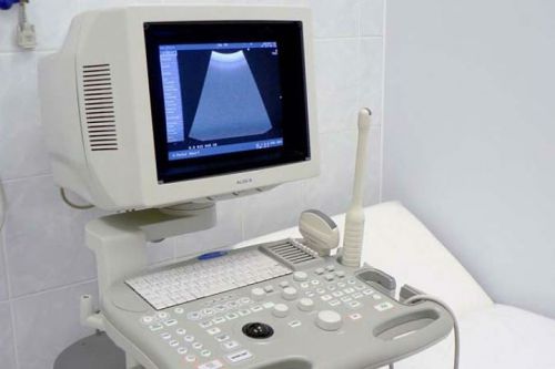
Ultrasound diagnostics is still one of the most common and familiar research methods. internal organs, which is used along with other, more modern methods.
Ultrasound of the kidneys and adrenal glands is a study that allows you to collect comprehensive information about the state of both the kidneys and the endocrine glands. It is with its help that specialists can identify at an early stage and stop the development of various malignant formations, as well as benign tumors, as well as all kinds of inflammation, disruption of the body, hematomas. This is the primary method of examining the patient, after which additional procedures are prescribed, unless, of course, the doctor considers that they are necessary.
Ultrasound diagnosis of the adrenal glands does not require almost any preparation on the part of the patient, you just need to know a few simple rules. Everything else is done by the doctor. After conducting a study, he studies the size and shape of the glands, which can tell about the development of the tumor process in the body and other unpleasant pathologies which are best identified as early as possible.
The adrenal glands, despite their small size, perform very important features, and in case of failures in their work, suffers general health. it endocrine glands responsible for metabolic processes, as well as various adaptation mechanisms that the body uses in adverse external conditions. Most often, these organs look "at the same time" when examining the kidneys, but sometimes a specialized study is also prescribed.
So, the patient was assigned an ultrasound of the adrenal glands. What is this procedure? That's what they call special study carried out using ultrasound. adrenal glands in human body produce certain hormones, and if for some reason the hormonal background suddenly changes, and there is also a malfunction of the excretory system, the doctor will first refer such a patient to an ultrasound diagnosis of the adrenal glands. Typically, this procedure requires the following indications:
- The patient complains about the sudden appearance of pigmentation on the skin.
- There is constant weakness, lethargy.
- It is completely incomprehensible why weight suddenly increases noticeably, obesity develops, and doctors cannot find any obvious reasons.
- A lot of stretch marks appear on the skin, also in the absence of noticeable causes.
- In the event that the patient is diagnosed with infertility.
- For unknown reasons, not identified by other doctors, blood pressure rises or falls markedly.
- If a specialist has a suspicion that a patient has an adrenal tumor.
- If adrenal injury is suspected or the person is injured abdominal cavity. Such cases should not be ignored, so as not to miss the development of harmful processes in the body.
- In cases where the patient has cysts or inflammatory phenomena in the glands.
- If hyperplasia is detected.
It should be noted that the ultrasound diagnostic procedure itself is not suitable for all people. The fact is that due to the deep location of these organs in the body (under the diaphragm itself), the device’s signal needs to penetrate not only through the skin, fat layer and muscles, but also through the intestines, the retroperitoneal layer of adipose tissue, in order to get to the very end adrenal glands. The signal at this point becomes weak. Therefore, in order for the study that doctors do to show the maximum amount of information, the patient must have a normal or even insufficient body weight, not have in the past abdominal operations(after all surgical interventions adhesions are left in the abdominal cavity, which are able to scatter ultrasound).
If the patient does not have the above contraindications, the doctor will be able to safely conduct an ultrasound of the adrenal glands, the preparation for which includes a decrease in gases in the intestines. It is difficult for people with increased body weight to be examined using ultrasound. That's why overweight body is a contraindication for the appointment of ultrasound. There are no other restrictions dictated by the patient's state of health.
Preparing for an ultrasound
So, what needs to be done so that the procedure is as successful as possible and gives the best top scores? The recommendations are very simple, and the doctor must explain to the patient all the details in order to avoid gross errors that can distort the course and results of the study. Preparation includes several important points:
- Three days before the scheduled ultrasound of the adrenal glands, the patient must adhere to lung nutrition menu, as clean as possible from slags. It is forbidden to eat any foods that increase the formation of gases, any fried or fatty foods. You will also have to remove meat from the diet, as well as any confectionery sweets, pastries, sweets. Any vegetables, fruits, a variety of nuts, legumes and all cereals are allowed, as well as varieties of bread with a rough composition (best with seeds or bran). Dried fruits are allowed herbal teas, honey, natural (preferably vegetable, not fruit) juices.
- Preparing for ultrasound diagnostics adrenal glands provides for the lightest dinner on the day preceding the procedure (no later than seven o'clock in the evening). Later, after it, you can’t eat anything anymore.
- Preparation includes taking laxatives to enhance bowel movements. It is best to take them in the evening, before the day on which the procedure is scheduled. It could be Castor oil, "Bisacodyl" or other drugs prescribed by your doctor.
- To prepare for an adrenal ultrasound the best way, you can’t have breakfast in the morning: the stomach should remain empty.
- Immediately before the ultrasound of the adrenal glands, the person who came to the study donates blood for hormones and visits an endocrinologist.
How is an ultrasound performed?
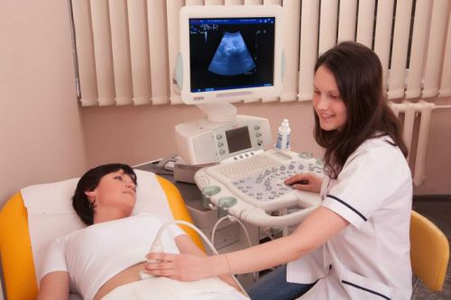
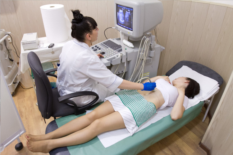
So, the preparation is over, the person comes to the study itself. The patient will just have to get up or lie down, expose the lower back and stomach: the doctor will apply a special gel on them. At the same time, it should be taken into account that the possibilities differ significantly for different adrenal glands: the left one is detected only in half of the cases, and the right one in 90 percent of studies. So the effectiveness of the study, unfortunately, cannot be called one hundred percent successful.
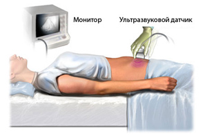
Conducting an ultrasound of the kidneys and adrenal glands, the specialist examines a special triangle formed by the right kidney, the right hepatic lobe and the inferior vena cava. Then the patient lies on his right side, and the doctor goes to the left adrenal gland. Sometimes it is possible to get to it only in those cases when the patient lies on his stomach or just stands straight. In this case, the patient must breathe deeply and calmly: in this way, the diaphragm is lowered, and then the ultrasound penetrates to the upper poles of the kidneys. The whole procedure takes only about fifteen minutes, it is completely painless and safe for health.
What can be detected with an ultrasound?
When everything is over, the patient asks the question: what does an ultrasound of the adrenal glands reveal? There are several options here. If the patient managed to prepare optimally, then the study will show whether the person has one of the four variants of diseases:
- Hyperplasia. This is a disease that can develop in a person even before birth. The tissues begin to grow, and as a result, the organs increase without changing their shape. This pathology is caused by toxicosis, which is caused by inflammatory processes in the mother's body functional disorders in her body, as well as receiving cortical medicines. The disease can manifest itself by the appearance of acne, early hair growth, late onset of menstruation, excessive pigmentation of the external genital organs.
- The procedure can also reveal hematomas, as well as various inflammatory processes that may develop due to closed injuries abdominal cavity.
- Indications are able to highlight the appearance of a cyst in the body - extremely unpleasant education with liquid content, which is difficult to diagnose and can cause a lot of trouble to the patient.
- The presence of benign formations (they are called adenomas) or malignant (their name is "sarcoma") tumors.
It should also be borne in mind that ultrasound of the adrenal glands can help the doctor in cases where the patient is diagnosed with problems such as narrowing of the ureters, prolapse and cysts of the kidneys, vascular inflammation, various abscesses, dystrophic changes, inflammatory processes, kidney stones.
It should be remembered that only a specialist, but not a patient, can fully evaluate and study all indications. So do not read your own card with horror, it is better to contact your doctor, who will tell you in detail about the results obtained and further actions.
How is the diagnosis and what to do after the ultrasound?
So, the ultrasound of the adrenal glands shows only a thin line on the screen instead of the adrenal glands. This means that everything is in order, and the endocrine glands are healthy, there is nothing more to worry about. It is worth preparing for trouble when organs are clearly visible on the monitor: enlarged adrenal glands indicate the presence of neoplasms.
After the procedure, the patient is sent with the results to the endocrinologist, who studies the information received, prescribes suitable treatment, for example, using hormonal drugs. If the patient is suspected of having a tumor, then after an ultrasound he is sent to needle biopsy, MRI (or CT) with contrast agent and row special analyzes blood. Thus, if the adrenal glands are sick, the ultrasound examination is only a preparation for further, more deep research, since the procedure itself does not provide all the information necessary for treatment.
Conclusion
Ultrasound of the adrenal glands is one of the main studies that are most often prescribed when it is necessary to examine a patient for a number of diseases. It remains quite informative, of high quality and affordable. Despite the fact that the effectiveness and efficiency of this study is not always one hundred percent, it is it that serves as the initial means of studying the state of health of organs and identifying various pathologies. Only after it, specialized, more accurate and in-depth studies, such as MRI, are prescribed.
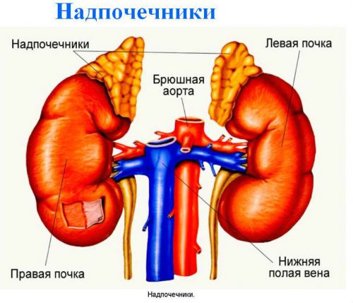
The adrenal glands are paired organs belonging to the endocrine system. They are responsible for the production of the most important hormones necessary for normal functioning body, namely: cortisol, adrenaline, norepinephrine, testosterone. If we talk about anatomy, then they are closely related to the kidneys and got their name due to the fact that they are located on upper parts kidneys. Ultrasound is helpful in diagnosing and treating many problems in both women and men.
Ultrasound of the adrenal glands shows the following parameters of the organs:
- location;
- condition;
- contours;
- dimensions.
In what cases is it necessary to carry out diagnostics?
An ultrasound examination is carried out with the aim of:
- diagnosis of diseases;
- estimates painful changes;
- control of the dynamics of the treatment.
Ultrasound can assess the need for additional methods diagnosis and determine the further specifics of therapeutic therapy.
Ultrasound of the kidneys and adrenal glands is a highly informative and absolutely painless procedure.
Indications for ultrasound examination are the following symptoms and diseases, namely:
- identification of suspected neoplasms;
- establishing true reasons arterial hypertension;
- determination of the reasons that led to the appearance of excess weight;
- the presence of hyperfunctional or hypofunctional activity of organs;
- identifying the causes of infertility;
- determining the causes of muscle weakness.
If the patient complains of the following symptoms, then he should definitely do an ultrasound:
- unreasonable weight gain;
- strong jumps in blood pressure;
- pain in the lower back or abdomen;
- seals.
Ultrasound will help identify:
- tumor;
- cysts;
- hematomas;
- cancerous lesions of the glands;
- inflammatory process.
Contraindications for ultrasound
Due to the safety and painlessness of the procedure, it has no significant limitations. However, there are some limiting restrictions, namely:
- Pregnancy period. If, nevertheless, there is a great need for diagnostics, then during this period ultra sound waves with the least effect on the fetus.
- Skin diseases. If there are inflammatory processes on the skin, then this will be an obstacle for the contact of the sensor with the patient's skin.
- Skin wounds.
It is believed that pregnant women should only have a routine examination if the risk to the woman exceeds possible risks for the development of the fetus, the specialist may decide on the need for an ultrasound.
Ultrasound helps to identify serious violations on the early stages
How to properly prepare?
Preparation for an ultrasound of the adrenal glands includes the following points:
- You should prepare in a few days, or rather, in three days, you need to follow a diet. It is necessary to exclude foods that cause flatulence from your diet, this includes carbonated drinks, beer, Rye bread, fatty, fried food. It is permissible to consume juices, tea, potatoes, cereals, vegetables and fruits.
- How to prepare for an adrenal ultrasound the night before? It is necessary to drink a laxative to get rid of excess slagging.
- It is best to carry out the procedure on an empty stomach or two to three hours after eating.
- As an additional preparation before an ultrasound, you can donate blood for hormones.
- Before the procedure, it is important to come to a consultation with an endocrinologist.
- With arterial hypertension, preparation consists in drinking two liters of natural water.
If one of the adrenal glands is not visualized, then the study should be repeated.
The adrenal glands play a vital role in the functioning of the body, and their violation can lead to such serious problems like infertility and obesity
If we talk about preparing for ultrasound of the kidneys and adrenal glands, then it expands somewhat. You can eat food eight to twelve hours before the examination. In addition, in order for the doctor to better examine the kidneys, there should be no gas formation in the intestines. To do this, after the last meal, you can take activated charcoal.
How to prepare for an ultrasound of the kidneys with medication?
Prepare for diagnosis as follows:
- First of all, microclysters are made or a glycerin suppository is placed. You can also use drugs such as Picolax or Guttolax.
- For two or three days they drink Smecta, Espumizan or Sorbeks.
- Each meal for several days should be accompanied by the use of Mezim or Pancreatin.

Do not eat cabbage before kidney ultrasound
Conducting research
First of all, the patient should lie on his back or on his stomach, it does not matter. The lumbar region and lower abdomen must be freed from clothing. Next, the doctor applies a gel to the site of the projection of the adrenal glands, with the help of which further research is possible.
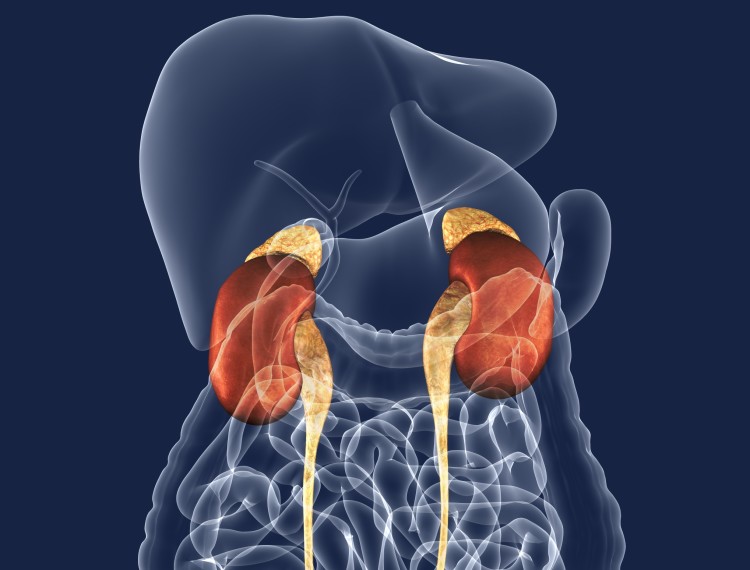
The probability that the doctor will see both adrenal glands is not as high as statistics show, the left adrenal gland is visualized only in half of the cases. Generally healthy adrenal glands are not visible, because if we talk about their structure, then they do not differ from the fiber of the retroperitoneal space. Normally, it will only be possible to see a section of their location.
For effective scanning, the patient is asked to take a breath. To view the left adrenal gland, the patient must lie on the right side, and to diagnose the right organ, respectively, on the left.
Healthy adrenal glands are not visualized
The ability to detect glands and make a diagnosis is possible only in a child or young people who have an asthenic body type.
Features of the study of children
Diagnosis in children is carried out in the following cases:
- suspicion of disorders of the kidneys or adrenal glands;
- the presence of an already formed pathology to clarify the degree of severity of the process, development possible complications and spread of affected foci;
- after laboratory research with a contradictory and ambiguous clinical picture;
- pain syndrome.
Preparing for an ultrasound of the kidneys
At normal condition The child is not pre-prepared. However, gas formation and overweight may hinder research. Just like adults, children should follow a three-day diet that eliminates foods that cause flatulence.
Preparing for an Adrenal Exam
Be sure to enter within three days diet food which includes avoiding animal products. The procedure must be carried out on an empty stomach. It is better to give the child a laxative before the study.
Ultrasonography
High frequency sound waves are used to produce images. This diagnostic procedure can be performed on pregnant women. Ultrasonography helps to examine not deeply located organs and tissues. During the procedure, the doctor may ask the patient to take a deep breath, hold their breath, or change position.
Ultrasonography is a harmless diagnostic procedure
With ultrasonography of the abdominal organs, you can not eat six hours before the study, you can only drink water. You can also safely take your usual medications. The procedure allows you to diagnose not only organs, but also blood vessels and lymph nodes.
So early and accurate diagnosis is the key to your health. You should not engage in self-diagnosis, but it is better to entrust your health to professionals. Ultrasound is painless, safe and available method research!
If adrenal dysfunction is suspected, ultrasound is almost always prescribed. Ultrasound of the adrenal glands is the primary research method, and allows you to get the maximum possible information about changes in the organ. The procedure does not require special preparation, but its implementation is somewhat difficult due to anatomical features location of the adrenal glands. Ultrasound is done with calm breathing. Changes in the size or shape of the glands may indicate a tumor process. Sometimes the deformation of nearby organs and a change in the number and pattern of the vessels of the adrenal glands are determined, this also indicates the presence pathological process. Ultrasound examination is relatively inexpensive and has good feedback patients and doctors.
The technical capabilities of ultrasound devices make it possible to determine only the location of the adrenal gland, but no other details are visualized due to the acoustic characteristics of the tissues of the adrenal glands. The fact is that on the echogram, iron is almost no different from the tissue of the retroperitoneal tissue that surrounds it. There must be good reasons for ordering an ultrasound examination. For example, violation hormonal background person or problem excretory system, since the adrenal gland is associated with the kidney, an ultrasound of the kidneys and adrenal glands is prescribed immediately.
Indications for research and preparation
The photo shows the adrenal glands
The most common reasons for referring a patient to an ultrasound of the adrenal glands are:
- The appearance of pigmentation
- Constant weakness
- Obesity
- female infertility
- Formation of stretch marks on the skin
- Increase or decrease in blood pressure
- Suspicion of tumors
- Weakness in the muscles
- Complaints indicating hyper- or hypofunction of the organ
- Injuries of the abdominal cavity, ultrasound of the adrenal glands in this case is mandatory
- cysts
- Hematomas
- Inflammatory processes in the gland
- Hyperplasia.
Preparation for ultrasound of the adrenal glands is simple, the patient is given recommendations that he must follow a few days before the study:
- 3 days before the procedure, you need to follow a light, slag-free diet. Exclude gas generating products, fatty and fried foods, as well as meat products. It is allowed to eat vegetables, legumes, fruits, nuts, cereals, seeds, wholemeal or bran bread. Confectionery products are not recommended, you can eat dried fruits and honey. From drinks, juices and herbal teas will be useful.
- Dinner before the day when the ultrasound is scheduled should be as light as possible. You can't eat anything after.
- In the evening of the same day, it is recommended to take a laxative for additional bowel cleansing.
- On the day of the study, you can’t have breakfast, they do an ultrasound of the adrenal glands on an empty stomach.
- Before the diagnosis itself, a blood test for hormones is done and referred for a consultation with an endocrinologist.
How is an ultrasound of the adrenal glands done?
The organs themselves are difficult to access for diagnosis, but the patient does not feel any discomfort. The patient during ultrasound of the adrenal glands can lie on his back, stomach or side, sometimes holding in a standing position is indicated. The skin at the site of the organ projection and the ultrasonic sensor are lubricated with a special gel, it is evenly distributed over the lumbar region. Ultrasound begins with the definition right kidney, inferior vena cava and right lobe of the liver. In the triangle between these organs is the adrenal gland.
Usually, the right adrenal gland is easier to see on ultrasound than the left. When the right one is discovered and studied, one starts looking for the left one. To do this, the doctor asks the patient to lie on his left side and do deep breath. In some cases, the left gland is available for ultrasound diagnosis from the left hypochondrium. The angle of the colon from the side of the spleen can cause difficulties during ultrasound of the adrenal glands if it contains gases.
For more information about the adrenal glands, see the video:
What can be determined during ultrasound diagnostics?
Hyperplasia of the organ is well defined by ultrasound of the adrenal glands. It is formed in the intrauterine period. Causes include maternal toxicity during pregnancy, functional impairment, and corticosteroid treatment. The disease is manifested by too early hair growth of the armpits and pubis at 4-5 years. As well as the appearance of a rough male voice in boys in early childhood, acne, early menses in girls and premature stunting in height.
In case of injuries, ultrasound of the adrenal glands is done to determine the inflammatory process and hematomas. Rarely, cysts of the adrenal glands are found. They are also laid down during embryonic development and for a long time not amenable to diagnosis. Cyst - what is it? This is a cavity filled with liquid. Most often, cysts are solitary.
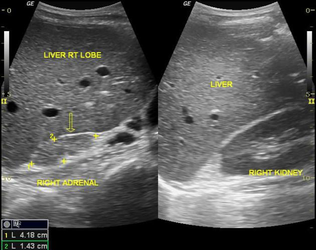
Malignant and benign tumors, usually adenomas and sarcomas are also well visualized by ultrasound of the adrenal glands. By structure healthy organs homogeneous, the capsules are not visible, the shape of the right adrenal gland is triangular, the left is lunate. Normal sizes adrenal glands: 2-7 cm long, 1.5-4 cm wide, 5-12 mm thick. If the adrenal glands are not visible on ultrasound, this is also the norm.
May 2, 2017 VrachIT'S IMPORTANT TO KNOW! Alexander Myasnikov in the program "About the Most Important": The only remedy for KIDNEY DISEASES that really helps almost immediately...
One of the most common methods for diagnosing the condition of internal organs is ultrasound. This procedure is carried out in relation to the adrenal glands. However, it cannot be called the most informative. This is due to the peculiarities of the location of this body. Nevertheless, ultrasound of the adrenal glands is often used to obtain primary information about the size of the gland, its structure.
When appointed
Because the adrenal glands are the organ that produces various hormones, then the presence of signs of hormonal imbalance may cause the need for their diagnosis. The first step is an ultrasound. The reason is the symptoms characteristic of hormonal disorders. In some cases, these manifestations develop slowly, in others suddenly and are very pronounced.
Indications
- General weakness for no apparent reason.
- Change in skin color.
- Obesity.
- Infertility in women.
- Stretch marks on the skin.
- Decreased potency in men.
- Cycle failures in women.
- Persistent hypertension, uncontrollable, or pressure spikes.
- Abdominal injuries.
Contraindications and restrictions
There are no absolute contraindications for ultrasound of the adrenal glands, since the radiation exposure during this procedure is minimal. However, doctors do not recommend such an examination during pregnancy, and damage is also a temporary limitation. skin in the sensor area. Adhesions formed after abdominal surgery can become an obstacle, as they scatter ultrasound.
Information in the diagnosis of an organ
What does an ultrasound of the adrenal glands show? If the organ was visualized, then its dimensions can be determined. The norm of the adrenal glands is 1-1.5 cm in length and 0.3-1.6 cm in width for the right, 1.5-2 cm in length and 0.8-1.5 cm in width for the left.
It is also normal that the left and right parts of the body have different sizes, and the left one is not always determined, which is possible in half the cases. If an increase in the size of the adrenal gland is detected, this indicates organ hyperplasia, the presence of an inflammatory process, hematomas, tumors or cysts.
Study preparation
In order for the survey to be successful, you need to prepare for it. Such preparation is due to the peculiarities of the location of the organ, which complicate the ultrasound of the glands. The adrenal glands are located at the very top of the kidneys and are located under the diaphragm deep in the space behind the peritoneum. It will not work to carry out a qualitative examination of the organ from the back, because the muscles, ribs and vertebral structures interfere. Therefore, visualization is carried out from the side of the abdomen, through the front wall.
However, in this examination, the ultrasound signal passes through several layers of different tissues before reaching the target. These are skin, subcutaneous fat, intestinal loops, retroperitoneal adipose tissue. Such a number of obstacles for ultrasound leads to the fact that it weakens. Therefore, to improve the diagnosis, it is necessary to ensure the maximum possible visualization. It is for this that preparation for the procedure is necessary.
In addition, surplus subcutaneous fat interfere with the normal conduct of the study, the best results are obtained in patients with a lean physique. It is important to ensure as little gas in the intestines as possible.
- Espumizan intake.
- Special diet a few days before the procedure.
- Taking a laxative in the evening on the eve of the day of the ultrasound.
- Dinner on the eve of the procedure should be the last meal.
- Fluid restriction on the day of the ultrasound.
The diet before the ultrasound is observed for three days, it should be light and slag-free. On diet days, it is recommended to take espumizan so that by the time the study is performed, there is no accumulation of gases in the intestines. The diet excludes the use of products that promote gas formation:
- everything fatty and fried;
- meat;
- legumes;
- bakery products;
- strong coffee;
- carbonated drinks;
Ultrasound procedure
Ultrasound examination of the adrenal glands is performed transabdominally, that is, through the anterior abdominal wall. The organ itself is not echogenously visualized, therefore, for examination, you need to find its location. The doctor begins the study on the right, he determines the right kidney, inferior vena cava and right side liver, since the right adrenal gland is located between these points.
It is scanned with the patient lying on his back or on his side during a deep breath. To study the left side of the organ, the doctor may ask the patient to change position. Left side organ is more accessible for ultrasound through the left hypochondrium.
Scanning is very easy. For him, the patient releases the lower part of the body from clothing, the skin on the abdomen in the projection of the adrenal glands is lubricated with a special gel, which provides better ultrasound conductivity. Next, the doctor guides the sensor of the device over the surface of the skin of the abdomen and examines the information received on the screen. The patient does not feel any discomfort during this examination, its duration is no more than 20 minutes. After the end of the procedure, the gel is wiped off with a napkin.
What the results mean
The specialist in ultrasound diagnostics takes notes in accordance with the received data. They are the basis for setting preliminary diagnosis endocrinologist.
If hyperplasia is detected, then tissue proliferation occurs. Such changes may lead to hormonal disorders. An increase in an organ can be a consequence of its inflammation, injury, or the formation of neoplasms. Any deviation from the norm is a reason for additional surveys. You will need a hormonal analysis, CT scan adrenal glands, as well as some other diagnostic procedures that will allow an accurate diagnosis to be made.
Despite the fact that ultrasound of the adrenal glands does not make it possible to obtain full information about the state of this body, it is widely used. The point is that the devices ultrasound research are available in almost all clinics and do this procedure accessible. Plus, it's completely safe. The possibility of making a preliminary diagnosis allows us to assume how serious the deviations are and to prescribe further examinations if necessary. Early detection adrenal dysfunction allows you to get help on time, avoid aggravation of the situation and the development of complications.
How to cure kidneys at home?

Edema face and legs PAIN in the lower back PERMANENT weakness and fast fatiguability, painful urination? If you have these symptoms, then there is a 95% chance of kidney disease.
If you care about your health, then read the opinion of a urologist with 24 years of experience. In his article, he talks about RENON DUO capsules. This is a fast-acting German kidney repair remedy that has been used all over the world for many years. The uniqueness of the drug is:
- Eliminates the cause of pain and brings the kidneys to their original state.
- German capsules eliminate pain already during the first course of use, and help to completely cure the disease.
- Missing side effects and no allergic reactions.
The adrenal glands are paired organs. endocrine system, producing a number of hormones necessary to ensure the vital activity of the body; are located on the upper parts of the kidneys and are closely related to them anatomically. The glands have different shape: left - lunar, and right - triangular.
In the structure of the adrenal gland, two layers can be distinguished: cortical and cerebral. In turn, the cortical layer has glomerular, fascicular and reticular zones. Each layer is characterized by the production of certain hormones. Hormones such as adrenaline, norepinephrine, testosterone, cortisol, aldosterone are secreted by the adrenal glands.
With the help of ultrasound, an analysis is made of the location, condition, contours and size of the adrenal glands. Ultrasound of the adrenal glands is a highly informative and completely painless procedure.
Reasons for an ultrasound
An ultrasound examination is carried out to diagnose diseases, abnormalities, pathologies, as well as to assess painful changes and their stages. The data obtained make it possible to establish a further examination scheme and the specifics of treatment. In the presence of the following symptoms and diseases, research is recommended:
- detection of suspected tumors;
- establishing the cause of hypertension;
- determining the cause of excessive weight gain;
- the presence of hypofunction or hyperfunction of the adrenal glands;
- identification of sources of infertility;
- determining the cause of muscle weakness.
Ultrasound of the adrenal glands is able to determine in the early stages such types of diseases as hyperplasia, different kinds inflammatory formations, hematomas, cysts.
 Despite their small size, the adrenal glands play a very important role in the human body. important role. A change in their work can lead to hypertension, obesity and even infertility.
 Hyperplasia- endocrine disorder. Characterized by hair covering the axillary and pubic areas in early age, late menstrual cycle, inhibition or growth arrest. To establish the presence or absence of hyperplasia is possible only by conducting an echographic study and using other types of diagnostics. Glandular hematomas occur in newborns due to trauma at birth.
Adrenal cysts happen rarely. The disease is formed during the period of embryonic development. The identification of such a cyst is a very long and complex process, since the difference between an adrenal cyst and a subcapsular cyst of the kidney is determined only at the time of ultrasound diagnosis.
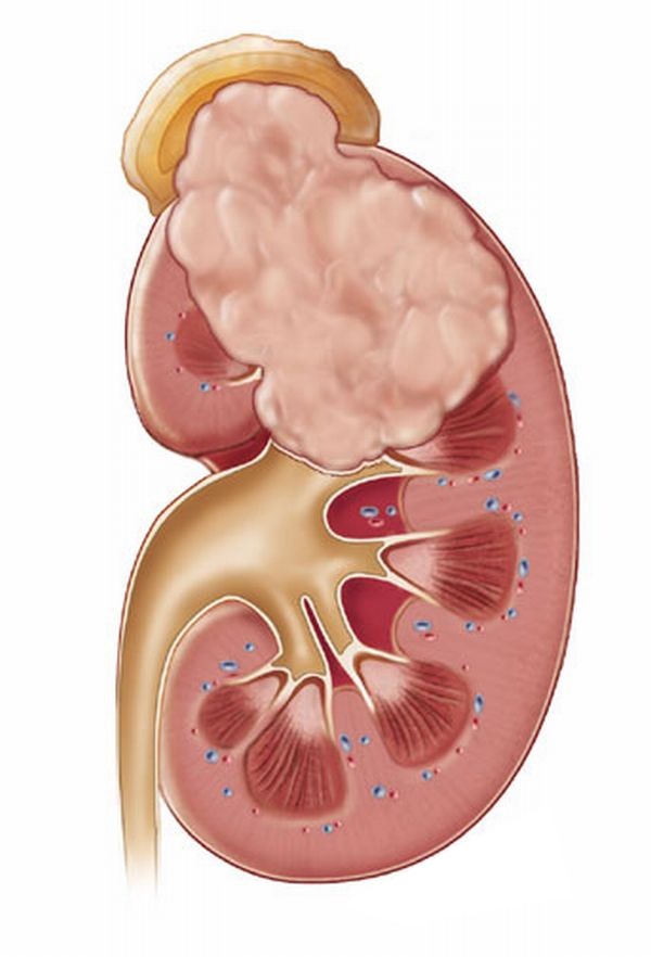
Tumors: the disease is diagnosed using ultrasound only after the tumor reaches a size of more than 2 cm. Often, the tumor causes significant harm to the body. Namely:
- if the tumor has reached 4-5 cm in size, then it is able to compress the organs located nearby and impede their functioning;
- some tumors can produce a significant amount of hormones that interfere with the work of the whole body;
- the probability that the tumor is benign is higher, however, the appearance malignant tumor is also possible. According to modern medical research a pattern can be traced that the larger the tumor, the more likely it is that the patient will be diagnosed with cancer.
 As a rule, it is possible to diagnose a tumor in the adrenal glands only when its size reaches 2 cm. This is due not so much to the shortcomings of diagnosis, but to the late handling of complaints - initial stages the tumor is simply not felt and can only be detected during a routine examination
 Benign formations- adenoma and aldosteroma - are formed on the adrenal cortex. These are homogeneous (homogeneous) formations on average 4-5 cm with clear edges and low echogenicity. If a tumor is found, a conclusion is made about focal formation in the projection of the adrenal glands. Obtaining a conclusion about the disease provides a basis for the preparation and conduct of further examinations.
Contraindications for ultrasound
There are no serious restrictions for performing an ultrasound of the adrenal glands, since the procedure is absolutely safe and painless. However, attention should be paid to the following deterrent indications:
- Pregnancy. Ultrasound during pregnancy is considered undesirable. If there is a need for a study, then it is carried out with the least influence of ultrasonic waves on the fetus.
- Skin diseases. Various skin inflammatory processes prevent the sensor from closely contacting the patient's skin.
- Damage to the skin in the form of wounds also does not allow the sensor to come into close contact with the skin.
 It is believed that during pregnancy, ultrasound, except for planned ones, is undesirable. However, if the risk outweighs the threat to the health of the fetus, then such a study is carried out
 Study preparation
Preparation for an ultrasound of the adrenal glands includes the following items:
- Preparation for the procedure requires the patient to follow a diet 3 days before the procedure. It is required to avoid eating foods that cause excessive gas formation: fatty and fried foods. The preparation also includes the rejection of drinks with gas, alcoholic beverages. Allowed the use of potatoes, cereals, pasta, vegetables, fruits. From drinks: juices and tea.
- In advance, you need to take a laxative to get rid of excess toxins. It is better to do this preparation option in the evening the day before the procedure.
- The study is done on an empty stomach, so a few hours before it is not recommended to eat.
- Additionally, before the study, it is possible to conduct hormonal analysis blood.
- Also, the preparation for the ultrasound of the adrenal glands includes a visit to the endocrinologist for a consultation.
Carrying out an ultrasound examination
To start the procedure, the subject must lie on his back or on his stomach. From time to time it is allowed that the patient maintains a “standing” position or lies on his side. lumbar region and Bottom part the abdomen is released from clothing in preparation for the examination. After that, the doctor applies a gel to the required area of the body in order to examine it using an ultrasound device.
The probability of identifying the adrenal glands from two sides is not always successful: the right adrenal gland appears in 90% during the study, and the left one only in 50%. During the examination, healthy adrenal glands are not visible, since they do not differ in structure from retroperitoneal tissue, therefore, it is only possible to identify the area where the adrenal glands are located.
When starting an ultrasound, the doctor finds with right side kidney, right lobe liver and inferior vena cava. In the area that is formed extreme points of these organs, the right adrenal gland is located. For a successful scan, the patient needs to take a deep breath. Then the subject must roll over to the right side so that the specialist continues the examination of the left adrenal gland. It is best to view the right organ from the front access, the left one is detected by examination from the side.
Adrenals that are not affected by various disorders are not visualized on the study. The ability to detect glands and conduct an examination is possible only in childhood or people of young years with an asthenic physique.
 Ultrasound of the adrenal glands is performed transabdominally - through the anterior abdominal wall. Since the organ itself is not echogenically visualized, the doctor simply finds the place where it is located and examines its structure for homogeneity, possible neoplasms
 Ultrasound of the adrenal glands can not always help in identifying their pathology. Selective angiography provides more opportunities for accurate diagnosis.
Dimensions of healthy adrenal glands
What does an ultrasound of the adrenal glands show? The study shows that the right adrenal gland is located in the space of the upper medial surface of the right kidney and vena cava from below. Its dimensions:
- length - 1-1.5 cm;
- width - 0.3-1.6 cm;
- height - 1-2 cm.
The left gland is placed in the zone of the left kidney and aorta. Dimensions of a healthy adrenal gland:
- length - 1.5-2.5 cm;
- width - 0.8-1.5 cm;
- height has approximately the same characteristics with length.


