Features of bladder cystography. Preparation for the procedure. How to make an x-ray of the urethra and bladder using a contrast agent
There are several ways to conduct research Bladder. One of them is ultrasound (ultrasound). This procedure is quite safe.
bladder ultrasound
How is an ultrasound of the bladder performed?
Ultrasound examination of the bladder helps to assess the volume of the bladder, urinary retention in a patient for one reason or another, the volume of residual urine in patients who have decreased urine production due to any reason. Bladder ultrasound can also be used to find out and evaluate the structure of the bladder.
Bladder ultrasound is also used to assess bladder function in incontinence and retention. urinary tract.
How does the process of examining the bladder take place? Uses special ultrasound scanner which is battery powered. It consists of a portable unit and applied ultrasound. During the examination of the patient's abdomen, a special sensor, which is ultrasonic, transmits sound waves. They, in turn, are displayed on the converter. Data on the state of the bladder during the study are transferred to a computer. The bladder volume is calculated automatically. The procedure takes about two minutes. It eliminates the complications associated with catheterization. It should also be said that this procedure is non-invasive.
It is very important that the ultrasound examination of the bladder does not have any complications.
After the process is completed, it is necessary to unhook the sensors and wipe the gel from the skin.
Bladder examination
The diagnosis of a disease such as cystitis is made on the basis of a doctor's examination and laboratory data.
What about laboratory data? First, this general analysis urine, in which the doctor can detect the presence of pus. Next item laboratory research genitourinary system is a urine culture. This analysis helps to find out the nature of the pathogen that caused the inflammation. It is also necessary to pass a general blood test in order to find out the severity of inflammation in the body.
Sometimes it is required to make an immunogram in order to identify violations of the immune system. However, most often the study of the bladder is carried out by the method of cystoscopy. This examination allows you to examine the bladder from the inside, as well as determine the to a large extent probability of the nature of the disease and its prevalence.
Ultrasound of the bladder - preparation for the study

One of the most informative methods of research and diagnosis of diseases is the ultrasound of the bladder. His training is minimal. In addition, this method is absolutely painless and safe. Ultrasound can be performed even for the smallest patients. That is why ultrasound has become so widespread and is used in various fields of medicine. For ultrasound of some organs, special preparation is not required, for others there are special rules. So, if the doctor has prescribed an ultrasound for you, preparation is simply necessary. If you ignore it, then the study will be uninformative, and the results will be unreliable. Since a number of diseases are diagnosed with the help of ultrasound, it is better to take the time to prepare for the procedure.
So, ultrasound of the bladder is prescribed for bladder injuries, tumors and cystic formations, suspected bladder disease, stones, blood in the urine, infravesical obstruction, to study the urodynamics of the upper urinary tract. In addition, ultrasound of the bladder is performed to monitor the treatment process, for research and during a preventive medical examination.
What do you need to know if you are going to do this research? Remember that before an ultrasound of the bladder, the preparation is as follows:
What time of day should I go for an ultrasound of the bladder? If you have the opportunity to choose the time of the procedure, it is better to stop at the morning hours. In this case, the study is carried out on an empty stomach. If an ultrasound of the bladder is due in the second half, then let's say light breakfast, but no later than 6 hours before the procedure.
What foods should not be eaten before undergoing an ultrasound of the bladder? A couple of days before the upcoming test, eliminate foods from the diet that can cause increased gas formation in the intestine. it whole milk, legumes, raw vegetables and fruits, carbonated drinks, black bread, grapes, mayonnaise, high-calorie confectionery.
What to do with increased gas formation? In case of increased gas formation - a few days before the examination, it is necessary to start taking enterosorbents (espumizan, Activated carbon).
What should I tell the doctor before performing an ultrasound of the bladder? If you had a colonoscopy or fibrogastroscopy two days before the ultrasound of the bladder, tell the doctor about it, in which case the ultrasound should be rescheduled. After an X-ray of the stomach or intestines with the use of contrast, it is necessary to pause for 3-4 days.
How much liquid do you need to drink for an ultrasound of the bladder? Ultrasound is performed with a full bladder, an hour before the study, you need to drink about a liter of liquid.

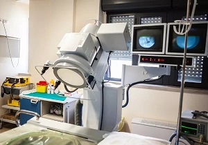
Bladder x-ray
How is a bladder x-ray performed?
X-rays are taken in hospitals or clinics to diagnose diseases related to the urinary system.
There are two types of bladder x-rays:
- urethrography;
- cystography.
With urethrography, it is possible to diagnose diseases associated with the urethra. On this type of x-ray examination of the bladder, with an x-ray, a specialist can see injuries to the urethra, sand in the canals, foreign bodies and education.
Cystography is used to diagnose diseases associated with the prostate gland and directly with bladder. This study gives a complete picture of the described organs, with an X-ray examination, you can see tumors, stones and sand, foreign bodies and chronic cystitis. Cystography is used to treat urinary incontinence problems.
These studies can be carried out in calm state or while urinating. An x-ray examination of the bladder is carried out in a special x-ray room, when the patient lies on the couch. The entire study is carried out under the supervision of a urologist, who comments on the state of the organs under study. For complete picture during the study, a contrast agent is injected into the urethra, and an x-ray is taken at the doctor's command.
Features of the bladder x-ray
The study is not very painful and most often passes without much suffering of the patient. But if the patient is too sensitive to pain, the study of the bladder is carried out under local anesthesia. Special training before the study from the patient is required. Before starting an X-ray examination, the patient must observe for several days special diet. The diet should contain a minimum of carbohydrate. Before the actual bladder X-ray, the patient must have a complete bowel movement. An enema is done the day before the x-ray examination of the bladder, and this procedure is also repeated in the morning before the x-ray. Depending on the condition of the bowel, the urologist may prescribe charcoal or a laxative, which must be taken a few days before the examination.
For a more accurate diagnosis of the disease, an X-ray examination of the bladder is carried out together with ultrasound or computed tomography. All these studies are carried out both in a complex and independently to obtain reliable information and the patient's disease.
X-ray studies of the organs of the urinary and reproductive systems are usually started with a survey radiograph of the kidneys and all urinary tracts. However, this method gives doctors only a part of necessary information about the state of these organs and the ability to perform their function. Apply many more precise methods, for example, an x-ray of the bladder, which allows you to study the sighting of a specific area of the urinary tract.
X-ray examination of the bladder using X-ray contrast
To do this, they use the ability of the kidneys to capture contrast agents from the blood, which then enter the urine, so that formations such as the renal calyces, the pelvis system, the ureters and the bladder can be examined. Such a study is called excretory (infusion) urography.
Indications for the procedure
The bladder looks on the x-ray as a shadow in the form of a transverse oval, the lower edge of which coincides with top edge pubic bone. In the case of urography, its shadow has an average intensity and an even contour. The advantages of urography when examining the bladder are:
- The procedure is available to all patients.
- The study has a low cost.
- It is non-invasive (no penetration).
- The doctor has the opportunity to study the structure of the kidneys, ureters and bladder in one study.
- The possibility of diagnosing calcifications in different segments of the urinary tract.
However, urography also has a number of disadvantages.
It provides only limited information about the structural features of the kidney parenchyma, and cannot completely replace other methods for their study. It does not provide information about functional state urinary excretion, and it is not possible to perform a study with reduced renal filtration. It is contraindicated in case of insufficiency of the heart, liver, kidneys and intolerance to iodine preparations.
Other studies
Ascending cystography
An alternative study option is the introduction contrast medium through the urethra itself. In this case, it is understood that a cystography is done, or directly an x-ray of the bladder. Since the concentration of contrast in this case is much higher, you will get a shadow of high intensity, which stands out against the background of the bones.
In the case of a normal x-ray picture, the bladder shadow is homogeneous, its contour is even and regular. If there are stones or a tumor inside, then either the uniformity of the shadow changes, or the clarity and evenness of the contours.
Indications for cystography are:
- Traumatic extravasation.
- postoperative extravasation.
- Suspicion of bladder diverticula.
- Suspicion of vesicoureteral reflux.
Also this procedure may be given after past injuries abdomen, when there is no exact information about the state of the bladder. It is also carried out in case of violation of innervation this body which may appear various types urinary disorders. It is also useful in diagnostics. urolithiasis, recurrent cystitis. In the latter case, cystography can also prevent the development of neoplasms, since in the case of chronic course hypertrophy of the walls of the bladder develops, from which cancer can develop.
During urination, the contrast medium passes from the bladder into the space of the urethra. The survey, which is carried out at this time, is called voiding cystography. It allows you to get an image primary departments urethra and assess the function of the sphincters of the bladder.
Quantitative diagnostic methods
If there is a suspicion that there is an obstruction to the outflow of urine from the bladder, doctors radiometrically determine the amount of urine remaining. This is due to the fact that in such cases, after urination, there is still some urine left inside the bladder.
To measure its amount, a radiopharmaceutical is injected into the patient, and after one and a half or two hours, a measurement of the intensity of radiation over the bladder is taken. After the patient is asked to empty it, and the radiation level is measured again, and the volume of residual urine is calculated. The most common and informative alternative to this study is ultrasound sonography. However, sometimes these methods are used together, as this makes it possible to get a complete picture. pathological process.
What can be seen in the pictures?

Normal urethrocystogram (pictures taken in different projections)
One of the main pathologies that are detected by X-ray examination are tumors. Cysts or neoplasms may long time develop secretly, without disrupting the functioning of body systems. Most informative method used to diagnose neoplasms of the bladder is computed tomography.
Doctors can make an accurate diagnosis of cancer only after performing a cystoscopy with a biopsy, but there are a number of factors that make an X-ray examination necessary. Firstly, the malignancy of papillomas most often occurs in the depth of the organ wall, and this cannot be established by examining the biopsy. Secondly, cystoscopy does not provide information about the germination of the tumor in the wall and neighboring organs, does not show the presence of metastases in regional lymph nodes.
Therefore, if cancer is suspected diagnostic search better to start with radiodiagnosis. When an artifact is found, it cannot be said whether it is benign or malignant. A sure sign cancer on radiographs is its germination deep into the wall of the bladder and invasion into the paravesical tissue.
Tomography is valuable because it can accurately visualize tumors of the fundus and apex of the bladder. Cystography is also able to visualize the process, however, for this, a double contrast procedure must be performed.

Lacunar cystogram of a patient with adenoma prostate, papillary tumor and bladder stones
Another use of radiography in the study of the bladder is the diagnosis of congenital anomalies.
The most common of these is the diverticulum. He represents himself abdominal education, which is connected to the bladder through a thin neck. It is best determined by cystography. There may also be a ureterocele. At first glance, it may be similar to a diverticulum, however, its main difference is that it does not lie away from the organs, but is, in fact, a hernia of the ureter.
Procedure procedure
The study is quite safe and can be carried out for all patients, except for pregnant women, since the influence of x-rays to the fruit. Cystography, in which a contrast agent is injected into the bladder through the urethra, is not recommended for people with acute infectious diseases and debilitated patients. This procedure should not be done if a traumatic rupture of the organ is suspected, in which case excretory urography should be prescribed.
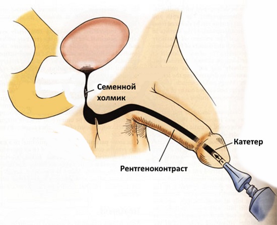
Schematic representation of ascending urethrocystography
All studies are carried out on an empty stomach. The patient should be warned in advance about the procedure, the doctor needs to collect a detailed history, clarify the tolerance of iodine and preparations containing it, if urography is planned. The night before, the patient must cleanse the intestines, the procedure is repeated in the morning on the day of the study. A person should come to the radiology room on an empty stomach. Then the patient is laid on his back, and a survey radiograph is taken. After proceed to additional methods research.
Addresses medical centers and clinics in Moscow where you can do cystography.
What is cystography
Cystography is a method diagnostic examination bladder through the use of an X-ray machine and special contrast agents that fill the bladder.
There are two ways to introduce contrast:
- Descending, implying the introduction of contrast into the bloodstream, followed by the release of the substance into the bladder through the kidneys.
- Ascending - the contrast agent enters the bladder through the catheter inserted into the urethra.
Cystography allows you to get reliable information about the shape, localization and pathologies of the bladder.
Indications
Diagnostic examination of the bladder using x-rays is ordered to detect the following diseases and organ pathologies:
- malformations of the bladder;
- urolithiasis disease;
- violation of the innervation of the bladder;
- recurrent cystitis;
- foreign bodies;
- neoplasms, including malignant ones;
- mechanical damage to the bladder;
- prostate tumors;
- protrusion of the walls of the bladder (diverticula);
- complications after infectious diseases.
Contraindications and effects on humans
Methods of diagnostic examination of human internal organs using X-ray waves, including cystography, suggest radiation exposure organism.
Despite the fact that rays with low activity are used in medicine, their influence can adversely affect intrauterine development fetus. Accordingly, cystography of the bladder is contraindicated in pregnant women, and is also not performed during breastfeeding child - there is a high probability of a small dose of radiation to a newborn with mother's milk.
In the presence of acute infectious diseases of the bladder, mechanical damage urethra, obstruction, rupture of the urethra, allergy to contrast agent and in rehabilitation period after surgical operations cystography on the bladder is prohibited.
Training
Before the examination procedure, the doctor must tell the patient how to carry out cystography, warn about possible side effects.
The patient signs consent to the manipulations and reports an allergic reaction (if any) to the drugs that are planned to be used during the procedure: iodine, contrast agents, latex, anesthetics.
The day before the cystography and directly on the date of the planned events, the patient is recommended to consume only a clear non-carbonated liquid. Be sure to cleanse the intestines with laxatives and enemas. Avoid foods that cause gas and bloating.
Three hours before the procedure, you can not eat or drink.
The attending physician may prescribe other preparatory procedures based on individual characteristics patient.
How is the procedure
X-ray diagnostics of the bladder is carried out both in stationary conditions and on an outpatient basis.
The algorithm of medical manipulations is as follows:
1. The patient is freed from clothing and accessories that impede the study. In return, they can provide a special sterile set of clothes.
2. Be sure to empty the bladder before the procedure.
3. The patient is placed with his back on the X-ray machine.
4. Take a picture on an empty bladder.
After the first shot:
- with ascending cystography, a 10–30% solution of a radiopaque substance is injected into the bladder through the urethra through a catheter. The total volume for adults is from 150 to 300 ml, for children under 12 years old - no more than 100 ml.
- if descending cytography is used, then the study is started one hour after the injection of a contrast agent into the vein. During this time, the bladder fills with the contrast released by the kidneys, which allows you to clearly visualize the contours of the organ and get optimal images.
However, ascending and descending cystography does not allow the formation of images of low-contrast or low-contrast bladder stones, as well as minor neoplasms. In this case, the double pneumocystography method is used: the bladder is filled with 15-20 milliliters of a contrast agent and nitrous oxide, oxygen, or carbon dioxide.
With conventional pneumocystography, no contrast agent is used, only gas.
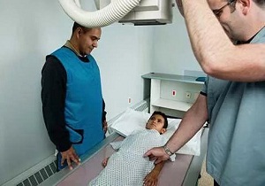
5. If X-ray diagnostics assigned to a man - protection of the testes is provided by overhead plates.
6. Pictures are taken during the introduction of contrast and immediately after filling the bladder with the substance, the next - after the removal of fluid.
7. For a complete examination, the patient may be asked to take a certain position (on the back, stomach, on the side, raise the legs at an angle of 90 degrees) in order to photograph the organ in various projections.
8. At the end of the manipulation, the medical staff removes the catheter.
Cystography is quite painful and unpleasant procedures therefore, it is prescribed in cases where other methods do not allow to fully assess the condition of the bladder.
To the number possible complications from the procedure include:
- bladder infection,
- gas embolism,
- the appearance of blood in the urine after catheterization,
- some pain when urinating.
The only remedy for CYSTITIS and its prevention, recommended by our subscribers!
Cystography is a diagnostic endoscopy, which is produced using x-ray radiation. AT modern medicine most often it is the methods of cystographic examination that are used to determine various pathologies structures and shapes of the bladder, urethra and ureters.
Revealing pathological conditions bladder is an essential step in the treatment process various diseases urinary system. Endoscopy of the genitourinary organs is prescribed for both adults and children. X-ray of the bladder allows the doctor to put correct diagnosis the patient, as well as prescribe the necessary course of treatment to eliminate the existing pathology.
What is a cystography?
As mentioned above, cystography of the genitourinary organs is a diagnostic examination method used in urology to identify pathologies of the genitourinary organs. Similar diagnostic methods are carried out by introducing a special contrast agent into the cavity of the bladder, followed by x-rays. The injected contrast agent can be both gaseous and liquid. The X-ray contrast agent is injected into the body through a catheter. There are two forms of administration of a contrast agent, which are most often used in modern medicine:
- Ascending cystography – state of the art diagnostic technique, which is based on the introduction of a radiopaque substance directly into the cavity of the bladder. A special drug is administered by placing a catheter - this is done in the interval between emptying the bladder from urine and the subsequent urge to urinate.
- Descending cystography - this diagnostic technique is based on the introduction of a special contrast agent intravenously, by injection. Of course, before the moment the drug enters the cavity of the bladder, a lot of time will pass - usually at least an hour. And only then it is possible to carry out cystography, otherwise the results of the examination will be untrue. It is thanks to such a long and laborious process of determining the existing pathologies of the urogenital area that all large quantity experts consider the ascending cystography technique to be more effective. In addition, during the ascending cystography, you can get better and more reliable results, in turn, the descending technique is considered less effective.
- In some cases, voiding cystography is recommended - this technique involves an examination only at the time of urination. Of course, such x-ray examination quite difficult, and therefore today is not widely used.
With the introduction of a contrast agent, internal organs, such as the bladder, acquire a clearer and brighter outline, after which it becomes possible to examine stones or other pathologies in the cavity internal organ. Bladder x-rays are also widely used to detect benign or malignant neoplasms in the region of the urinary tract.
Indications and contraindications for cystography
Endoscopy of the bladder is performed in the following cases:
- If you suspect tuberculosis of the genitourinary system.
- Cystography is prescribed to determine the presence of tumors of a benign or malignant nature in the pelvic area.
- If stones are suspected, or X-ray methods are considered the most informative.
- Revealing congenital pathologies urinary system, which is most often used in the case of diagnosis in young children.
- If vesicoureteral reflux is suspected or serious, cystography is most often used as a diagnostic examination.
- Indications for cystography of the bladder are considered various complications after past illnesses infectious nature.
- Also, it is cystography of the bladder that is performed in the case of diagnosing enuresis in a patient. Most often, this problem is faced by children and adolescents, and cystography allows you to establish exact reason diseases and prescribe the necessary course of treatment.
Despite all its many advantages, this technique also has several contraindications, in which the diagnostic procedure is strictly prohibited.
- Diagnosis is not applied to pregnant women.
- This x-ray procedure not prescribed to patients who have inflammatory processes in the region of the bladder and urinary canals.
- If the patient has urination with blood impurities, cystography is strictly prohibited.
Carrying out cystography
In case of ascending cystography urinary organs, approximately 0.2 l of a special contrast agent is injected directly into the cavity of the organ, while the patient is in the supine position. All jewelry and accessories should be removed at the time of the diagnostic procedure, as they may distort the information content of the result. In the vast majority of cases, during the procedure, it is recommended to release the body under study from clothing and put on special medical underwear.
After the x-ray preparation is introduced into the cavity of the bladder, the catheter is clamped to avoid leakage of the drug. Next, X-ray images are taken from different positions - when the patient lies on his back, on his side, at the time of urination or after it.
It should be noted that cystography is accompanied by noticeable painful sensations, and therefore, if it is necessary to conduct small children, cystography is carried out with the simultaneous use of painkillers medicines. After the procedure, the doctor compares the images taken before the X-ray of the bladder and the images obtained during the procedure - this makes it possible to put accurate diagnosis and prescribe the necessary treatment.

Preparation for bladder cystography
The first rule of proper preparation for the study of the bladder is to eliminate the increased gas formation in the intestine, which can significantly distort the result of the study.
2-3 days before the procedure, you should begin to observe strict diet with the complete exclusion of products that promote increased gas formation. Such products include strong tea and coffee, carbonated drinks and mineral water, beans and other legumes, white cabbage, dairy products, whole milk, corn. In the morning, before the cystography, the patient is given cleansing enema, which contributes to the complete release of the intestine from the contents.
Before carrying out, consultations with a nephrologist, radiologist and urologist are mandatory. They will give everything necessary recommendations, thanks to which the results of cystography of the bladder will be as effective and informative as possible.
Consequences of the study of the bladder
The main danger after this study organs of the genitourinary sphere is to remove the contrast agent from human body. In order to facilitate this process, after the diagnostic procedure, it is recommended to observe strict bed rest– thanks to this, the removal of the x-ray specimen is easier and painless.
In extremely rare cases it is possible to develop such dangerous complication, as an infection of the urinary tract, which results from the placement of a catheter. This complication develops in extremely rare cases and requires immediate treatment. Also among rare complications cystography can be attributed to accidental injury to the mucous membrane of the urethra or directly to the bladder, which most often results from a lack of experience among medical personnel. In order to avoid similar situation should only be referred to experienced doctors, to large, respected diagnostic centers.
By secret
- Incredible… Chronic cystitis can be cured forever!
- This time.
- No antibiotics!
- This is two.
- During the week!
- It's three.
Follow the link and find out how our subscribers do it!
Cystography - difficult diagnostic procedure. The study of the bladder is carried out strictly according to indications, if other methods do not give an accurate answer to the question about the causes of pathological processes in urinary tract.
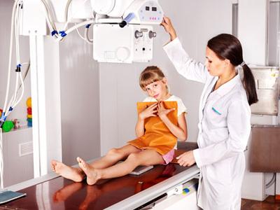
It is important to know the indications and contraindications for cystography in children. How is a bladder exam done with a contrast agent? Is there any discomfort during the procedure? What does the study show? Answers in the article.
general information
The procedure involves the introduction of a radiopaque substance into the cavity of the bladder. Doctors fill the cavity with a solution (from 50 to 200 ml depending on the age of the child) containing 10-30% Urografin, Iodamine, Triombrast.
At the beginning of the procedure and after filling all areas with a contrast agent, the doctor takes x-rays to assess the condition of the organs of the genitourinary system. A special drug stains the tissues, all affected areas stand out well in the pictures. According to the results of the study, a clear radiographic picture of the bladder cavity is visible. The procedure, depending on the variety, takes from 30 to 60 minutes, with preparation - up to two hours.
Features and types of research
Cystography is carried out using two main methods:
- ascending method. The X-ray contrast agent is injected into the bladder through the urethra using a catheter. To reduce discomfort, apply the Cathejel gel with analgesic effect;
- descending method. The composition is injected into a vein, then the blood carries the substance throughout the body, after certain time the drug enters the bladder, stains the tissues. From this point on, x-rays can be taken. The method is less painful, but the penetration of contrast into the blood increases the risk allergic reactions.
On a note:
- with the development of non-contrast and small tumors, another type of study is required - pneumocystography. The main difference is that the introduction into the bladder cavity is not liquid form drug, but gas. For the procedure, oxygen, carbon dioxide or nitrous oxide are used;
- sometimes doctors combine gas and liquid to diagnose complex cases of diseases, anomalies in the development of the urinary tract and the tumor process. Lacunar cystography is a highly informative method;
- if the child has problems with urination, then the urologist may prescribe a voiding cystography. The study is carried out directly during the excretion of urine. The technique provides accurate data on the areas of leakage of the radiopaque substance. An unpleasant moment is psychological discomfort that is difficult for a child to cope with.
Advantages and disadvantages
Diagnostic studies using contrast have both strong and weak sides. Despite the high information content of cystography, the method has some negative points to which it is important to draw the attention of parents.
The doctor should explain how to prepare the child for the procedure to reduce the risk of complications and inaccurate results. Physicians must warn Negative consequences, exactly follow the rules of diagnosis, take into account the age of the patient, the condition of the patient's problematic organs.
Advantages:
- on the x-rays all deviations are noticeable, which are difficult to recognize at and;
- for the most complete picture in complex cases, doctors may choose best method: lacunar or pneumocystography;
- the result of the study is ready after a short period of time after the procedure. The doctor assesses tissue damage, gives a preliminary conclusion, and refers to a pediatric urologist;
- New digital-resolution X-ray machines allow you to study the entire process, from bladder filling to urine excretion. With dynamic cystography, radiation exposure is significantly reduced, which is especially important when examining children.
Flaws:
- psychological discomfort;
- the child does not always understand the instructions of the doctor;
- during the introduction of the catheter, painful sensations appear;
- possible complications in the form of allergic reactions to the drug, nephrotoxic effect. With retrograde cystoscopy, these complications are absent, but other problems are possible: acute delay urine against the background of spasm of the sphincter and trauma to the urethral mucosa (more often in elderly patients);
- when the walls of the bladder rupture, the penetration of a contrast agent into the bloodstream can provoke sepsis.
Indications for diagnostics
complex diagnostic study appointed after preliminary survey little patient. It is important to collect blood tests, urine, do an ultrasound scan, conduct.
Injection of a radiopaque substance into the bladder cavity childhood undesirable, the child and parents are often afraid of the procedure, conditions are created for the development of stress. If, according to the results of other types of examination, there is no exact data on the causes negative symptoms, localization, severity of the pathological process in the urinary tract, then it is necessary to carry out cystography.
Main indications:
- pathology of bean-shaped organs and bladder;
- bladder rupture or reflux;
- detection of a tumor of unknown etiology;
- accumulation and salt stones;
- anomalies in the development of the organs of the genitourinary system.
Contraindications
Ascending cystography is not performed in the following cases:
- inflammation in the scrotum, urethra, bladder;
- urinary excretion blood clots or massive hematuria.
On a note! Descending cystography has the same limitations as excretory urography: severe pathologies liver and kidneys, allergy to iodine preparations, kidney failure, diseases thyroid gland, poor clotting blood.
In most cases, doctors avoid cystography, use other diagnostic methods. If descending or ascending cystography is indispensable, parents will have to explain to the child how the study will take place, and why you will have to suffer a little during the procedure.
Important psychological preparation, an accessible explanation of the essence of the method and the importance of the survey. The less parents panic, the lower the risk of developing fear in a son or daughter.
For getting reliable results You will have to change the diet of a young patient:
- for a week, exclude from the menu all items that provoke increased gas formation in the intestines. You should not give legumes, carbonated drinks, muffins, fresh milk;
- if the child suffers from constipation, then for two weeks before the procedure little patient takes a mild laxative;
- babies receive tea that prevents the accumulation of gases or dill water;
- two days before the examination of the bladder, the doctor categorically prohibits products that cause flatulence;
- on the day of the study, an enema is required to maximize the removal of food debris and gases from the intestines.
How to treat ? Check out the selection effective options therapy.
About how it is done excretory urography kidney with the use of a contrast agent is written on the page.
How is the procedure carried out
All types of cystography cause psychological discomfort to the patient; with an ascending method of research, pain is felt when placing a urethral catheter. It is important to follow all the commands of the doctors in order to get the most exact result. After the procedure, the urine often changes color: a radiopaque substance comes out.
Study information:
- after preparation, the patient lies down on the x-ray machine (position "on the back"). During the first phase, you need to lie still. The doctor takes a general picture of the organs of the genitourinary system;
- then the doctor proceeds to the most unpleasant part - he inserts a catheter through which the bladder cavity is filled with liquid with a radiopaque substance or gas. Up to 12 years, 50-100 ml of the drug is enough, adolescents are supposed to adult norm- 200-300 ml;
- during the introduction of contrast and during the examination, the doctor presses the catheter, which provokes soreness and the urge to urinate. You can’t do without this stage - it is important to hold the contrast inside the cavity so that the substance does not leak out of the bladder during the procedure;
- after filling the bladder with a special substance, the radiologist takes several pictures in different projections: on the side, from the abdomen and from the back. A small patient should raise his legs to the level of 90 degrees, additionally raise his shoulders. In this position, the pictures are the most clear and informative;
- at the end of the procedure, the doctor removes the catheter, takes a picture of an empty bladder;
- after the examination, the doctor examines the finished images, analyzes the image. If fistulas or ruptures of the walls of the urethra or bladder are detected, hospitalization of the child is required to prevent sepsis: through the zones of ruptures, the contrast enters the blood;
- after the procedure, in the absence of complications, a hospital stay is not required; most often, cystography is performed on an outpatient basis. Observation by doctors of the urological department is necessary if a small patient was previously admitted to a medical institution with severe pathologies of the urinary tract, and cystography is one of the planned methods of examination.
Diagnostic results

What does a cystogram show? The procedure allows you to accurately determine the area of localization of the pathological focus, the severity of the changes and the spread of inflammation, the type of tumor process or the size of the stones. In the first picture, the radiologist sees the exact location of the kidneys and other organs of the urinary system. After filling the cavities with a radiopaque substance, pathologies that are difficult to recognize on ultrasound and cystoscopy can be detected.
In the stained organs, deviations are clearly distinguishable:
- congenital defects of the bladder and bean-shaped organs;
- and renal ureteral reflux;
- stones in the urinary tract;
- change in the structure and thickness of the bladder walls;
- fistula, traumatic injuries the walls of the bladder and other parts of the urinary system;
- tumors and calculi of any size;
- inflammatory processes in natural filters, bladder, ureters, other pathologies.
At proper preparation, following the instructions of the radiologist, cystography in children provides accurate data on the nature, stage, localization of pathological foci in the urinary tract. Parents should properly prepare the child psychologically, change the diet to eliminate interference during the study.


