What does endoscopic examination mean in Laura. Endoscopy of ENT organs. Nasal endoscopy at the Ear, Nose and Throat Clinic
Nasal endoscopy is an important diagnostic method for identifying a number of pathologies. This method of research is considered budgetary and highly informative.
The procedure is carried out using a miniature endoscope, which is a thin wire with a camera at the end. This device allows you to study in detail the condition of the mucous membrane of the nasal passages. Manipulation is painless, may be accompanied by minor discomfort. Endoscopic examination of organs is more informative than standard examination.
The camera magnifies the image, which allows you not to miss even the slightest deviations from the norm. The equipment is equipped with a flashlight, which makes it possible to study all the details and not to miss pathological changes in tissues. Inspection of ENT organs does not take much time.
There is no pain during the procedure. Discomfort occurs only in individuals with a deviated nasal septum. In this case, the movement of the camera becomes difficult, and the doctor can make little effort and change the trajectory, which can contribute to the occurrence of discomfort during the examination.
nose
The study of the maxillary sinuses using optical devices allows you to identify polyps, inflammatory diseases and various neoplasms.
Larynx
Examination of the pharyngeal cavity is necessary to identify diseases associated with voice changes, the formation of neoplasms, tumors.
ear
Examination of the ear area reveals inflammatory processes that often lead to deafness and other hearing impairments.
Types of endoscopy
Inspection of the mucous membrane of the nasopharynx can be done in several ways. It all depends on the nature of the symptoms and the age of the patient.
Front
Rhinoscopy is performed by introducing an endoscope to a depth of no more than 2 cm. Local anesthetics can be used to get rid of discomfort.
rear
Inspection is carried out through the mouth. The device is inserted deeply, up to the pharyngeal wall. Despite the discomfort of manipulation, this type of study allows you to identify adenoids, tumors and polyps in the early stages. This method is used only in rare cases and when dangerous diseases are suspected.
Medium
This method of research allows you to diagnose the condition of the anterior paranasal sinuses. Manipulation is carried out using an elongated instrument through the nasal passages. In this case, local anesthetics and vasoconstrictor drops are often used, which eliminate the swelling of the mucous membrane.
Straight
Laryngoscopy is performed using a movable instrument that is inserted into the larynx cavity. The procedure can be unpleasant for the patient and often provokes vomiting, so the throat is irrigated with Lidocaine before manipulation. The use of microlaryngoscopy allows to identify a wide range of diseases of the larynx.
indirect
The procedure is carried out using a special mirror, which is placed in the larynx area. At the same time, a frontal reflector is fixed on the doctor's head, which reflects light. The manipulation lasts no more than 5 minutes, but does not provide such detailed information as the direct research method.
Surgical
The surgical method is used not only for diagnostic, but also for therapeutic purposes. Manipulation may be accompanied by minor incisions and punctures. Often, using this method, pathological foci are eliminated, tissue biopsy is performed for histological examination. This method involves the use of anesthesia.
Indications for the procedure
The procedure is used for symptoms and pharynx. Endoscopic examination is carried out with suspicion of neoplasms: benign and malignant. Additional indications:
- the presence of inflammatory processes occurring in the nasal cavity and pharynx;
- polyps;
- enlarged adenoids;
- difficulty breathing through the nose;
- pain in the sinuses;
- voice change, hoarseness;
- sensation of a foreign body in the larynx during a conversation or when swallowing food.
The examination shows the presence of purulent foci, the amount of altered tissue and other transformations of the mucous membrane, including microdamages.
Survey Rules
The study is recommended to be carried out on an empty stomach. There are no special rules, the procedure is quick and painless. If there is a mucous secret in the sinuses, then it is important to blow your nose so that nothing interferes with the study. The doctor performs the procedure in gloves, having previously disinfected the endoscope. Diagnosis is carried out with the patient sitting or lying down.
Training
The doctor informs the patient that on the eve of the study it is forbidden to instill any solutions into the nose, use nasal ointments and other means that may complicate the manipulation.
It is important to refrain from smoking. The child must be set up for endoscopy, explaining to him how the study will take place. It is important that during the diagnosis the person was in a stationary state.
Carrying out technology
Most often, during the procedure, the patient is in a special chair. Each type of research is carried out differently. When using the direct method, 2 thin and closed jaws are used. The patient is asked to tilt his head back, and the instrument is inserted a few centimeters into the nasal passage. Then the branches are slightly moved apart and the sinuses are examined using special optical equipment.
The posterior method of research is carried out using a spatula, which removes the tongue from the larynx. Then the device is inserted as deeply as possible, reaching the pharyngeal wall. To reduce the chance of vomiting, breathe only through your nose. Before the procedure, it is forbidden to eat and drink.
The average type of technique involves the introduction of branches through the nasal passages and examination using an optical device. Before manipulation, the nasopharynx is irrigated with an anesthetic solution, and a vasoconstrictor is instilled into the nose.
The surgical method requires more preparation. In this case, various types of anesthesia can be used. Often, during the manipulation, an incision is made in the tissues of the nasal mucosa in order to get rid of chronic rhinitis. A small fragment of the material in the presence of polyps is sent to the laboratory for a more thorough diagnosis.
An indirect type of research is used in any clinic. The patient sits on a chair, slightly throwing back his head and sticking out his tongue. The doctor inserts a mirror into the larynx and examines the palatine tonsils and pharynx. At the same time, the slightest deviations from the norm are well visualized.
The direct method is often performed using a movable laryngoscope. The rigid technique with a rigidly fixed apparatus is used during surgical intervention. Before starting the procedure, the patient is explained the sequence of steps. This method is carried out using general anesthesia.
The laryngoscope is inserted through the larynx and advanced deeply. This method is considered the most informative.
Features of endoscopy in children
The study of the nasal cavity and pharynx in a child is carried out in the presence of parents. Often the implementation of manipulation is complicated by the fact that it is most difficult for children to be immobile for 5-10 minutes. For diagnosis, the most painless methods are chosen, which are practically devoid of discomfort.
If, nevertheless, the procedure involves additional surgical intervention, then the child should be carefully prepared. To begin with, determine whether there is an allergy to any medications. Conduct special tests. In order for the procedure itself not to cause shock in the child, he is told and shown what tools will be used during the study and what they are for.
It is important to pay attention to anesthesia. Therefore, even when using the most minimally invasive techniques, local anesthetics are used. It is important for a child, like an adult, to refrain from eating and drinking. Children are explained the rules of behavior during the introduction of the endoscope. If this is not enough, then in extreme cases resort to the use of general anesthesia.
For manipulation in children, an endoscope no more than 2 mm in diameter is used. It does not create discomfort, easily moves through the sinuses and does not injure them. The specialist tries to introduce the instrument very carefully so that there is no sensation of a foreign body. At the end of the procedure, it is important to ensure that the child does not pick his nose.
What are the contraindications
The main contraindications are persistent nosebleeds. If the vessels are too thin and weak, then the risk of damage is high. Therefore, before resorting to endoscopy, it is important to check the condition of the venous apparatus, as well as donate blood for the rate of platelet aggregation.
An additional contraindication is an increased gag reflex. In this case, methods based on deep insertion of the instrument into the larynx are often not used. The procedure is not performed during pregnancy. Endoscopy is contraindicated in infancy, as the sinuses are easily injured.
With a strong increase in the tonsils, manipulation is not carried out, since such a clinical picture makes it difficult to visualize the tissues. A contraindication is an allergic reaction to painkillers. The procedure is not performed in the treatment of anticoagulants, since in case of accidental damage to the vessel, there is a high probability of bleeding that will be difficult to stop.
With a deviated nasal septum, a pediatric endoscope is used, which reduces the risk of discomfort. If a person is allergic to local anesthetics and endoscopy is necessary, then an easy option is chosen that can be performed without the use of anesthesia.
A contraindication is the unstable mental state of the patient, the presence of schizophrenia, disorders of the central nervous system.


Year by year, diagnostic medical techniques are being improved to provide timely and most complete assistance to the patient. Qualified ENT specialists are increasingly using nasal endoscopy in their practice. makes it possible to establish an accurate diagnosis based on the examination data. Before the examination, the patient may have questions. To exclude unnecessary experiences, we will try to reveal the essence of the procedure.
What it is?
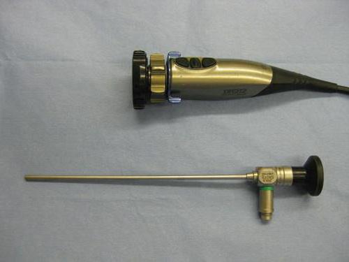
An endoscope is a light-conducting device with a device that looks like a thin rigid or flexible tube, the thickness of which does not exceed 4 mm. Flashlight and camera on one end, eyepiece on the other. Endoscopy is the ability to examine some of the internal organs by inserting an endoscope into the cavity. It is administered through natural routes or by puncture. Nasal endoscopy - examination with a thin endoscope through the nose.
Why is this needed?
The survey is carried out to achieve the following goals:
- detection of the presence of pathologies of the paranasal sinuses;
- identifying the presence or absence of pathologies of the nasal septum;
- monitoring the presence or absence of the effect of medical procedures;
- detection of tumors, the presence of foreign bodies, the presence of wounds in the nasal cavities (carrying out microsurgical manipulations to eliminate them);
- collection of secretions for bacteriological studies;
- monitoring the condition of ENT organs after operations;
- treatment of wound surfaces and elimination of interference for drainage of the sinuses;
- determining the state of the mucous membrane of the nasal passages, the size of the content structure;
- the most accurate diagnosis of major ENT diseases.

When is endoscopy of the nose and nasopharynx indicated?
The otolaryngologist prescribes endoscopy to the patient in many cases. A doctor can conduct such an examination when contacting:
- with epistaxis of unknown origin;
- sinusitis;
- runny nose;
- polyposis;
- changes in the nasal septum;
- injuries of the face and skull;
- unexplained headaches;
- during the rehabilitation period after rhinoplasty or other interventions.
So, for example, with sinusitis, endoscopy of the sinuses helps to determine which departments are affected by the inflammatory process. And if you suspect the presence of polyps or minor tumors, the doctor decides on surgical endoscopy. As you understand, the price for nasal endoscopy of varying complexity will differ. It can be from 450 to 3500 rubles. The exact cost must be clarified with the specialist who will carry out the procedure.

Preparation for manipulation
Before endoscopy, special preparation of the patient is not required. The doctor may irrigate the mucosa with a vasoconstrictor drug to reduce swelling. This will increase the overview during the manipulation.
Does it hurt or not?
Most of all, patients are nervous because they are afraid of pain. To avoid pain, the doctor irrigates the mucous membrane with a local anesthetic. If minimally invasive surgery is planned, then general anesthesia can be used.
If the patient has wide nasal passages, then the doctor can perform a routine examination with a thin endoscope without the use of anesthesia. In addition, endoscopy of the nose and nasopharynx can be performed without anesthesia for severe allergic reactions to anesthetics.

How is the procedure?
Inspection begins with an examination of the lower nasal passage. Then the endoscope is passed to the nasopharynx and its thorough examination is performed. The mouth of the auditory tube and choana is also examined. The next stage is an examination of the sphenoidal pocket, upper and
Features of nasal endoscopy in children
Doctors are confident that this type of examination is the most effective in carrying out nasal endoscopy in children - to ensure their calmness and immobility. To do this, before starting the procedure, the doctor talks with a small patient, explaining to him that the procedure is fast, it will be a little unpleasant, but not painful. The main thing is to convince the child that it is impossible to break out, twitch and scream so as not to interfere with the doctor. In many cases, nasal endoscopy for children is performed in the hands of the parents. This way they feel more relaxed.

Is it necessary...
Sometimes patients doubt the need for nasal endoscopy. However, it should be borne in mind that when examining with a conventional expander and a mirror, the doctor cannot get a complete picture. In order not to be mistaken in the diagnosis and not to prescribe unnecessary drugs, the doctor needs to perform an examination with an endoscope. In addition, this will allow timely detection of the appearance of neoplasms, notice the curvature of the septum and assess the state of the adenoids.
Removal of polyps
A nasal polyp is formed when histamine and inflammatory mediators are released, which destroy the mucous membrane, and swelling and changes in the glandular tissues occur. Endoscopy of nasal polyps has replaced mechanical removal with a metal wire loop. Thanks to modern advances, the doctor can expand the fistulas of the sinuses and remove the polyposis tissue as much as possible. At the same time, the invasiveness of the manipulation is significantly reduced, the surgeon can visually assess the progress of the procedure by watching it on the monitor, the patient will be discharged from the hospital in 3-5 days.

It should be borne in mind that endoscopy of nasal polyps does not eliminate the cause of the growth of polyposis tissue. The patient should continue the treatment of the underlying disease, otherwise the problem will return in a few years. Previously, with mechanical removal, polyps grew again much faster.
Contraindications for endoscopy
Examination with an endoscope does not worsen the patient's condition, therefore, there are no special contraindications. The only difficulty is an allergy to anesthesia. Be sure to warn the doctor about nosebleeds and high sensitivity. In this case, the procedure will be carried out with an ultra-thin (children's) apparatus.
Video endoscopy of ENT organs (or endovideoscopy) is a method of instrumental diagnostics that allows you to study the state of this group of organs using an endoscope equipped with a video camera. Endoscopy in ENT diseases is used as one of the most modern examination methods available to the otolaryngologist.
Who needs this procedure
This method makes it possible to effectively carry out endoscopy of the nasopharynx in children and adults, as well as to carry out a detailed examination of other ENT organs (nasal cavity, pharynx, ears, entrance to the nasal sinuses). The most common reasons for its use are:
- nose bleed
- nasal congestion
- feeling of tightness and pain in the sinuses
- suspected sinusitis, polyps, or foreign bodies
- hoarseness
- swallowing problems
- hearing impairment.
For videoendoscopy of ENT organs there are no absolute contraindications, however, if the patient has problems with blood clotting, it is necessary to notify the specialist conducting the examination.
How is video endoscopy performed?
Endoscopy of the nose, larynx and other ENT organs is performed by an otolaryngologist. During the examination, the doctor collects an anamnesis, carefully questions the patient, performs an examination and palpation of the ENT organs, and conducts video endoscopy. Based on the collected data, the otolaryngologist can send for an additional examination, if necessary.
This type of examination usually takes up to 10 minutes and is relatively painless. Before starting an endoscopy of the ENT organs, the otolaryngologist explains the details of the procedure and clarifies how to behave during the examination. With mucosal edema, the doctor may instill a vasoconstrictor drug to facilitate the insertion of the endoscope and improve the analysis of the posterior nasal cavity and nasopharynx.
The patient sits in a chair, and the doctor in turn inserts a video endoscope into each nasal passage. An endoscope equipped with a video camera and a backlight is carefully moved to the desired section of the ENT organ, and the specialist carefully assesses its condition. Modern video endoscopes are equipped with a highly sensitive high-resolution camera, which allows you to instantly display the image of the organ under study on the monitor during the examination. Thanks to this approach, the doctor and the patient immediately see a high-quality image of the area under study, which significantly expands the diagnostic possibilities.
With the help of a video endoscope, an otolaryngologist can:
- perform a nasopharyngeal endoscopy
- detect the presence of small polyps that are indistinguishable when examined with a standard endoscope
- clarify the features of secretions and their origin
- perform sinus endoscopy
- if necessary, take a biopsy
- examine the pharynx, ear and larynx
- perform ENT manipulations and / or operations with a high level of control.
Endoscopy result
Based on the study, the otolaryngologist issues a written conclusion. If the data obtained are sufficient, then a diagnosis is made, and treatment can be prescribed. Otherwise, additional types of examination may be recommended to clarify the diagnosis.
Benefits of the procedure
The provided service of video endoscopy has a number of advantages compared to traditional endoscopy of ENT organs and other types of instrumental examination:
- no special training required
- improved examination quality, facilitating extremely accurate diagnosis and prescribing the most effective treatment
- early detection of pathological formations in the ENT organs
- the ability to examine hard-to-reach areas in the nasopharynx and tympanic cavity
- with increased sensitivity, it is possible to use a local anesthetic applied to the mucous membrane, which increases the comfort of the procedure
- the ability to record the study on digital media for subsequent analysis
- the absence of radiation, characteristic of CT and radiography of ENT organs.
A-Media Clinic has highly qualified specialists and modern equipment, which guarantees fast, high-quality and safe endoscopy of ENT organs, as well as other types of body examinations.
Almost every procedure for examining patients for pathologies of the ENT organs necessarily includes, in addition to examination and anamnesis, also instrumental diagnostic methods aimed at visualizing the internal state of the organs. Various types of micro- and endoscopies act as such instruments.
Microscopy of ENT organs
Microscopy is the examination of mucous membranes. ENT organs under a microscope.
Microscopy is the only accurate tool for making an accurate diagnosis. It is carried out using specialized ENT microscopes (link to our models), which allow diagnosing both atypical and sluggish inflammatory processes of the middle ear, tympanic membrane and the walls of the auditory canal itself. The most important difference between these microscopes and ordinary ones is the possibility of carrying out medical procedures and microsurgery simultaneously with the examination.
Figure 1. Specialized otolaryngological microscope Haag-Streit Surgical ALLEGRA 50
A similar procedure is performed without any anesthesia or surgical intervention. During the procedure, the doctor sits in front of the patient and sets the microscope with the light source in the required position so that the eardrum can be effectively examined.
With this diagnostic, you can determine:
- injuries caused by a sharp drop in pressure (rupture of the membrane);
- perforation of the tympanic membrane;
- inflammation of the tympanic membrane and middle ear.
Endoscopy of ENT organs
Endoscopy of ENT organs- this is an examination of the mucous membranes of the upper respiratory tract with the help of endoscopes. This procedure is carried out both for the respiratory tract (nasopharynx, larynx, trachea), and for the auditory organs.
According to the method of image transmission, all endoscopes are divided into:
fiberscopes- a cheaper type of endoscope, in which the image is transmitted through the optical system to the viewing eye on the endoscope. As a result, the image is not processed and only the doctor can see it.
Video endoscopes- a device with a camera installed at the end of the endoscope, the image from which is displayed on a specialized medical monitor through an image processing system.
Depending on the area (ear, throat, nose), apply:
- pharyngoscopy - examination of the oral cavity and pharynx ;
- laryngoscopy - examination of the larynx;
- stroboscopy - examination of the vocal cords;
- rhinoscopy - examination of the nasopharynx;
- otoscopy - examination of the middle ear and auditory canal;
- nasopharyngoscopy - an examination conducted to exclude malformations of the tonsils and adenoids;
- sleep endoscopy - examination of the upper respiratory tract with a flexible endoscope during medication sleep. This is an innovative solution in the diagnosis of snoring and apnea.
An interesting feature: not all such devices can be attributed to endoscopes. For example, the Heine Mini 3000 viewing otoscope is not one of them, because it cannot be connected to a cold light source or an image capture system.
The use of endoscopy in otolaryngology opens up wide opportunities for access to previously closed sections of the nasal cavity, maxillary and frontal sinuses, as well as the larynx, where the doctor cannot physically look. This technique makes the detection of mucosal polyps and deformities of the nasal septum easier, allowing an accurate diagnosis of the patient's condition. In addition, the endoscopic technique used for the ear section makes it possible to describe the inflammatory processes of the middle ear, to assess the state of the tympanic membrane.
For example, the pharynx is quite successfully examined using the fibrolaringoscopic technique. It allows you to visually evaluate all the features of its structure, as well as take pictures / videos. Another method of examining the larynx is known, it is called microlaryngoscopy. The technique involves the analysis of the vocal cords and the larynx as a whole under optical magnification using a rigid video endoscope (stroboscope), often inserted into the trachea using an intubation laryngoscope (through the laryngoscope, the endotracheal tube passes through the oral cavity and larynx, entering the trachea between the vocal cords).
Today, stroboscopes are the only imaging tool that allows complex examinations of the trachea and voice-forming apparatus. The use of such a technique allows you to accurately determine the source of the problem and purposefully influence local areas of the larynx. It is very important that no additional incisions on the neck are required to insert the stroboscope into the larynx, everything happens through the natural respiratory tract.
As for the nasopharynx, a method called fibrorhinopharyngoscopy is actively used today. This technique only allows visualization of the nasopharynx with simultaneous biopsy, which is very convenient when verifying suspicions of a tumor.
Auxiliary equipment
For endoscopic diagnostics and therapy, in addition to the ENT combine with endoscopy support, as well as related ENT equipment, the following devices are needed:
For video endoscopy
- specialized video endoscope (Atmos also uses video capture of images from fiberscopes, i.e. the endoscope can be used simultaneously as both a video and a fiberscope);
- cold light source with light guide;
- medical monitor.
For fibroscopy
- Specialized fiberscope;
- Cold light source with light guide.
With the help of endoscopic diagnostics, it is possible to identify and determine:
- the level and degree of obstruction of the respiratory tract;
- pathology of narrowing of the oropharynx;
- the physical condition of the epiglottis;
- shape, size of the root of the tongue, distant palate and uvula.
Indications for endoscopic examination:
- trouble breathing through the nose;
- copious discharge from the nose;
- smell problems;
- snore;
- stops breathing in a horizontal position (during sleep);
- problems with the function of the auditory tube;
- recurrent nasal bleeding;
- tumors of the nasal cavity;
- chronic tonsillitis;
- acute and chronic laryngotracheitis;
- dysphonia (violation of voice functions).
Conclusion
The use of the latest technologies of microscopy and endoscopy in the treatment of ENT diseases allows several times to simplify not only the definition of the diseases themselves, but also provide a reasonable explanation to the patient regarding the pathology, showing this on a video image.
To diagnose various pathologies, many different research methods are used. However, endoscopy of ENT organs belongs to the most modern, informational one. It allows you to recognize the pathological focus at the initial stage of its development, to determine its nature, which helps to prescribe adequate treatment.
Method characteristic
Endoscopic examination of ENT organs refers to a low-traumatic method that allows not only to diagnose diseases, but also to carry out some procedures. The endoscope has the appearance of a device equipped with a flexible tube with a thickness of not more than 4 mm. One end has a camera with a flashlight, the other has an eyepiece.
This method allows you to examine the internal organs through the nasal passages or after a puncture. It is possible to perform the procedure in different angles with a strong exaggeration of the image. This greatly facilitates the diagnosis. The study lasts a few minutes, after which the patient has the opportunity to immediately go home.
Indications
Endoscopy of the nasopharynx is required in the presence of:
- difficult breathing;
- deterioration of the sense of smell;
- nasal discharge;
- nosebleeds;
- frequent headaches;
- constant tinnitus;
- delayed speech development in children;
- snoring.
In addition, the procedure is necessary in the presence of the following diseases:
- sore throats;
- sinusitis;
- rhinitis;
- hay fever;
- pharyngitis;
- frontitis;
- sinusitis;
- inflamed cribriform labyrinth.
Endoscopy is often prescribed for enlarged adenoids. It allows you to determine the degree of growth of lymphoid tissue. In addition, indications for diagnostics are trauma to the soft tissues of the face, deviated nasal septum. It is prescribed before the surgical intervention of the nasopharynx, in the postoperative period.
Endoscopy is used to treat adenoids in children
What determines
Endoscopy of the nasopharynx is performed if there is doubt about the diagnosis or if it is necessary to determine the degree of the disease. By endoscopic examination
- it is possible to detect even the slightest pathological changes in the mucosa. Using the procedure, you can determine the presence of:
- tumor processes of various origins;
- proliferation of adenoid tissue;
- pathological processes of the maxillary sinus;
- polyposis of different sizes;
- violations of the structure of the nasopharynx.
Through endoscopy, the doctor receives an image magnified 30 times. Due to this, a better surgical intervention is possible.
How the study is done
Endoscopy is allowed for children from 3 years of age. Before the procedure, the nasal cavity is anesthetized with lidocaine. The drug in the form of a spray is used for spraying in the nasopharynx, while the tip of the equipment is lubricated with a gel. After anesthesia, the patient experiences temporary discomfort in the form of tingling, burning.
Next, the patient needs to sit comfortably in a chair with his head thrown back. This posture allows you to better view the organs as a result of straightening the pharynx. After anesthesia, the endoscope is inserted with careful movements.
The image is displayed on the screen. Each sinus of the nose is viewed in turn. If there are indications, then with the help of the procedure, surgical intervention is performed, in which there is no injury to the mucosa.
During this operation, there is no risk of opening severe bleeding, it does not leave scars, scars. If the patient takes drugs that thin the blood, then it is necessary to warn the doctor about this so as not to provoke nosebleeds.
During the procedure, the patient must be in a calm state, he should not move. If discomfort or pain occurs, it is possible to inform the doctor performing the procedure. There are endoscopes specialized for adults and children. If there is a desire, the patient can look at the image on the screen.
Contraindications
There are no absolute contraindications for an endoscopic procedure. Relative contraindications include:
- the presence of an allergic reaction to lidocaine and other anesthetic drugs used for local anesthesia;
- tendency to nosebleeds. If the patient is prone to nosebleeds, then the doctor should be warned about this. In this situation, the procedure is performed with the thinnest endoscope;
- excessive sensitivity of the mucous membrane;
- reduced blood clotting;
- weakening of blood vessels;
- some neurotic disorders.

A thinner tube is used for endoscopic examination of children
Consequences of the procedure
Many patients are afraid of the negative consequences of nasopharyngeal endoscopy. Usually the procedure is carried out without any problems, but sometimes it may occur:
- allergic reaction to painkillers;
- discomfort during the procedure, pain in the nasopharynx or for a short time after it;
- difficulty swallowing, swelling of the pharynx, as a result of which respiratory function may slightly deteriorate;
- nausea, hoarseness. To alleviate the condition, use a warm soda rinse;
- nosebleeds;
- if tissue was taken during the procedure, a cough with blood clots may occur. Unpleasant symptoms will pass on their own, after a few days;
- dizziness.
When an endoscopic procedure is performed by an experienced specialist, such troubles are extremely rare. Pain is relieved by anesthesia. During the insertion of the endoscope, the patient may feel a slight pressure that does not pose a danger. For young patients, a flexible instrument is used that eliminates trauma to the mucous membrane. Endoscopy of the nasopharynx allows you to detect various pathologies, determine their nature, and conduct a study.


