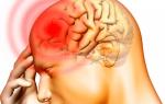
What function is not present in the digestive system. General scheme of the digestive tract. Diagram of the structure of the human stomach
The given sequence of digestion processes provides the most complete mechanical and chemical processing of the food bolus in order to extract all the necessary substances. The stages of the digestion process are discussed in this article. You can learn about the process of digestion in the human body, starting from the oral cavity and ending with the large intestine. It is very difficult to overestimate the importance of the digestion process, in fact it is a factor in maintaining the organic life of the body. normal process human digestion provides all the needs for proteins, fats and carbohydrates. WITH energy point view, the process of digestion in the body is necessary to extract calories in order to direct them to the work of the muscles and internal organs. On the same principle, the work of the brain and the entire central nervous system, including its function of thermoregulation.
Fundamentals of the physiology of digestion
Nutrition is a complex process of intake, digestion and absorption of nutrients. In recent decades, a special science of nutrition, nutriciology, has begun to actively develop. Consider the basics of the physiology of digestion in the human oral cavity, stomach and intestines.
Digestive system- a set of organs that ensure the absorption of the nutrients the body needs as an energy source for cell renewal and growth. Distinguish between cavity and membrane digestion. Abdominal is carried out in the oral cavity, stomach, small and large intestines. Membrane - at the level of the surface of the cell membrane and the intercellular space, characteristic of the small intestine.
Dietary proteins, fats, carbohydrates, vitamins, minerals cannot be absorbed by the body, its tissues and cells unchanged. Complex food substances are broken down by hydrolase enzymes released into the cavity digestive tract in certain areas of it. In the process of digestion, from high-molecular compounds, they gradually turn into low-molecular, soluble in water. Proteins are broken down by proteases into amino acids, fats by lipases into glycerol and fatty acids, carbohydrates - amylases to monosaccharides.
All these substances are absorbed in the digestive tract and enter the blood and lymph, i.e., into the liquid media of the body, from where they are extracted by tissue cells. The end products of digestion that are absorbed into the blood are simple sugars, amino acids, fatty acids and glycerol.
Vitamins, macro- and microelements in the digestive system can be released from bound state in which they are in food products, but the molecules themselves do not split.
The digestive system consists of several parts: the mouth, pharynx, esophagus, stomach, small intestine, large intestine, and rectum.
The essence, physiology and features of the processes of digestion in the human oral cavity
The essence of digestion in the oral cavity is that food is crushed. In the oral cavity, the processes of digestion conclude that there is an active processing of food with saliva (0.5-2 l is formed per day), the interaction of microorganisms and enzymes (amylases, proteinases, lipases). In saliva, some substances dissolve and their taste begins to appear. The physiology of digestion in the oral cavity is based on the fact that saliva contains the enzyme amylase, which breaks down starch into sugars.
So, the action of amylase is easy to trace: if you chew bread for 1 minute, you feel a sweet taste. Proteins and fats do not break down in the mouth. Average duration digestion in the oral cavity is minimal and is only 15-20 s.
Features of digestion in the oral cavity are that further food bolus(usually 5-15 cm3) moves into the stomach. The act of swallowing includes the oral (voluntary), pharyngeal (fast involuntary), esophageal (slow involuntary) phases. On this, the process of digestion in the human oral cavity is considered to be actually completed. The average duration of the passage of the food bolus through the esophagus is 2-9 s and depends on the density of the food. The digestive tract is provided with special valves to prevent backflow, as well as to differentiate the effects of digestive enzymes.
The processes of digestion that occur in the human stomach
The stomach is the widest part of the digestive tract, it can grow in size and contain a large number of food. Due to the rhythmic contraction of the muscles of the walls, digestion in the stomach begins with the fact that the food is thoroughly mixed with sour gastric juice.
The food bolus, once in the stomach, stays in it for 3-5 hours and is subjected to mechanical and chemical processing. Digestion processes in the stomach begin with the fact that food is exposed to gastric juice (2-2.5 liters are excreted per day) and hydrochloric acid present in it (provides an acidic environment), pepsin (digests proteins) and other acidic proteases such as rennin ( chymosin).
Pepsinogens (precursors of pepsin) are divided into two groups. The first, after activation with hydrochloric acid and transformation into pepsins, hydrolyzes certain types of proteins for the processes of digestion occurring in the stomach with the formation of large peptides at pH 1.5-2.0. The second fraction, after activation with hydrochloric acid, turns into gastrixin, which hydrolyzes food proteins at pH 3.2-3.5.
Enzymes in the process of digestion in the human stomach digest proteins to low molecular weight peptides and amino acids. The digestion of carbohydrates, which began in the mouth, stops in the stomach, because in acidic environment amylase loses its activity.
Features of the physiology of digestion in the cavity of the human stomach
Digestion in the human stomach is based on the action of gastric juice, which contains lipase, which breaks down fats. In digestion in the stomach cavity, hydrochloric acid of gastric juice plays an important role. Hydrochloric acid increases the activity of enzymes, causes denaturation and swelling of proteins, and has a bactericidal effect.
Normally, the acidity of gastric juice ranges from pH 1.6 to 1.8. Deviation of gastric juice from the norm is used in the diagnosis of stomach ulcers, anemia, tumors. Features of digestion in the stomach is that under the action of hydrochloric acid, many pathogens are deactivated.
The physiology of digestion in the stomach is such that food rich in carbohydrates stays in the stomach for about two hours, is evacuated faster than protein or fatty food, which lingers in the stomach for 8-10 hours.
Mixed with gastric juice and partially digested food in small portions, at certain intervals, when its consistency becomes liquid or semi-liquid, it passes into the small intestine.
Functions and features of the digestion process in the human small intestine
From the stomach, the food bolus enters the small intestine, the length of which in an adult reaches 6.5 meters. Digestion in small intestine is the most important from the biochemical point of view of the assimilation of substances.
Intestinal juice in this section of the digestive tract has alkaline environment due to the entry into the small intestine of bile, pancreatic juice and secretions of the intestinal walls. In some individuals, there is a slow process of digestion in the small intestine, due to a deficiency of the lactase enzyme, which hydrolyzes milk sugar(lactose), which is associated with indigestibility whole milk. In total, more than 20 enzymes are used in digestion in the human small intestine (enterokinases, peptidases, phosphatases, nucleases, lipase, amylase, lactase, sucrase, etc.).
The functions of digestion in the small intestine depend on its departments. The small intestine has three sections that pass into each other - the duodenum, jejunum and ileum. Bile is secreted into the duodenum, which is produced in the liver. In the duodenum, food is exposed to pancreatic juice, bile. The juice secreted by the pancreas is a colorless clear liquid with pH 7.8-8.4. Pancreatic (pancreatic) juice contains enzymes that break down proteins and polypeptides: trypsin, chymotrypsin, elastase, carboxypeptidases and aminopeptidases.
The pancreatic juice contains: lipase, which breaks down fats; amylase, which completes the complete breakdown of starch to a disaccharide - maltose; ribonuclease and deoxyribonuclease, splitting ribonucleic and deoxyribonucleic acids. The secretion of pancreatic juice, depending on the composition of the food, lasts 6-14 hours, it is the longest when taking fatty foods.
An important role in the process of digestion is played by the liver, where bile is formed (0.5-1.5 liters per day). Features of digestion in the small intestine are that bile promotes the emulsification of fats, the absorption of triglycerides, activates lipase, stimulates peristalsis, inactivates pepsin in the duodenum, has a bactericidal and bacteriostatic effect, enhances hydrolysis and absorption of proteins and carbohydrates.
Bile does not contain digestive enzymes, but is necessary for the dissolution and absorption of fats and fat soluble vitamins. With insufficient production of bile or its release into the intestine, the digestion and absorption of fats are disturbed, and their excretion increases unchanged with feces.
The final digestion of carbohydrates, protein residues, fats occurs in the jejunum and ileum with the help of enzymes that are produced by the cells of the mucous membrane of the intestine itself. Outgrowths of the wall of the small intestine are covered with enterocytes - villi. Through many villi from its surface, the breakdown products of proteins and carbohydrates enter the blood, and the breakdown products of fats enter the lymph. Due to the large number of special folds and villi, the total suction surface of the intestine is about 500 m2.
In the small intestine, the majority of simple chemical fragments of food are absorbed.
Physiology, functions and processes of digestion in the large intestine
Undigested food remains are then taken to the colon, in which they can be from 10 to 15 hours. In this section of the digestive tract, such processes of digestion in the intestine as the absorption of water and microbial metabolization of nutrients are carried out.
The length of the large intestine in an adult is on average 1.5 m. It consists of three parts - the blind, transverse colon and rectum.
Digestion in the large intestine is dominated by mechanisms reverse suction. It absorbs glucose, vitamins and amino acids produced by bacteria in the intestinal cavity.
An important role in the processes of digestion in the large intestine is played by dietary ballast substances. These include indigestible biochemical components: fiber, hemicellulose, lignin, gums, resins, waxes.
The basis of ballast components are substances plant origin, included in the structure of the walls of plants and contained in wood, seed husks, bran. Most of ballast substances are cellulose and branched polysaccharides based on xylose, arabinose, mannose, galactose. Ballast ingredients of animal origin include non-disposable human body elements connective tissue animals.
Collagen protein, resistant to the action of proteolytic enzymes, performs physiological functions digestion in the large intestine, similar to dietary fiber. Mucopolysaccharides that are not hydrolyzed in the intestine and contained in the intercellular substance of animal tissues have the same properties. The largest number of these structural polysaccharides is found in the connective tissue, lungs, blood.
The structuring of food affects the rate of absorption in the small intestine and the duration of transit through the gastrointestinal tract.
Dietary fibers and products of collagen thermohydrolysis have the ability to retain a significant amount of water, which significantly affects the pressure, mass and electrolyte composition of feces, contributing to the formation of soft feces.
Dietary fiber and indigestible connective tissue proteins are among the main components that make up the environment in which beneficial intestinal bacteria live.
Dietary fibers and connective tissue elements have great importance For electrolyte metabolism in the gastrointestinal tract. This is due to the fact that collagen, like polysaccharides, has cation exchange properties and helps to eliminate various harmful compounds from the body.
Dietary fiber in human diets reduces the risk of developing neoplastic diseases, peptic ulcer, diseases duodenum, diabetes, cardiovascular disease, render beneficial effect on the body of people overweight bodies suffering from atherosclerosis, hypertension and other diseases.
Dietary fibers that are not broken down by the enzymes of the gastrointestinal tract are partially destroyed under the influence of microflora.
Formed in the large intestine stool, consisting of undigested residues food, mucus, dead cells of the mucous membrane and microbes that continuously multiply in the intestine, causing fermentation and gas formation processes.
total weight intestinal microflora a person is 1.5-2.0 kg. The flora of the large intestine contains anaerobic species microorganisms: bifidobacteria (108-1010 cfu/g in adults, 109-10sh cfu/g in children), bacteroids (109-1010 cfu/g in adults, 106-108 cfu/g in children), lactobacilli (106-107 cfu/g /g in adults, 106-10 CFU/g in children), peptostreptococci, clostridia, which is up to 99% of the total composition. About 1% of the microflora of the large intestine is represented by aerobes: coli, enterobacteria (proteus, enterobacter, etc.), enterococci, staphylococci, yeast-like fungi. The amount of each species ranges from 104-108 CFU/g.
The process of splitting and absorption of substances in digestion
The process of absorption in digestion is the passage of nutrients from the cavity of the digestive tube into the cells intestinal epithelium and then into the blood. Preliminary breakdown of substances in the process of digestion is necessary to obtain products at the cellular and molecular level.
Absorption is carried out throughout the digestive tract, the surface of which is covered with villi. There are 30-40 villi per 1 mm2 of the mucosa. At the same time, 50-60% of the products of protein metabolism are absorbed in the duodenum; 30% - in the small intestine and 10% - in the large intestine. Carbohydrates are absorbed only in the form of monosaccharides. The products of fat metabolism, as well as most of the water- and fat-soluble vitamins that come with food, are absorbed in the small intestine.
"Anatomy digestive system»
Topic study plan:
General data on the structure of the organs of the digestive system.
The oral cavity and its contents.
The structure of the throat. lymphoepithelial ring. Esophagus.
The structure of the stomach.
Thin and colon, structural features.
The structure of the liver. Gallbladder.
Pancreas.
General information about the peritoneum.
General data on the structure of the organs of the digestive system.
The digestive system is a complex of organs whose function is to mechanically and chemically process food substances, absorb processed substances and remove the remaining undigested parts of food. The organs of the digestive system include the oral cavity with its contents, pharynx, esophagus, stomach, small intestine, colon, liver and pancreas.
The oral cavity and its contents.
The oral cavity is divided into the vestibule of the mouth and the oral cavity proper. The vestibule of the mouth is the space located between the lips and cheeks on the outside, the gums and teeth on the inside. Through the mouth opening, the vestibule of the mouth opens outward. The oral cavity itself is limited, respectively, in front - by teeth and gums, behind - communicates with the pharynx with the help of a pharynx, at the top - by a hard and soft palate, from below - by the tongue and the diaphragm of the oral cavity.
IN  The oral cavity contains teeth, tongue and ducts of the salivary glands. A person in the process of life has 20 dairy and 32 permanent tooth. They are divided into incisors (2), canines (1), small molars (2), large molars (2-3); milk teeth formula: 2 1 0 2, that is, there are no small molars. Formula permanent teeth: 2 1 2 3. Crown, neck and root are distinguished in each tooth. The crown is covered with enamel on the outside, the root is covered with cement, and the entire tooth consists of dentin, inside of which there is a cavity filled with pulp (containing nerves, blood vessels, connective tissue). With the help of teeth, mechanical processing of food takes place. The tongue is a muscular organ. He participates in the processes of formation of the food bolus and the acts of swallowing, speech formation; due to the presence of specific nerve endings on its mucous membrane, the tongue is also an organ of taste and touch. The basis of the tongue is striated voluntary muscles. They are distinguished by two groups: own muscles of the tongue (upper and lower longitudinal, vertical, transverse) and skeletal muscles (styl-lingual, genio-lingual and hyoid-lingual muscles). The contraction of these muscles makes the tongue mobile, easily changing shape. In the language, the body, apex, root, upper surface (back) and lower surface are distinguished. Outside, the tongue is covered with a mucous membrane. On the upper surface of the tongue there are papillae: mushroom-shaped, trough-shaped, conical, filiform and foliate. With the help of these
  structures, the perception of the taste of the food taken, its temperature and texture is carried out. On bottom surface tongue has a frenulum, on the sides of which there is a sublingual meat. They open a duct common to the sublingual and submandibular salivary glands. Additionally, in the thickness of the mucous membrane, oral cavity and tongue, a large number of small salivary glands are laid. In the vestibule of the oral cavity, the duct of the third major salivary gland, the parotid gland, opens. The mouths of the duct open on the buccal mucosa at the level of the upper second large molar. The salivary glands differ from each other in structure and in secret. So, the parotid gland belongs to the alveolar in structure and serous in secret; the submandibular gland, respectively, to the alveolar-tubular and mixed; sublingual - to the alveolar-tubular and mucous membranes.
The structure of the throat. lymphoepithelial ring. Esophagus.
G  tray - a hollow muscular organ. The pharyngeal cavity is divided into three parts: nasal, oral and laryngeal. The nasal part of the pharynx communicates with the nasal cavity through the choanae, with the middle ear cavity through auditory tube; the oral part of the pharynx communicates with the oral cavity through the pharynx, and the laryngeal part communicates with the vestibule of the larynx, and then passes into the esophagus. By function, the nasal part of the pharynx is respiratory, because. serves only to conduct air; the oral part of the pharynx is mixed - both respiratory and digestive, because. conducts both air and food bolus, and the laryngeal part is only digestive, tk. carries only food. The wall of the pharynx consists of mucous, fibrous, muscular and connective tissue membranes. The muscular membrane is represented by striated muscles: three pairs of muscles that compress the pharynx and two pairs of muscles that lift the pharynx. In the pharynx, a number of accumulations of lymphoid tissue are focally located. So, in the area of its arch lies the pharyngeal tonsil, in the place where the auditory tubes open - the tubal tonsils, the lingual tonsil is localized on the root of the tongue, and two palatine tonsils lie between the arches of the soft palate. The pharyngeal, palatine, lingual, and tubal tonsils form Pirogov's pharyngeal lymphoepithelial ring.
The esophagus is a flattened tube from front to back, 23-25 cm long. It begins at level VI cervical vertebra and passes into the stomach at level XI thoracic vertebra. It has three parts - cervical, thoracic and abdominal. There are five constrictions and two expansions along the esophagus. Three constrictions are anatomical and are preserved on the cadaver. These are pharyngeal (at the point where the pharynx passes into the esophagus), bronchial (at the level of the tracheal bifurcation) and diaphragmatic (when the esophagus passes through the diaphragm). Two narrowings are physiological, they are expressed only in a living person. Aortic (in the aortic region) and cardiac (when the esophagus passes into the stomach) narrowing. Extensions are located above and below the diaphragmatic constriction. The wall of the esophagus consists of three membranes (mucous, muscular and connective tissue). The muscular membrane has a peculiarity: in the upper part it consists of striated muscle tissue and gradually it is replaced by smooth muscle tissue. In the middle and lower thirds of the esophagus, there are only smooth muscle cells.
The structure of the stomach.
AND  stomach - muscular hollow organ, in which the cardial part, arch, body, pyloric part are distinguished. The stomach has an inlet (cardiac) and an outlet (pyloric), anterior and posterior walls, two curvatures - large and small. The wall of the stomach consists of four membranes: mucous, submucosal, muscular and serous. The mucous membrane is lined with a single-layer epithelium, has numerous tubular gastric glands. There are three types of glands: cardiac, gastric and pyloric. They consist of three types of cells: main (produce pepsinogen), parietal (produce hydrochloric acid) and accessory (produce mucin). The submucosa of the stomach is well developed, which contributes to the formation of numerous folds on the mucous membrane. This ensures close contact of food with the mucous membrane and increases the area of absorption of nutrients into the blood. The muscular layer of the stomach is presented unstriated muscle tissue and consists of three layers: outer - longitudinal, middle - circular and inner - oblique. The most pronounced circular layer is on the border between the pylorus and the duodenum and forms a muscular ring - the pyloric sphincter. The outermost layer of the stomach wall is formed by the serosa, which is part of the peritoneum. The stomach is located in the abdominal cavity. Under the action of gastric juice in the stomach, food is digested, all the enzymes of which act only in an acidic environment (pH = 1.5-2.0), and it is created by the presence of hydrochloric acid up to 0.5%. Food stays in the stomach from 4 to 10 hours, and in that part of the food bolus that has not yet been saturated with gastric juice, saliva enzymes break down carbohydrates, but this is a trace reaction. The stomach breaks down complex proteins into simpler ones. varying degrees complexity, under the action of pepsin, which was formed from pepsinogen as a result of activation with hydrochloric acid. Chymosin curdles milk proteins. Lipase breaks down emulsified milk fat. The formation and secretion of gastric juice is regulated by the neurohumoral pathway. I.P. Pavlov identified two phases - reflex and neurohumoral. In the first phase, secretion occurs when the receptors of smell, hearing, vision are stimulated, while eating and when swallowing. In the second phase, the secretion of the stomach is associated with irritation of the receptors of the gastric mucosa by food and the excitation of the brain centers of digestion.
Humoral regulation occurs due to the appearance in the blood of stomach hormones, products of digestion of proteins and various minerals. The nature of secretion depends on the quality and quantity of food, on the emotional state and health, and continues as long as there is food in the stomach. By contracting the walls of the stomach, food is mixed with gastric juice, which contributes to its better digestion and transformation into a liquid slurry. The transition of food from the stomach to the duodenum occurs in a dosed manner, and through neurohumoral regulation is dosed by the pyloric sphincter. The sphincter opens when the environment of the food that has left the stomach becomes neutral or alkaline, and after the release of a new portion with an acidic reaction, the sphincter contracts and stops the passage of food.
Small and large intestine, structural features.
The small intestine begins at the pylorus and ends at the beginning of the large intestine. The length of the small intestine in a living person is about 3 m, its diameter ranges from 2.5 to 5 cm. The small intestine is divided into duodenum, jejunum and ileum. The duodenum is short - 27-30 cm. Most of the intestine lies to the right of the bodies of I-II lumbar vertebrae in the region of the posterior wall abdominal cavity and for the most part it turns out to be located retroperitoneally, i.e. covered by the peritoneum only in front. The common bile duct and the pancreatic duct flow into the intestine, which, before flowing into the intestine, are connected and opened by a common opening for them on the major duodenal papilla. The duodenum consists of four parts: upper, descending, horizontal and ascending parts, and looks like a horseshoe that covers the head of the pancreas.
T  small intestine and ileum have considerable mobility, as they are covered with peritoneum on all sides and are attached to back wall abdominal cavity through the mesentery. The wall of the small intestine is composed of the mucosa, submucosa, muscularis and serosa. A distinctive feature of the small intestine is the presence of villi in the mucous membrane that covers its surface. In addition to the villi, the mucosa of the small intestine has numerous circular folds, due to which the area of absorption of nutrients increases. The small intestine has its own lymphatic apparatus, which serves to neutralize microorganisms and harmful substances. It is represented by single and group lymphatic follicles. The muscular membrane of the small intestine consists of two layers: the outer one is longitudinal and the inner one is circular. Thanks to the muscle layers in the intestine, peristaltic and pendulum movements are constantly carried out, which contribute to the mixing of the food mass. The reaction of the intestinal environment is alkaline, the main digestion takes place here. Enterokinase, an enzyme from the intestinal glands, converts inactive trypsinogen into active trypsin, which, together with chymotrypsin, breaks down proteins into amino acids. Lipase, activated under the influence of bile, breaks down fats to glycerol and fatty acids. Amylase, maltase, lactase break down carbohydrates into glucose (monosaccharides). In the jejunum and ileum, the digestion of food ends and the resulting products of digested food are absorbed. For absorption, the mucous membrane has a huge number of microvilli. Outside, the villi are covered with epithelial cells, in their center there is a lymphatic sinus, and along the periphery - blood capillaries 18-20 per 1 mm 2. Amino acids and monosaccharides are absorbed into the blood of the capillaries of the villi. Glycerin and fatty acids are absorbed mainly into the lymph and then into the blood. In the small intestine, food is almost completely digested and absorbed. Undigested residues enter the large intestine, mainly plant fiber by 50% unchanged.
The large intestine is divided into a number of parts: caecum with appendix, ascending colon, transverse colon, descending colon, sigmoid colon, and rectum. The length of the large intestine ranges from 1 to 1.5 m, its diameter is from 4 to 8 cm. The large intestine has a number of distinctive features from the small intestine: the walls have special longitudinal muscle cords - ribbons; swellings and omental processes. The wall of the large intestine is composed of the mucosa, submucosa, muscularis and serosa. The mucous membrane does not have villi, but has semilunar folds. The latter increase the absorption surface of the mucous membrane, in addition, there are a large number of group lymphatic follicles in the mucous membrane. A feature of the structure of the intestinal wall is the location of the muscular membrane. The muscular layer consists of outer - longitudinal and inner - circular layers. The circular layer of all sections of the intestine is continuous, and the longitudinal one is divided into three narrow ribbons. These ribbons begin at the point of origin of the appendix from the caecum and extend to the beginning of the rectum. At the same time, the ribbons of the longitudinal muscle layer are much shorter than the length of the intestine, which leads to the formation of swellings, separated from each other by furrows. Each furrow corresponds to inner surface semilunar fold intestine. The serous membrane covering the large intestine forms protrusions filled with adipose tissue - omental processes. The large intestine is separated from the small intestine by the ileocecal sphincter. The function of the large intestine is to absorb water, ferment carbohydrates, putrefy proteins, and form feces. In the large intestine, peristaltic and pendulum movements are performed. The large intestine does not have villi, and the glands produce a small amount of juice. Bacteria in the large intestine contribute to the breakdown of fiber and the synthesis of a number of vitamins. Putrefactive bacteria from the products of protein decay can form toxic substances - indole, skatole, phenol.
The large intestine absorbs water, decay products, fermentation, and the formation of feces. Blood from the intestines passes through the liver, where nutrients undergo a series of transformations and neutralization of toxic substances occurs.
The structure of the liver. Gallbladder.
P  the liver is the most big gland organism (its weight is about 1.5 kg). The functions of the liver are diverse: antitoxic function (neutralization of phenol, indole and other decay products that are absorbed from the lumen of the colon), participates in protein metabolism, synthesis of phospholipids, blood proteins, converts ammonia into urea, cholesterol into bile acids, is a blood depot and in the embryonic period of the liver, the function of hematopoiesis is inherent. In the liver, glucose is converted to glycogen, which is deposited in the liver cells and excreted into the blood as needed. Bile is also produced in the liver cells, which enters the lumen of the duodenum through the bile ducts. Excess bile accumulates in gallbladder. Up to 1200 ml of bile is formed and secreted per day. When digestion does not occur, bile accumulates in the gallbladder and enters the intestine as needed, depending on the presence and composition food taken. The color of bile is yellow-brown and is due to the pigment bilirubin, which is formed as a result of the breakdown of hemoglobin. Bile emulsifies fats, facilitating their breakdown, and also activates the digestive enzymes of the intestines. The liver is located in the abdominal cavity, mainly in the right hypochondrium. The liver has two surfaces: diaphragmatic and visceral. Divided into right and left lobe. The gallbladder lies on the lower surface of the liver. IN back section The inferior vena cava passes through the liver. The transverse groove on the lower surface of the liver is called the gates of the liver. The gate of the liver includes its own hepatic artery, portal vein and their accompanying nerves. From the gates of the liver exit: the common hepatic duct and lymphatic vessels. The structural unit of the liver is hepatic lobule, which has the shape of a prism and consists of numerous liver cells that form crossbars - trabeculae. Trabeculae are oriented radially - from the periphery of the lobule to the center, where the central vein lies. Along the edges of the prism lie the interlobular artery, vein and bile duct, which form hepatic triad. In the thickness of the trabeculae, which are formed by two rows of liver cells, there are bile ducts into which bile is produced. Through these grooves, it enters the interlobular bile ducts. Bile exits the liver through the common hepatic duct. As mentioned above, it serves as a reservoir for the accumulation of bile. gallbladder. The gallbladder is a hollow muscular organ that stores bile. It distinguishes the bottom, body and neck. The cystic duct leaves the neck and joins the common hepatic duct to the common bile duct. The wall of the gallbladder consists of mucous, muscular and serous membranes.
Pancreas.
P  The pancreas is not only a large gland of external secretion, but also an endocrine gland. It has a head, body and tail. The pancreas is located so that its head is covered by the duodenum (at the level of I-II lumbar vertebrae, to the right of them), and the body and tail go from the head to the left and up. The tail of the gland is directed towards the spleen. The length of the pancreas is 12-15 cm. Inside the gland, along its length, the pancreatic duct passes, into which the ducts from the lobules of the gland flow. The gland duct connects with bile duct and opens with a common opening for them into the duodenum at the top of the major papilla. Sometimes there is an additional duct. Most of the substance of the pancreas consists of alveolar-tubular glands that produce pancreatic juice. The lobules consist of glandular cells, where digestive enzymes are synthesized - trypsin, chymotrypsin, lipase, amylase, maltase, lactase, etc., which, as part of the pancreatic juice, enter the duodenum through the duct. Pancreatic juice is colorless, transparent, has an alkaline reaction, about 1 liter is produced per day. It is involved in the breakdown of proteins, fats and carbohydrates. In addition, in the substance of the gland there are specially arranged islets of Langerhans, which release hormones into the blood - insulin (reduces blood glucose) and glucagon (increases blood glucose). The pancreas lies retroperitoneally (extraperitoneal position).
The role of I.P. Pavlov in the study of the functions of the digestive system. Before Pavlov, the effect of individual enzymes and juices on many products was known, but it was not clear how these processes proceed in the body. A detailed study of the secretion of the glands became possible after the introduction of the fistula technique. For the first time, a Russian surgeon V.A. Basov in 1842. A fistula is a connection of organs with the external environment or other organs. I.P. Pavlov and his collaborators improved and applied new operations to create fistulas of the salivary glands, stomach, and intestines in animals to obtain digestive juices and determine the activity of these organs. They established that salivary glands reflexively excited. Food irritates the receptors located in the oral mucosa and excitation from them through the centripetal nerves enters the medulla oblongata, where the center of salivation is located. From this center, along the centrifugal nerves, excitation reaches the salivary glands and causes the formation and secretion of saliva. This is an innate unconditioned reflex.
Along with unconditioned salivary reflexes, there are conditioned salivary reflexes in response to visual, auditory, olfactory and other stimuli. For example, the smell of food or a sight causes salivation.
To obtain pure stomach juice, I.P. Pavlov proposed a method of imaginary feeding. In a dog with a gastric fistula, the esophagus was cut at the neck and the cut ends were sutured to the skin. After such an operation, food enters the stomach, and falls out through the opening of the esophagus, and the animal can eat for hours without being satiated. These experiments make it possible to study the influence of reflexes from the receptors of the oral mucosa on the gastric glands. But this operational technique cannot fully reproduce the conditions and processes in the stomach, since there is no food in it. To study the processes of digestion in the stomach, I.P. Pavlov performed the operation of the so-called small ventricle. The small ventricle was cut out from the wall of the stomach so that neither the nerves nor the vessels connecting it with the large one were damaged. The small ventricle represents a section of the large one, but its cavity is isolated from the latter by a wall of fused mucous membrane, so that food digested in the large ventricle cannot enter the small one. With the help of a fistula, the small ventricle communicates with the external environment, and the function of the stomach was studied by secretion of juice. Works by I.P. Pavlov on the study of the digestive organs formed the basis for the treatment of these organs, the system of therapeutic nutrition and food regimen healthy person.
Suction is a complex physiological process, as a result of which nutrients pass through the layer of cells in the wall of the digestive tract into the blood and lymph. The most intensive absorption occurs in the jejunum and ileum. In the stomach, monosaccharides, minerals, water and alcohol are absorbed, in the large intestine - mainly water, as well as some salts and monosaccharides. Medicinal substances, depending on the chemical and physico-chemical properties, as well as on one or another dosage form, can be absorbed in all parts of the digestive tract. The absorption process is provided by filtration, diffusion and active transfer, regardless of the difference in the concentration of dissolved substances. Of great importance is the motor activity of the villi. The total surface of the mucous membrane of the small intestine due to villi is 500 m 2 . Amino acids and carbohydrates are absorbed into the venous part of the capillary network of the villi and enter portal vein passing through the liver, enter the general circulation. Fats and their breakdown products enter the lymphatic vessels of the villi. In the epithelium of the villi, the synthesis of neutral fats occurs, which, in the form of tiny droplets, enter the lymphatic capillaries, and from there with the lymph into the blood.
Suction water by diffusion begins in the stomach and intensively occurs in the small and large intestines. A person consumes about 2 liters of water per day. Besides, in gastrointestinal tract receives about 1 liter of saliva, 1.5-2.0 liters of gastric juice, about one liter of pancreatic juice, 0.5-0.7 liters of bile, 1-2 liters of intestinal juice. In just a day, 6-8 liters of fluid enter the intestines, and 150 ml is excreted with feces. The rest of the water is absorbed into the blood. Minerals dissolved in water are absorbed mainly in the small intestine by active transport.
HYGIENIC CONDITIONS FOR NORMAL DIGESTION
Diseases of the digestive system are quite common. The most common are gastritis, peptic ulcer of the stomach and duodenum, enteritis, colitis and cholelithiasis.
Gastritis is an inflammation of the lining of the stomach. It occurs under the influence of various pathogenic factors: physical, chemical, mechanical, thermal and bacterial agents. Of great importance in the development of the disease is a violation of the regimen and quality of nutrition. With gastritis, secretion is disturbed and the acidity of gastric juice changes. Disorder of the function of the stomach with gastritis is often reflected in the activity of other organs of the digestive system. Gastritis is often accompanied by inflammation of the small intestine (enteritis), and inflammation of the large intestine (colitis) and inflammation of the gallbladder (cholecystitis). Peptic ulcer disease is characterized by the fact that non-healing ulcers form in the stomach or duodenum. Peptic ulcer disease is not a local process, but the suffering of the whole organism. In the development of the disease, neuropsychic injuries, increased excitability of the receptor apparatus of the gastrointestinal tract, and reduced resistance of the mucous membrane to the digestive action of gastric juice play a role. A certain role in the development of peptic ulcer is given to hereditary factors.
Serious diseases such as typhoid fever, dysentery, cholera, poliomyelitis and others. These diseases usually occur with poor water supply, the use of unwashed vegetables and fruits with which pathogenic microbes are transmitted, and personal hygiene is not followed.
Regulation of digestive processes. Physiological studies of digestion were carried out by I.P. Pavlov. The entire cycle of his published works is called "Works on the Physiology of Digestion", which included such as "On the reflex inhibition of salivation" (1878), "On a surgical method for studying the secretory phenomena of the stomach" (1894), "On the digestive center" ( 1911) and others.
Prior to Pavlov's work, only unconditioned reflexes were known, and Pavlov established the enormous importance of conditioned reflexes. He found that gastric juice is secreted in two phases. The first begins as a result of food irritation of the receptors of the oral cavity and pharynx, as well as visual and olfactory receptors (the type and smell of food). The excitation that has arisen in the receptors along the centripetal nerves enters the digestive center located in medulla oblongata, and from there - along the centrifugal nerves to the salivary glands and glands of the stomach. Juice secretion in response to irritation of the receptors of the pharynx and mouth is unconditioned reflex, and the secretion of juice in response to irritation of the olfactory and taste receptors is a conditioned reflex. The second phase of secretion is caused by mechanical and chemical stimuli. In this case, acetylcholine, hydrochloric acid, gastrin, as well as food components and products of protein digestion serve as irritants. You should have an idea about the concept of "hunger" and "appetite". Hunger is a condition that requires a certain amount of food to be eliminated. Appetite is characterized by a selective attitude to the quality of the food offered. Its regulation is carried out by the cerebral cortex, it depends on numerous mental factors.
The digestive system performs a number of functions:
-mechanical function, or crushing of food, is carried out with the help of teeth in the oral cavity and due to mixing in the stomach and small intestine, as well as transporting the food bolus through the digestive tract due to contraction of the muscular membrane (peristalsis);
-secretory function consists in the synthesis and secretion of digestive enzymes by the digestive glands;
-chemical function It consists in the chemical processing of food (digestion) with the help of digestive enzymes. The primary chemical processing of food begins in the oral cavity and ends in the small intestine, where the final chemical processing takes place. In the large intestine and at the junction of the large and small intestines inhabited by intestinal microflora- symbiotic microorganisms that help us digest plant and dairy foods;
- suction function ensures the absorption of digestion products into the blood and lymph. Partial absorption of carbohydrates begins in the oral cavity, continues in the stomach, where protein breakdown products begin to be absorbed. The main absorption occurs in the small intestine. It should be noted that the products of lipid digestion are absorbed into the lymph;
-excretory function- excretion of undigested food residues and waste products;
-endocrine- secretion of digestive hormones.
Oral cavity, or oral cavity(Fig. 1)
Rice. 1.The oral cavity and pharynx: 1 - upper and 2 - lower lip; 3 - pharynx; 4 - language; 5 - palatoglossal and 6 - palatopharyngeal arches; 7- palatine tonsil; 8 - tongue; 9 - soft and 10 - hard palate; 11 - gums
Teeth(Fig. 2). Main function- capture and primary mechanical processing of food (grinding).
In humans, there are two types of teeth, depending on the time of appearance:
-baby teeth(temporary). A child has 20 milk teeth, which function until they are replaced by permanent teeth at the age of 7 to 13-14 years. On each half of the jaw, 2 incisors, 1 canine, 2 large molars are distinguished;
-permanent teeth. A person has 32 permanent teeth: in each half of the jaw there are 2 incisors, 1 canine, 2 small molars and 3 large molars.

Rice. 2.Scheme of the structure of the tooth: I - enamel; 2 - dentin; 3 - tooth pulp; 4 - gum; 5 - cement; 6 - periodontal; 7-bone; I - tooth crown; II - the neck of the tooth; III - tooth root; IV - root canal
Language. A mobile muscular organ, dressed in a mucous membrane, richly supplied with blood vessels and nerves.
The mucosa is rich in taste buds - papillae(Fig. 3). Distinguish: filiform And fungiform papillae- scattered over the entire upper surface of the tongue; papillae, rolled, - in the amount of 7-11 are located on the border of the body and the root of the tongue; foliate papillae - clearly visible along the edges of the tongue. On bottom side there is no papillary tongue.
The tongue is involved in the process of sucking, swallowing, speech articulation, is an organ of taste (fungiform and foliate papillae perceive sour, sweet and salty tastes, and papillae with a roller - bitter).

Rice. 3.Language: 1 - the root of the language; 2 - filiform, 3 - mushroom-shaped, 4 - surrounded by a roller and 5 - foliate papillae; 6 - blind fossa; 7 - palatine-lingual fold; 8 - palatine and 9 - lingual tonsils; 10 - epiglottis
Pharynx
Muscular organ that connects the mouth to the esophagus nasal cavity with the larynx, i.e. in the pharynx the digestive and Airways . The pharynx is divided into three parts: nasopharynx, oropharynx And guttural part. Located in the throat six tonsils. Nasopharynx through choanae communicated with nasal cavity. On the side walls are openings of the auditory (Eustachian) tubes, which connect it with the cavity middle ear, helping to equalize pressure in the middle ear with external pressure. tonsils perform important protective and partly hematopoietic functions. Sharp increase tonsils - the first sign of angina, scarlet fever, diphtheria.
Esophagus
It is a muscular tube about 25 cm long (Fig. 4). It begins without sharp boundaries from the pharynx at the level of the VI cervical vertebra and at the level of the XI thoracic vertebra opens into the stomach. The muscular layer has the following features: in the upper third it consists of striated muscles, A in the lower third - only from smooth muscles. The main function of the esophagus is to carry the food bolus to the stomach. Partially the esophagus performs protective function with the help of three constrictions (it is in these constrictions that accidentally swallowed foreign objects). It does not have its own digestive glands, digestion is carried out by saliva enzymes. It has an alkaline environment.

Rice. 4.The structure of the wall of the esophagus. Mucosa (I), muscular (II) and serous (III) membranes: 1 - multilayer squamous epithelium; 2 - own and 3 - muscular layers of the mucous membrane; 4 - submucosal layer; 5 - mucous gland; 6 - layer of circular and longitudinal (7) muscles
Stomach
The only expanded part of the digestive tube up to 5 liters (Fig. 5). Distinguish inlet (cardia), bottom, body And exit (gatekeeper). At the entrance and exit there are circular muscles-contactors (sphincters). The muscular layer has three types of muscles: longitudinal, ring And oblique.
The stomach performs several functions: mechanical processing of food due to mixing, temporary storage and chemical processing of food, and partial absorption. Chemical processing of food is carried out by gastric juice secreted by own glands. Gastric juice It has acidic environment(pH 2). glands are made up of three types of cells: main that secrete digestive enzymes lining, highlighting hydrochloric acid, And additional that secrete mucus.

Rice. 5.Stomach with an open anterior wall (A) and its muscular membrane (B): 1 - cardial part; 2 - cardiac opening; 3 - bottom of the stomach; 4 - the body of the stomach; 5 - small and 6 - large curvature of the stomach; 7 - pyloric (pyloric) part; 8 - gatekeeper; 9 - pylorus hole; 10 - muscular membrane; 11 - longitudinal (outer) layer; 12 - circular layer; 13 - pyloric sphincter; 14 - oblique fibers
Small intestine
The longest part of the digestive tract (up to 5 m) is divided into three parts: duodenum, skinny And ileum. characteristic feature is the presence villi formed by the mucous membrane (Fig. 6, 7). villi have microvilli, formed villus epithelium. On the border with the stomach and large intestine there are sphincters. Ducts open into the duodenum pancreas And gallbladder.

Rice. 6.The mucous membrane of the small intestines. A - skinny; B - iliac: 1 - muscular membrane; 2 - mesentery; 3- serosa; 4 - single follicles; 5 - circular folds; 6 - mucous membrane; 7 - group follicles

Rice. 7.Scheme of the structure of the villi of the small intestine: 1 - intestinal epitheliocytes; 2 - goblet cells; 3 - central lymphatic sinus; 4 - arteriole; 5 - venule; 6 - blood capillaries
The small intestine is the organ in which the breakdown of proteins, fats and carbohydrates is finally completed And digestion products are absorbed as well as salts and water. Digestion takes place under the influence intestinal juice allocated intestinal glands, pancreatic juice secreted by the pancreas, and bile. Available abdominal And parietal digestion.
Colon
It has a length of up to 2 m and a diameter of up to 5-7 cm. It consists of three sections: the caecum with the appendix (Fig. 8), the colon and rectum. There is a large number of symbiotic bacteria here. The main functions of the large intestine are the absorption of water and the formation of feces. Due to the presence of bacteria, fiber fermentation And protein putrefaction, a number of bacteria synthesize vitamins.

Rice. 8.Caecum with appendix (appendix): 1 - appendix(appendix); 2 - opening of the appendix; 3 - caecum; 4 - opening of the small intestine; 5 - large intestine; 6 - colon
digestive glands
Salivary glands . The salivary glands secrete saliva, which is protein secretion(serous) and mucous component. Protein secretion is isolated parotid glands , slimy - palatine And back lingual; submandibular And sublingual- mixed secret. The main components of saliva are: mucin- mucous protein substance, lysozyme- bactericidal agent, amylase enzymes And maltase.
Distinguish small And major salivary glands. The small ones are labial, buccal, dental, lingual, palatine. These glands are located in the corresponding parts of the oral mucosa. There are three pairs of major salivary glands: parotid, submandibular And sublingual; they lie outside the oral mucosa, but excretory ducts open into the mouth.
Liver - the largest gland (weight up to 1.5 kg). Most of it is in the right hypochondrium, the smaller one goes into left side abdominal cavity. The main secret that the liver secretes into the digestive system is bile. Bile emulsifies fats, activates fat-splitting enzymes of the pancreas, but does not contain enzymes itself. Carbohydrates are converted to glycogen in the liver. The liver also performs a barrier function, neutralizing toxic substances that appear in the body in the process of metabolism. Outside of the digestion process, bile is collected in the gallbladder.
Pancreas - digestive gland 20 cm long and 4 cm wide, located behind the stomach. The pancreas is related to mixed glands. The exocrine part produces pancreatic juice, containing trypsinogen, amylase, maltase, lactase, lipase, nuclease. The endocrine part produces hormones: insulin And glucagon.
Digestive enzymes
The main function of the digestive system - digestive - is performed by specialized proteins - digestive enzymes. In each section of the digestive tract, specific enzymes function that contribute to the digestion of certain substances.
Digestive enzymes
|
glands |
Enzymes |
What is splitting |
Final product |
|
Starch. Glycogen |
Maltose |
||
|
Maltase |
Maltose |
Two molecules of glucose |
|
|
Glands of the stomach |
|||
|
milk protein |
Denaturation - curdling |
||
|
pancreas |
Protein. Peptides |
Dipeptides. Amino acids |
|
|
Maltose |
|||
|
Fatty acid. Glycerol |
|||
|
Liver and gallbladder |
Bile salts and bile alkalis do not contain digestive enzymes |
Activation of digestive enzymes, emulsification of fats, absorption of fatty acids |
|
|
Glands of the small intestine |
Sucrase |
sucrose |
Fructose. Glucose |
|
Maltase |
Maltose |
||
|
Glucose. Galactose |
|||
|
Phosphatase |
organic phosphates |
free phosphate |
vitamins
vitamins called a group of biologically active organic compounds of various chemical nature, entering the body with food of plant and animal origin. Some vitamins are synthesized microbial flora of the intestine. Vitamins are present in food in negligible amounts, and the body also needs them in small quantities, but at the same time they play a very important role in exchange processes, often being integral part enzymes. In the absence of any vitamin or its precursor in the body, disease occurs - avitaminosis. But, although vitamins are important for the body, their overdose (intoxication) due to intake higher doses also leads to painful manifestations and called hypervitaminosis.
Vitamins are divided into two groups depending on the solvents in which they dissolve: fat-soluble(vitamins A, D, E, K) and water soluble(vitamins of group B, PP, C, etc.).
Almost everyone believes that of all the organs of the digestive system, the main role in the digestion of food is assigned to the stomach. Not exactly the right guess. The stomach is indeed an important and necessary organ of the digestive system, but still the main digestion takes place in the lesser known.
Despite this, each organ of the digestive system is important in its own way and performs strictly assigned functions. Therefore, it is difficult to single out one of the departments of the digestive system and call it the main one.
Gastrointestinal organs
The gastrointestinal tract (GIT) includes:
- oral cavity;
- pharynx;
- esophagus;
- stomach;
- (duodenum, jejunum, ileum);
- (caecum, colon, rectum).
The length of the digestive tract is about 9-10 meters. The whole process of digestion of one meal takes about 12-48 hours, in some cases it can be more. This is due to the fact that each section of the digestive tract performs strictly defined functions in the digestion of food, bypassing which the entire digestive process is disrupted.
What happens in the stomach
From the mouth through the esophagus, the food bolus (chyme) enters the stomach. Previously, it is already moistened with saliva and crushed with teeth, partially processed with enzymes contained in saliva, moistened and warmed to the desired temperature in the esophagus. Food is stored in the stomach for 2-4 hours. During this time, it is partially processed, subjected to careful crushing for subsequent shipment in small portions to next department GIT.
 Cells of the stomach secrete pepsinogen, hydrochloric acid, mucin, a small amount lipase and amylase. Hydrochloric acid disinfects food masses from pathogens, activates stomach enzymes, denatures proteins and participates in neurohumoral regulation passage of food into the duodenum. In an acidic environment, pepsinogen is converted to pepsin, which triggers the initial breakdown (hydrolysis) of proteins to amino acids. Amylase is involved in the breakdown of carbohydrates to glucose, lipase breaks down fats into fatty acids and glycerol. Mucin is involved in mucus formation to protect the walls of the stomach from the effects of hydrochloric acid. Partially digested food enters the duodenum.
What happens in the duodenum
The main part of carbohydrates, fats, nucleic bases, part of proteins and other compounds have yet to be split into separate components. For these purposes, the ducts of the pancreas and gallbladder open into the intestinal lumen.
The pancreas synthesizes and secretes up to 2 liters of juice per day, which contains the following active compounds:
- Trypsin. Under its action, proteins are hydrolyzed to amino acids.
- Lipase, phospholipase and esterase are involved in fat metabolism.
- amylase and maltase. These enzymes break down carbohydrates into glucose.
- Lactase. Affects lactose in dairy products.
- Nucleases hydrolyze nucleic bases.
- Bicarbonates create alkaline reaction for enzymes, help in neutralizing hydrochloric acid.
 Bile enters from the gallbladder, which activates pancreatic enzymes, creates a special weakly alkaline reaction for their work, promotes the absorption of fatty acids into the systemic circulation, enhances intestinal motility, neutralizes gastric hydrochloric acid, and participates in the hydrolysis of fats.
Food molecules, breaking up into fragments with a lower molecular weight are absorbed into the blood from the small intestine. Digestion in the small intestine takes 1-4 hours.
The large intestine is involved in water-salt exchange, forms fecal masses from undigested food residues for subsequent excretion from the body. Right here. Food remains in the large intestine for 24-48 hours.
The coordinated work of all departments of the digestive system provides the body with the necessary substances for further life. Disabling any organ leads to qualitative and quantitative disturbances in the processes of digestion, absorption and excretion, therefore it is difficult to single out one of the departments and call it the main one.
Proper functioning of all organs of the human body is the key to health.
At the same time, the digestive system is one of the most important, as it involves the daily performance of its functions.
The structure and functions of the human digestive system
The components of the digestive system are the gastrointestinal tract (GIT) and auxiliary structures . The whole system is conditionally divided into three sections, the first of which is responsible for mechanical processing and processing, in the second section the food is subjected to chemical processing, and the third is designed to remove undigested food and surpluses from the body.

Based on this division, the following functions of the digestive system follow:
- Motor. This function involves the processing of food mechanically and its movement along the gastrointestinal tract (food is crushed, mixed and swallowed by a person).
- Secretory. As part of this function, special enzymes are produced that contribute to the formation of conditions for the chemical processing of incoming food.
- Suction. To perform this function, the intestinal villi absorb nutrients, after which they enter the bloodstream.
- excretory. As part of this function, substances are removed from the human body that are not digested or are the result of metabolism.
Human gastrointestinal tract
It is advisable to start the description of this group with the fact that the gastrointestinal tract involves a composition of 6 separate elements (stomach, esophagus, etc.).

As functions of the tract, they separately study the motor, secretory, absorption, endocrine (consists in the production of hormones) and excretory (consists in the release of metabolic products, water and other elements into the body).
Oral cavity
Cast initial department The gastrointestinal tract protrudes from the oral cavity. It becomes the beginning of the food processing process. Produced mechanical processes cannot be imagined without the participation of the tongue and teeth.

Such processes cannot do without the work of auxiliary structures.
Pharynx
The pharynx is an intermediate link between oral cavity and esophagus. The human pharynx is presented in the form of a funnel-shaped canal, which narrows as it approaches the esophagus (the wide part is at the top).

The principle of the pharynx is that food enters the esophagus by swallowing in portions, and not all at once.
Esophagus
This section connects the pharynx and stomach. Its location starts from chest cavity and ends in the abdominal cavity. Food passes through the esophagus in seconds.

Its main purpose is to prevent the reverse movement of food up the alimentary canal.
Diagram of the structure of the human stomach
Physiology assumes such a structure of the stomach, the functioning of which is impossible without the presence of three membranes: the muscular membrane, the serous membrane and the mucous membrane. The mucosa produces useful material. The other two shells are for protection.

In the stomach, processes such as the processing and storage of incoming food, the breakdown and absorption of nutrients occur.
Diagram of the structure of the human intestine
After the processed food stays in the stomach and performs a number of functions in the corresponding departments, it enters the intestines. It is arranged in such a way that it involves the division into the large and large intestine.

The sequence of passage of food is as follows: first it enters the small intestine, and then into the large intestine.
Small intestine
The small intestine consists of the duodenum (where the main stage of digestion takes place), the jejunum, and the ileum. If we briefly describe the work of the duodenum, then acid is neutralized in it, and substances and enzymes are broken down. Both the jejunum and the ileum take an active part in the process of absorption of important elements by the body.
Colon
The final part of food processing takes place in the large intestine. The first section of the large intestine is the caecum. Then food mixture Fall into colon, after which the principle of the sequence of passage through the ascending, transverse, descending and sigmoid colon works.
Then the food mixture enters the rectum. In the colon, substances are finally absorbed, the process of formation of vitamins takes place and feces are formed. The large intestine is by far the largest section of the digestive system.
Subsidiary Bodies
Auxiliary organs consist of two glands, the liver and gallbladder. The pancreas and liver are considered large digestive glands. The main function of excipients is to promote the digestive process.
Salivary glands
Job location salivary glands- oral cavity.

With the help of saliva, food particles are soaked and easier to pass through the channels of the digestive system. At the same stage, the process of splitting carbohydrates begins.
Pancreas
Iron belongs to the type of organs that produce hormones (such as insulin and glucagon, somatostatin and ghrelin).

In addition, the pancreas secretes an important secret, it is necessary for normal operation food digestion systems.
Liver
One of the most important organs digestive systems. It cleanses the body of toxins and unnecessary substances.

The liver also produces bile, which is necessary for the digestion process.
gallbladder
Helps the liver and serves as a kind of container for the processing of bile. At the same time, it removes from bile excess water, thereby forming a concentration that is suitable for the digestion process.
When studying human anatomy, it is important to know and understand that the successful functioning of each of the organs and sections of the digestive system is possible with the positive work of all other interconnected parts.