Edwards disease. Edwards syndrome - causes, symptoms, diagnosis and treatment. Deviations in the syndrome
Most women are well aware of what Down syndrome is. And during pregnancy, many learn that very rarely, but there is another chromosomal disorder called Edwards Syndrome. And many are concerned about how to find out how high the risk of having a child with Edwards syndrome is and how such a pathology is diagnosed during pregnancy?
What is Edwards Syndrome?
Edwards syndrome is a genetic disease characterized by duplication (trisomy) of the ХVIII chromosome and is manifested by a number of characteristic malformations in the fetus during pregnancy, often leading to the death of the child or his disability. That is, in a child, instead of 46 chromosomes, 47 are formed, this extra chromosome gives another name for the disease - trisomy 18. The syndrome was named after the researcher John Edwards, who first described it in 1960.
Why does Edwards syndrome occur - causes of pathology
Even if the parents are healthy, and there is no such pathology in the family history, a child with chromosome 18 can be born to any woman.
As you know, in each human cell there are 46 chromosomes, and in female and male germ cells, 23 chromosomes each, which, when combined during the fertilization of an egg, also give a total of 46 chromosomes. If we talk about Edwards syndrome, the causes of its appearance are unknown.
Currently, it is only known that as a result of certain genetic mutations, an extra 47th chromosome appears (an additional chromosome in the 18th pair of chromosomes, which thus becomes not 2, but 3).
In 95% of all cases of the development of Edwards syndrome, it is the extra 18th chromosome (trisomy) in the cells, but in 2% there is only an "elongation" of the 18th chromosome (translocation), when the total number of chromosomes remains normal and equals 46.
In 3% of cases of Edwards syndrome, “mosaic trisomy” occurs, when an additional 47th chromosome is found in the body not in all cells, but only in a certain part of it. Clinically, all 3 variants of Edwards syndrome proceed in almost the same way, however, the first variant may differ in a more severe course of the disease.
How common is this pathology?
Children with Edwards syndrome die in utero in approximately 60% of cases. Despite this, among genetic diseases, this syndrome in surviving infants is quite common, second only to Down's syndrome in frequency of occurrence. The prevalence of Edwards syndrome is 1 case in 3 - 8 thousand children.
It is believed that in girls this disease occurs 3 times more often, and the risk of Edwards syndrome increases significantly if a pregnant woman is 30 years of age or older.
Mortality in Edwards syndrome in the first year of life is about 90%, and the average life expectancy in severe cases of the disease in boys is 2-3 months, and in girls - 10 months, and only a few survive to adulthood. Most often, children with Edwards syndrome die from suffocation, pneumonia, cardiovascular failure or intestinal obstruction - complications caused by congenital malformations.
How does the syndrome manifest itself in a child?
The symptoms of Edwards syndrome can be divided into several large groups:
The first group includes symptoms characteristic of the appearance of the child:
- At birth, low body weight (about 2100 - 2200 grams)
- Disproportionately small head
- Defects in the development of the upper or lower jaws (micrognathia)
- Distortion of the shape of the face and the formation of malocclusion
- Cleft palate (cleft palate) or cleft lip (cleft lip)
- The fingers of the hand are clenched, they are uneven in the fist
- Low set ears
- Webbing or complete fusion of the fingers of the lower extremities
- congenital clubfoot
- "Rocking foot"
- Relatively small oral fissure (microstomy)
The second group includes signs of disruption of the internal organs, motility and neuropsychic development:
- The presence of congenital heart defects, for example, an open foramen ovale, a ventricular septal defect, an open ductus arteriosus, etc.
- Development of inguinal or umbilical hernias.
- Digestive organs: gastroeosophageal reflux disease, violation of the swallowing and sucking reflex, atresia of the esophagus or anus, Meckel's diverticulum, violation of the location of the intestine.
- Central nervous system: delayed neuropsychic development, mental retardation, underdevelopment of the cerebellum, corpus callosum, smoothing or atrophy of the cerebral convolutions.
- Genitourinary system: cryptorchidism, hypospadias in boys, clitoral hypertrophy, underdevelopment of the ovaries in girls, regardless of gender - horseshoe or segmented kidney, doubling of the ureters.
- Strabismus, scoliosis, muscle atrophy.
How to find out the pathology during pregnancy - diagnosis
Patau syndrome, Edwards syndrome and other trisomies are best identified before the baby is born. As a rule, prenatal diagnosis of this syndrome is carried out in 2 stages:
- For a period of 11–13 weeks (screening, which is based on various biochemical tests in a woman).
- Determination of the karyotype of the fetus in pregnant women of their risk group.
At 11–13 weeks, the level of certain blood proteins is determined in the woman’s blood: β-hCG (β-subunit of human chorionic hormone) and plasma protein A associated with pregnancy. Then, taking into account these data, the age of the pregnant woman, the risk of having a child with Edwards syndrome is calculated, and a risk group for pregnant women is formed.
Further, in the risk group at a later date, material is taken from the fetus to make an accurate diagnosis: at 8–12 weeks it is a chorionic villus biopsy, at 14–18 - amniocentesis (study of amniotic fluid), after 20 weeks - cordocentesis (intrauterine blood sampling from the umbilical cord fetus with ultrasound guidance). After that, the presence or absence of an additional 18th chromosome is determined in the obtained material using KF-PCR (quantitative fluorescent polymerase chain reaction).
If the pregnant woman has not passed a genetic screening examination, then at a later date, a preliminary diagnosis of Edwards syndrome is carried out using ultrasound. Other indirect signs, on the basis of which Edwards syndrome can be suspected at a later date:
- The presence of anomalies in the development of bones and soft tissues of the head (“cleft palate”, low-set auricles, “cleft lip”, etc.).
- Detection of defects on the part of the cardiovascular, genitourinary system, as well as the musculoskeletal system.
Diagnostic signs of the syndrome in a child
After the birth of a child, the main diagnostic signs of the presence of Edwards syndrome are the following:

Signs of a characteristic dermatographic picture:
- undeveloped distal flexion fold on the fingers
- the presence in 1/3 of cases of a transverse palmar groove
- arches on the fingertips
- change in the skin pattern of the palm: the distal location of the axial triradius and an increase in the ridge count.
Edwards syndrome on ultrasound - choroid plexus cysts
In the early stages, it is extremely difficult to suspect Edwards syndrome on ultrasound, however, at 12 weeks of pregnancy, symptoms of an indirect nature characteristic of it are already revealed. :
- Signs of fetal growth retardation
- Bradycardia (decreased fetal heart rate)
- Omphalocele (presence of a hernia of the abdominal cavity)
- No visualization of the nasal ossicles
- There is one artery in the umbilical cord, not 2
Also on ultrasound, vascular plexus cysts, which are cavities with fluid contained in them, can be detected. By themselves, they do not pose a threat to health and disappear by the 26th week of pregnancy. However, such cysts very often accompany various genetic diseases, for example, Edwards syndrome (in this case, cysts are found in 1/3 of children suffering from this pathology), therefore, if such cysts are found, the doctor will refer the pregnant woman to a genetic consultation for examination.
Treatment
Since children with Edwards syndrome rarely live up to a year, treatment is first aimed at correcting those malformations that are life-threatening:
- restoration of the passage of food in atresia of the intestine or anus
- feeding through a tube in the absence of swallowing and sucking reflexes
- antibacterial and anti-inflammatory therapy for pneumonia
With a relatively favorable course of the disease, some anomalies and malformations are corrected: surgical treatment of the "cleft palate", heart defects, inguinal or umbilical hernia, as well as symptomatic drug treatment (prescription of laxatives for constipation, "foam suppressants" for flatulence, etc.) . 
Children with Edwards syndrome are prone to diseases such as:
- otitis media
- kidney cancer (Wilms tumor)
- pneumonia
- conjunctivitis
- pulmonary hypertension
- apnea
- high blood pressure
- frontitis, sinusitis
- urinary tract infections
Therefore, the treatment of patients with Edwards syndrome includes timely diagnosis and treatment of these diseases.
Prognosis for a child
In most cases, the prognosis is poor. The few children with Edwards syndrome who survive to adulthood have severe mental disabilities and constantly require outside care and supervision. However, with appropriate activities, they are able to respond to comfort, smile, eat independently, and interact with caregivers, acquiring various skills and abilities.
The vast majority of people know that there is such a disease as Down syndrome. Someone knows that this is a chromosomal disease, and someone believes that this is a mental or neurological deviation from the norm. But few people know that there are much more chromosomal diseases, and one of them is Edwards syndrome. Perhaps this is due to the short period of life of children born with Edwards syndrome, and the relatively low percentage of such children to healthy children (1 child with the syndrome per 7000 live births and 6000 all newborns).
Information The percentage of natural miscarriage with a genetically defective fetus and the percentage of abortions after the diagnosis of the fetus is high.
What it is?
Edwards syndrome is a severe chromosomal disorder also called trisomy 18. The disease is accompanied by severe pathologies of all organs and multiple malformations. Most often, girls are born with Edwards syndrome: there are three times more of them than boys with the same pathology. Perhaps this is due to the fact that the natural life-support mechanism of female fetuses “works” better.
Additionally Australian medical scientists studied placentas and came to the conclusion that they have a different structure and carry out their protective functions in different ways depending on the sex of the gestating fetus. Previously, statistics noted that the gestation rate of girls with malformations is higher than that of boys with similar deviations, and the mortality among newborn girls is lower than among boys, all other things being equal.
Causes of Edwards Syndrome
The cause of Edwards syndrome is the presence of an extra 18th chromosome in the karyotype of the zygote. Normally, a diploid set has two chromosomes. In the human body, all cells are diploid, that is, they contain two chromosomes in the nucleus. Haploid - have one chromosome in the nucleus - only germ cells (gametes). Haploid sex cells are due to the fact that during fertilization, each cell brings its own set of 23 chromosomes. Thus, the fetus will have the necessary 46 chromosomes - half from each parent.
Sometimes at the time of fertilization, a failure occurs and the wrong number of chromosomes is formed. This "irregularity" just acts as the cause of chromosomal diseases: Edwards syndrome,. All these diseases cause serious deviations in the development of children, affect their health and significantly reduce the quality of life of patients. All these children, regardless of the degree of anomalies, are incapable of independent living and need constant care.
Information Sometimes the number of sex chromosomes is disturbed. This is the cause of a number of other pathologies. Rarely, a germ cell already contains the wrong number of chromosomes. Such diseases lead to sexual infantilism, the inability to have sex, infertility, but in general they do not have such a catastrophic effect on the general health of patients and allow them to lead an almost full life.
Any chromosomal abnormalities are associated with the age of the mother at the time of the first birth. It is believed that after 45 years, a woman has a chance to give birth to a child with the pathology in question, equal to 0.7%. However, the number of young mothers aged 20-30 years who gave birth to a child with Edwards syndrome is higher than the number of mothers of the same age who gave birth to children with Down syndrome and Patau syndrome. The age of the mother is not so pronounced associated with such a chromosomal pathology as Edwards syndrome. Presumably, the risk of genetic abnormalities in children conceived in adulthood is associated with possible gene mutations in the mother's cells during life under the influence of adverse environmental factors. However, this is only an assumption and does not explain all cases of birth of children with chromosomal diseases.

Signs of the disease
Diagnosis of children born with Edwards syndrome is carried out primarily by appearance. Newborns with any chromosomal abnormality look specific. Each disease has its own phenotypic features. Infants with normal and even excess pregnancy are born with a low weight - approximately 2200 g. This is probably due to circulatory and trophic disorders of the infant's tissues, as well as umbilical cord nutrition deficiency throughout the entire duration of pregnancy.
The phenotypic manifestations of the disease are very diverse:
- anomalies of the skull and facial bones;
- the brain skull is usually dolichocephalic - long and narrow - in shape;
- the lower jaw is underdeveloped, the mouth opening is irregular and small;
- palpebral fissures narrow and short, auricles of irregular shape with missing parts and low set;
- sometimes there is no ear canal or it is very short, because of which children are born deaf;
- the sternum is short, wide, the intercostal spaces are significantly reduced;
- the foot is usually deformed - the big toe is thick and short, the heel protrudes sharply, the arch sags.
Other nonspecific manifestations are possible - polydacty, oligodacty and other deviations. Nonspecificity means that these abnormalities occur in other genetic disorders.
In the course of a detailed morphological examination with Edwards syndrome, the newborn reveals malformations of the heart and large vessels, pathologies of other internal organs. Without intervention, food often cannot pass through the digestive tract, and the ability to empty the bowels and bladder is impaired.
Information All patients with Edwards syndrome have cerebellar hypoplasia (because of this, the movements of the child are uncoordinated and uncontrolled, which persists for the entire period of life), hypoplasia of the corpus callosum (causing convulsions, impaired sensory reactions, inability to thermoregulate, low modulation of cry), violation of the structures of olives - parts of the medulla oblongata (because of this, there are problems with spontaneous breathing and blood circulation, in particular, respiratory arrest is one of the most common causes of death in children with Edwards syndrome).
Children with Edwards syndrome also have pronounced muscle weakness, abnormal relaxation of all muscles, no reaction to stimuli - light, sound, touch, reflexes are not developed, sometimes even sucking and swallowing. In the case of children surviving to the age of one and older, which happens extremely rarely, almost never, there is a deep mental retardation up to idiocy and a complete lack of intelligence.
Edwards disease on ultrasound
All pregnant women are required to be screened for the presence of chromosomal abnormalities in the fetus. Edwards syndrome is not diagnosed in any way until 12 weeks, although there are deviations from the moment of fertilization of the egg, but starting from 12 weeks it is possible to detect symptoms characteristic of this disease. These include:
- Bradycardia (violation of the fetal heart rate in the form of a decrease in heart rate),
- Omphalocele (presence of a hernia in the abdominal cavity),
- Absence of visually defined nasal ossicles,
- The absence of one artery in the umbilical cord (normally, there should be two arteries in the umbilical cord),
- The presence of a vascular plexus cyst (the cyst itself does not pose a danger to the fetus and spontaneously disappears by the 26th week of pregnancy, but it often occurs against the background of various genetic diseases in the fetus, including Edwards syndrome).
The presence of at least one of these deviations is the reason for sending a pregnant woman for additional research. There is no forced examination of a woman; she must give voluntary consent to all diagnostic procedures and manipulations. The doctor is obliged to explain the meaning and importance of diagnostic procedures in this case, as well as the procedure for their implementation.
For more, fetal spina bifida due to a neural tube defect, an increase in fluid in the collar space, shortening of the bones, deformity of the skull, and changes in brain structures can be detected. These deviations are characteristic of many chromosomal abnormalities.
Ultrasound to detect chromosomal diseases in the fetus is called and performed by doctors who have received special training in the field of genetic abnormalities. For this, a woman is usually sent to another institution for examination, since not every polyclinic has the necessary equipment and a specialist with the appropriate level of training.
Additionally The 1st trimester screening program for pregnant women also includes a blood serum test, since not a single most accurate ultrasound device can show with absolute accuracy the presence or absence of diseases in the fetus.
The risk of having a child with a chromosomal disease is calculated according to the following indicators:
- amount of pregnancy-associated plasma protein A (serum analysis)
- the amount of free β-subunit of human chorionic gonadotropin (serum analysis),
- morphological features (ultrasound).
The individual risk of having a child with a genetic developmental anomaly is 1/100 or more.
The most accurate diagnosis can be made on the basis of a study of the fetal material.
For this . Which procedure will be carried out depends on the gestational age:
- At 8-12 weeks (the chorion tissue has the same set of chromosomes as the fetus, therefore it accurately shows the presence or absence of genetic abnormalities,
- At 14-18 weeks (collection and analysis of amniotic fluid),
- At 18-20 weeks and later (collection and analysis of cord blood).
Amniocentesis and cordocentesis are often performed together during the same diagnostic procedure. This allows for more comprehensive and accurate results.
Important In the case of a diagnosis of chromosomal disorders, including Edwards syndrome in the fetus and congenital malformations with an unfavorable prognosis for the life and health of the child, by decision of the perinatal council and the voluntary consent of the pregnant woman, an abortion is performed regardless of the gestational age.
A woman must be informed about the diagnosis of the fetus, the course of the diagnosed disease, the developmental features and the quality of life of children born with such an anomaly.
After 16-18 weeks of pregnancy, a triple screening test for fetal chromosome abnormalities is performed. During a biochemical blood test, the following indicators are evaluated:
- α-fetoprotein (alpha-FP);
- free β-subunit of human chorionic hormone (beta-hCG);
- estriol free.
Types of the syndrome
There is a type of complete trisomy of Edwards syndrome, when there is an additional chromosome in all cells of the child's body (this is 95% of all cases), mosaic trisomy, when an additional chromosome is not found in all cells (3% of all cases), a type of partial trisomy of Edwards syndrome, when part chromosome is attached to another chromosome (2% of all cases). The first and second variants of trisomy 18 develop in one of the germ cells of the parents before conception. The third variant develops after fertilization.
In the second and third types of development of the Edwards syndrome, they are slightly less pronounced, but also do not allow the child to lead a full life.
The course of Edwards syndrome
Due to the fact that Edwards syndrome is associated with multiple pathologies in the development of internal organs, his prognosis for life is extremely unfavorable. In the first three months after birth, about 60% of children die. About 5-10% survive to a year, and less than 1% to adolescence. These children are deep oligophrenics. They do not speak, practically do not understand spoken speech (there are often deaf people among them), do not remember anything, do not coordinate their actions, do not control the urge to urinate and defecate, cannot eat on their own (according to some reports, some adolescents have mastered independent eating without help). cutlery), children with Edwards syndrome practically do not walk or walk with great difficulty and support. They have a sharply reduced sensitivity, including pain. In most cases, it is difficult for them to change the position of their body. Meaningful activity is not available to the vast majority of patients with Edwards syndrome. They do not distinguish people from each other, their attention is almost never focused. They cannot cry or laugh, but there have been cases when children with this disease were able to smile and even responded positively to affection.
The most common causes of death in children with Edwards syndrome are respiratory arrest or cardiac arrest.
Treatment and consequences of Edwards syndrome
Correction and treatment of chromosomal disorders is impossible. Due to the strongest pathologies in the development of almost the entire body of children with Edwards syndrome, surgical treatment to restore the structures and functioning of internal organs is almost ineffective. All medical care comes down to constant monitoring of the child's life and support of his family.
Immediately after birth, treatment is aimed at correcting those defects that pose the most obvious threat to the life of the child: the passage of food is surgically restored in case of anomalies in the development of the esophagus and / or intestines, feeding through a tube is established, since sucking and swallowing reflexes are often absent or extremely weakly expressed. If necessary, anti-inflammatory and antibacterial therapy is carried out.
Information If the prognosis for the baby is relatively favorable, then measures are taken for the surgical correction of heart and vascular defects, hernias are eliminated, and the palate is completely or partially eliminated (). Medications are also prescribed to stimulate bowel movements and the removal of intestinal gases, since these processes are difficult due to intestinal pathologies.
Children throughout their lives suffer from sinusitis, conjunctivitis, otitis media, pneumonia, infections of the genitourinary system. They often develop kidney cancer. Treatment in such cases is symptomatic.
Edwards syndrome is one of the genetic diseases resulting from a violation in the structure of chromosomes. The fetus has a duplicated 18th chromosome, resulting in a whole complex of defects in its development. Instead of the intended 46 chromosomes, the child develops 47.
Edwards syndrome was named after the scientist John Edwards, who first described it in the 60s of the last century.
It should be noted that a child with the syndrome can be born even to completely healthy parents, so the main causes of the disease are still unknown. Only one thing is clear that pathology arises as a result of genetic mutations and can develop in three main forms:
- Trisomy (95% of cases);
- Translocation (2% of cases);
- Mosaic trisomy (3% of cases).
Clinical manifestations in all three forms of pathology are absolutely identical, but trisomy, as a rule, is more severe.
Symptoms
With the development of Edwards syndrome in a pregnant woman, in 60% of cases the fetus dies even inside the womb. But it also happens that babies survive. Cases of the prevalence of pathology are approximately 1 in 5,000 babies. In girls, the disease occurs three times more often. It is believed that in older expectant mothers the risk of its development is more increased.
Usually, the symptoms of Edwards syndrome are detected at the stage of an ultrasound examination, which is carried out during pregnancy. Already at a period of 12 weeks, signs characteristic of pathology may be detected. In particular:
- signs of fetal growth retardation;
- reduced heart rate;
- hernia of the peritoneum;
- lack of nasal bones;
- one artery in the umbilical cord instead of two.
In addition, a symptom of the development of the syndrome in pregnant women may be vascular plexus cysts. That is, cavities with liquid. They do not pose a threat to the expectant mother or fetus and often disappear by the 26th week of pregnancy. But it is cysts of this kind that are the main sign of various genetic abnormalities in the fetus. They appear in a third of children with pathology, so the doctor, when cysts are detected, sends the expectant mother for an additional genetic examination.
Diagnosis of Edwards syndrome during pregnancy
Edwards syndrome is defined in two main stages:
- when undergoing screening for a period of 11 to 13 weeks of pregnancy;
- when determining the fetal karyotype.
At the first stage, the pregnant woman undergoes laboratory diagnostics. After all the tests, taking into account the data obtained and the age of the expectant mother, the doctor can calculate the risk of having a baby with genetic abnormalities.
If a pregnant woman is at risk, material is taken from the fetus at a later date, with the help of which an accurate diagnosis is established. From 14 to 18 weeks, amniocentesis is done, examining the amniotic fluid, then, by the end of pregnancy, cordocentesis. During this examination, blood is taken from the umbilical cord of the fetus, and then they look for the presence of an extra chromosome in the cells.
Also, the examination of a pregnant woman can be carried out with the help of ultrasound. During such an examination, anomalies in the development of the child's musculoskeletal system, the bones of his skull are revealed. Malformations of the genitourinary and cardiovascular systems are also found.
All of these diagnostic methods are highly informative and accurate by about 90%.
Complications
The prognosis of pathology can be called disappointing. The mortality rate for infants born with Edwards syndrome is about 95% in the first year of life. Death occurs due to disorders incompatible with life. Defects can occur in the development of the organs of the respiratory system and the cardiovascular system. Babies who managed to survive usually suffer from a severe form of mental retardation.
From the future mother, who is ready for the birth of a child with such deviations, increased attention to him and control over his condition will be required. Such children lag behind in physical and mental development. With proper care, a child can learn to eat and raise his head on his own.
Treatment
What can you do
The expectant mother should understand that if the diagnostic results showed the presence of Edwards syndrome in the fetus, then the diagnosis will remain with the baby for life. Therefore, it is up to her to decide whether she is ready to take on such responsibility, or the only right option is to terminate the pregnancy in the early stages.
What does a doctor do
Edwards syndrome is a genetic disorder, and it can affect the cells of the child's body to varying degrees. That is, in order to cure the pathology, it is necessary to “correct” violations in the structure of chromosomes. At the moment, this is impossible, so the disease is classified as incurable. Although in the future, according to doctors and scientists, it will still be possible to find a universal cure for chromosomal disorders of a different nature.
Since the disease is incurable, therapy is limited to supportive measures. The doctor tries to strengthen the morale of the expectant mother, gives her instructions on the future upbringing and care of a baby with such a pathology. If a genetic abnormality is detected early in pregnancy, the doctor usually advises to terminate it. What to do - parents decide. If they are ready to take on the responsibility of raising a baby with Edwards syndrome, they should be prepared for the fact that such children rarely live up to a year and always have some defects in the work and structure of systems and organs.
Prevention
There are no preventive measures to prevent the development of the syndrome during pregnancy. The only preventive measure is aimed at early detection of a genetic disorder, so all types of diagnostics are usually carried out in the 1-2 trimester of pregnancy.
Edwards syndrome is a disease caused by chromosomal abnormalities, which is accompanied by a whole complex of various abnormalities and developmental disorders. The cause of this disease is trisomy on chromosome 18, that is, an extra copy of the chromosome is present in the body, which leads to various complications of a genetic nature.
The average risk of having a baby with Edwards syndrome is 1 in 5,000, and most newborns with this syndrome die within the first few weeks of life. Less than 10% live one year. Edwards syndrome means profound mental retardation and numerous congenital malformations that affect both external and internal organs. The most common are heart, brain, kidney, and cleft lip and / or palate, small head, clubfoot, small jaw.
For the first time the symptoms of this disease were investigated and formulated in 1960 by Dr. John Edwards. He established a relationship between the manifestation of certain symptoms, and also indicated over 130 defects that are observed during this disease. Almost all of these signs of the disease are noticeable quite clearly, and it is not possible to eliminate them with the help of existing methods of treatment. Perhaps in the future there will be such an opportunity, but it is too early to talk about it.
Why Edwards Syndrome Occurs
If the diagnosis of "Edward's syndrome" was made during pregnancy, then the likelihood of miscarriage or stillbirth is extremely high. Edwards syndrome quite often occurs by accident, and, unfortunately, it cannot be prevented.
Currently, there are no preventive measures to prevent this chromosomal abnormality, since the exact causes of its development have not been clarified, however, there are some reasons why the risk of fetal disease may be increased. These include:
- adverse environmental factors;
- exposure to radiation and toxic chemicals, as well as radiation;
- tobacco and alcohol;
- heredity - a predisposition to certain diseases can be passed down through generations;
- exposure to certain drugs;
- consanguinity of spouses;
- the most significant factor is the age of the expectant mother - starting from the age of 35, the likelihood of the fetus developing Edwards syndrome and some other chromosomal diseases increases significantly.
Forms of Edwards syndrome
The consequences of this chromosomal anomaly are largely influenced by the stage of development of the embryo at which it occurs. The full form of Edwards syndrome is the most severe, it develops on the condition that three chromosomes appeared at a time when there was only one cell. During the subsequent division, extra copies will be transferred to the following cells. Accordingly, a disturbed set of chromosomes will be observed in every cell.
Another form of Edwards syndrome is called mosaic, because in this case, healthy and mutated cells are like a mosaic. It occurs in 10% of cases. The signs inherent in Edwards syndrome, in this case, will be weaker, but normal development will be difficult. An excess chromosome occurs at a time when the embryo already has several cells. The distorted genetic set will be contained only in a part of the body, and the rest of the cells will be healthy. Occasionally, it happens that cells concentrate in organs that can be removed - in this case, the disease can be prevented.
Another option for partial trisomy is a possible translocation. In this case, not only nondisjunction of chromosomes is observed, but the so-called translocation rearrangement, which leads to information redundancy. The genetic sequence on two chromosomes can be partially swapped. In the event that one of these chromosomes is the 18th, then its genes will move to another site. The translocation form of Edwards syndrome can occur even at the stage of gamete maturation or already during the formation of the embryo. During translocation in one cell, in addition to a pair of 18 chromosomes, there is an extra copy of information on the other chromosome. In this case, deviations do not appear to such a significant extent, since the genes located on it are not duplicated.
Researchers of Edwards syndrome have concluded that 80% of cases of the disease occur in the full form of trisomy, 10% in mosaic. The remaining cases are represented by translocation forms of the disease and disorders, due to which 2 extra chromosomes appear in the karyotype.
How common is Edwards syndrome?
This information has different meanings in different sources: the lower limit can be called the value of 1 in 10,000, and the upper one - 1: 3300 newborns. Currently, the average value is 1:7000, which is almost 10 times rarer than cases of Down syndrome.
In the course of research, it was noted that the likelihood that a newborn will have an extra chromosome 18 increases in proportion to the age of the woman. This also applies to other abnormalities caused by trisomy. It is worth noting that with Edwards syndrome, the incidence of the disease does not depend as much on the age of the mother as in cases of Down syndrome and Patau syndrome, that is, with trisomy 13 and 21 chromosomes, respectively.
According to statistics, after 45 years the risk increases to 0.7%, that is, the probability that a child with such a syndrome will be born increases several times - up to 1:140-150. The average age of spouses is 32.5 years for a woman and 35 years for a man. It is important to remember that the probability of occurrence and the probability of having a sick child with Edwards syndrome are different concepts. The average risk rate is 1:7000 in the case of newborns, and at conception it is 1:3000, that is, more than 2 times more frequent. The birth of a child with chromosomal disorders is possible both at 30 and at 20 years old, this disease can appear in a child of a healthy couple.
Edwards syndrome is an anomaly that has a direct relationship with the sex of the child - in boys it is much less common. According to research, Edwards syndrome occurs in girls 3 times more often than in boys. Some researchers hypothesize that such statistics can be explained by the influence of the X chromosome. There is a possibility that in the presence of an extra chromosome 18, such a combination gives a stabilizing effect, and male zygotes with trisomy 18 are “rejected” by the body.
Are test results always accurate?
Although there may be some symptoms during pregnancy that may be characteristic of Edwards syndrome, in fact, it does not develop so often. It is very important to remain calm if any individual symptoms or some ambiguities have been found. Quite often, women in various forums share their experiences and test results. The main thing is to listen to the opinion of competent specialists who will conduct detailed studies before making a diagnosis.
According to the results of genetic testing, the syndrome is diagnosed much more accurately than according to the results of ultrasound and blood tests, so you should not worry until thorough research is carried out. Often there are cases when a blood test shows that the level of hormones does not correspond to the norm during pregnancy, but at the same time a healthy baby is born.
If there is any doubt about the results of classical tests, the doctor prescribes additional testing - most often one of the invasive methods. Cases where the results of genetic testing turned out to be incorrect are extremely rare, their accuracy is more than 99%.
Edwards syndrome during pregnancy
The development of a fetus with an excess chromosome 18 proceeds differently than in a normal embryo. This can affect the timing of gestation: quite often with such a diagnosis, post-term children are born - their gestation period exceeds 42 weeks. As a rule, pregnancy proceeds with complications. There are a number of signs by which doctors may suspect the presence of a disease in the fetus.
One of the signs that may accompany Edwards syndrome is the lack of fetal activity. In particular, the heart rate (bradycardia) may be reduced. Polyhydramnios is quite common. Since the size of the placenta in Edwards syndrome is usually out of date and smaller, the woman's body cannot ensure the normal development of the fetus. However, all these signs cannot be a sufficient basis for the diagnosis.
Quite often there are cases when 1 umbilical artery develops instead of 2, as a result of which there is a lack of oxygen in the embryo - for this reason, many children experience asphyxia at birth. In this case, a hernia of the abdominal cavity (omphalocele) can be observed. Ultrasound diagnostics can detect formations from the vascular plexuses - in fact, these are fluid-filled cavities that do not pose a threat. They usually disappear by the 26th week. The situation is complicated by the fact that quite often such formations are accompanied by all sorts of genetically determined diseases. For example, with Edwards syndrome, cysts are found in almost 30% of children. If such formations are found, a woman is sent to a genetic consultation.
Another common occurrence in Edwards syndrome is a lack of weight (on average, a little more than 2 kg) and pronounced malnutrition (chronic eating and digestion disorders).
This disease is characterized by high intrauterine mortality - up to 60% of fetuses diagnosed with Edwards syndrome die in the womb.
prenatal diagnosis
Since Edwards syndrome is a very severe chromosomal disorder, it is very important to diagnose the disease in time during pregnancy.
The whole further fate of the patient will depend on the correct diagnosis. It is possible and necessary to carry out diagnostics at the prenatal stage in order to take the necessary measures or terminate the pregnancy in a timely manner. There are several ways to conduct prenatal diagnosis of the fetus.
An ultrasound examination may reveal indirect signs that only indicate the possibility of a chromosomal abnormality. These include various internal malformations, low fetal weight, large volume of amniotic fluid, and some others.
In order for the diagnostic results to correctly reflect the current condition, it is highly desirable for a woman to undergo a prenatal screening procedure. This diagnostic method is aimed at identifying with maximum accuracy the risk of chromosomal and other abnormalities in the fetus. Such diagnostics are carried out for all women - it allows you to identify among them those who belong to the high-risk group. If a threat is detected, the specialist gives a referral for invasive testing, the results of which will confirm or refute suspicions.
Prenatal screening is divided into two stages. The first falls on the 11-13th week of pregnancy, during this period a study of biochemical parameters is carried out. At this time, ultrasound is still difficult to judge the presence of anomalies, and the information received about the development of the fetus cannot be considered accurate enough. The final diagnosis is not made, the woman only falls or does not fall into the risk group. At this stage, a blood test for hormone levels is performed. In the first trimester, the screening results are not yet final, they only indicate that there is a possibility that the child has Edwards syndrome. Calculations are based on the analysis of blood proteins, which are reliable indicators of the state of the fetus during pregnancy.
These proteins include protein A (PAPP-A), produced by plasma, as well as β-hCG. This is human chorionic gonadotropin (hCG), a protein produced by the cells of the membranes of the embryo, and then by the placenta. The alpha unit is a common indicator for hCG and some other hormones, and their beta subunits are different. For this reason, it is the β-subunit of hCG that is determined in the blood.
The second stage of prenatal screening is to obtain accurate information about the condition of the fetus. For analysis, a tissue sample is taken, which is subjected to genetic research. Any method can be used to take a sample. The most accurate are amniocentesis (taking a sample of amniotic fluid) and cordocentesis (taking a sample of umbilical cord blood).
Based on the test results, one can judge the state of the karyotype. A negative result will mean that no chromosomal abnormalities have been detected, but a positive answer will confirm the fears and the basis for the diagnosis.
Invasive testing methods
The most accurate and reliable methods are considered to be invasive methods that require surgical intervention and penetration into the membrane of the fetus. They differ from each other in terms of the time required for the study of samples and the risks to which the fetus is exposed. The optimal method of sampling is determined by the doctor's recommendations and depends largely on the development of the fetus and the anatomical features of the woman. When carrying out invasive procedures, there is a risk of abortion and complications. Such procedures are prescribed only when the risk of disease is higher than the risk of complications.
Already in the early stages, it is possible to perform a chorionic villus biopsy (VVK) - starting from the 8th week. This is the main advantage of CVS, because the sooner the results of the diagnosis are known, the more chances there will be to prevent complications. For analysis, a sample of one of the layers of the placental membrane, the chorion, is taken from the uterus. Its structure practically coincides with the structure of the fetal tissue. With the help of analysis, it is possible to diagnose the risk of not only chromosomal and genetic diseases, but also intrauterine infections. Sample size should not significantly affect the course of pregnancy.
Another sampling method, amniocentesis, is suitable for later dates, starting from the 14th week. Its essence lies in the fact that a probe penetrates into the amniotic membranes of the fetus, with the help of which amniotic fluid is taken - they contain fetal cells. Since the degree of penetration in this case is higher, the likelihood of various complications is somewhat greater.
Another invasive method is cordocentesis. It is recommended that it be carried out no earlier than the 20th week. Its essence is to take a sample of the cord blood of the fetus. This method is complicated by the fact that the needle must enter the vessel of the umbilical cord, and inaccuracies are unacceptable. The procedure is as follows: a special puncture needle is inserted through the anterior abdominal wall, with which a small amount of blood is taken - usually about 5 milliliters. The whole process is controlled by an ultrasound machine. This method can be combined with amniocentesis if there is doubt about the test results.
All these procedures are not completely painless and 100% safe for the unborn fetus, but genetic diseases, especially such as Edwards syndrome, can be much more dangerous. For this reason, women are strongly encouraged to get tested.
After the material is taken by one of the three listed methods, it will be sent for verification. Specialists will conduct a thorough chromosomal analysis of the genetic material, after which it will be possible to confirm or refute the diagnosis.
Due to the fact that invasive methods involve penetration into the membranes, the risk of adverse effects on the fetus increases. Errors during these procedures are unacceptable, however, complications may occur. In particular, the appearance of serious diseases and the development of congenital malformations are possible.
The situation is complicated by the fact that in some cases, after such invasive procedures, the risk of spontaneous termination of pregnancy may increase. One way or another, future parents must decide for themselves whether it is worth exposing the fetus to the risk of miscarriage in order to obtain information about its genetic characteristics.
Non-invasive testing methods
Such methods are absolutely safe for the fetus, since they do not require penetration into the membranes, so there is no direct effect on the unborn child. The accuracy of risk determination is not lower than when using invasive methods. So, for example, when karyotyping requires a sample of the mother's blood, since it contains free fetal DNA - specialists extract them, then copy them and analyze them. This method allows you to detect the presence of abnormal chromosomal changes with a fairly high accuracy.
Postpartum diagnosis
Since Edwards syndrome is characterized by a fairly large number of pronounced abnormalities, it is quite easy to diagnose it even by external manifestations. However, this is not enough, and a procedure is required on the basis of which a diagnosis can be made.
You can apply additional research methods, which include:
- Ultrasound, since to identify pathologies, it is necessary to examine the internal organs, including the heart;
- brain tomography, which can reveal abnormalities;
- consultations with pediatric doctors: ophthalmologist, endocrinologist, neurologist, otolaryngologist; it is important to find specialists who have an understanding of this syndrome and have worked with patients who have been diagnosed with this syndrome.
- To find out whether surgical treatment of the disease is possible, a consultation with a qualified surgeon is necessary.
To confirm (or refute) the diagnosis, it is important to obtain all the necessary genetic information. It can be obtained using the karyotyping procedure, during which the analysis of the genetic code is carried out.
Karyotyping is an analysis of the chromosome set, which consists in the analysis of the venous blood of the parents. After receiving the cells needed for analysis, they are placed in an incubator and copied. When a sufficient number of cells are obtained, their division is stopped, they are stained and the chromosomes in the cell nucleus are examined.
The analysis requires increased attention and some preparatory procedures, so the results become known after about 2 weeks.
How accurate are genetic testing results?
All of the listed methods of genetic testing are highly accurate, especially in comparison with other methods of analysis. The accuracy of the result can exceed 90%, while the probability of a false answer remains quite low.
In the case of a mosaic form, the probability of detecting a disease will be lower, since it is impossible to trace which cells will fall into the test material. This is what is most difficult in determining the results. So, if only healthy cells were taken for analysis, then it will be impossible to detect a mosaic form. And vice versa: cells with a disturbed chromosomal composition can get into the sample. Then there will be grounds for a positive answer, but the information will not be complete enough.
In any case, it will be better to check the data obtained using other methods - from ultrasound to direct postpartum examination.
It is very important that parents understand that if a genetic anomaly is found in a child as a result of testing, then it will remain for life. Cells cannot change karyotype, and there are no such predictions for the near future.
How long do children with Edwards syndrome live?
The disorders caused by trisomy 18 are much more serious than those caused by trisomy 21 (i.e., Down's syndrome). With Down syndrome, people can live for decades, they adapt to some extent to social life.
The life expectancy of a child with Edwards syndrome is extremely short: most children do not live up to a year, only 10% of children survive to this age. Approximately 50% of patients die within the first 2 months, while there is a relationship with gender. Boys with this syndrome live for about 60 days, and girls for about 280 days.
External deviations in Edwards syndrome
External and internal manifestations of the disease can vary significantly depending on the characteristics of the development of the fetus. Most often, chromosomal disorders appear at the initial stages of embryo development, so they affect the development of the whole organism. There are several external signs that are highly likely to suggest the presence of Edwards syndrome in newborns.
One of the most characteristic features of this disease is the distorted shape of the cranial bones: the skull is elongated from the top of the head to the chin, however, the diagnosis of microcephaly (reduction in the size of the skull and brain) or hydrocephalus (fluid accumulation in the brain) is often made. The forehead is narrow and the occiput is broader and more prominent, with the ears set lower than in normal development. The jaw bones are deformed - often this leads to a significant decrease in the lower jaw, it becomes narrow and underdeveloped. As a consequence, the mouth is also small and often triangular due to the shortening of the upper lip. The sky is high, sometimes there is a gap. The neck may be shortened, with a characteristic fold.
The palpebral fissures are narrower and shorter than necessary, the bridge of the nose is expanded and depressed - this is especially noticeable despite the fact that the nose is usually narrowed, the bones of the nose may be visually absent. The eyeball is also subject to changes and disorders that lead to cataracts and coloboma, that is, the absence of part of the eye membrane. In addition, there may be other visual impairments.
The ears are set low and deformed, often in a horizontal plane. The ear lobe is often absent, and sometimes the tragus is absent. The external auditory meatus is often narrowed, sometimes it may be completely absent.
A wide range of disorders concerns the bone skeleton. First of all, the joints cannot function properly, so the feet and hands cannot flex and extend as they should. In addition, the feet are underdeveloped, because of this, their shape changes, they become less mobile. The thumb is shortened, and the second and third grow together, sometimes so strongly, sometimes flipper-like limbs are formed. In 80% of cases, a foot with a sagging arch, a protruding heel and a short thumb is formed.
Due to excessive mobility of the hip joint, dislocations often occur.
On the pads of the fingers, the number of arches can be 10 times more than normal, but the fingers do not have a flexion fold. In almost 30% of patients, transverse furrows and many scallops appear on the palms.
Among other things, with Edwards syndrome, the shape of the chest is deformed - it expands, and the intercostal spaces decrease, so it becomes shorter and wider.
Significant changes are undergoing and internal organs. Almost all patients have heart disease. As a rule, it is characterized by insufficient development of valves in the arteries and aorta. In this case, quite often there is a defect of the interventricular septum.
There are very serious disturbances in metabolic processes, such as the work of the endocrine system. Due to chromosomal disorders, the glands cannot function normally, so growth slows down significantly. Hormonal disorders lead to underdevelopment of the subcutaneous tissue. Every tenth has a malfunction of the adrenal glands or the thyroid gland.
Reduced muscle tone usually increases over time, and blood circulation improves.
Approximately at a half of patients abnormal development of intestines is observed. Most often, this anomaly lies in its unusual location, while a bag formed from the layers of the intestinal wall appears, and the esophagus narrows too sharply. The kidneys are often segmented or irregularly arched, and there may be duplication of the ureters.
Changes also affect the genitals. In boys, the testicle may not descend into the scrotum (cryptorchidism) and the structure of the penis changes. In girls, a hypertrophied clitoris is formed, and the ovaries are underdeveloped.
In general, the picture of external and internal deviations in Edwards syndrome is as follows. In 100% of cases, anomalies in the structure of the skull and a change in the shape of the face are observed. Almost 97% have a decrease in the jaw (microgenia), in a little more than 95% of cases, the structure and location of the auricles are disturbed. Skull elongation is observed in almost 90% of patients, a high palate in 78%, and a reduced mouth in 71% of cases.
As for limb disorders, they are present in 98% of patients. The most common changes in the shape of the hands (more than 91%) and feet (76%).
The development of the cardiovascular system is impaired in more than 90% of patients. About 1/3 of patients have disorders of the genitourinary system, and 55% of the digestive system.
Feeding infants with Edwards syndrome
Since developmental anomalies are specific and quite serious, feeding children with Edwards syndrome is very difficult. The most serious problems are caused by the absence or violation of the sucking and swallowing reflex. The child either cannot drink milk or may choke when swallowing.
Deviations in mental development
In all patients, the brain is underdeveloped, especially the corpus callosum and cerebellum. This inevitably leads to disturbances in mental development. If the child survives, then over time, a noticeable lag behind peers is revealed.
As a rule, with the full form of Edwards syndrome, oligophrenia develops to a complicated degree. With a mosaic form of the disease, it may not manifest itself so clearly. Often, patients develop convulsive syndrome (brain dysfunction, accompanied by involuntary muscle contractions).
The consequences of the mosaic form of the syndrome are not so severe, but the violations are still numerous and noticeable. At the same time, the severity of the form of the disease does not depend on the ratio of healthy and mutated cells. Another difficulty lies in the variety of forms of the disease.
The life of a child with Edwards syndrome requires increased attention and control. The development of mental abilities proceeds with complications, there are difficulties in communication. Most often they are able to recognize and respond to comfort, they can learn to smile. If the child learns to recognize some people, then in some cases very limited communication is possible. With proper care, a child can learn to raise his head and eat on his own.
Can Edwards syndrome be cured?
The situation is complicated by the fact that Edwards syndrome appears as a result of genetic disorders that can affect the cells of the body to varying degrees. It turns out that for a complete cure, it is necessary to “correct” the material in all diseased cells. At the moment, scientific discoveries do not allow such a procedure, so genetic diseases are still incurable. Experts do not exclude that such an opportunity will appear in the future. At the moment, it is only possible to smooth out the consequences of pathological changes.
Since the disease cannot be completely cured, therapy is usually limited to supportive measures. In particular, doctors try to boost the morale of the patient and his family as much as possible. Even if the measures taken are as effective as possible, the chances that the child will live at least up to a year do not exceed 5-10%. Children who survive have a large number of various deviations and disorders.
The development of children who are diagnosed with Edwards syndrome at birth is always characterized by the presence of any anomalies. The biggest difficulty for the doctor is to determine the form of the disease and select therapy. External changes can be corrected by surgery, however, due to early mortality, such measures will most often be unjustified.
External disorders could be corrected by surgery, but the risks increase, as malfunctions in the cardiovascular system lead to complications. Serious disorders of the nervous and muscular system are the reason that the musculoskeletal system cannot develop normally - this leads to scoliosis, strabismus and even muscle atrophy.
Patients with Edwards syndrome have low tone of the peritoneal walls and intestinal atony; in combination with the distortion of the facial bones, all this greatly complicates breastfeeding. Some improvement may come from formula milks, defoamers, and laxatives.
Patients with Edwards syndrome are at risk of developing kidney cancer, so regular ultrasound examinations are necessary. There may be complications caused by improper functioning of the genitourinary system. There is a high probability of otitis, sinusitis, conjunctivitis, pneumonia and a number of other diseases.
Since quite a lot of malfunctions appear with such a diagnosis, it is important to monitor the patient's condition all the time in order to notice them in a timely manner and begin treatment.
Is Edwards syndrome hereditary?
Edwards syndrome is accompanied by a large number of serious developmental abnormalities, so the question arises, is this disease and a tendency to it inherited? The answer lies in the very cause of the disease.
The development of numerous anomalies is the result of the appearance of extra chromosomes: the 18th chromosome is formed either in the germ cell or during the development of the embryo. This means that the parents themselves are healthy, and there are no prerequisites for the development of Edwards syndrome in their genetic material.
Another topical question: is it possible to transfer an altered set of chromosomes to subsequent generations? The answer to it will be negative, but mainly only because the majority of patients do not live to reproductive age. In addition, the transfer of an extra chromosome is impossible even theoretically, since the genital organs are underdeveloped, and the reproductive abilities are completely undeveloped.
Based on these facts, as well as numerous medical studies, it is safe to conclude that Edwards syndrome will not be inherited.
Can Edwards syndrome appear in the next child?
If children with Edwards syndrome have ever been born in a family, then it is quite logical that the spouses will have a question about the recurrence of such an anomaly. The researchers argue that such a phenomenon is unlikely. Such a deviation in itself is quite rare - it occurs in about 1% of cases. At the same time, the probability that Edwards syndrome will be diagnosed again during the next pregnancy is about 0.01%.
The risk of mutations in germ cells or germ cells may depend on some aggressive factors. These include alcohol consumption, contact with cigarette smoke, etc. It is important that a woman carefully monitor her health and refrain from the negative influence of factors that can affect the development of the fetus, both directly and indirectly. Of great importance is not only the period of pregnancy, but also the period before conception.
(trisomy on chromosome 18) is the second most common chromosomal disorder after Down syndrome. The frequency of Edwards syndrome is 1:5000-1:7000 newborns. Girls with Edwards syndrome are born three times more often than boys.
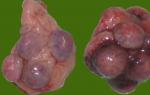
| Picture 1. An example of the diagnosis of Edwards syndrome by CF-PCR. | Figure 2. Sick child with Edwards syndrome. |
| An example of the diagnosis of Edwards syndrome (trisomy on chromosome 18). Highlighted in purple are markers located in the region of chromosome 18, which is critical for the development of Edwards syndrome. In the genotype of the test sample, there is 1 peak for the D18S978 marker (the marker is not informative), for the D18S535 and D18S386 markers - 3 peaks (trisomy), for the D18S390 and D18S819 markers - the dose effect - an unequal ratio of the height of the two peaks (trisomy). Thus, for four markers (D18S535, D18S386, D18S390 and D18S819), trisomy was detected, which makes it possible to establish the diagnosis of Edwards syndrome. The genetic sex corresponds to male - there are peaks corresponding to the Y-chromosome for the markers Amelogenin, 4SH, ZFXY, TAFL and there is a peak of the SRY gene. |
|

The "gold standard" for detecting chromosomal disorders throughout the world has long been and continues to be the method of karyotyping with differential staining of chromosomes. This method makes it possible to analyze the karyotype as a whole and to determine large (at least 5–10 million base pairs) chromosomal rearrangements. However, it has a number of limitations, such as labor intensity, duration (1-2 weeks), high requirements for the qualifications and experience of the specialist conducting the study, and, in some cases, technical problems (insufficient quantity and quality of the material under study, the absence of mitoses or culture growth).
The method of quantitative fluorescent polymerase chain reaction (QF-PCR), which is increasingly used to diagnose aneuploidies, including Edwards syndrome, is deprived of these shortcomings (Fig. 1). This method has a reliability comparable to that of standard karyotyping, is faster, cheaper, less demanding on the quantity and quality of the material (since it is not associated with the growth of cell culture) and allows you to simultaneously analyze a large number of samples. However, the CF-PCR method also has limitations: in mosaic cases, it can detect only high-level mosaicism (from 20%), in addition, it cannot exclude the presence of rarer chromosomal disorders that may be associated with fetal malformations. When conducting prenatal diagnosis of Edwards syndrome, in addition to fetal material, it is necessary to provide biological material to the mother in order to exclude the possibility of obtaining a false negative result due to improper sampling of fetal material. Analysis of the fetal material is performed within three working days.
With Edwards syndrome, there is a pronounced delay in prenatal development, children are born with prenatal malnutrition (average body weight at birth is 2340 g). External manifestations of Edwards syndrome are diverse (Fig. 2). The most typical are delayed psychomotor development, hypoplasia of the skeletal muscles and subcutaneous adipose tissue, congenital heart defects, anomalies in the structure of the face and skull (dolichocephaly, microphthalmia, shortening of the palpebral fissures, low position of the auricles, micrognathia, sloping chin), multiple deformities of the hands and feet, anomalies in the development of the gastrointestinal tract, genitourinary system and central nervous system (spinal hernia, hypoplasia of the corpus callosum and cerebellum). The life expectancy of children is sharply reduced: 90% of them die before a year from complications caused by congenital malformations (asphyxia, pneumonia, intestinal obstruction, cardiovascular insufficiency).
The reason for the development of Edwards syndrome is the tripling of chromosome 18. Trisomy on chromosome 18 is a special case of aneuploidy - the presence in the genome of a set of chromosomes that is different from the standard for this species and not multiple of it. Trisomy 18 is usually caused by nondisjunction of chromosomes during the formation of the parent's germ cells (eggs and sperm), as a result of which the child receives an extra 18th chromosome from the mother or from the father. In this case, all cells of the child's body will carry the anomaly. In the case when nondisjunction of chromosomes occurs during the division of any cell of the embryo, a mosaic variant of Edwards syndrome is observed (10% of cases).
The risk of having children with Edwards syndrome, according to various literature data, does not change or slightly increases with the age of the pregnant woman.
Prenatal diagnosis of Edwards syndrome includes two stages. At the first stage, at a gestational age of 11-13 weeks, screening is carried out, which is based mainly on biochemical parameters, since in the early stages, ultrasound does not allow detecting any gross developmental anomalies in the case of Edwards syndrome, which can be detected only by 20-24 week. Biochemical analysis of the level of certain proteins in the blood of a pregnant woman (free β-subunit of human chorionic hormone (β-hCG) and pregnancy-associated plasma protein A (pregnancy associated plasma protein-A, PAPP-A)), taking into account her age, allows you to calculate for her the risk of having a sick child. However, these methods do not allow to make an accurate diagnosis, and as a result of the screening, a risk group of pregnant women with an increased probability of giving birth to a patient with Edwards syndrome is formed. At the second stage, an invasive procedure is performed in the risk group to obtain the fetal material necessary to accurately determine the status of the fetus. Depending on the gestational age, this may be a chorionic villus biopsy (weeks 8-12), amniocentesis (weeks 14-18) or cordocentesis (after the 20th week). In the obtained tissue samples of the fetus, the chromosome set is determined.
The Center for Molecular Genetics performs diagnostics of Edwards syndrome (including prenatal) by CF-PCR.