What can mri. Magnetic resonance imaging So which is better
Magnetic resonance imaging (MRI) is one of the most modern diagnostic methods that allows you to study almost any system of the body. The most important characteristic of an MRI machine is the magnetic field strength, which is measured in Tesla (T). The quality of visualization directly depends on the field strength - the higher it is, the better the image quality, and, accordingly, the higher the diagnostic value of the MRI study.
Depending on the power of the device, there are:
■ low-field tomographs - 0.1 - 0.5 T (Fig. 1);
■ high-field tomographs - 1 - 1.5 T (Fig. 2);
■ ultra-high-field tomographs - 3 T (Fig. 3).

At the moment, all major manufacturers produce MR scanners with a field of 3 T, which differ little in size and weight from standard systems with a field of 1.5 T.
Safety studies in MR imaging have not shown any negative biological effects of magnetic fields up to 4 T used in clinical practice. However, it should be remembered that the movement of electrically conductive blood creates an electrical potential, and in a magnetic field it will create a small voltage through the vessel and cause an elongation of the T wave on the electrocardiogram, therefore, in studies in fields above 2 T, ECG monitoring of patients is desirable. Physical studies have shown that fields above 8 T cause genetic changes, charge separation in liquids, and changes in the permeability of cell membranes.
Unlike the main magnetic field, gradient fields (magnetic fields perpendicular to the main, main, magnetic field) are switched on at certain time intervals in accordance with the chosen technique. Rapid switching of gradients can induce electrical currents in the body and lead to stimulation of peripheral nerves, causing involuntary movements or tingling in the limbs, but the effect is not dangerous. Studies have shown that the threshold for stimulation of vital organs (for example, the heart) is much higher than for peripheral nerves, and is about 200 T/s. When the threshold [rate of change of gradients] dB/dt = 20 T/s is reached, a warning message appears on the operator console; however, since the individual threshold may differ from the theoretical value, constant monitoring of the patient's condition is necessary in strong gradient fields.
Metals, even non-magnetic ones (titanium, aluminium), are good conductors of electricity and will heat up when exposed to radio frequency [RF] energy. RF fields induce eddy currents in closed loops and conductors, and can also create significant stress in extended open conductors (eg, rod, wire). The length of electromagnetic waves in the body is only 1/9 of the wavelength in air, and resonance phenomena can occur in relatively short implants, causing their ends to heat up.
Metallic objects and external devices are generally erroneously considered safe if they are non-magnetic and are labeled "MP compatible". However, it is important to make sure that objects that are scanned inside the working area of the magnet are immune to induction. Patients with implants are only eligible for MR examination if the implants are both non-magnetic and small enough to heat up during scanning. If the object is longer than half the length of the RF wave, the patient's body may experience high heat resonance. The limiting dimensions of metal (including non-magnetic) implants are 79 cm for a field of 0.5 T and only 13 cm for 3 T.
Switching gradient fields creates a strong acoustic noise during an MR examination, the value of which is proportional to the power of the amplifier and the field strength and, according to regulatory documents, should not exceed 99 dB (for most clinical systems it is about 30 dB).
based on the article "Possibilities and limitations of high-field magnetic resonance imaging (1.5 and 3 Tesla)" A.O. Kaznacheeva, National Research University of Information Technologies, Mechanics and Optics, St. Petersburg, Russia (journal "Radiology and Therapy" No. 4 (1) 2010)
read also the article "Safety of magnetic resonance imaging - the current state of the issue" by V.E. Sinitsyn, Federal State Institution "Treatment and Rehabilitation Center of Roszdrav" Moscow (Journal "Diagnostic and Interventional Radiology" No. 3, 2010) [read]
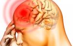
MRI DURING PREGNANCY - IS IT SAFE?

Currently, MRI is a widely used method of radiation diagnostics, which is not associated with the use of ionizing radiation, as in X-ray examination (including CT), fluorography, etc. MRI is based on the use of radio frequency pulses (RF pulses) in a high magnetic field. The human body consists mainly of water, consisting of hydrogen and oxygen atoms. At the center of each hydrogen atom is a small particle called a proton. Protons are very sensitive to a magnetic field. Magnetic resonance imaging scanners use a constant strong magnetic field. After the object under study is placed in the magnetic field of the tomograph, all its protons line up in a certain position along the external magnetic field, like a compass needle. An MRI scanner sends a radio frequency pulse to the part of the body being examined, causing some of the protons to move out of their original state. After turning off the radio frequency pulse, the protons return to their previous position, emitting the accumulated energy in the form of a radio frequency signal that reflects its position in the body and carries information about the microenvironment - the nature of the surrounding tissue. Just as a million pixels form an image on a monitor, the radio signals from millions of protons, after complex mathematical processing, form a detailed image on a computer screen.
However, certain precautions must be strictly observed when performing an MRI. Potential hazards for patients and staff in MRI rooms may be related to factors such as:
■ constant magnetic field generated by the tomograph's magnet;
■ changing magnetic fields of the instrument (gradient fields);
■ RF radiation;
■ devices and substances included with the scanner, such as cryogens (liquid helium) and electrical cables.
Due to the "youth" of the technique, a small (worldwide) amount of accumulated safety data, the FDA (Food and Drug Administration, USA), together with the World Health Organization, impose a number of restrictions on the use of MRI, due to the possible negative impact strong magnetic field. The use of a magnetic field up to 1.5 T is considered acceptable and absolutely safe, except for cases when there are contraindications for MRI (MR tomographs up to 0.5 T - low-field, from 0.5 to 1.0 T - medium-field, from 1.0 - 1.5 T and more - high-field).
Speaking about long-term exposure to constant and alternating magnetic fields, as well as radio frequency radiation, it should be noted that there is no evidence of the existence of any long-term or irreversible effects of MRI on human health. So, female doctors and radiologists are allowed to work during pregnancy. Monitoring of their health showed that no abnormalities were noted in their health or in their offspring.
In magnetic resonance imaging of women of childbearing age, it is necessary to obtain information about whether they are pregnant or not. There is no evidence of the harmful effects of magnetic resonance imaging on the health of pregnant women or the fetus, but it is strongly recommended to perform MRI for women in position only with obvious (absolute) clinical indications, when the benefits of such an examination clearly outweigh the risks (even if very low).
If there are only relative indications for MRI, then doctors recommend abandoning this study in the first three months (up to 13 weeks of gestation, I trimester) of pregnancy, since this period is considered fundamental for the formation of the internal organs and systems of the fetus. During this period, both the pregnant woman and the child itself are very sensitive to the effects of teratogenic factors that can cause disruption of the embryogenesis process. In addition, according to most doctors, the first three months, the pictures of the fetus are not clear enough due to their small size.
Moreover, during the diagnosis, the tomograph itself creates a background noise and emits a certain percentage of heat, which can also potentially affect the fetus in early pregnancy. As mentioned above, MRI uses RF radiation. It can interact both with body tissues and with foreign bodies in it (for example, metal implants). The main result of this interaction is heating. The higher the frequency of RF radiation, the more heat will be released, the more ions are contained in the tissue, the more energy will be converted into heat.
To evaluate the thermal effects of RF radiation, specific absorption rate - SAR (specific absorbtion rate), displayed on the display screen of the device, helps. It increases with increasing field strength, RF pulse power, decreasing slice thickness, and also depends on the type of surface coil and patient weight. MRI systems are protected to prevent the SAR from rising above a threshold, which could result in tissue heating of more than 1°C.
During pregnancy, MRI can be used to diagnose pathology either in a woman or in a fetus. At the same time, MRI is prescribed according to ultrasound diagnostics when certain pathologies are detected in the development of the unborn child. The high sensitivity of MRI diagnostics makes it possible to clarify the nature of the deviations and helps to make an informed decision on whether to continue or terminate the pregnancy. MRI becomes especially important if it is necessary to study the development of the fetal brain, diagnose malformations of cortical development associated with a violation of the organization and formation of brain convolutions, the presence of areas of heterotopia, etc. Thus, the reasons for MRI can be:
■ various developmental pathologies of the unborn child;
■ deviations in the activity of internal organs, both the woman herself and the unborn child;
■ the need to confirm indications for artificial termination of pregnancy;
■ as evidence or, conversely, refutation of a previously diagnosed diagnosis based on tests;
■ the lack of the possibility of ultrasound due to the obesity of the pregnant woman or the inconvenient location of the fetus in the last stage of pregnancy.
On the territory of Russia, there are no restrictions on MRI in the first trimester, however, the Commission on Ionizing Radiation Sources at WHO does not recommend any exposure to the fetus, which may in any way affect its development (despite the fact that studies have been carried out during which children under 9 years of age were observed, subjected to MRI in the first trimester of intrauterine development, and no deviations in their development were found). It is important to remember that the lack of information about the negative impact of MRI on the fetus does not mean the complete elimination of the harm of this type of study for the unborn child.
note: pregnant [ !!! ] it is forbidden to conduct an MRI with intravenous administration of MR contrast agents (they penetrate the placental barrier). In addition, these drugs are excreted in small amounts with breast milk, so the instructions for gadolinium drugs indicate that when they are administered, breastfeeding should be stopped within a day after the administration of the drug, and the milk secreted during this period should be expressed and poured out. .
Literature: 1. article "Safety of magnetic resonance imaging - the current state of the issue" V.E. Sinitsyn, Federal State Institution "Therapeutic and Rehabilitation Center of Roszdrav" Moscow; journal "Diagnostic and interventional radiology" Volume 4 No. 3 2010 pp. 61 - 66. 2. article "MRI diagnostics in obstetrics" Platitsin I.V. 3. materials of the site www.az-mri.com. 4. materials from the site mrt-piter.ru (MRI for pregnant women). 5. materials from the site www.omega-kiev.ua (Is MRI safe during pregnancy?).
From the article: "Obstetric aspects of acute cerebrovascular disorders during pregnancy, childbirth and the postpartum period (literature review)" R.R. Harutamyan, E.M. Shifman, E.S. Lyashko, E.E. Tyulkina, O.V. Konysheva, N.O. Tarbaya, S.E. Flock; Department of Reproductive Medicine and Surgery, FPDO, Moscow State University of Medicine and Dentistry. A.I. Evdokimova; City Clinical Hospital №15 named after O.M. Filatov; Department of Anesthesiology and Resuscitation, FPC MR, Peoples' Friendship University of Russia, Moscow (Reproduction Problems magazine No. 2, 2013):
“MRI does not use ionizing radiation, and no harmful effects on the developing fetus have been noted, although long-term effects have not yet been studied. A recently published guideline from the American Radiological Society states that MRI should be performed on pregnant women if the benefit of the study is clear and the necessary information cannot be obtained by safe methods (for example, using ultrasound) and cannot be waited until the end of the patient's pregnancy. MRI contrast agents easily cross the uteroplacental barrier. No studies have been conducted on the removal of contrast from amniotic fluid, just as their potential toxic effects on the fetus are not yet known. It is assumed that the use of contrast agents for MRI in pregnant women is justified only if the study is undeniably useful for making a correct diagnosis in the mother [read source].”
From the article"Diagnostics of acute disorders of cerebral circulation in pregnant women, puerperas and women in childbirth" Yu.D. Vasiliev, L.V. Sidelnikova, R.R. Arustamyan; City Clinical Hospital №15 named after O.M. Filatov, Moscow; 2 SBEE HPE "Moscow State University of Medicine and Dentistry named after A.I. A.I. Evdokimov" of the Ministry of Health of Russia, Moscow (magazine "Problems of reproduction" No. 4, 2016):
“Magnetic resonance imaging (MRI) is a modern diagnostic method that allows you to identify a number of pathologies that are very difficult to diagnose using other research methods.
In the first trimester of pregnancy, MRI is performed according to vital indications on the part of the mother, since organo- and histogenesis has not yet been completed. There is no evidence that MRI has a negative effect on the fetus or embryo. Therefore, MRI is used for research not only in pregnant women, but also for fetography, in particular, for examining the fetal brain. MRI is the method of choice in pregnancy if other non-ionizing medical imaging methods are insufficient, or if the same information is needed as with x-rays or computed tomography (CT) but without the use of ionizing radiation.
There are no restrictions on MRI during pregnancy in Russia, however, the Commission on Non-Ionizing Radiation Sources at WHO does not recommend any exposure to the fetus from the 1st to the 13th week of gestation, when any factor can in any way affect its development.
In the II and III trimesters of pregnancy, the study is safe for the fetus. Indications for MRI of the brain in pregnant women are: [ 1 ] stroke of various etiologies; [ 2 ] vascular diseases of the brain (anomalies in the development of blood vessels of the head and neck); [ 3 ] trauma, bruises of the brain; [ 4 ] Tumors of the brain and spinal cord; [ 5 ] paroxysmal conditions, epilepsy; [ 6 ] infectious diseases of the central nervous system; [ 7 ] headache; [ 8 ] violations of cognitive functions; [ 9 ] pathological changes in the sellar region; [ 10 ] neurodegenerative diseases; [ 11 ] demyelinating diseases; [ 12 ] sinusitis.
For MR angiography in pregnant women, the introduction of a contrast agent in most cases is not necessary, in contrast to CT angiography, where it is required. Indications for MR angiography and MR venography in pregnant women are: [ 1 ] cerebrovascular pathology (arterial aneurysms, arteriovenous malformations, cavernomas, hemangiomas, etc.); [ 2 ] thrombosis of large arteries of the head and neck; [ 3 ] thrombosis of venous sinuses; [ 4 ] identification of anomalies and variants of development of the vessels of the head and neck.
There are few contraindications for the use of MRI in the general population, and in pregnant women in particular. [ 1 ] Absolute contraindications: artificial pacemaker (its function is disturbed in the electromagnetic field, which can lead to the death of the examined patient); other electronic implants; periorbital ferromagnetic foreign bodies; intracranial ferromagnetic hemostatic clips; conductive pacemaker wires and ECG cables; pronounced claustrophobia. [ 2 ] Relative contraindications: I trimester of pregnancy; serious condition of the patient (an MRI is possible when the patient is connected to life support systems).
In the presence of heart valves, stents, filters, the study is possible if the patient provides the accompanying documents of the manufacturer, which indicate the possibility of an MRI indicating the magnetic field strength, or an epicrisis of the department where the device was installed, which indicates the permission conducting this survey” [read source].
There are no equals
MRI is a research method based on obtaining images of tissues and organs using electromagnetic waves. It has no equal when examining the brain and spinal cord, cranial nerves, intervertebral discs, bile ducts, the reproductive system of men and women, the mammary glands, joints, and blood vessels. The magnetic resonance tomograph makes it possible to conduct research in any planes, taking into account the anatomical features of the patient's body, and, if necessary, to obtain three-dimensional images for an accurate assessment of the relative position of various structures. At the same time, unlike computed tomography, ionizing radiation is not used, the patient is not exposed to radiation exposure, and, therefore, there is no risk of side effects that are associated, albeit to a small extent, with exposure to x-rays with frequent repeated studies. The advantage of this method over ultrasound examinations (ultrasound) is that for MRI, air and bones are not a hindrance to imaging. The undoubted advantage of MRI is the good natural tissue contrast.
MRI is performed according to indications, both for emergency diagnostics and in a planned manner. It best of all “shows” the condition of soft tissues and bone marrow to the doctor in comparison with other methods. Muscle, fat, fluid, tendons, and cartilage are clearly visible and differentiated from each other on magnetic resonance images. In most cases, the method is indispensable in the diagnosis of traumatic, tumor and inflammatory diseases of bones and joints.
MRI and the brain
If we compare magnetic resonance imaging with computed tomography, then MRI is superior to computed tomography in detecting and assessing lesions in the brain substance, especially in identifying pathological processes in the posterior cranial fossa, in the pituitary gland, and in visualizing cranial nerves. Allows you to see and assess the state of the vessels of the brain and neck without the introduction of a contrast agent. In addition, on modern tomographs, with a magnetic field induction of 3 Tesla, there is a function of accelerated scanning. In the case when a patient has a traumatic brain injury, a stroke or an attack with an increased pain syndrome, just a few minutes are enough to scan the brain.
MRI and spine
Most often, MRI is in demand in the diagnosis of diseases of the spine - this is the only method that allows you to see pathological changes in the intervertebral discs of the cervical, thoracic and lumbosacral spine, as well as the spinal cord and nerve roots, to diagnose inflammatory, tumor and vascular lesions with confidence. Evaluation of the ratio of herniated discs and nerve roots in patients with osteochondrosis allows you to determine the indications for surgery.
Its difference from the classical model is that this tomograph can be used to scan the entire spine in 35 minutes in one examination, which, of course, saves time and money for patients. In addition, this device opened up new possibilities, for example, in the study of the heart.
MRI and cardiovascular system
Despite the fact that the study of the arteries of the heart (coronary arteries) is the prerogative of angiography, including CT angiography, it is possible to examine the state of the heart muscle, valves and heart function only with the help of ECHO-KG and MRI. These methods complement each other in some way, but MRI allows you to get the most unique information about the state of the heart muscle, membranes. To do this, a contrast agent is injected and during the study the blood flow in the heart muscle is assessed, which means that the viability of the myocardium is determined in patients who have had a heart attack, the condition of the heart valves, the contractility of each of its walls are assessed, inflammatory changes, hereditary pathology and many other diseases are detected.
Studies of the vessels of the brain and neck, shoulder girdle, legs, as well as the aorta and its branches with intravenous contrasting allows you to get the most detailed information necessary for vascular surgeons. We see a three-dimensional picture of the state of the vessels in a particular area, evaluate the extent and severity of, for example, narrowing of the vessel by an atherosclerotic plaque, track anomalies and variations in the structure of the vessels, which makes it possible to plan surgical treatment taking into account the individual characteristics of each individual patient.
MRI and pelvic organs
In most city clinics, tomographs with a magnetic field induction of 1.5 Tesla are installed. Only two clinics in St. Petersburg are equipped with the latest generation devices with a capacity of 3 Tesla - "Medem" and Clinical Hospital No. L.G. Sokolov. Another new generation tomograph has been installed at the Diagnostic and Treatment Center of the International Institute for Biological Problems (MIBS).
The new 3-tesla tomograph is additionally equipped with an endorectal coil, which expands the possibilities in the study of prostate lesions, the state of adjacent organs and tissues.
For the study of the pelvic organs, women have additional unique programs that allow you to get very thin sections, examine the organs in all planes, and, accordingly, make the correct diagnosis.
MRI and gastrointestinal tract
MRI makes it possible to qualitatively examine the organs of the abdominal cavity, including the biliary tract. Magnetic resonance cholangiography allows you to see the bile ducts without the introduction of instruments (laparoscopy), to assess the condition of those parts of the bile ducts that are hidden from ultrasound.
Thin sections, multi-phase scanning, and the use of special contrast agents make it possible to accurately diagnose focal changes in the liver, to identify even the smallest foci of several millimeters in size, which in some cases radically changes the treatment tactics. It is possible to assess the spread of tumors of the pancreas, kidneys, adrenal glands and spleen, to identify inflammatory changes in the pancreas.
MRI and all, all, all
The screening technique for examining the whole body (scanning) gives an idea of the state of all organs and systems. This study gives the doctor information about whether there are changes that make it possible to suspect the presence of malignant tumors at a stage when they do not yet give clinical manifestations and are not detected in other ways. The data obtained during the screening require additional examination. This diagnostic method is effective for patients already suffering from oncological diseases, as it allows to detect metastases.
The appearance of a new tomograph allowed doctors to solve many diagnostic problems that could not be solved before. Due to the fact that the opening of the device, in which the patient is placed during the scan, has a larger diameter than most tomographs, this makes the examination comfortable even for those suffering from claustrophobia and for large overweight patients. Previously, they could only be examined on low-field tomographs, the capabilities of which are limited.
Anna Rogozina
Dr. Peter
Opportunities for a detailed examination of all organs and tissues of the human body in modern medicine are many. One of the reliable and reliable methods is magnetic resonance imaging, which has long moved from the category of high-tech assistance to the category of routine, affordable diagnostics. The article will provide answers to the most common questions about MRI - what is it, how it is performed and in what cases it is prescribed.
How MRI works
What is MRI in medicine? This is a research technique based on the physical phenomenon of magnetic resonance. The “resonator” in this case is the patient himself, or rather, his tissues and organs. Despite the fact that the MRI examination is called "nuclear", it has nothing to do with radiation.
"Nuclear" in this case means that the nuclei of hydrogen atoms present in all tissues respond to a combination of a constant magnetic field and electromagnetic waves, the source of which is a special scanner. These responses are recorded and ordered by the apparatus, which adds them up to a high-quality, clear image.
Types of magnetic resonance imaging (MRI) =
Diagnostics by MRI is carried out on devices of various types.
Diagnostics by MRI is carried out on devices of various types. The classification that is important for the patient is open and closed devices.
- Open. What is an open MRI? The space in which the patient is located during the examination remains open. The device itself consists of two parts - the upper one, hanging over the patient, and the lower one, on which it rests. Both parts are equipped with magnets. An open MRI study is indicated for those who suffer from clautrophobia, obese patients or those with physical limitations.
- Closed. Traditional instruments, which are a tunnel and a moving table.
Some types of MRI studies are carried out only in closed devices. For example, if you need to take an MRI of the head, it is important to ensure that it is completely immobile. To do this, the head is fixed, and in open-type devices, fixation is not provided.
Another difference between MRI machines is the power measured in Tesla. Depending on this parameter, they are divided into:
- Low-floor (0.5 T).
- Mid-field (up to 1 T).
- High-field (up to 1.5 T).
The power will depend on the scanning time of a particular MRI area, the quality of imaging and the cost of the study. The higher the power of the equipment installed in the clinic, the higher the speed and the higher the price.
Having figured out what MRI diagnostics is, it is worth taking the time to study the equipment of the selected medical center. Low-field devices produce a picture with less accurate visualization than high-field devices.
What does an MRI show?

The study is completely non-invasive and non-contact.
MRI is a unique study, because it allows you to see a wide range of pathologies of different organs.
- Inflammatory diseases.
- Infections.
- Tumors.
- Pathology of blood vessels and heart.
- Injuries and their consequences.
The structure of tissues, the configuration of organs, blood supply, biochemical processes - all these phenomena can be assessed using a magnetic resonance tomograph.
Benefits of an MRI Scan
Magnetic resonance imaging has many advantages over other types of medical research:
- Obtaining very high-quality, detailed images.
- The principle of operation of MRI does not involve radiation, and therefore it can also be in childhood.
- Allows you to visualize structures that are difficult to study - for example, the spinal cord and brain.
- You can get images in several projections. Due to this, the diagnosis of some diseases is carried out earlier than is possible on computed tomography (for example, cerebral ischemia).
In comparison with other methods of studying the state of health, this diagnostic method has both advantages and disadvantages:
- CT is a more dangerous study because it involves X-ray exposure. However, if it is necessary to make a diagnosis of the state of the musculoskeletal system, it is more appropriate to conduct computed tomography.
- ultrasound. There are no contraindications for ultrasound, so it can be performed on any patient. However, ultrasound will not cope with such tasks as assessing the condition of the bones, stomach, and lungs. In addition, MRI images are more accurate.
- EEG (electroencephalography) - diagnosis of diseases. According to the encephalogram, it is very difficult to diagnose the presence of tumors and other organic diseases. In addition, the method cannot be called accurate, since the result is influenced by the emotions experienced by the patient.
How is an MRI done?
The study is completely non-invasive and non-contact. An unpleasant sensation during scanning can only be delivered by the sounds emitted by the device. To prevent the patient from hearing them, he is offered headphones with pleasant music. How is an MRI performed? The algorithm is the following:
- The patient removes all metal jewelry, watches.
- The subject lies on the table. , legs, and sometimes the head are comfortably fastened with straps.
- The table drives into the tunnel, where scanning is carried out for the required time (from 15 to 60 minutes).
- See also: o.
In the presence of claustrophobia, this must be reported to the doctor. How is an MRI done in this case? Most likely, it will be proposed to undergo diagnostics on an open device.
Types of diagnostics

MR angiography can be done without the use of a contrast agent.
The MRI procedure has several varieties:
- MR diffusion. This is a type of magnetic tomography that measures the speed of movement of water molecules. The method allows to determine disorders of cerebral circulation and to identify oncological formations.
- MR perfusion visualizes the features of the passage of blood through tissues, the speed of this process, and vascular permeability. Due to this, it is possible to differentiate healthy tissues from pathological ones.
- MR spectroscopy to detect biochemical changes in tissues. The value of such an MRI analysis lies in the fact that biochemical changes occur even when there are no clinical manifestations of the disease. So, it can be detected at the earliest stage.
- Angiography is a study that allows you to see the lumen of blood vessels and evaluate blood flow.
MR angiography can be done without the use of a contrast agent. But most often, contrast is used to improve the visibility of blood vessels. MRI with contrast is a method that allows you to see what is happening with the vessels that permeate each organ. The so-called paramagnets, primarily gadolinium, are used as a contrast agent.
How does an MRI with contrast work? Most often, it is introduced after pictures without contrast have been taken. The substance is administered intravenously, then repeated pictures are taken. In what cases and why is it recommended to do such a study?
- Aneurysm suspected.
- There are reasons to assume the presence of tumors.
- Stroke.
- Diagnosis after some operations (for example, prostate).
- Head injury.
- to detect metastases.
Allergy to gadolinium is rare, unlike an allergic reaction to iodine, which is used as the basis of a contrast agent in CT scans.
Indications and contraindications

After an MRI scan, the interpretation of the results usually takes 1-2 days.
Indications for MRI vary depending on the area of the body that needs to be examined. We list some of the indications sufficient for MRI:
- The brain is subject to examination in case of neurological symptoms, visual or hearing impairments, after injuries. What is the brain?
- The organs of the abdominal cavity are examined for pain, jaundice, severe dyspeptic symptoms.
- The heart is subject to study in ischemic heart disease, pain and arrhythmias, after a heart attack.
- The genitourinary system is examined for urination disorders, pain, and the appearance of blood.
Despite the abundance of diagnoses that can be made to children, they are referred for MRI only in emergency cases, when another examination was ineffective. All types of oncology or suspicion of it, epilepsy, neurological disorders, spinal problems, cardiovascular diseases, various severe injuries, kidney or liver diseases - all this is an indication for magnetic resonance imaging in children.
Magnetic resonance imaging (MRI) is one of the most informative and safe methods of instrumental diagnostics. The study is non-invasive, and therefore atraumatic. Its essence lies in the specific reaction of hydrogen atoms, concentrated in the tissues of the human body, to a powerful magnetic field generated by the device.
Vascular ultrasound (doppler ultrasound) is an informative and safe diagnostic method used to study the volume, intensity and speed of blood flow in veins and arteries. In clinical practice, the study is most often used to identify pathologies of the vessels of the lower extremities, since they are the most common among diseases of the circulatory system.
Multiple myeloma is an oncological pathology of the hematopoietic system associated with malignant division and reproduction of mature plasma cells, which provokes increased production of monoclonal immunoglobulins, bone resorption and secondary immunodeficiency.
MRI (magnetic resonance imaging) is an informative and safe research method used in the diagnosis of diseases of the spinal cord and brain, internal organs of the peritoneum, various parts of the spine and other structures of the musculoskeletal system. Along with MRI, CT (computed tomography) is also popular.
If cardiovascular diseases take the rightful first place among the causes of death, then cancer rightfully ranks second. In the last decade, there has been a noticeable trend towards an increase in the number of patients with various malignant tumors. In the process of curing a person from an oncological disease, the most important place is occupied by the early diagnosis of the disease, since it can be cured only at the initial stages.
If the doctor conducting the pregnancy recommends an MRI, then he has reason to be concerned about the condition of the woman or the development of the fetus. At the same time, all expectant mothers inevitably have a question: how safe is such a procedure for the health of the child?
MRI is the most advanced and highly accurate type of instrumental diagnostics, which has no alternative in detecting many severe pathologies, including neoplasms and some severe diseases of the central nervous system. Therefore, there are cases when the passage of this procedure is vital for a patient with claustrophobia.
MRI is by far the most accurate of all instrumental diagnostic methods in modern medicine. With its help, layered images of tissues and internal organs of the body are made with a thickness of only a few microns, then transforming them into three-dimensional images and displaying them on the screen.
We have come close to the issue of the MRI device. The apparatus includes the main magnet, gradient coils, RF coils, phase-sensitive detector, data analysis device, power supply and system cooling equipment.
Depending on the created magnetic field strength tomographs are divided into:
- ultra-low-field (voltage less than 0.1 T);
- low-field (up to 0.5 T);
- mid-field (up to 1 T);
- high-field (up to 2 T);
- ultrahigh-field (more than 2 T).
Currently, high-field and ultra-high-field MR tomographs are the most common.
By type of magnets devices are divided into:
- with a permanent magnet (used in open tomographs);
- resistive electromagnets (also used in open apparatus, but less and less common);
- superconducting electromagnets (capable of creating high intensity fields, but are quite expensive and require cooling with liquid helium).
By degree of "openness" MR-devices are:
- closed (tunnel type). Examination in such tomographs is not recommended for patients suffering from claustrophobia;
- open type (they usually have less power, but they allow diagnosing patients who are afraid of confined spaces).
In medical practice, there are several separate MRI technologies: MR angiography, MR perfusion, functional MRI, MR diffusion, etc.
MR angiography is a way to study the human vascular system. The method allows evaluating the functional and anatomical features of blood flow, identifying disorders of the blood supply to the brain and internal organs, identifying atherosclerosis, thrombosis, stenosis, vascular aneurysms, heart defects and other disorders.
MR perfusion allows you to evaluate the passage of blood through the tissues of the body, including through the tissues of the liver and brain.
The main task of functional MRI is to study the cerebral cortex to determine the features of the brain regions responsible for vision, memory, movement, speech and other functions. The principle of the method is that when the patient performs certain tasks, the blood flow in the corresponding parts of the brain increases.
MR diffusion is a method for determining the movement of intracellular water molecules in tissues.
In modern diagnostics, MRI is an indispensable method for examining and detecting pathologies, which allows planning surgical interventions, treatment and monitoring its effectiveness.
In our center you can perform all types of tomographic studies. More information about the prices for MRI and CT diagnostics can be found on our website or by contact numbers.