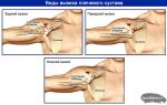
Strangulated hernia types. Classification, symptoms and treatment of strangulated hernia. Treatment of hernial bulges with the process of infringement
- compression of the hernial sac in the hernial orifice, causing a violation of the blood supply and necrosis of the organs forming the hernial contents. Infringement of a hernia is characterized by sharp pain, tension and soreness of the hernial protrusion, irreducibility of the defect. Diagnosis of incarcerated hernia is based on the history and physical examination, plain radiography abdominal cavity. During hernia repair for a strangulated hernia, resection of the necrotic intestine is often required.

General information
Incarcerated hernia is the most frequent and severe complication of abdominal hernia. Strangulated hernias are an acute surgical emergency and are second in incidence only to acute appendicitis, acute cholecystitis, and acute pancreatitis. In operative gastroenterology, strangulated hernia is diagnosed in 3-15% of cases.
Incarceration of a hernia is associated with a sudden compression of the contents of the hernial sac (omentum, small intestine and other organs) in the hernial orifice (defects of the anterior abdominal wall, apertures of the diaphragm, pockets of the abdominal cavity, etc.). Any abdominal hernias can be infringed: inguinal (60%), femoral (25%), umbilical (10%), less often - hernias of the white line of the abdomen, esophageal opening of the diaphragm, postoperative hernias. Infringement of a hernia is associated with the risk of developing necrosis of compressed organs, intestinal obstruction, peritonitis.
Types of infringement of a hernia
Depending on the organ squeezed in the hernial orifice, hernias are distinguished with infringement of the intestines, omentum, stomach, bladder, uterus and its appendages. The degree of overlapping of the lumen of a hollow organ in case of infringement of a hernia can be incomplete (parietal) and complete. In some cases, for example, when Meckel's diverticulum or the appendix is infringed, the lumen of the organ does not overlap at all. According to the peculiarities of development, antegrade, retrograde, false (imaginary), sudden (in the absence of a hernia history) strangulation of the hernia are distinguished.
There are two mechanisms of hernia incarceration: elastic and fecal. Elastic infringement develops in the case of a simultaneous exit through a narrow hernial orifice of a large amount of hernial contents. The internal organs enclosed in the hernial sac cannot retract into the abdominal cavity on their own. Their infringement by a narrow ring of hernial ring leads to the development of ischemia, severe pain syndrome, persistent muscle spasm hernial orifice, which further aggravates the incarceration of the hernia.
Fecal infringement develops with a sharp overflow of the afferent loop of the intestine, which has fallen into the hernial sac, with intestinal contents. At the same time, the discharge section of the intestine is flattened and infringed in the hernial orifice along with the mesentery. Fecal infringement often develops with long-term irreducible hernias.
Infringement of a hernia can be primary and secondary. Primary infringement is less common and occurs against the background of a one-time emergency effort, as a result of which there is a simultaneous formation of a hernia that did not previously exist and its compression. Secondary infringement occurs against the background of a previously existing hernia of the abdominal wall.
Causes of infringement of a hernia
The main mechanism of hernia incarceration is a sharp simultaneous or periodically recurring increase in intra-abdominal pressure, which may be associated with excessive physical effort, constipation, coughing (with bronchitis, pneumonia), difficulty urinating (with prostate adenoma), difficult childbirth, crying, etc. Development and infringement of the hernia contributes to the weakness of the muscles of the abdominal wall, intestinal atony in the elderly, traumatic injuries abdomen, surgery, weight loss.
After normalization of intra-abdominal pressure, the hernial gates decrease in size and infringe on the hernial sac that has gone beyond them. At the same time, the probability of development of infringement does not depend on the diameter of the hernial orifice and the size of the hernia.
Symptoms of a strangulated hernia
Infringement of a hernia is characterized by the following symptoms: a sharp local or diffuse pain in the abdomen, the inability to set the hernia, tension and soreness of the hernial protrusion, the absence of the “cough push” symptom.
The main signal of hernia incarceration is pain, which develops at the height of physical effort or tension and does not subside at rest. The pain is so intense that the patient often cannot help moaning; his behavior becomes restless. In the objective status, pallor of the skin is noted, the phenomena of pain shock are tachycardia and hypotension.
Depending on the type of strangulated hernia, pain may radiate to the epigastric region, the center of the abdomen, groin, and thigh. When intestinal obstruction the pain becomes spasmodic. The pain syndrome, as a rule, is expressed within a few hours, until necrosis of the strangulated organ develops and death occurs. nerve elements. With fecal infringement, pain and intoxication are less pronounced, necrosis of the intestine develops more slowly.
When a hernia is infringed, a single vomiting may occur, which initially has a reflex mechanism. With the development of intestinal obstruction, vomiting becomes constant and acquires a fecal character. In situations of partial infringement of the hernia, obstruction phenomena, as a rule, do not occur. In this case, in addition to pain, tenesmus, gas retention, dysuric disorders (increased painful urination, hematuria) may disturb.
A long-term infringement of a hernia can lead to the formation of a hernial sac phlegmon, which is recognized by characteristic local symptoms: edema and hyperemia of the skin, soreness of the hernial protrusion and fluctuation over it. This condition is accompanied by general symptoms - high fever, increased intoxication. The outcome of a hernia incarceration that has not been eliminated in time is diffuse peritonitis, caused by the transition of inflammation to the peritoneum or perforation of the stretched section of the strangulated intestine.
Diagnosis of strangulated hernia
In the presence of a hernia history and a typical clinic, the diagnosis of a strangulated hernia is not difficult. During the physical examination of the patient, attention is paid to the presence of a tense, painful hernial protrusion that does not disappear with a change in body position. A pathognomonic sign of hernia incarceration is the absence of a transmission cough impulse, which is associated with the complete delimitation of the hernial sac from the abdominal cavity by a restraining ring. Peristalsis over the restrained hernia is not auscultated; sometimes there are symptoms of intestinal obstruction (Val's symptom, splashing noise, etc.). Often there is asymmetry of the abdomen, positive peritoneal symptoms.
In the presence of intestinal obstruction, plain radiography of the abdominal cavity reveals Cloiber cups. For the purpose of differential diagnosis, ultrasound of the abdominal organs is performed. Infringement of the femoral and inguinal hernia should be distinguished from hydrocele, spermatocele, orchiepididymitis, inguinal lymphadenitis.
Treatment of strangulated hernia
Regardless of the type, localization and timing of infringement, complicated hernias are subject to immediate surgical treatment. At the prehospital stage, attempts to reduce a strangulated hernia, self-administration of antispasmodics and analgesics, and taking laxatives are categorically unacceptable. The operation for infringement of a hernia is performed according to vital indications.
Surgical intervention for incarcerated hernia aims at releasing the compressed organs, examining the incarcerated organ for its viability, resection of the necrotic area, and performing hernioplasty (hernioplasty with local tissues or with the help of synthetic prostheses).
The most crucial moment of the operation is to assess the viability of the strangulated bowel loop. The criteria for the viability of the intestine are the restoration of its tone and physiological coloration after release from the restraining ring, the smoothness and luster of the serous membrane, the absence of a strangulation furrow, the presence of pulsation of the mesenteric vessels, and the preservation of peristalsis. In the presence of all these signs, the intestine is recognized as viable and is immersed in the abdominal cavity.
Otherwise, if the hernia is incarcerated, a resection of a section of the intestine is required with the imposition of an end-to-end anastomosis. If it is impossible to perform resection of the necrotic intestine, an intestinal fistula is superimposed (enterostomy, colostomy). Carrying out primary plastic surgery of the abdominal wall is contraindicated in case of peritonitis and phlegmon of the hernial sac.
Forecast and prevention of strangulated hernia
Mortality in the incarcerated hernia among elderly patients reaches 10%. Late appeal for medical care and attempts at self-treatment of hernia incarceration lead to diagnostic and tactical errors, significantly worsen the results of treatment. Complications of operations for strangulated hernia may be necrosis of the altered intestinal loop with an incorrect assessment of its viability, intestinal anastomosis failure, and peritonitis.
Prevention of infringement consists in the planned treatment of any identified abdominal hernias, as well as the exclusion of circumstances that contribute to the development of a hernia.
Before we analyze the infringement of a hernia of the abdomen, let's look at what a hernia is and what they are. External hernia of the abdomen is the protrusion (protrusion) under the skin of the viscera along with the parietal sheet of the peritoneum through various openings in the muscular-aponeurotic layer of the abdominal wall. Components of a hernia: hernial orifice, hernial sac, hernial contents.
Hernia gates can be:
- natural (congenital) - anatomical formations (umbilical ring, inguinal, femoral, obturator canals, etc.);
- artificial (acquired) - defects in the muscular-aponeurotic layer of the abdominal wall.
hernial sac, as a rule, is the parietal sheet of the peritoneum, only in rare cases (sliding hernia) one of the walls (posterior or lateral) of the hernial sac can be a hollow organ (caecum, bladder).
hernial contents in the vast majority of cases, the intestines and omentum are, in rare cases there may be the bladder, uterine appendages, appendix, Meckel's diverticulum and other organs.
Hernias are divided into uncomplicated and complicated.
Uncomplicated hernias are otherwise called free or reducible - this is a condition when the contents of the hernial sac freely move (reset) into the abdominal cavity.
Complicated hernias are of two types: irreducible and restrained.
Irreducible hernias- this is a condition when the hernial contents, due to the development of the adhesive process in the hernial sac, are not reduced or not completely reduced into the abdominal cavity.
Infringement of a hernia of the abdomen (infringement of any hernia) is a condition when there is an acute or subacute discrepancy between the size (cross-sectional area) of the hernial orifice and hernial contents at this level. In this regard, there is squeezing (infringement) in the hernial orifice of the hernial contents.
According to the pathogenesis, the infringement can be elastic and fecal.
Elastic strangulation of abdominal hernia occurs suddenly with a sharp increase in intra-abdominal pressure. Fecal infringement of a hernia of the abdomen occurs subacutely, more often with large, especially postoperative hernias.
Infringement of abdominal hernia symptoms
With elastic infringement, a very intense constant or growing pain of a cutting nature suddenly appears in the area of hernial protrusion with irradiation to the epigastric region and lower back. With fecal infringement, the pain appears gradually, but progresses rapidly and within 1 to 2 hours also reaches a significant intensity. The pain may be accompanied by a single or repeated vomiting and severe weakness. Hernia before an attack of pain, reducible or partially reducible, ceases to be reduced, increases in size
On examination the skin over the hernial protrusion is not changed. On palpation, a sharply painful dense-elastic formation is determined. The greatest pain in the early period is noted in the area of the hernial ring. When coughing and straining, the hernial protrusion does not increase. The cough impulse symptom is negative (when the patient coughs, pressure on the hernial contents is not transmitted). Percussion often determines tympanitis, since in 70 - 80% of patients the intestines are infringed. Auscultatory bowel sounds over the hernial protrusion are not detected.
With strangulated inguinal, femoral and obturator hernias, Baryshnikov's symptom is very characteristic, which consists in the fact that when the outstretched leg is raised on the side of the infringement, the pain in the area of the hernia gate increases sharply. Since intestinal loops are most often infringed, after 2-3 hours, symptoms of intestinal obstruction naturally appear and progress in patients with intestinal obstruction: cramping pain, flatulence, frequent vomiting, abdominal asymmetry, Valya, Sklyarova and others.
When the bladder is infringed in patients, against the background of a pain syndrome localized above the womb, dysuric disorders appear: frequent and / or painful urination.

Diagnosis of infringement of a hernia of the abdomen
- Anamnesis - the presence of a hernial protrusion.
- Sudden development of the disease at the time of heavy physical exertion, straining, coughing.
- Primary localization of pain in the projection of natural or artificial holes in the muscular-aponeurotic layer of the abdominal wall.
- Change in the nature and localization of pain: at first, intense pain of a cutting nature in the area of the hernia orifice, later cramping pain in the abdomen.
- The presence of a sharply painful dense-elastic formation in the projection of natural or artificial holes in the muscular-aponeurotic layer of the abdominal wall.
- Absence of local and general signs of inflammation.
- In the vast majority of patients, 2-3 hours after the onset of the disease, symptoms of intestinal obstruction appear and progress.
- In rare forms of strangulated hernias: parietal strangulation of one of the walls of the intestine (Richter's hernia), strangulation of the appendix, uterine appendages, fatty suspensions of the colon, Meckel's diverticulum (Littre's hernia), there are certain difficulties in diagnosis, since they are not accompanied by a clinic of ileus, but all others signs of infringement are always present.
- Laparoscopy: the internal opening of the hernial orifice is tightly closed by the intestine and/or omentum.
Infringement of abdominal hernia: doctor's tactics
When the diagnosis is established, an emergency hernia repair is performed. In order to prevent premature repositioning of strangulated organs into the abdominal cavity during surgery without proper revision of them, as well as the diagnosis of retrograde strangulation ( Maydl's hernia), immediately after opening the hernial sac, the strangulated organs are fixed, and only after that the hernial orifice (infringing ring) is dissected. The revision of the intestine is carried out by successively examining its 01 leading dch outlet loop or vice versa. At the same time, the loop above the afferent end of the strangulated intestine and the loop of the outlet end located in the abdominal cavity are also examined.

In the absence of signs of non-viability of the restrained organs, a typical hernia repair is performed.
If there are signs of non-viability of the strangulated intestinal loop (hemorrhagic effusion, colibacillary odor, dark color of the intestine, absence of vascular pulsation), the affected intestine is resected within the limits of obviously healthy tissues. Resection is performed either through an incision for herniotomy (herniolaparotomy) or through a laparotomy incision. After the main stage of the operation is completed, the hernial orifice plasty is performed.
With neglected strangulated hernias, a phlegmon of the hernial sac develops (the infection exits beyond the lumen of the intestine and the hernial sac), manifested by severe endotoxicosis, fever, high leukocytosis, hyperemia and swelling of the skin and subcutaneous tissue. In these cases, laparotomy is immediately performed, resection of the strangulated intestine with the imposition of an anastomosis. After that, the skin and subcutaneous tissue over the hernial protrusion is widely dissected, the hernial sac is opened, necrotic tissues are removed, and the wound is drained. Hernioplasty in these cases is contraindicated.
From the point of view of the mechanism of occurrence of this complication of hernias, there are two fundamentally different types of infringement: elastic and fecal.
Elastic restraint occurs after a sudden release of a large volume of the abdominal viscera through a narrow hernial orifice at the time of a sharp increase in intra-abdominal pressure under the influence of strong physical stress. The released organs do not retract back into the abdominal cavity on their own. Due to compression (strangulation) in the narrow ring of the hernial orifice, ischemia of the restrained organs occurs, which leads to a pronounced pain syndrome. In turn, it causes a persistent spasm of the muscles of the anterior abdominal wall, which aggravates the infringement. Unliquidated elastic infringement leads to rapid (within several hours, at least 2 hours) necrosis of the hernial contents.
At fecal incarceration compression of the hernial contents occurs as a result of a sharp overflow of the leading section of the intestinal loop located in the hernial sac. The efferent section of this loop is sharply flattened and compressed in the hernial orifice along with the adjacent mesentery. Thus, eventually, a pattern of strangulation develops, similar to that observed with elastic infringement. At the same time, for the development of intestinal necrosis with fecal infringement, a longer period (several days) is needed.
An indispensable condition for the occurrence of elastic infringement is the presence of narrow hernial orifices, while fecal incarceration often occurs with wide hernial orifices. In the case of fecal infringement, physical effort plays a lesser role than with elastic strangulation; much more important is the violation of intestinal motility, slowing down peristalsis, which is often found in the elderly and senile age. Along with this, with fecal infringement, kinks, twisting of the intestine located in the hernia and its fusion with the walls of the hernial sac play a significant role. In other words, fecal infringement usually occurs as a complication of a long-term irreducible hernia.
Various organs that are hernial contents can be infringed. Most often, the small intestine or the area of the greater omentum is infringed, less often the large intestine. Very rarely, organs located mesoperitoneally are infringed: the caecum, bladder, uterus and its appendages, etc. The most dangerous is the infringement of the intestine, since it can necrosis and develop severe strangulation intestinal obstruction, which, along with pain shock, causes progressive intoxication.
Pathogenesis (what happens?) during strangulated hernia
At the moment of infringement, a closed cavity is formed in the hernial sac, containing an organ or organs in which the blood supply is impaired. At the site of compression of the intestinal loop, omentum and other organs, a so-called strangulation furrow, which remains clearly visible even after the elimination of the infringement. It is usually clearly visible both in the region of the adductor and efferent sections of the intestine, and in the corresponding sections of the mesentery.
Initially, as a result of impaired blood supply in the intestine, venous stasis occurs, which soon causes swelling of all layers of the intestinal wall. Simultaneous diapedesis shaped elements blood and plasma both inside the lumen of the strangulated intestine and into the cavity of the hernial sac. In the closed lumen of the ischemic intestine, the process of decomposition of the intestinal contents begins, characterized by the formation of toxins. Strangulated bowel loop rather quickly, within a few hours (with elastic infringement), subject to necrosiswhich begins with the mucosa, then strikes under slime layer, muscular and lastly serosa. This must be remembered when assessing its viability.
The fluid that accumulates when infringed in the closed cavity of the hernial sac (due to trans- and exudation) is called hernial water. At first, it is transparent and colorless (serous transudate), but as the formed elements are sweated, the hernial water becomes pink, and then red-brown in color. The necrotic intestinal wall ceases to serve as a barrier for the microbial flora to go beyond its limits, as a result of which the exudate eventually acquires a purulent character with a colibacillary odor. A similar purulent inflammation that developed in the late stages of infringement, spreading to the tissues surrounding the hernia, received an ingrained, but not entirely accurate name. "phlegmon of the hernial sac".
In case of infringement, not only the part of the intestine located in the hernial sac suffers, but also its leading section, located in the abdominal cavity. As a result of the development of intestinal obstruction, intestinal contents accumulate in this section, which stretches the intestine, and its wall becomes sharply thinner. Further, all the disorders characteristic of this pathological condition arise.
As a result of strangulation, strangulation obstruction is known to be one of the most severe types of intestinal obstruction, especially when the small intestine is strangulated. In this case, early repeated vomiting quickly leads to dehydration, loss of vital electrolytes and protein ingredients. In addition, compression of the nerve elements of the mesentery leads to severe pain shock up to the point where necrosis of the intestine and the strangulated mesentery occurs. These changes and damage to the adducting intestine are associated with the risk of developing not only phlegmon of the hernial sac, but also purulent peritonitis.
These factors determine high level lethality, which persists with strangulated hernias, which indicates the need not only for early surgical intervention but also conducting vigorous corrective postoperative therapy.
As special types of infringement There are retrograde (W-shaped) and parietal (Richter) infringement, Littre's hernia.
Retrograde infringement characterized by the fact that in the hernial sac there are at least two intestinal loops in a relatively safe condition, and the third loop connecting them, which is located in the abdominal cavity, undergoes the greatest changes. She is in the worst conditions of blood supply, because her mesentery kinks several times, entering and exiting the hernial sac. This type of infringement is observed infrequently, but it proceeds much harder than usual, since the main pathological process does not develop in a closed hernial sac, but in a free abdominal cavity. IN this case there is much great danger occurrence of peritonitis. With retrograde infringement, the surgeon during the operation must without fail examine the loop of the intestine located in the abdominal cavity.
parietal infringement known in the literature also under the name of Richter's hernia. With this type of infringement, the intestine is not compressed to the full extent of its lumen, but only partially, usually in the area opposite its mesenteric edge. In this case, there is no mechanical intestinal obstruction, but there is a real danger of necrosis of the intestinal wall with all the ensuing consequences. At the same time, it is quite difficult to diagnose such an infringement, due to the absence of severe pain (the mesentery of the intestine is not infringed). The small intestine is more often exposed to parietal infringement, however, cases of parietal infringement of the stomach and large intestine are described. This type of infringement never occurs with large hernias, it is typical for small hernias with narrow hernial orifices (femoral, umbilical hernia, hernia of the white line of the abdomen).
hernia littre - This is a strangulation of Meckel's diverticulum in an inguinal hernia. This pathology can be equated to the usual parietal infringement, with the only difference being that, due to the worse conditions of blood supply, the diverticulum undergoes necrosis faster than the usual intestinal wall.
Symptoms of Strangulated Hernia
When complaining of sudden abdominal pain (especially if they are accompanied by symptoms of intestinal obstruction), it is always necessary to exclude the infringement of the hernia. That is why, when examining any patient with suspected acute abdomen it is necessary to examine the anatomical zones of the possible exit of hernias.
There are four hallmarks of abuse:
1) sharp pain in the hernia or throughout the abdomen;
2) irreducible hernia;
4) lack of transmission of the cough impulse.
Pain is the main symptom of abuse. It occurs, as a rule, at the moment of strong physical stress and does not subside, even if it stops. The pain is so strong that it becomes difficult for the patient to resist moaning and screaming. His behavior is restless, the skin turns pale, often the phenomena of a real pain shock with tachycardia and a decrease in blood pressure develop.
The pain most often radiates along the hernial protrusion; when the mesentery of the intestine is infringed, irradiation is observed in the center of the abdomen and the epigastric region. In the vast majority of cases, the pain remains very severe for several hours until the moment when necrosis of the strangulated organ occurs with the death of intramural nerve elements. Sometimes the pain can take on a cramping character, which is associated with the development of intestinal obstruction.
Hernia irreducibility - a sign that can only matter if a free, previously reducible hernia is infringed.
Tension of hernial protrusion and a slight increase in its size is accompanied by infringement of both reducible and irreducible hernia. In this regard, this feature is more important for recognizing the infringement than the irreducibility of the hernia itself. Usually, the protrusion becomes not only tense, but also sharply painful, which is often noted by the patients themselves when they feel the hernia and try to reduce it.
No cough transmission in the area of hernial protrusion - the most important sign of infringement. It is due to the fact that at the moment of infringement, the hernial sac is disconnected from the free abdominal cavity and becomes, as it were, an isolated formation. In this regard, the increase in intra-abdominal pressure that occurs at the time of coughing is not transmitted to the cavity of the hernial sac (negative symptom of a cough shock). This symptom is difficult to assess in large ventral hernias, which contain a significant part of the abdominal organs. In such situations, when coughing, it is difficult to determine whether a cough impulse is transmitted to the hernia, or it shakes along with the entire abdomen. For the correct interpretation of this symptom in such cases, you should not put your hand on the hernial protrusion, but cover it with both hands. In the case of a positive symptom of a cough shock, the surgeon feels an increase in the hernia.
Percussion over the strangulated hernia, dullness due to hernial water is usually determined (if the hernial sac contains a gut, then tympanitis is heard in the first hours of the infringement).
Infringement is often accompanied by a single vomiting, which at first is reflex in nature. In the future, with the development of intestinal obstruction and gangrene of the intestine, it becomes permanent. The vomit turns greenish-brown in color. bad smell. Since the incarceration of the intestine (excluding Richter's hernia) is complicated by acute intestinal obstruction, it is accompanied by all the characteristic symptoms.
Partial infringement of the large intestine, for example, the caecum in a sliding inguinal hernia, does not cause obstruction, but soon after the infringement, along with pain, there are frequent false urges to defecate (tenesmus). Parietal infringement of the bladder in a sliding hernia is accompanied by dysuric disorders: frequent painful urination, hematuria.
In elderly patients suffering from hernia for many years, in cases of long-term use of the bandage, a well-known addiction to painful and other unpleasant sensations in the area of the hernia is developed. In such patients, if there is a suspicion of infringement, it is important to identify changes in the nature of the pain syndrome, the moment of onset of intense pain and other unusual symptoms.
Prolonged infringement, as already mentioned, leads to the development of phlegmon of the hernial sac. Clinically, this is manifested by a syndrome of systemic inflammatory response and characteristic local signs: edema and hyperemia of the skin, severe pain and fluctuation over the hernial protrusion.
Ultimately, prolonged infringement ends, as a rule, with the development of diffuse peritonitis due to the transition of the inflammatory process to the abdominal cavity, or due to perforation of a sharply stretched and thinned adductor section of the strangulated intestine.
Above, a picture was presented that is mainly inherent in elastic infringement. Fecal infringement has the same patterns of development, but it proceeds less rapidly. In particular, with fecal infringement, the pain syndrome is not so pronounced, the phenomena of intoxication develop more slowly, and necrosis of the strangulated intestine occurs later. Nevertheless, fecal infringement is just as dangerous as elastic, since the final outcome of these two types of infringement is the same, so the treatment tactics for them are the same.
Separate types of strangulated hernias
Strangulated inguinal hernia. Incarcerated inguinal hernia occurs in 60% of cases in relation to the total number of infringements, which corresponds to the highest frequency of inguinal hernia in surgical practice. Oblique inguinal hernias are more likely to be infringed, since they pass along the entire length of the inguinal canal, while direct hernias pass only through its distal part.
The clinical picture of an incarcerated inguinal hernia is quite characteristic, since all signs of infringement are easily visible. Difficulties occur only when the canal hernia is infringed in the deep inner ring of the inguinal canal, which can only be detected with a very careful examination. Usually, in this case, in the thickness of the abdominal wall, according to the localization of the lateral inguinal fossa, it is possible to feel a dense, rather painful small formation, which helps to establish the correct diagnosis.
It is necessary to differentiate the incarceration of the inguinal hernia from inguinal lymphadenitis, acute orchiepididymitis, tumor and dropsy of the testicle or spermatic cord, and strangulated femoral hernia. The first two are usually not anamnestic indications on the previous hernia, there is no pronounced pain syndrome and vomiting, and pain is most often accompanied by an early increase in body temperature. A regular physical examination helps to establish the correct diagnosis, in which it is possible to determine the unchanged outer ring of the inguinal canal, the presence of abrasions, scratches, abscesses of the lower limb or prostatitis, proctitis, phlebitis of the hemorrhoid, which are the causes of concomitant lymphadenitis. In cases of orchiepididymitis, it is always possible to determine the presence of an enlarged, painful testicle and its epididymis.
Oncological diseases of the testis and spermatic cord are not accompanied by sudden appearance clinical symptoms indicating a strangulated inguinal hernia. Careful digital examination inguinal canal allows you to exclude this pathological condition. The testicular tumor is palpable dense, often bumpy. Palpation of hydrocele and funiculocele is painless, unlike strangulated hernia.
In women, it is not always easy to distinguish an infringement of an inguinal hernia from a femoral one, especially with a small, hernial protrusion. Only with a very careful and careful examination can it be established that the femoral hernia comes from under the inguinal ligament, and the external opening of the inguinal canal is free. However, the error in the preoperative diagnosis is not of decisive importance here, since in both cases it is shown urgent operation. Having found out during the intervention the true localization of the hernial ring, choose the appropriate method of plasty.
If there are difficulties in the clinical verification of the cyst of the round ligament of the uterus, the patient must be subjected to emergency surgical intervention, since in such a difficult diagnostic situation, a strangulated inguinal hernia can be missed.
In case of infringement of the inguinal hernia after dissection of the skin and subcutaneous fatty tissue (the projection of the incision is 2 cm higher and parallel to the pupart ligament), a hernial sac is isolated in the bottom area. The wall is carefully opened. It is not necessary to dissect the hernial sac near the place of infringement, since here it can be soldered to the hernial contents.
Thickening of the outer wall of the hernial sac in patients with right-sided strangulation may indicate the presence of a sliding hernia. To avoid wounding the caecum, the thinnest-walled part of the hernial sac should be opened on its anterior medial surface.
If during the operation muscle fibers are found in the inner wall of the hernial sac, an infringement of the bladder should be suspected. The presence of dysuric phenomena in the patient reinforces this suspicion. In such a situation, it is necessary to open the most thin-walled lateral part of the hernial sac in order to avoid iatrogenic damage to the bladder.
Having opened the hernial sac, the transudate is aspirated and the culture is taken. Fixing the hernial contents by hand, dissect the infringing ring. Usually it is the external opening of the inguinal canal. Therefore, along the fibers, the aponeurosis of the external oblique muscle of the abdomen is dissected on a grooved probe in the outward direction (Fig. 6.6). If an infringement is found in the internal opening of the inguinal canal, the infringing ring is also cut lateral to the spermatic cord, remembering that the lower epigastric vessels pass from the medial side.
If necessary, in particular, to perform resection of the small intestine or greater omentum, a herniolaparotomy is performed - the posterior wall of the inguinal canal is dissected and the tendon part of the internal oblique and transverse muscles is crossed. In most patients, this access is quite enough to bring out for the purpose of inspection and resection a sufficient part of the small intestine and the greater omentum.
It is necessary to make an additional median incision of the abdominal wall in such situations:
1) in the abdominal cavity, a pronounced adhesive process that prevents the removal of the sections of the intestine necessary for resection through the available access in the inguinal region;
2) it is necessary to resect the terminal section ileum with the imposition of ileotransverse anastomosis;
3) necrosis of the caecum and sigmoid colon was revealed;
4) phlegmon of the hernial sac was found;
5) diffuse peritonitis and/or acute intestinal obstruction was diagnosed.
Having completed the stage of hernia repair, after isolating, bandaging and removing the hernial sac, proceed to the plastic part of the operation. Regardless of the type of strangulated inguinal hernia (oblique or direct), it is better to perform plastic surgery of the posterior wall of the inguinal canal. Such a tactical approach to the choice of surgical intervention is pathogenetically correct and justified, since the development of any inguinal hernia is based on the structural failure of the transverse fascia. In emergency surgery, the simplest and most reliable methods of hernia repair should be used. These conditions are met Bassini method(Fig.6.7). Under the raised spermatic cord, the first three sutures fix the edge of the sheath of the rectus abdominis muscle and the connected muscle tendon to the periosteum of the pubic tubercle and the Cooper's ligament, which is located on the upper surface of the symphysis. Then the edges of the internal oblique and transverse muscles are sutured with the capture of the transverse fascia to the pupart ligament. Non-absorbable suture material is used. Svy impose at a distance of 1 cm from each other. Tissue tension in the plasty area with a high inguinal gap is eliminated by dissecting the anterior wall of the vagina of the rectus abdominis muscle for several centimeters. The cord is placed over the stitches on the newly created back wall. Then, the dissected leaves of the aponeurosis of the external oblique muscle are sutured edge to edge. At the same time, an external opening of the inguinal canal is formed so that it does not compress the spermatic cord.
In cases of significant "destruction" of the posterior wall of the inguinal canal, the use of a modified Bassini operation is justified - methodsPostempsky. The internal oblique and transverse muscles are dissected laterally from the deep opening of the inguinal canal in order to move the spermatic cord to the upper lateral angle of this incision. Under the elevated spermatic cord from the medial side, the connected tendon of the internal oblique and transverse muscles and the edge of the sheath of the rectus muscle are sutured to the pubic tubercle and Cooper's superior pubic ligament. To the inguinal ligament, not only the overhanging edge of the muscles and the transverse fascia are fixed with sutures, but also the upper medial leaf of the aponeurosis with Kimbarovsky sutures (Fig. 6.8). The spermatic cord is transferred under the skin into the thickness of the subcutaneous fat, forming a duplication under it from the inferolateral leaf of the aponeurosis. With such plastic surgery, the inguinal canal is eliminated.
Plastic surgery of the inguinal canal in women is carried out using the same methods listed above. Strengthen the back wall under the round ligament of the uterus or, quite justified, capturing it in the seams. A laxative incision on the anterior wall of the sheath of the rectus abdominis muscle is most often not needed, because. the inguinal gap is slightly expressed, the internal oblique and transverse muscles are closely adjacent to the pupart ligament. The external opening of the inguinal canal is closed tightly.
In cases of infringement of recurrent hernias and structural "weakness" of the natural muscular-fascial-aponeurotic tissues, a synthetic mesh patch is sewn in to strengthen the posterior wall of the inguinal canal.
Strangulated femoral hernia occurs on average in 25% of cases in relation to all strangulated hernias. Differential Diagnosis carried out between acute femoral lymphadenitis, strangulated inguinal hernia and thrombophlebitis of the aneurysmal expansion of the mouth of the great saphenous vein.
Establishing the diagnosis of acute lymphadenitis is helped by anamnestic data indicating the absence of a hernia and the results of an objective study. Attention should be paid to the presence of abrasions, ulcers and abscesses on the lower extremities, which served as the entrance gate for infection. However, sometimes lymphadenitis is correctly diagnosed only during the intervention, when in the area of the subcutaneous ring of the femoral canal (oval fossa), not a hernial protrusion is found, but a sharply enlarged, hyperemic Rosenmuller-Pirogov lymph node. In these cases, the inflamed lymph node should not be excised in order to avoid prolonged lymphorrhea and impaired lymph circulation in the limb. The intervention is completed by partial suturing of the wound.
The usual thorough physical examination of the patient helps to identify the restrained femoral, and not the inguinal hernia. An error in the diagnosis, as noted above, is not fundamental, since the patient is somehow indicated for emergency surgery. It should take into account the presence of phenomena of intestinal obstruction, which develop when the intestine is infringed and dysuric disorders caused by infringement of the bladder.
The diagnosis of varicothrombophlebitis at the level of the saphenofemoral transition in most cases does not cause significant difficulties. It is necessary to take into account the presence of local signs of a thrombotic process in the underlying saphenous veins (hyperemia, tenderness and cord-like cord). The contours and dimensions of the palpable infiltrate do not change when the patient is transferred from a vertical to a horizontal position, the cough impulse is negative. For the purpose of accurate topical diagnosis, ultrasonic duplex angioscanning with color flow mapping is used.
The operation for a strangulated femoral hernia is one of the most technically difficult interventions due to the narrowness of the operational access to the neck of the hernial sac and the proximity of important anatomical structures: femoral vessels, inguinal ligament.
The elimination of the infringement is possible almost only in the medial direction due to the dissection of the lacunar (gimbernate) ligament. However, one must be extremely careful here, since in 15% of cases the lacunar ligament is perforated by a large obturator artery, which abnormally departs from the inferior epigastric artery. The indicated anatomical variant in the old manuals was called the “crown of death”, since if an artery was accidentally injured, severe bleeding occurred, which was difficult to cope with.
Careful and careful dissection of the ligament strictly under visual control avoids this extremely unpleasant complication. If, however, an injury to the abnormal artery has occurred, then it is necessary to press the bleeding site with a swab, cross the inguinal ligament, isolate the lower epigastric artery and tie either its main trunk or the obturator artery immediately at the place of its discharge. The dissection of the inguinal ligament is also resorted to in cases where it is not possible to eliminate the infringement due to the dissection of the lacunar ligament alone.
Many surgeons, operating on patients with strangulated femoral hernia, prefer femoral methods performing hernia repair and plasty. These techniques are characterized by the approach to the femoral canal from the side of its external opening. Of the many proposed methods, only practically acceptable Bassini method, which is as follows. After excision of the hernial sac, the inguinal ligament is sutured with two or three sutures to the superior pubic (Cooper) ligament, i.e., to the thickened periosteum pubic bone. Thus, the internal opening of the femoral canal is closed. More than three stitches are not recommended, as this may lead to compression of the outwardly lying femoral vein.
The main disadvantages of the Bassini method are: the difficulty of isolating the neck of the hernial sac, in connection with which its long stump is left; technical difficulties at the stage of elimination of the femoral canal and, especially, bowel resection. All these negative consequences can be avoided using inguinal access.
We believe it is advisable to use more Ruji-Par methodlaveccio, first of all, with prolonged infringement of the intestine, when the need for its resection is very likely. The incision is made, as with an inguinal hernia or in the form of a hockey stick, passing to the thigh, which facilitates the selection of the hernial sac. The latter is opened and the restrained organ is fixed. The external opening of the femoral canal is dissected on the thigh, the lacunar ligament from the side of the opened inguinal canal. Having immersed the insides into the abdominal cavity, the selected hernial sac is transferred into the inguinal canal, passing it under the pupart ligament. The hernial sac is excised after the neck is isolated and ligated. Sutures are applied, departing from the femoral vein, between the pubic and pupart ligaments. Produce plastic inguinal canal and suturing the wound. For bowel resection, laparotomy is performed through the inguinal canal.
Strangulated umbilical hernia occurs in surgical practice in 10% of cases in relation to all strangulated hernias.
The clinical picture of the infringement that arose against the background of a reducible hernia is so characteristic that it is almost difficult to confuse it with another pathology. Meanwhile, it must be taken into account that umbilical hernias are most often irreducible, and the presence of an adhesive process in this area can cause pain and adhesive intestinal obstruction, which is sometimes incorrectly regarded as an infringement of the hernia. The only distinguishing diagnostic feature is the presence or absence of a cough impulse transmission.
With small umbilical hernias, Richter's infringement is possible, which presents known difficulties for recognition, since parietal incarceration of the intestine is not accompanied by symptoms of acute intestinal obstruction.
Use online access with excision of the navel, because. around it there are always pronounced changes in the skin. Two bordering incisions are made around the hernial protrusion. In this regard, the hernial sac is opened not in the region of the domed bottom, but somewhat from the side, i.e., in the body region. Dissection of the aponeurotic ring is performed in both directions in the horizontal or vertical direction. The latter is preferable, as it allows the transition to a full-fledged midline laparotomy to perform any required operative intervention.
With phlegmon of the hernial sac, Grekov's operation is performed (Fig. 6.9). The essence of this method is as follows: the fringing skin incision is continued, somewhat narrowing, through all layers of the abdominal wall, including the peritoneum, and thus the hernia is excised in a single block along with the infringing ring within healthy tissues. Entering the abdominal cavity, the strangulated organ is crossed proximal to the strangulation and the entire hernia is removed without releasing its contents. If the intestine was infringed, then an anastomosis is applied between its inlet and outlet sections, preferably "end to end". If the omentum is infringed, a ligature is applied to its proximal section, after which the hernia is also removed in a single block.
Of the methods of plastic surgery of the aponeurosis of the anterior abdominal wall, either the Sapezhko method or the Mayo method is used. In both cases, a duplication of the aponeurosis is created by applying U-shaped and interrupted sutures.
Strangulated hernia white line of the abdomen. Classical infringement of hernias of the white line of the abdomen in surgical practice is quite rare. Much more often, an infringement of the preperitoneal fatty tissue, which protrudes through slit-like defects in the aponeurosis of the white line of the abdomen, is taken for a strangulated hernia. However, there are also true infringements with the presence of a bowel loop in the hernial sac, most often of the type of Richter's hernia.
In this regard, during surgical intervention for the alleged infringement of a hernia of the white line of the abdomen, it is necessary to carefully dissect the preperitoneal prolapsing through the defect of the white line of the abdomen. adipose tissue. If a hernial sac is found, it should be opened, the organ in it should be inspected, and then the hernial sac should be excised. In the absence of a hernial sac, a suture ligature is applied to the base of the lipoma and cut off. For plastic closure of the hernial orifice, a simple suturing of the aponeurosis defect with separate sutures is usually used. Rarely, in the presence of multiple hernias, plastic surgery of the white line of the abdomen is used according to the Sapezhko method.
Strangulated postoperative ventral hernia is relatively rare. Despite the large hernial orifice, infringement can occur in one of the many chambers of the hernial sac through the fecal or, much less frequently, through the elastic mechanism. Due to the existing extensive adhesions, kinks and deformities of the intestine, acute pains and adhesive intestinal obstruction often occur in the area of postoperative hernias, which are regarded as the result of infringement of the hernia. Such an error in the diagnosis is not of fundamental importance, since in both cases one has to resort to emergency operation.
Surgical intervention for a strangulated postoperative hernia is usually performed under anesthesia, which allows for a sufficient revision of the abdominal organs and suturing the defect of the abdominal wall.
The skin incision is made bordering, since it is sharply thinned over the hernial protrusion and is directly fused with the hernial sac and underlying intestinal loops. After opening the hernial sac, the infringing ring is dissected, its contents are inspected, and viable organs are immersed in the abdominal cavity. Some surgeons do not isolate the hernial sac due to the significant trauma of this manipulation, but sutured the hernial orifice inside it with separate sutures. With small defects, the edges of the aponeurosis or muscles are sutured "edge to edge". With huge ventral hernias, including most of the contents of the abdominal cavity, especially in the elderly, the hernial orifice is not sutured, but only skin sutures are applied to the surgical wound. Complex plasty methods, especially with the use of alloplastic materials, are not used so often in such cases, since they greatly increase the risk of surgical intervention in this severe group of patients.
You can count on the success of alloplasty only by strictly observing the rules of asepsis. The synthetic “mesh”, if possible, is fixed in such a way that the edges of the aponeurosis are sewn over it (the intestine must be “fenced off” from the synthetic material by a part of the hernial sac or a large omentum). If this is not possible, the "patch" is sewn to the outer surface of the aponeurosis. It is mandatory to carry out drainage of the postoperative wound (with active aspiration for 2-3 days). All patients are prescribed antibacterial drugs wide spectrum of activity.
The surgeon may face infringement in his work. spige hernia leva (lunar) line. The hernial orifice with it is localized on the line connecting the navel with the anterior superior axis of the ilium near the outer edge of the sheath of the rectus abdominis muscle. The hernial sac can be located both subcutaneously and interstitially between the internal oblique muscle and the aponeurosis. Surgical correction of such a hernia is performed from an oblique, pararectal or transverse approach.
Infringement of the lumbar, obturator, ischial hernia, etc. is extremely rare. The principles of their surgical treatment are set out in special guidelines.
Strangulated internal hernias occupy a modest place in urgent surgery. Compression of organs can occur in the folds and pockets of the peritoneum near the caecum, in the mesentery of the intestine, at the ligament of Treitz, in the lesser omentum, in the region of the broad ligament of the uterus, etc. With a diaphragmatic hernia, the intra-abdominal viscera are infringed in the apertures of the diaphragm of congenital or traumatic origin. More often, such a hernia is "false" in nature, since there is no hernial sac.
A strangulated internal hernia may present with symptoms of acute intestinal obstruction (with abdominal pain, vomiting, stool and gas retention, and other clinical and radiological symptoms). Preoperative diagnosis of parietal infringement of hollow organs is extremely difficult. Radiologically, a strangulated hernia of the diaphragm is recognized by the presence of a part of the stomach or other organ in the chest cavity above the diaphragm.
As a rule, this kind of infringement is found during the revision of the abdominal cavity, operating on the patient for intestinal obstruction. Volume surgical intervention in this case, it is determined by the specific anatomical "situation" and the severity of pathological changes on the part of the strangulated organ. Any damage to the integrity of the diaphragm must be repaired. Small holes are sutured from the transabdominal access, connecting their edges with interrupted sutures. Extensive diaphragm defects are “closed” with various grafts from the side of the pleural cavity.
Postoperative management of the patient
Postoperative period with a strangulated hernia, it requires significantly more attention than with a planned hernia repair. This is due to the fact that, on the one hand, patients are admitted in a rather serious condition, on the other hand, the advanced age of most patients. In this regard, in addition to the usual painkillers and cold on the operating area, patients are prescribed the necessary cardiotropic and other drugs. Carry out adequate detoxification therapy, necessary measures to combat violations of water-electrolyte and acid-base balance. In case of resection of the intestine, patients are transferred for 2-3 days to complete parenteral nutrition. Antibiotics are prescribed as indicated. It is extremely important to restore the peristaltic activity of the intestine.
In order to prevent venous thromboembolic complications, anticoagulants and drugs that improve rheological properties blood. The patient should get out of bed as early as possible, after putting on a bandage. Active motor mode is required already on the day of surgery.
Treatment of developed complications is carried out in accordance with their nature. After operations performed without hernioplasty, repeated planned interventions are performed after 3-6 months.
Concluding this chapter, it should be recognized that only timely surgical debridement of hernias in a planned manner will reduce the number of emergency interventions. Complicated hernia should be operated on as soon as possible from the moment of infringement. Adequate surgical tactics and the correct technique for performing all stages of the operation contribute to a decrease in postoperative complications, provide a good functional result and prevent the recurrence of the disease.
Diagnosis of strangulated hernia
Diagnosis of strangulated hernia in typical cases is not difficult. First of all, it is necessary to take into account the history, from which it is possible to identify the presence of a hernia in the patient, which until the onset of pain was reducible and painless. It should also be taken into account that the moment of infringement is preceded, as a rule, by strong physical stress: lifting weights, running, jumping, defecation, etc.
The physical examination of the patient should be very careful, as initial picture infringement has similar features with some other acute diseases of the abdominal organs. In this regard, with pain in the abdomen, first of all, it is necessary to examine all those “weak” places of the abdominal wall that can serve as hernia gates. The urgent need for such an examination arises because sometimes there are so-called primary strangulated hernias. This concept includes hernias that are infringed immediately at the time of their initial appearance, without a previous hernia history. Hernias of rare localizations are especially often subjected to primary infringement: Spigelian (lunate) line, lumbar regions, obturator canal, etc.
On examination, the hernial protrusion is usually clearly visible, it does not disappear and does not change shape when the position of the patient's body changes. On palpation, the protrusion is sharply tense and painful, especially in the area of the hernial orifice. There is no transmission cough impulse. Percussion of the protrusion in the early stage of infringement of the intestine can reveal tympanitis, but later, due to the appearance of hernial water, tympanitis is replaced by a dull percussion sound. During auscultation over the strangulated hernia, peristalsis is not auscultated, but over the abdominal cavity it is often possible to identify increased peristalsis leading section of the strangulated intestine. When examining the abdomen, it is sometimes possible to note splashing noise, Val's symptom and other symptoms of intestinal obstruction. The presence of the latter in the case of a strangulated hernia can also be established with a survey roentgenoscopy of the abdominal cavity, in which fluid levels in the intestinal loops with accumulation of gas above them (Kloiber's cups) are usually clearly visible.
Differential Diagnosis when a hernia is infringed, it is necessary to carry out with a number of pathological conditions associated both with the hernial protrusion itself and not directly related to it. Of course, in typical cases, the diagnosis of strangulation is simple, but sometimes due to a number of circumstances (primarily strangulated hernia, the presence concomitant pathology organs of the abdominal cavity, etc.), its recognition is very difficult.
First of all, it is necessary to differentiate strangulated hernia from non-reducible. The latter, as a rule, is not tense, not painful, and transmits a cough impulse well. In addition, completely irreducible hernias are rare, usually part of the hernial contents can still be reduced. Particular difficulties in the differential diagnosis may arise in the case of a multi-chamber hernia, when infringement occurs in one of the chambers. Nevertheless, in this case, there are obligatory signs of infringement: pain, tension and lack of transmission of the cough impulse.
In practical surgery, sometimes it becomes necessary to differentiate a hernia incarceration from coprostasis. The latter condition occurs mainly in irreducible hernias in the elderly, who have a physiological slowdown in peristalsis and a tendency to constipation. This leads to stagnation of the contents in the intestinal loop located in the hernial sac, but unlike fecal infringement, coprostasis never compresses the mesentery of the intestine. Clinically, coprostasis increases gradually without previous physical stress with the slow development of the pain syndrome. The pains are never intense, the first place is the retention of stool and gases, the tension of the hernial protrusion is not expressed, the symptom of a cough shock is positive. Coprostasis does not require surgical treatment; a conventional siphon enema is used to eliminate it. Meanwhile, it should be borne in mind that non-eliminated caprostasis can lead to fecal incarceration of the hernia.
In clinical practice, there are situations that are usually denoted by the term false prejudice. This concept includes a symptom complex resembling a picture of infringement, but caused by some other acute disease of the abdominal organs. This symptom complex causes an erroneous diagnosis of incarcerated hernia, while the true nature of the disease remains hidden. The most common diagnostic errors occur with strangulation intestinal obstruction, hemorrhagic pancreatic necrosis, peritonitis of various nature, hepatic and renal colic. Misdiagnosis leads to incorrect surgical tactics, in particular, herniotomy instead of the necessary wide laparotomy or, unnecessary herniotomy for urolithiasis or biliary colic. A guarantee against such an error is only a careful examination of the patient without any omissions. Particular attention should be paid to pain outside the hernia.
The clinician may also face such a situation when the incarceration of the hernia, as the true cause of intestinal obstruction, remains unrecognized, and the disease is regarded as a consequence of strangulation of the intestine in the abdominal cavity. The main reason for such an error is an inattentive examination of the patient. It should be remembered that a strangulated hernia does not always look like a clearly visible protrusion on the anterior abdominal wall. In particular, with an initial inguinal hernia, an infringement occurs in the inner ring of the inguinal canal. In this case, an external examination, especially in obese patients, does not give any results; only with careful palpation in the thickness of the abdominal wall slightly above the inguinal ligament can a dense painful formation small size. We should also not forget about the possibility of infringement of rare hernias: the obturator canal, Spigelian line, lumbar, perineal, etc., which, when infringed, most often give a picture of acute intestinal obstruction. Here it is appropriate to recall the statement of the famous French clinician G. Mondor: "When not propatency of the intestines, it is necessary, first of all, to examine the hernial orificeand look for a strangulated hernia."
Undoubtedly, if there are any doubts about the diagnosis, they should be resolved in favor of a strangulated hernia. Surgeons who have great experience in the treatment of hernias, formulate this setting as follows: “In doubtful cases, it is much more correct to lean towards the infringement and urgently operate on the patient. It is less dangerous for the patient to recognize an infringement where there is none, than to mistake an infringement for some other disease.
at prehospital and stationary stages you should do the following.
Prehospital stage:
1. With pain in the abdomen, a targeted examination of the patient for the presence of a hernia is necessary.
2. In case of hernia incarceration or suspicion of infringement, even in the case of its spontaneous reduction, the patient is subject to emergency hospitalization in a surgical hospital.
3. Dangerous and unacceptable are attempts to forcefully reduce strangulated hernias.
4. The use of painkillers, baths, heat or cold for patients with strangulated hernias is contraindicated.
5. The patient is taken to the hospital on a stretcher in the supine position.
Stationary stage:
1. The basis for the diagnosis of strangulated hernia are:
a) the presence of a tense, painful and self-reducing hernial protrusion with a negative cough shock;
b) clinical signs of acute intestinal obstruction or peritonitis in a patient with a hernia.
2. Determine: body temperature and skin temperature in the area of the hernial protrusion. If signs of local inflammation are detected, a differential diagnosis is made between the phlegmon of the hernial sac and other diseases (inguinal adenophlegmon, acute thrombophlebitis of the aneurysmically dilated mouth of the great saphenous vein).
3. Laboratory research: general analysis blood, blood sugar, urinalysis and others according to indications.
4. Instrumental studies: radiography chest, ECG, plain radiography of the abdominal cavity, according to indications - ultrasound of the abdominal cavity and hernial protrusion.
5. Consultations of a therapist and an anesthesiologist, if necessary - an endocrinologist.
Treatment of strangulated hernia
Surgical tactics unequivocally indicates the need for prompt surgical treatment of a strangulated hernia, regardless of the type of hernia and the period of infringement. The only contraindication to surgery is the agonal state of the patient. Any attempt to reduce a hernia at the prehospital stage or in a hospital seems unacceptable because of the danger of moving an organ that has undergone irreversible ischemia into the abdominal cavity.
Of course, there are exceptions to this rule as well. We are talking about patients who are in extremely serious condition due to the presence of concomitant diseases, in whom no more than 1 hour has passed since the infringement that occurred in front of the doctor. In such situations, surgery poses a significantly greater risk to the patient than attempting to reduce the hernia. Therefore, it can be done with caution. If a little time has passed since the infringement, then the reduction of the hernia is also acceptable in children, especially of an early age, since their muscular aponeurotic formations of the abdominal wall are more elastic than in adults, and destructive changes occur much less frequently in the restrained organs.
In some cases, the patients themselves, who have some experience in reducing their hernia, because of the fear of the upcoming operation, make repeated and often rather rude attempts to reduce the strangulated hernia at home. As a result, a state of so-called imaginary reduction, being one of the most severe complications this disease. Much less often, imaginary reduction is the result physical impact doctor. We list the options for "imaginary reduction":
1. In a multi-chamber hernial sac, it is possible to move the strangulated viscera from one chamber to another, which lies deeper, most often in the preperitoneal tissue.
2. You can separate the entire hernial sac from the surrounding tissues and set it, together with the strangulated viscera, into the abdominal cavity or preperitoneal tissue.
3. There are known cases of neck separation both from the body of the hernial sac and from the parietal peritoneum. In this case, the strangulated organs are “reset” into the abdominal cavity or preperitoneal tissue.
4. A rupture of the strangulated intestine can be a consequence of rough reduction.
Typical clinical symptoms of a strangulated hernia after "imaginary" reduction cease to be determined. Meanwhile, the presence of severe pain when examining the location of the hernia and abdomen, combined with anamnestic information about attempts to forcefully reduce the patient, makes it possible to establish the correct diagnosis and subject the patient to emergency surgical intervention.
In doubtful cases (irreducible hernia, multi-chamber postoperative hernia), the issue should be resolved in favor of an emergency operation.
In case of false infringement syndrome caused by another acute surgical disease of the abdominal organs in patients with a hernia, the necessary operation is performed, and then hernioplasty, if there are no peritonitis phenomena.
We will especially dwell on the surgical tactics in the case of spontaneous reduction of a strangulated hernia. If it happened before hospitalization: at home, in an ambulance on the way to the hospital or in the emergency room, then the patient should still be hospitalized in surgery department.
The existing irrefutable fact of infringement with a disease duration of more than 2 hours, especially with symptoms of acute intestinal obstruction, is an indication for emergency surgery (performed by median laparotomy) or diagnostic laparoscopy. The injured organ must be found and assessed for its viability.
In all other cases of spontaneous reduction: 1) the period of infringement is less than 2 hours; 2) doubts about the reliability of the infringement that took place - dynamic monitoring of the patient's condition is necessary. In those situations when the state of the abdominal cavity in the next day after the infringement does not cause alarm: there are no pains and signs of intoxication, the patient can be left in the hospital and, after the necessary examination, undergo a planned hernia repair.
If during the observation the patient's body temperature rises, pain in the abdomen persists and symptoms of peritoneal irritation appear, an emergency median laparotomy is performed and the organ subjected to infringement and necrosis is resected. Spontaneous reduction of the hernia can occur on the way to the operating room, during induction of anesthesia or the onset of local anesthesia . Despite this, proceed to the operation. After opening the hernial sac (if necessary, a herniolaparotomy is performed), nearby organs are examined. Having found the organ that has been infringed, it is removed into the wound and its viability is assessed. If it is difficult to find the strangulated organ, they resort to laparoscopy through the mouth of the opened hernial sac. Then the operation is continued and completed according to the rules generally accepted for a strangulated hernia.
Preoperative preparation before a surgical intervention for a strangulated hernia, it is most often minimal: the patient is asked to urinate or urine is removed using a catheter, the area of the surgical field is shaved and its hygienic preparation is carried out. If necessary, empty the stomach with a tube.
Patients with long-term infringement, with symptoms of severe intoxication and with severe concomitant diseases are subject to hospitalization in the intensive care unit for the appropriate correction of impaired homeostasis within 1.5-2 hours (or it is carried out on the operating table), after which the operation is performed. The question of necessity special training the patient for surgery is decided jointly by the senior surgeon and the anesthesiologist. Particular attention should be paid to elderly and senile patients with serious pathology of the cardiovascular system. Regardless of the nature of the preparation, the operation must be performed as soon as possible (no later than the first 2 hours after hospitalization), since with each subsequent hour the risk of intestinal necrosis increases. Delaying the operation by expanding the scope of the examination of the patient is unacceptable.
Anesthesia. Many surgeons prefer local anesthesia. It is believed that it does not lead to undesirable reduction of the hernia. Meanwhile, experience shows that this danger is clearly exaggerated. Preference for any localization of a strangulated hernia, no doubt, should be given to epidural (spinal) anesthesia, or intubation endotracheal anesthesia.
The latter is urgently needed in cases of expanding the scope of surgical intervention due to intestinal obstruction or peritonitis.
Features of the surgical intervention. An emergency operation for a strangulated hernia has a number of fundamental differences from a planned hernia repair. It should be remembered that the primary task of the surgeon in this case is to expose and fix the strangulated organ as quickly as possible in order to prevent it from slipping into the abdominal cavity during subsequent manipulations in the area of the hernia orifice and eliminating the strangulation. The incision is made directly above the hernial protrusion in accordance with the localization of the hernia. The skin, subcutaneous adipose tissue are dissected and, without completely releasing the hernial sac, its bottom is dissected. Usually, yellowish or dark brown hernial water is poured out. In this regard, before opening the hernial sac, it is necessary to isolate the wound with gauze napkins. Immediately after opening the hernial sac, the assistant takes the strangulated organ (most often a loop of the small intestine) and holds it in the wound. After that, you can continue the operation and cut the infringing ring, that is, the hernial orifice (Fig. 6.3). Do this in the safest direction in relation to the surrounding organs and tissues. The restrained organ can be released in two ways: dissection of the aponeurosis begins either directly from the side of the hernial orifice, or goes in the opposite direction from the unchanged aponeurosis to the scar tissues of the restraining ring. In both cases, in order to avoid damage to the underlying organ, the dissection of the aponeurosis must be performed by placing a grooved probe under it.
Once again, we recall the possibility of retrograde infringement. Due to this, if there are two or more intestinal loops in the hernial sac, thenit is necessary to remove and inspect the intermediate loop, which is located in the abdominal cavity.
Having freed the strangulated intestine, its viability is assessed according to the following criteria:
1) normal pink color intestinal wall;
2) the presence of peristalsis;
3) determination of the pulsation of the vessels of the mesentery involved in strangulation.
If all of these signs are present, then the intestine can be considered viable and immersed in the abdominal cavity. In doubtful cases, 100-150 ml of a 0.25% solution of novocaine is injected into the mesentery of the intestine and the strangulated area is warmed for 10-15 minutes with napkins moistened with warm isotonic sodium chloride solution. If, after this, at least one of the above signs is absent and there are doubts about the viability of the intestine, then this serves as an indication for resection of it within healthy tissues, which in most cases is performed through herniolaparotomy access.
In addition to the strangulated loop, 30-40 cm of the leading section of the intestine (above strangulation) and 15-20 cm of the outlet (below it) are subject to removal. The longer the infringement, the more extensive the resection should be. This is due to the fact that with infringement of the intestine, which is essentially one of the types of strangulation obstruction, the leading section, which is located above the obstacle, suffers to a much greater extent than the outlet. In this regard, the imposition of an intestinal anastomosis near the strangulation groove is associated with the risk of its failure and the development of peritonitis.
Resection of the strangulated small intestine is performed according to general surgical rules, first the mesentery is dissected in stages and ligatures are applied to its vessels, and then the mobilized part of the intestine is excised. Anastomosis between the leading and abducting departments is preferable to impose "end to end". With a sharp discrepancy between the diameters of the afferent and efferent sections of the intestine, they resort to the imposition of an anastomosis "side to side".
If the distal border during resection of the ileum is located less than 10-15 cm from the caecum, one should resort to imposing an ileoascendo - or ileotransverse anastomosis.
In some cases, the strangulated bowel itself appears to be quite viable, but has pronounced strangulation furrows, in place of which local necrosis may develop. In such a situation, they resort to circular immersion of the strangulation furrow with nodal serous-muscular silk sutures, with mandatory control of the patency of the intestine. With deep changes in the region of the strangulation furrow, the intestine should be resected.
It must be remembered that in the strangulated loop of the intestine, the mucous membrane and submucosal layer, which are not visible from the side of the serous membrane, are primarily affected, and the defeat of which can only be judged by indirect evidence. The literature describes cases of ulceration of the mucous membrane and perforation of ulcers of the small intestine, subjected to infringement. Also described is cicatricial stenosis of the small intestine after infringement, soldering it with the surrounding organs, which subsequently led to the occurrence of intestinal obstruction.
The situation is much simpler with necrosis of the restrained omentum. In this case, its necrotic part is removed, and the proximal part is set into the abdominal cavity. If the fatty suspension is infringed, the nutrition of the corresponding part of the intestine may be disturbed. Therefore, when resected, it is necessary to carefully examine the adjacent intestinal wall and assess its viability.
The tactics of the surgeon in cases of infringement of other organs (fallopian tube, appendix, etc.) is determined by the severity of morphological changes in these anatomical formations. For example, when operating on a patient with necrosis of the sigmoid colon, it is necessary to significantly expand the scope of surgical intervention and perform the Hartmann operation from an additional median laparotomy access.
Having immersed in the abdominal cavity a viable or resected organ that has undergone infringement, the hernial sac is completely isolated from the surrounding tissue, ligated at the neck and excised. Excision of the hernial sac is not resorted to with extensive hernias, in the elderly, burdened by concomitant diseases and in children. In these cases, they only tie up and cross the hernial sac at the neck, and its inner surface is smeared with alcohol in order to cause adhesion of the peritoneal sheets.
In the future, depending on the type of hernia, proceed to hernial ring plasty. From this point on, the operation does not fundamentally differ from planned hernia repair, except that with a strangulated hernia, it is necessary to use the simplest, least traumatic methods of hernioplasty, which do not significantly complicate or burden the surgical intervention. To date, tension-free methods of hernioplasty using various allografts have been developed. In emergency surgical practice, they are rarely used, usually in patients with strangulated hernias who have large hernia gates (recurrent inguinal, umbilical, postoperative, etc.).
Primary plastic surgery of the abdominal wall cannot be performed with phlegmon of the hernial sac and peritonitis (due to the severity of the condition of the patients and the danger purulent complications), large ventral hernias that have existed in patients for many years (possible development of severe respiratory failure). In these cases, after suturing the peritoneum, the surgical wound should only be partially sutured and the skin sutured.
The volume and sequence of surgical intervention for strangulated hernia, which led to the development of acute intestinal obstruction, are determined by the characteristics and severity of the clinical situation.
Separately, it is necessary to dwell on the principles of surgical intervention for special types of strangulated hernia. Finding infringement sliding hernia, the surgeon must be especially careful when assessing the viability of the strangulated organ in that part of it that does not have a serous cover. Most often, the caecum and bladder “slip off” and are infringed. In case of necrosis of the intestinal wall, a median laparotomy and resection of the right half of the colon are performed with the imposition of an ileotransverse anastomosis. After the end of this stage of the operation, plastic closure of the hernial orifice begins. In the case of necrosis of the bladder wall, the operation is no less difficult, since it is necessary to undertake its resection with the imposition of an epicystostomy.
With a restrained hernia littre Meckel's diverticulum should be excised in any case, regardless of whether its viability is restored or not. The need to remove the diverticulum is due to the fact that this rudiment is deprived, as a rule, of its own mesentery, comes from the free edge of the small intestine and is poorly supplied with blood. In this regard, even its short-term infringement is associated with the danger of necrosis. To remove the diverticulum, either a ligature-purse-string method, similar to appendectomy, is used, or wedge resection intestines, including the base of the diverticulum.
When phlegmon of the hernial sac the operation is carried out in 2 stages. First, a median laparotomy is performed under general anesthesia. With this complication, the strangulated organ is so firmly soldered to the hernial orifice that there is practically no danger of it slipping into the abdominal cavity. At the same time, the presence purulent inflammation in the area of the hernia creates a real risk of infection of the abdominal cavity, if the operation is started in the usual way by opening the hernial sac.
Having made a laparotomy, they approach the restrained organ from the inside. If the gut is restrained, then it is mobilized within the above limits. The ends of the restrained part of the intestine to be removed are also cut off, leaving small stumps that are sutured tightly. An anastomosis is performed between the inlet and outlet sections of the viable intestine with a single-row intranodal suture. The question of how to complete the resection of the colon is decided individually. As a rule, an obstructive resection is performed with a colostomy.
After the formation of the interintestinal anastomosis, a purse-string suture is applied to the peritoneum around the strangulated ring (the intestinal stumps are first immersed under the peritoneum), thereby delimiting the abscess from the abdominal cavity. Then the laparotomic wound is sutured and proceed to the 2nd stage of intervention directly in the area of the hernial protrusion. The skin, subcutaneous fatty tissue are dissected, the bottom of the hernial sac is opened, and then the hernial orifice is notched just enough to allow the strangulated organ to be removed and removed, including the blind ends of the intestine left outside the peritoneum. After that, the necrotic intestine is removed, the abscess cavity is drained and plugged. There can be no talk of any plasty of the hernial orifice in these cases.
Naturally, the refusal to repair the hernia ring leads to a recurrence of the hernia, but it must always be remembered that the primary task of the surgeon is to save the life of the patient, and the operation for recurrent hernia can then be performed in a planned manner. The indicated surgical tactics are used in almost all cases of phlegmon of the hernial sac, with the exception of purulent inflammation of the strangulated umbilical hernia, in which the circular through method of hernia repair, proposed by I.I. Grekov. The essence of this method is described below in the section on umbilical hernias.
In patients who are in an extremely serious condition, which does not allow performing a wide laparotomy, it is permissible to resort to the so-called exteriorization of the strangulated organ. In these cases, under local anesthesia, the hernial sac and the infringing hernial ring are dissected, after which the necrotic intestine subjected to infringement is removed and fixed outside the hernial sac. It is also possible to excise the necrotic part of the intestine and fix the ends of the intestine in the circumference of the wound like a double-barreled stoma.
26.11.2018Folk, "grandmother's methods", when the sick person is confused to wrap blankets and close all windows, not only can be ineffective, but can aggravate the situation
19.09.2018
A huge problem for a person taking cocaine is addiction and overdose, which leads to death. Blood plasma produces an enzyme called...
31.07.2018
In St. Petersburg, the AIDS Center, in partnership with the City Center for the Treatment of Hemophilia and with the support of the Society of Hemophilia Patients of St. Petersburg, launched a pilot information and diagnostic project for patients with hemophilia infected with hepatitis C.
Medical Articles
Almost 5% of all malignant tumors are sarcomas. They are characterized by high aggressiveness, rapid hematogenous spread and a tendency to relapse after treatment. Some sarcomas develop for years without showing anything ...
Viruses not only hover in the air, but can also get on handrails, seats and other surfaces, while maintaining their activity. Therefore, when traveling or in public places, it is advisable not only to exclude communication with other people, but also to avoid ...
Returning good vision and saying goodbye to glasses and contact lenses forever is the dream of many people. Now it can be made a reality quickly and safely. New opportunities for laser vision correction are opened by a completely non-contact Femto-LASIK technique.
Cosmetic preparations designed to care for our skin and hair may not actually be as safe as we think.
What it is? The main impetus to this condition is a sharp increase in intra-abdominal pressure as a result of a simultaneous excessive physical effort. Strong tension of the abdominal wall contributes to the expansion of the hernial ring, into which the organs fall out. After the termination of the load, the hole in the peritoneum contracts and compresses them.
For a doctor, such a diagnosis means a critical condition that requires immediate surgery.
Emergency measures are taken, because the hernial ring compresses the organs in the hernia cavity, and access to them is terminated nutrients and oxygen, which leads to rapid tissue death. Necrotic substances (necrosis - necrosis) rapidly enter the general circulation and cause severe intoxication of the body.
Almost always, infringement of a hernia occurs suddenly:
- a person feels an unexpected attack unbearable pain which does not go away even after relaxation;
- hernial protrusion is tense and cannot be reduced;
- the general condition deteriorates sharply, blood pressure drops, the pulse quickens.
Within 7-8 hours, the first symptoms occur, indicating the development of necrosis in the strangulated tissues.
Delay in contacting a surgeon and attempting self-treatment can be fatal.
I am glad that modern methods of surgical treatment of strangulated hernia allow intervention in a minimally invasive way. The laparoscopy method significantly reduces the recovery period, reduces the risk of postoperative infection and does not leave vicious scars on the body. Operations to remove a secondary strangulated hernia usually pass without complications.
Primary and secondary infringement
In operational practice, strangulated hernia is the fourth most common pathology. It can be primary and secondary.
It is extremely rare, but it is the most dangerous. At the same time, a pathological hole is formed in the tissues, the internal organs are extruded there and they are infringed.
The danger of primary infringement is in the wrong attitude towards one's condition of patients, an attempt at self-treatment and a late appeal for qualified help. As a result, the course of the operation is aggravated by such complications:
- necrosis of compressed tissues,
- peritonitis,
- severe intoxication of the body.
Occurs against the background of an existing hernia. Patients adequately assess their condition and quickly turn to the surgeon.
Causes of infringement of a hernia
The risk of infringement threatens any kind of pathological protrusion, regardless of its location, anatomical location and hernial contents. The main provoking factor is a rapid and strong contraction of the abdominal muscles, in which the pressure on the hernial ring from the inside increases sharply.
(if the table is not fully visible, scroll to the right)
Lifting a heavy object with a jerk
Frequent difficult births
Starting push during high jump
Straining during constipation or difficulty urinating with prostate adenoma
Excessive power loads
Classification of hernias subject to infringement
Incarcerated hernias are clearly classified according to various indicators. This helps the surgeon accurately determine the type and extent of the operation.
1. By anatomical location
(if the table is not fully visible, scroll to the right)
- inguinal (60% of cases),
- femoral (25%),
- umbilical (10%).
- white line of the abdomen,
- postoperative.
- hernia of the Spigelian line,
- hernia of the lumbar triangle.
- Supradiaphragmatic;
- subphrenic;
- preperitoneal (supravesical, epigastric);
- intraperitoneal (mesenteric parietal, Douglas pocket);
- area hernia pelvic floor peritoneum.
2. According to the organ, restrained in the hernial orifice
(if the table is not fully visible, scroll to the right)
- Stuffing box,
- loops of the small intestine
- colon,
- cecum,
- bladder.
- uterine appendages,
- uterus body,
- seminal canal,
- stomach,
- lower esophagus.
3. By the nature of the infringement
(if the table is not fully visible, scroll to the right)
Compression of one loop of the intestine or other organ.
Two intestinal loops enter the cavity of the hernial sac at once, and the connecting loop remains in the abdominal cavity. With a relatively favorable condition of the loops that have fallen into the cavity, the connecting one is significantly compressed.
Only one wall of the intestine is infringed - opposite to the level of the mesentery.
4. According to the degree of compression of the prolapsed organ
(if the table is not fully visible, scroll to the right)
Complete. It proceeds according to the type of antegrade and retrograde infringement.
Incomplete. It is formed in the parietal type.
When clarifying the nature of the hernia and the type of infringement, the patient's complaints are important.
Symptoms
The main symptom of any type of strangulated hernia is pain, which differs depending on the location, type and degree of infringement. The pain can capture only the area of the hernia or spread throughout the abdominal cavity.
A strangulated hernia, depending on the anatomical location, can cause pain that radiates to the groin, thigh, anterior abdominal wall, center or one side of the abdomen. The pain that suddenly arose at the moment of infringement does not subside even at rest. The intensity of pain increases until the process of tissue necrosis touches the nerve endings. Then they gradually subside, replaced by intense anxiety and nervousness.
- The doctor at the same time notes a significant pallor of the skin, tachycardia of more than 120 beats per minute and a sharp drop in blood pressure. Taken together, these symptoms indicate the development of pain shock.
- If the infringement arose due to the accumulation of feces, then there will be no signs of pain shock. Tissue necrosis with this mechanism of compression develops much more slowly, and the pain is tolerable.
- The development of necrosis provokes the occurrence of intestinal obstruction, as evidenced by the appearance of repeated vomiting of a brown-green color, with a gradually increasing smell of feces.
- Partial (parietal) infringement develops without signs of intestinal obstruction and general intoxication. It is characterized by moderate pain, the absence of gases, the appearance of blood in the urine.
- A reliable indicator of infringement of organs in the hernial orifice is an increase in size and a strong protrusion tension.
- No cough impulse last symptom, which is checked by surgeons. If, when coughing, the general tension of the internal organs is not felt in the hernial protrusion, it means that the contents of the sac are separated from the common cavity and do not communicate with it.
Treatment of pathology
A patient with signs of infringement of a hernia is sent for surgery on an emergency basis. Surgical intervention has the status "for health reasons": this means that the only contraindication is the patient's apparent near-death condition.
The appearance of signs of infringement of a hernia is a signal to immediately call an ambulance. Before the arrival of the brigade, the patient should lie on his back, placing a small pillow under the pelvis. With unbearably severe pain, you can apply an ice pack. No further action can be taken.
- take a warm, and even more hot bath;
- apply a heating pad or warm compress;
- take analgesics, antispasmodics and laxatives.
- rupture of blood vessels and hemorrhage into the surrounding soft tissues,
- rupture of the hernia,
- penetration into the abdominal cavity of dead tissues,
- tearing off the neck of the hernia and repositioning it inside the peritoneum together with the strangulated organ.
Features of the operation for strangulated hernia
If the patient suffers from a severe heart disease or has recently had a heart attack, a cardiologist is called in and an operation is performed with his participation.
Preparatory measures are carried out at an accelerated pace, since every minute of delay increases the risk of developing tissue necrosis.
The main tasks of the surgeon during the operation:
- as soon as possible to detect, isolate and fix the infringed part of the organ;
- dissect the hernial orifice to release the clamped tissue;
- assess the condition of the affected area and make a decision on its liquidation;
- remove dead tissue and the body of the hernia;
- perform plasty of the hernia gate.
The use of a protective mesh in the plastic hernial orifice
Operation types
Traditional operational access
The procedure is performed under local, spinal or general anesthesia.
(if the table is not fully visible, scroll to the right)
Causes and treatment of pinched hernia in adults and children
The most serious complication of any hernia is its pinching. In this case, the contents, that is, the prolapsed organ, are squeezed in the hernial orifice or in the region of the neck of the hernial sac. The danger is that a pinched hernia leads to a violation of the blood circulation of the affected organ. Maybe complete cessation blood access to the affected area. This threatens with tissue necrosis and very dangerous consequences, up to death, since without timely assistance the patient develops phlegmon or peritonitis.
Infringement of a hernia of the abdomen
Most often, if we are talking about infringement, then we mean abdominal hernia, since this is the most common pathology. A hernia of any localization can be pinched. Elderly people are most susceptible to developing this complication.
Symptoms depend on the type of pathology, its size, the degree of infringement, as well as on the individual reaction of the body. Why does a pinched abdominal hernia occur? In general, the reasons are common for different localization of pathology. The bottom line is that parts of the prolapsed organ are squeezed in the narrowest place, usually this is the area of the hernial ring.
More often this happens quite abruptly and suddenly, but a gradual narrowing of the hernial opening is also possible. Usually this complication develops due to a strong sharp contraction of the abdominal muscles, which is accompanied by an increase in intra-abdominal pressure.
Causes of pinching
- Lifting excessive weights
- Sharp unsuccessful movements of the body,
- jumping from a height,
- Coughing,
- A large accumulation of feces in the intestinal loop with irreducible hernias.
Large hernial formations are more susceptible to pinching, especially those that are difficult to reduce or not reduce at all.
The larger the hernia, the greater the risk of infringement, so when rapid increase its size, it is worth considering the need for surgical treatment, which will prevent dangerous consequences.
The risk of infringement is especially high in the elderly, since their tissues are already weak and do not have sufficient elasticity and resilience for the abdominal wall to reliably support the internal organs. Signs in most depend on the type and location of the hernia, which organ is in the hernial sac, although there are common manifestations of this complication.
The most common is pinching of the inguinal hernia, in second place is the femoral, followed by the frequency of complications of umbilical hernia. In other cases, the risk of infringement is much less.
Signs and symptoms
Common symptoms of infringement of hernias of different localization are due to compression of the same organ. For example, pinching in the groin most often means squeezing the bowel loop. The same problem occurs with femoral or umbilical hernias. In these cases, the dominant intestinal symptoms, since certain sections of the intestines are "turned off" from the digestion process, and their contents cannot move forward normally. The result is intestinal obstruction.
There are general and local manifestations, the intensity of which depends on the time elapsed since the moment of pinching.
General signs
- Bad feeling,
- Nausea, vomiting,
- Paleness of the skin,
- drop in blood pressure,
- increased heart rate,
- signs of intoxication,
- No chair.
If the infringement occurred in a child, then he becomes restless, refuses to eat, cries. Young children may respond with a loud, intense cry as they are in intense pain.
local signs
- Sharp pain in the area of the hernia,
- Irreducibility of a previously reduced hernia,
- Compaction and tension of the hernial protrusion.
It doesn’t matter if an umbilical hernia is pinched or some other, but if the intestines are compressed, then it is required emergency help. This is explained by the fact that after 6-8 hours the process of tissue necrosis, that is, the necrosis of the strangulated fragment of the intestine, can begin. If help is even a little late, and peritonitis develops, then the consequences can be the most deplorable.
Experts distinguish three periods for infringement of the intestine. The first is shock, when a person feels acute pain. It is associated with the fact that the mesenteric nerves are clamped, as well as with the accumulation of feces and gases in the intestine due to the development of obstruction. The second period is called the phase of imaginary well-being, when the pain subsides somewhat. This occurs due to the onset of tissue necrosis. If at this stage the patient has not yet received help, then the third period begins - diffuse peritonitis.
Groin infringement
Most often, a pinched inguinal hernia occurs in men, since it is they who are most susceptible to this pathology, although it may develop in women and children. The main symptoms are acute pain in the groin, tension of the protrusion and the inability to correct it. You should not hesitate to contact a doctor, as an operation is needed, which must be carried out within a few hours from the moment of infringement.
Diagnosis usually proceeds without problems, as symptoms and examination by a surgeon give complete picture diseases. Pinching in a child is less common than in adults, but it requires more attention and efficiency, because in children all pathological processes develop faster.
umbilical infringement
With this pathology, part of the intestine or the greater omentum may be in the hernial sac. Pinched umbilical hernia in adults occurs much more often than in children. The children's body in most cases copes with this problem until the age of three, as the muscle and connective tissues develop.
The first symptoms mean that urgent help is needed.
Signs of infringement
- Sharp pain in the abdomen
- Nausea, possible vomiting
- Blood streaks in stool
- General weakness, deterioration of health,
- Intestinal obstruction.
Prevention of complications
Cardinal treatment of abdominal hernia in adults can only be surgical, since it is impossible to eliminate the hernial protrusion and eliminate the hernial orifice in another way. To avoid such a formidable complication as infringement, it is necessary to perform the operation in a planned manner. If you have a hernia, it is steadily increasing in size, and the surgeon recommends surgery, then you should not postpone it.
Types of strangulated abdominal hernias and their manifestations
A hernia is a disease accompanied by protrusion of parts of organs beyond the anatomical position. ICD code: K40-K46. The ventral hernial sac is formed in the abdominal cavity (ventrum - lat. cavity). One of the complications of a hernia is the compression of parts of the internal organs, as a result of which the compressed tissues suffer from circulatory disorders, there is a partial death of the epithelium, which calls for a strong inflammatory process. For timely treatment of a pinched abdominal hernia, it is important to recognize the symptoms of a complication in time.
How does pinching happen?
In the abdominal region there are anatomically weak points through which, for various reasons, internal organs can protrude. Most often, pathology occurs in the region of the umbilical ring, groin area, lumen of the white line of the abdomen, anatomical openings of the diaphragm.
Depending on the location of the protrusion, the movable organs of the abdominal cavity can get into the hernial orifice, namely the so-called hole in the muscles: intestinal loops, part of the stomach, greater omentum.
A terrible complication of the disease is the infringement of the internal organs located in the hernial sac. An inflammatory focus is formed in the compression zone, turning into gangrene, which spreads throughout the abdominal cavity.
Where can compression occur?
Pinched organs can occur anywhere in the abdomen where a protrusion has formed. The weakest areas of the peritoneal wall include:
- Ring in ligaments in the navel.
- The white line of the abdomen is a vertical line passing exactly in the middle: from the solar plexus to the pubis.
- Inguinal canal on the left and right side.
- Pathological formations - femoral canals.
- Scars after surgery.
The most commonly diagnosed infringement in the groin, navel, femoral fossa. Squeezing is less common in hernias of the white line of the abdomen and in the area of postoperative sutures.
The structure of the hernia
Each hernia has the same structure. The protrusion consists of the following parts:
- The pouch is a portion of the skin and internal fascia of the abdomen that surrounds organs that have undergone protrusion.
- The gate is an opening between the ligaments and muscles into which parts of the internal organs are pressed.
- Contents - intestinal loops, omentum, stomach.
Outwardly, the hernia resembles a rounded formation, which can be small or quite large. At rest, the protrusion may be hidden from view, the volume of formation increases with physical exertion, in a standing position, while running and walking.
Infringement can occur regardless of the location and size of the protrusion. A complication can happen at any time, so a hernia in medical practice called a time bomb.
Types of pathology
The clinical picture of the disease depends on the type of infringement and on which organ suffered due to compression. Infringements are classified according to the location (internal, external), the nature and degree of compression (retrograde, parietal, Littre's hernia), organs that have fallen into the hernial orifice (frequently falling organs, rare types of compression). Compression can be primary or secondary.
According to the mechanism of development, there is an infringement of an elastic and fecal nature.
elastic
In the presence of a protrusion, sudden squeezing of organs can occur under the influence of loads on the peritoneal region, when lifting weights, during severe coughing, sneezing and other types of stress. The consequence of muscle tension is a sharp expansion of the hernial ring, which provokes the exit of a significant part of the internal organ into the abdominal cavity. In this case, the pinched area is not reduced, since the gate closes and its contents are isolated. All this is accompanied severe pain and muscle spasm.
Fecal
The mechanism of fecal infringement is different in that here the causes of compression are not the physical tension of the muscles of the peritoneum, but the gradual accumulation of feces in the intestinal loops that have fallen into the hernial sac. Most often, this type of pinching occurs in elderly patients and people with impaired gastric motility.
According to the nature of compression, there are:
retrograde
It is characterized by impaired blood circulation in the area of the intestinal loop, which is located not in the hernial sac, but inside the abdominal cavity. During an emergency operation, the surgeon examines the contents of the hernial sac and finds that the strangulated end of the intestine is quite viable. Meanwhile, the damaged loop of intestine sinks deep into the abdominal cavity.
parietal
A feature is not the complete entry of the intestinal loop into the hernial orifice, but the squeezing of a certain part of it. Intestinal obstruction does not occur, but there is a high risk of death of one of the walls of the intestine.
hernia littre
This type of compression is very similar to parietal compression, with the difference that here the symptoms develop much faster. Necrosis and other complications may occur within the first hour of compression.
Regardless of the types of compression, the symptoms of pathology are similar. Pinching is accompanied by severe pain, the impossibility of self-reduction of the protrusion and dyspeptic disorders from the digestive system.
Common signs of a pinched abdominal hernia
When the organ is compressed, blood circulation is disturbed, which entails the development of many negative symptoms. Signs of pathology can be divided into early and late manifestations of the disease.
Early symptoms
Immediately after squeezing the organ, a person experiences a sharp pain, often a pain shock develops. The intensity of the symptoms depends on which organ has been compressed. When the greater omentum is pinched, the clinical manifestations may be mild, the patient experiences aching, cramping pain.
If the bowel loops are amenable to compression, the process may be accompanied by the following manifestations:
- Sharp intense pain of a paroxysmal nature.
- Repeated vomiting without relief.
- Severe bloating, no flatus.
- Decrease turning into complete absence intestinal peristalsis.
- Nausea, prolonged hiccups, belching, heartburn.
According to external signs, pinching is characterized by reddening of the skin, an increase in temperature in the area of the protrusion, the density of the hernia and its soreness. An important symptom by which compression can be diagnosed is the absence of a symptom on a cough shock.
late signs
In the absence of necessary medical care, a strangulated ventral hernia may be accompanied by the following manifestations:
- Hyperemia of the skin - local blood flow to the affected area.
- Strong rise in temperature.
- Accumulation of exudate at the site of pressure.
- Weakness, apathy, chronic fatigue.
Often patients have suppurative lesion(phlegmon) of the hernial sac, which can also be infringed with subsequent melting of the wall of the intestinal loop.
Infringement of internal hernias
Internal protrusions are rare, diagnosed by chance, when examining other internal organs. Pathology is formed due to weakness natural holes diaphragm. The manifestations of pathology are as follows:
- Slight pain on palpation of the affected area.
- Respiratory failure.
- The displacement of the heart is opposite from the side of pinching.
- The presence of peristaltic noise in the lower chest.
It is difficult to diagnose a pinched diaphragmatic hernia. This type of pathology is often found with its significant complication, since the signs of the disease are similar to the symptoms of a violation of the heart, lung disease, and stomach function.
Symptoms of strangulated inguinal hernia
Infringement of an inguinal hernia is often diagnosed with a protrusion in the region of the inguinal ring. With elastic pinching, the following symptoms appear:
- sharp pain;
- the impossibility of self-reduction;
- general deterioration of well-being.
With fecal compression, the patient has the following symptoms:
If a right-sided inguinal hernia is infringed, a differential diagnosis with acute appendicitis is necessary.
Signs of compression of the umbilical hernia
Pathology is more common in infants. Hernial formation in the navel may not bother the child long time, but with the development of pinching, the symptoms are pronounced. These include:
- intense cramping pain at the site of compression;
- malfunction of the organs of the gastrointestinal tract;
- nausea, vomiting;
- constipation;
- intestinal obstruction;
- the presence of blood in the feces;
- the impossibility of self-reduction of the protrusion.
Often there is an increase in body temperature, signs of intoxication, weakness.
Manifestations of a pinched femoral hernia
Squeezing of hernial contents during femoral protrusion is accompanied by such signs:
- tingling, cramping pains, aggravated by physical exertion;
- feeling of pressure in the groin area;
- flatulence, nausea, vomiting;
- swelling and redness of the tissues in the area of the protrusion;
- constipation, acute intestinal obstruction.
With the development of complications, such as necrosis and peritonitis, there may be a sharp increase in body temperature, general weakness, a sharp drop in blood pressure, confused consciousness, respiratory and cardiac arrest.
Pinched hernia of the white line of the abdomen
With this type of hernia, intestinal obstruction is rare. The main signs of organ compression in such hernias are:
- pain syndrome;
- pallor of the patient;
- general deterioration of well-being;
- nausea, vomiting;
- fever;
- bloating.
Often, the patient experiences a state of shock that occurs against the background of a strong pain syndrome, lowering blood pressure, increased heart rate.
Symptoms of compression of postoperative hernias
Postoperative hernias occur in places of scars formed after surgical treatment. A complication of the disease is the compression of internal organs by a hernial ring. The clinic of hernia incarceration after surgery includes the following symptoms:
- sudden onset pain
- increased sweating;
- tachycardia;
- nausea, vomiting;
- swelling and redness of the affected tissues;
- indigestion;
- bloating with the inability to pass gases;
- local or general increase in body temperature.
Regardless of the type of compression, if the above symptoms occur, you should immediately seek medical help.
Complications of pathology
With the development of pathology, the risk of complications is quite high. Severe consequences may appear if the patient seeks medical help too late. The most common complications are:
Necrosis
With an elastic form of pathology, necrosis occurs very quickly - tissue death due to a violation of blood and lymph flow in them. First, the mucous layer of the organ, submucosal tissues are affected, then the process of death spreads to the muscular and serous layers.
Peritonitis
A formidable complication that occurs with all types of infringements. With the development of pathology, the patient's condition deteriorates sharply, the functioning of all organs and systems is disrupted. There are signs of intoxication - weakness, nausea, vomiting, fever, apathy. In many cases, it is not possible to save the patient even in a hospital setting.
Phlegmon
Due to necrosis of the intestine that has entered the hernial orifice, a strong inflammatory process develops, which eventually affects all the surrounding tissues and passes to the organs of the peritoneum. Phlegmon develops in both elastic and fecal forms of pathology.
Diagnostics
Diagnosis of complications is not difficult. Pinching is easily detected by palpation. At visual inspection The patient's doctor pays attention to the following signs:
- Hardness of the hernial protrusion, soreness of the formation.
- The hernia does not disappear when the patient's body position changes.
- Negative cough symptom.
- Peristalsis is not audible.
Among instrumental methods, survey radiography is used, less often ultrasound and computed tomography.
After making a diagnosis, the doctor decides on the urgency of surgical intervention and other necessary manipulations to normalize the patient's condition.
Features of surgical treatment
Surgical intervention in case of infringement of the organ is carried out on an emergency basis and is as follows:
- Elimination of infringement and release of the organ by dissecting tissues in the area of the hernial orifice.
- Examination of the affected organ, making a decision on its excision, if necessary.
- Resection (removal) of tissues that have undergone necrosis.
- Reposition of the organ into the abdominal cavity.
- Hernioplasty.
After the operation, the patient goes through a rehabilitation period, which consists in taking medications to exclude infection of tissues, diet (proper nutrition), wearing a special bandage.
At timely treatment and compliance with preventive measures, the prognosis for recovery is favorable. Relapses of the disease are rare and the patient soon returns to normal life.
Both external (in various cracks and defects of the walls of the abdomen and pelvic floor) and internal (in the pockets of the abdominal cavity and apertures of the diaphragm) hernias can be infringed.
What is a hernia incarceration
What is a hernia incarceration? Infringement develops in 8-20% of patients with external abdominal hernias. If we consider that "hernia carriers" make up about 2% of the population, then the total number of patients with this pathology is quite large in the practice of emergency surgery. Patients are predominantly elderly and elderly. Their lethality reaches 10%.
From the point of view of the mechanism of occurrence of this complication of hernias, there are two fundamentally different types of infringement: elastic and fecal.
Elastic infringement occurs after a sudden release of a large volume of abdominal viscera through a narrow hernial orifice at the time of a sharp increase in intra-abdominal pressure under the influence of strong physical stress. The released organs do not retract back into the abdominal cavity on their own.
Due to compression (strangulation) in the narrow ring of the hernial orifice, ischemia of the restrained organs occurs, which leads to a pronounced pain syndrome. In turn, it causes a persistent spasm of the muscles of the anterior abdominal wall, which aggravates the infringement. Unliquidated elastic infringement leads to rapid (within several hours, at least 2 hours) necrosis of the hernial contents.
Diagnosis is difficult in elderly patients who have suffered from painful sensations for many years and eventually get used to them. With such manifestations, it is necessary to record changes in the intensity of pain and determine the presence of other symptoms that are not characteristic of the disease.
With fecal infringement, compression of the hernial contents occurs as a result of a sharp overflow of the leading section of the intestinal loop located in the hernial sac. The efferent section of this loop is sharply flattened and compressed in the hernial orifice along with the adjacent mesentery. Thus, eventually, a pattern of strangulation develops, similar to that observed with elastic infringement. At the same time, for the development of intestinal necrosis with fecal infringement, a longer period (several days) is needed.
An indispensable condition for the occurrence of elastic infringement is the presence of narrow hernial orifices, while fecal incarceration often occurs with wide hernial orifices. In the case of fecal infringement, physical effort plays a lesser role than with elastic strangulation; much more important is the violation of intestinal motility, slowing down peristalsis, which is often found in the elderly and senile age.
 Along with this, with fecal infringement, kinks, twisting of the intestine located in the hernia and its fusion with the walls of the hernial sac play a significant role. In other words, fecal infringement usually occurs as a complication of a long-term irreducible hernia.
Various organs that are hernial contents can be infringed. Most often, the small intestine or the area of the greater omentum is infringed, less often the large intestine. Very rarely, organs located mesoperitoneally are infringed: the caecum, bladder, uterus and its appendages, etc. The most dangerous is the infringement of the intestine, since it can necrosis and develop severe strangulation intestinal obstruction, which, along with pain shock, causes progressive intoxication.
Types of infringement of hernias
As special types of infringement of hernias, retrograde (W-shaped) and parietal (Richter) infringement, Littre's hernia, are distinguished.

Retrograde infringement
Retrograde incarceration of a hernia is characterized by the fact that in the hernial sac there are at least two intestinal loops in a relatively safe condition, and the third loop connecting them, which is located in the abdominal cavity, undergoes the greatest changes. She is in the worst conditions of blood supply, because her mesentery kinks several times, entering and exiting the hernial sac.
This type of infringement is observed infrequently, but it proceeds much harder than usual, since the main pathological process does not develop in a closed hernial sac, but in a free abdominal cavity. In this case, a strangulated hernia significantly increases the risk of peritonitis. With retrograde infringement, the surgeon during the operation must without fail examine the loop of the intestine located in the abdominal cavity.

parietal infringement
Parietal strangulated hernias are also known in the literature as Richter's hernias. Richter's infringement is when the infringement of the intestine is not compressed to the full extent of its lumen, but only partially, usually in the area opposite its mesenteric edge.
In the case of parietal infringement, mechanical intestinal obstruction does not occur, but there is a real danger of necrosis of the intestinal wall with all the ensuing consequences. At the same time, it is quite difficult to diagnose Richter's infringement, due to the absence of severe pain (the mesentery of the intestine is not infringed).
The small intestine is more often exposed to parietal infringement, however, cases of parietal infringement of the stomach and large intestine are described. This type of infringement never occurs with large hernias, it is typical for small hernias with narrow hernial orifices (femoral, umbilical hernia, hernia of the white line of the abdomen).
hernia littre
A strangulated hernia is a strangulated Meckel diverticulum in an inguinal hernia. This pathology can be equated to the usual parietal infringement, with the only difference being that, due to the worse conditions of blood supply, the diverticulum undergoes necrosis faster than the usual intestinal wall.
Symptoms of a strangulated hernia
 What are the symptoms of a strangulated hernia? Elastic infringement occurs at the moment of a sudden increase in intra-abdominal pressure during physical exertion, coughing, straining. In this case, overstretching of the hernial orifice occurs, as a result of which more than usual internal organs enter the hernial sac.
The return of the hernial orifice to its previous state leads to infringement of the contents of the hernia. With symptoms of elastic infringement of a hernia of the abdomen, the compression of the organs that have entered the hernial sac occurs from the outside.
With a long course of infringement, an inflammatory process occurs in the area of \u200b\u200bpinching, swelling, redness and soreness of the tissues are observed.
Fecal infringement of a hernia is more often observed in older people. Due to the accumulation of a large amount of intestinal contents in the afferent loop of the intestine located in the hernial sac, the discharge loop of this intestine is compressed, the pressure of the hernial gate on the contents of the hernia increases and the elastic is attached to the fecal infringement. So there is a mixed form of infringement.
With this pathology, reflex vomiting may occur. Then, with the development of necrosis of the intestine and obstruction, vomiting begins to be permanent. If there is a pinching of the caecum in the groin, then this causes a feeling of urge to defecate. If there is an infringement of the bladder, then urination becomes painful.
In the strangulated organ, blood and lymph circulation is disturbed, due to venous stasis, fluid is transuded into the intestinal wall, its lumen and the cavity of the hernial sac (hernial water). The intestine acquires a cyanotic color, the hernial water remains clear. Necrotic changes in the intestinal wall begin with the mucous membrane. The greatest damage occurs in the region of the strangulation furrow at the site of compression of the intestine by a restraining ring.
Over time, pathomorphological symptoms of infringement progress:
- Gangrene of the strangulated intestine sets in.
- The intestine acquires a blue-black color, multiple subserous hemorrhages appear.
- The intestine is flabby, does not peristaltize, the vessels of the mesentery do not pulsate.
- Hernial water becomes cloudy, hemorrhagic with a fecal odor.
- The intestinal wall may undergo perforation with the development of fecal phlegmon and peritonitis.
Incarceration of the intestine in the hernial sac is a typical example of strangulation ileus.
Almost any organ of the abdominal cavity can be strangulated in the hernial sac, but most often it is a loop of the small intestine or its wall, less often the omentum or the large intestine. Often a symptom of infringement occurs after a forced lifting of gravity, due to an increase in intra-abdominal pressure. There are primary strangulated hernias (the hernia occurs for the first time against the background of physical activity) and secondary (the infringement occurs against the background of an already existing hernia).
Early symptoms of a strangulated external abdominal hernia
 The early symptoms of a strangulated hernia can be characterized as follows: an external strangulation is characterized by the sudden appearance of sharp pains in it and its loss of the ability to be reduced into the abdominal cavity. The nature of the clinical manifestations in a strangulated hernia mainly depends on which abdominal organ has been compressed. When the intestinal loop is infringed, a picture of strangulation, usually small bowel obstruction appears with quite pronounced manifestations:
- sharp cramping pains
- vomit,
- gas retention,
- increased periodic intestinal peristalsis.
Infringement in the hernial sac of the omentum is characterized by less pronounced pain, intermittent single vomiting, which has a reflex character.
Local infringement is a dense, sharply painful formation, located in the area of the hernia gate under the skin of the anterior abdominal wall. Due to isolation from the abdominal cavity, unlike a free hernia, it does not increase with straining. For the same reason, another characteristic sign of pathology arises - the loss of the ability to transmit a cough shock by a hernial protrusion.
Percussion is determined by dullness (if the hernial sac contains an omentum) or tympanitis (when there is a gut containing gas in the hernial sac). In most cases, the diagnosis of a strangulated hernia is not difficult, especially since patients usually know that they have a hernia and themselves declare that, after the onset of sharp pains, they are not able to set the hernia, which used to be easily reduced into the abdominal cavity; with a hernia of the anterior abdominal wall, infringement is very rarely its first clinical manifestation.
Late signs of strangulated hernia
 Late signs of hernia incarceration are often recognized with a significant delay in elderly people with reduced reactivity, when pain in the area of hernia incarceration is mild and the main complaint is abdominal pain and vomiting (consequences of intestinal incarceration).
Difficulties in recognition are greatly aggravated in cases where the strangulated hernia is relatively small, especially in patients with a significantly developed subcutaneous fat layer. Examination and palpation of places of possible hernial protrusions (inguinal rings, femoral canal, navel, scars after previous operations) is a mandatory element of the examination of patients with abdominal pain.
In the first hours after the infringement of the hernia, the skin covering the hernial sac remains unchanged, however, in cases where patients seek medical help very late, on the 2-3rd day after the development of the infringement, phlegmon phenomena in the area of the hernia (skin hyperemia, tissue infiltration, severe pain, fever, local fever). This is due to the necrosis of the strangulated loop, its necrosis and the transfer of infection to the surrounding tissues (hernial sac and skin covering it).
Symptoms of strangulated internal abdominal hernia
In addition to external hernias, there are so-called internal strangulated abdominal hernias. Particularly noteworthy are hernias of the dome of the diaphragm, almost always on the left.
Infringement of the abdominal organs (most often the stomach or large intestine) in them when they penetrate into the left pleural cavity accompanied by sharp pains in the left side of the chest, excruciating vomiting (often with blood) or signs of intestinal obstruction.
In addition, the symptoms of infringement of a hernia of the abdomen in the form of damage to the abdominal organs are just as often accompanied by acute respiratory distress, severe tachycardia, a drop in blood pressure, pallor, cyanosis due to compression of the lung and displacement of the mediastinum by the abdominal organs that have fallen into the left pleural cavity.
On examination, the patient reveals:
- displacement of the heart to the healthy side,
- dullness of percussion sound or tympanitis,
- weakened breathing or its absence,
- sometimes - peristaltic noises over the lower parts of the chest on the left,
- moderate pain on palpation of the upper abdomen.
Infringement of a diaphragmatic hernia in the abdomen, as a rule, is not recognized or diagnosed with a significant delay (in patients, spontaneous pneumothorax, hemopneumothorax are suspected, extremely dangerous and contraindicated in these cases, pleural punctures are taken).
It should be remembered about the possibility of infringement of a diaphragmatic hernia of the abdomen in persons with a history of chest injuries or pelvic fractures. With these fractures, "closed" ruptures of the left dome of the diaphragm sometimes occur without damage to the outer integument. Free diaphragmatic hernias formed as a result of this can exist asymptomatically for several years and manifest themselves only as a formidable picture of sudden infringement. The diagnosis of diaphragmatic hernia in the hospital can be clarified by x-ray examination of the chest.
Symptoms of infringement are an indication for emergency hospitalization in the surgical department of the hospital. General state the patient may initially remain satisfactory, then progressively worsens due to the development of peritonitis, hernia phlegmon and manifestations of signs of pathology.
With the advanced form of parietal infringement in the femoral hernia, the inflammatory process in the tissues surrounding the hernial sac can simulate acute inguinal lymphadenitis or adenophlegmon.
Diagnosis of strangulated abdominal hernia
 Clinical manifestations depend on the type of infringement, the infringed organ, the time elapsed since the onset of the development of this complication. The main signs of pathology are pain in the area of the hernia and irreducibility of a hernia that had previously been freely reduced.
The intensity of pain is different, a sharp pain can cause a state of shock. Local signs of infringement are sharp pain on palpation, induration, tension of the hernial protrusion. The symptom of cough shock is negative. With percussion, dullness is determined in cases where the hernial sac contains an omentum, bladder, hernial water. If there is an intestine containing gas in the hernial sac, then a tympanic percussion sound is determined.
Thus, the diagnosis is made on the basis of the following clinical criteria:
- Sharp pain in the area of a pre-existing hernia or in the abdomen.
- The appearance or increase, compaction, soreness of an irreducible hernial protrusion.
- Lack of transmission of cough shock to hernial protrusion.
The symptoms of infringement have to be differentiated from an irreducible hernia, which usually has many years of prescription, and is a bag fused with the abdominal organs that have entered it. However, even with an irreducible hernia and persistent indications of patients on the stability of the type and size of the hernia, the appearance of sharp pains should be regarded as a possible infringement of the abdominal organs in it. In such cases, patients should also be urgently hospitalized.
The differential diagnosis of incarcerated inguinal and femoral hernia is carried out with inguinal or femoral lymphadenitis (it comes on gradually, proceeds against the background of high fever and chills, often has an entrance gate on the thigh or lower leg, is not accompanied by intestinal obstruction). In addition, the strangulated inguinal hernia of the abdomen is differentiated with acute hydrocele and acute orchiepididymitis (according to the same clinical signs) and with torsion of the testicle and spermatic cord (occurs at the age of 16-21 years, is characterized by a high standing of the painful testicle on palpation, the presence of a cough impulse and absence of hernia gates).
Infringement may occur in the internal opening of the inguinal canal. Therefore, in the absence of a hernial protrusion, it is necessary to conduct a digital examination of the inguinal canal, and not be limited to examining only its outer ring. With a finger inserted into the inguinal canal, you can feel a small, sharply painful seal at the level of the internal opening of the inguinal canal. This type of abuse is rare.
Separate types of strangulated hernias
Strangulated inguinal hernia
Incarcerated inguinal hernia occurs in 60% of cases in relation to the total number of infringements, which corresponds to the highest frequency of inguinal hernia in surgical practice. Oblique inguinal hernias are more likely to be infringed, since they pass along the entire length of the inguinal canal, while direct hernias pass only through its distal part.

The clinical picture of an incarcerated inguinal hernia is quite characteristic, since all signs of infringement are easily visible. Difficulties occur only when the canal hernia is infringed in the deep inner ring of the inguinal canal, which can only be detected with a very careful examination. Usually, in this case, in the thickness of the abdominal wall, according to the localization of the lateral inguinal fossa, it is possible to feel a dense, rather painful small formation, which helps to establish the correct diagnosis.
It is necessary to differentiate the incarceration of the inguinal hernia from inguinal lymphadenitis, acute orchiepididymitis, tumor and dropsy of the testicle or spermatic cord, and strangulated femoral hernia. In the first two cases, there are usually no anamnestic indications of a previous hernia, there is no pronounced pain syndrome and vomiting, and pain is most often accompanied by an early increase in body temperature.
A regular physical examination helps to establish the correct diagnosis, in which it is possible to determine the unchanged outer ring of the inguinal canal, the presence of abrasions, scratches, abscesses of the lower limb or prostatitis, proctitis, phlebitis of the hemorrhoid, which are the causes of concomitant lymphadenitis. In cases of orchiepididymitis, it is always possible to determine the presence of an enlarged, painful testicle and its epididymis.
Oncological diseases of the testis and spermatic cord are not accompanied by the sudden onset of clinical symptoms indicating a strangulated inguinal hernia. Careful digital examination of the inguinal canal eliminates this pathological condition. The testicular tumor is palpable dense, often bumpy. Palpation of hydrocele and funiculocele is painless, unlike strangulated hernia.
In women, it is not always easy to distinguish an infringement of an inguinal hernia from a femoral one, especially with a small, hernial protrusion. Only with a very careful and careful examination can it be established that the femoral hernia comes from under the inguinal ligament, and the external opening of the inguinal canal is free. However, the error in the preoperative diagnosis is not of decisive importance here, since an urgent operation is indicated in both cases. Having found out during the intervention the true localization of the hernial ring, choose the appropriate method of plasty.
If there are difficulties in the clinical verification of the cyst of the round ligament of the uterus, the patient must be subjected to emergency surgical intervention, since in such a difficult diagnostic situation, a strangulated inguinal hernia can be missed.
In case of infringement of the inguinal hernia after dissection of the skin and subcutaneous fatty tissue (the projection of the incision is 2 cm higher and parallel to the pupart ligament), a hernial sac is isolated in the bottom area. The wall is carefully opened. It is not necessary to dissect the hernial sac near the place of infringement, since here it can be soldered to the hernial contents.
Thickening of the outer wall of the hernial sac in patients with right-sided strangulation may indicate the presence of a sliding hernia. To avoid wounding the caecum, the thinnest-walled part of the hernial sac should be opened on its anterior medial surface.
If during the operation muscle fibers are found in the inner wall of the hernial sac, an infringement of the bladder should be suspected. The presence of dysuric phenomena in the patient reinforces this suspicion. In such a situation, it is necessary to open the most thin-walled lateral part of the hernial sac in order to avoid iatrogenic damage to the bladder.
Having opened the hernial sac, the transudate is aspirated and the culture is taken. Fixing the hernial contents by hand, dissect the infringing ring. Usually it is the external opening of the inguinal canal. Therefore, along the fibers, the aponeurosis of the external oblique muscle of the abdomen is dissected on a grooved probe in the outward direction (Fig. 6.6). If an infringement is found in the internal opening of the inguinal canal, the infringing ring is also cut lateral to the spermatic cord, remembering that the lower epigastric vessels pass from the medial side.
If necessary, in particular, to perform resection of the small intestine or greater omentum, a herniolaparotomy is performed - the posterior wall of the inguinal canal is dissected and
cross the tendon part of the internal oblique and transverse muscles. In most patients, this access is quite enough to bring out for the purpose of inspection and resection a sufficient part of the small intestine and the greater omentum.
It is necessary to make an additional median incision of the abdominal wall in such situations:
1) in the abdominal cavity, a pronounced adhesive process that prevents the removal of the sections of the intestine necessary for resection through the available access in the inguinal region;
2) it is necessary to resect the terminal ileum with the imposition of ileotransverse anastomosis;
3) necrosis of the caecum and sigmoid colon was revealed;
4) phlegmon of the hernial sac was found;
5) diffuse peritonitis and/or acute intestinal obstruction was diagnosed.
Having completed the stage of hernia repair, after isolating, bandaging and removing the hernial sac, proceed to the plastic part of the operation. Regardless of the type of strangulated inguinal hernia (oblique or direct), it is better to perform plastic surgery of the posterior wall of the inguinal canal.
Such a tactical approach to the choice of surgical intervention is pathogenetically correct and justified, since the development of any inguinal hernia is based on the structural failure of the transverse fascia. In emergency surgery, the simplest and most reliable methods of hernia repair should be used.
These conditions are met by the Bassini method. Under the raised spermatic cord, the first three sutures fix the edge of the sheath of the rectus abdominis muscle and the connected muscle tendon to the periosteum of the pubic tubercle and the Cooper's ligament, which is located on the upper surface of the symphysis. Then the edges of the internal oblique and transverse muscles are sutured with the capture of the transverse fascia to the pupart ligament. Non-absorbable suture material is used.
Sutures are placed at a distance of 1 cm from each other. Tissue tension in the plasty area with a high inguinal gap is eliminated by dissecting the anterior wall of the vagina of the rectus abdominis muscle for several centimeters. The cord is placed over the stitches on the newly created back wall. Then, the dissected leaves of the aponeurosis of the external oblique muscle are sutured edge to edge. At the same time, an external opening of the inguinal canal is formed so that it does not compress the spermatic cord.
In cases of significant “destruction” of the posterior wall of the inguinal canal, the use of a modified Bassini operation, the Postempsky technique, is justified. The internal oblique and transverse muscles are dissected laterally from the deep opening of the inguinal canal in order to move the spermatic cord to the upper lateral angle of this incision.
Under the elevated spermatic cord from the medial side, the connected tendon of the internal oblique and transverse muscles and the edge of the sheath of the rectus muscle are sutured to the pubic tubercle and Cooper's superior pubic ligament. To the inguinal ligament, not only the overhanging edge of the muscles and the transverse fascia are fixed with sutures, but also the upper medial leaf of the aponeurosis with Kimbarovsky sutures.
The spermatic cord is transferred under the skin into the thickness of the subcutaneous fat, forming a duplication under it from the inferolateral leaf of the aponeurosis. With such plastic surgery, the inguinal canal is eliminated.
Plastic surgery of the inguinal canal in women is carried out using the same methods listed above. Strengthen the back wall under the round ligament of the uterus or, quite justified, capturing it in the seams. A laxative incision on the anterior wall of the sheath of the rectus abdominis muscle is most often not needed, because. the inguinal gap is slightly expressed, the internal oblique and transverse muscles are closely adjacent to the pupart ligament. The external opening of the inguinal canal is closed tightly.
In cases of infringement of recurrent hernias and structural "weakness" of the natural muscular-fascial-aponeurotic tissues, a synthetic mesh patch is sewn in to strengthen the posterior wall of the inguinal canal.
Strangulated femoral hernia
 Strangulated femoral hernia occurs on average in 25% of cases in relation to all strangulated hernias. Differential diagnosis is carried out between acute femoral lymphadenitis, strangulated inguinal hernia and thrombophlebitis of the aneurysmal expansion of the mouth of the great saphenous vein.
Establishing the diagnosis of acute lymphadenitis is helped by anamnestic data indicating the absence of a hernia and the results of an objective study. Attention should be paid to the presence of abrasions, ulcers and abscesses on the lower extremities, which served as the entrance gate for infection.
However, sometimes lymphadenitis is correctly diagnosed only during the intervention, when in the area of the subcutaneous ring of the femoral canal (oval fossa), not a hernial protrusion is found, but a sharply enlarged, hyperemic Rosenmuller-Pirogov lymph node. In these cases, the inflamed lymph node should not be excised in order to avoid prolonged lymphorrhea and impaired lymph circulation in the limb. The intervention is completed by partial suturing of the wound.
The usual thorough physical examination of the patient helps to identify the restrained femoral, and not the inguinal hernia. An error in the diagnosis, as noted above, is not fundamental, since the patient is somehow indicated for emergency surgery. It should take into account the presence of phenomena of intestinal obstruction, which develop when the intestine is infringed and dysuric disorders caused by infringement of the bladder.
The diagnosis of varicothrombophlebitis at the level of the saphenofemoral transition in most cases does not cause significant difficulties. It is necessary to take into account the presence of local signs of a thrombotic process in the underlying saphenous veins (hyperemia, tenderness and cord-like cord).
The contours and dimensions of the palpable infiltrate do not change when the patient is transferred from a vertical to a horizontal position, the cough impulse is negative. For the purpose of accurate topical diagnosis, ultrasonic duplex angioscanning with color flow mapping is used.
The operation for a strangulated femoral hernia is one of the most technically difficult interventions due to the narrowness of the operational access to the neck of the hernial sac and the proximity of important anatomical structures: femoral vessels, inguinal ligament.
If the incarceration of the hernia is fully confirmed, and the duration of the incarceration is already more than 2 hours and intestinal obstruction is diagnosed, an emergency operation is performed.
The elimination of the infringement is possible almost only in the medial direction due to the dissection of the lacunar (gimbernate) ligament. However, one must be extremely careful here, since in 15% of cases the lacunar ligament is perforated by a large obturator artery, which abnormally departs from the inferior epigastric artery. The indicated anatomical variant in the old manuals was called the “crown of death”, since if an artery was accidentally injured, severe bleeding occurred, which was difficult to cope with.
Careful and careful dissection of the ligament strictly under visual control avoids this extremely unpleasant complication. If, however, an injury to the abnormal artery has occurred, then it is necessary to press the bleeding site with a swab, cross the inguinal ligament, isolate the lower epigastric artery and tie either its main trunk or the obturator artery immediately at the place of its discharge. The dissection of the inguinal ligament is also resorted to in cases where it is not possible to eliminate the infringement due to the dissection of the lacunar ligament alone.
Many surgeons, operating on patients with strangulated femoral hernia, prefer femoral methods of hernia repair and plasty. These techniques are characterized by the approach to the femoral canal from the side of its external opening. Of the many proposed methods, only the Bassini method is practically acceptable, which is as follows.
After excision of the hernial sac, the inguinal ligament is sutured with two or three sutures to the superior pubic (Cooper) ligament, i.e., to the thickened periosteum of the pubic bone. Thus, the internal opening of the femoral canal is closed. More than three stitches are not recommended, as this may lead to compression of the outwardly lying femoral vein.
The main disadvantages of the Bassini method are: the difficulty of isolating the neck of the hernial sac, in connection with which its long stump is left; technical difficulties at the stage of elimination of the femoral canal and, especially, bowel resection. All these negative consequences can be avoided using inguinal access.
We believe it is expedient to use the Ruggi-Parlavecchio method more often, first of all, with prolonged infringement of the intestine, when the need for its resection is very likely. The incision is made, as with an inguinal hernia or in the form of a hockey stick, passing to the thigh, which facilitates the selection of the hernial sac. The latter is opened and the restrained organ is fixed.
The external opening of the femoral canal is dissected on the thigh, the lacunar ligament from the side of the opened inguinal canal. Having immersed the insides into the abdominal cavity, the selected hernial sac is transferred into the inguinal canal, passing it under the pupart ligament. The hernial sac is excised after the neck is isolated and ligated. Sutures are applied, departing from the femoral vein, between the pubic and pupart ligaments. Produce plastic inguinal canal and suturing the wound. For bowel resection, laparotomy is performed through the inguinal canal.
Strangulated umbilical hernia
Strangulated umbilical hernia occurs in surgical practice in 10% of cases in relation to all strangulated hernias.
The clinical picture of infringement of an umbilical hernia in adults, which arose against the background of a reducible hernia, is so characteristic that it is difficult to confuse it with another pathology. Meanwhile, it must be taken into account that umbilical hernias are most often irreducible, and the presence of an adhesive process in this area can cause pain and adhesive intestinal obstruction, which is sometimes incorrectly regarded as an infringement of the hernia. The only distinguishing diagnostic feature is the presence or absence of a cough impulse transmission.
When diagnosing and prescribing treatment, it is necessary to know the main symptoms of infringement of the umbilical cord in adults.
With small umbilical hernias, Richter's infringement is possible, which presents known difficulties for recognition, since parietal incarceration of the intestine is not accompanied by symptoms of acute intestinal obstruction.
When an umbilical hernia is infringed, operative access is used with excision of the navel, because. around it there are always pronounced changes in the skin. Two bordering incisions are made around the hernial protrusion. In this regard, the hernial sac is opened not in the region of the domed bottom, but somewhat from the side, i.e., in the body region. Dissection of the aponeurotic ring is performed in both directions in the horizontal or vertical direction. The latter is preferable, as it allows the transition to a full-fledged midline laparotomy to perform any required operative intervention.
With phlegmon of the hernial sac, Grekov's operation is performed (Fig. 6.9). The essence of this method is as follows: the fringing skin incision is continued, somewhat narrowing, through all layers of the abdominal wall, including the peritoneum, and thus the hernia is excised in a single block along with the infringing ring within healthy tissues. Entering the abdominal cavity, the strangulated organ is crossed proximal to the strangulation and the entire hernia is removed without releasing its contents. If the intestine was infringed, then an anastomosis is applied between its inlet and outlet sections, preferably "end to end". If the omentum is infringed, a ligature is applied to its proximal section, after which the hernia is also removed in a single block.
Of the methods of plastic surgery of the aponeurosis of the anterior abdominal wall, either the Sapezhko method or the Mayo method is used. In both cases, a duplication of the aponeurosis is created by applying U-shaped and interrupted sutures.
Strangulated hernia of the white line of the abdomen
The classic strangulated hernia of the white line of the abdomen is quite rare in surgical practice. Much more often, for a strangulated hernia, they take an infringement of the preperitoneal
fatty tissue that protrudes through slit-like defects of the aponeurosis of the white line of the abdomen. Nevertheless, there are also true symptoms of infringement of a hernia of the white line of the abdomen with the presence of a loop of intestine in the hernial sac, most often of the type of Richter's hernia.
In this regard, during surgical intervention for a presumed infringement of a hernia of the white line of the abdomen, it is necessary to carefully dissect the preperitoneal fatty tissue prolapsing through the defect of the white line of the abdomen. If a hernial sac is found, it should be opened, the organ in it should be inspected, and then the hernial sac should be excised. In the absence of a hernial sac, a suture ligature is applied to the base of the lipoma and cut off. For plastic closure of the hernial orifice, a simple suturing of the aponeurosis defect with separate sutures is usually used. Rarely, in the presence of multiple hernias, plastic surgery of the white line of the abdomen is used according to the Sapezhko method.
Strangulated postoperative ventral hernia
Incarcerated postoperative ventral hernia is relatively rare. Despite the large hernial orifice, infringement can occur in one of the many chambers of the hernial sac through the fecal or, much less frequently, through the elastic mechanism.
Due to the existing extensive adhesions, kinks and deformities of the intestine, acute pains and adhesive intestinal obstruction often occur in the area of postoperative hernias, which are regarded as the result of infringement of the hernia. Such an error in the diagnosis is not of fundamental importance, since in both cases it is necessary to resort to an emergency operation.
Surgical intervention for a strangulated ventral hernia is usually performed under anesthesia, which allows for a sufficient revision of the abdominal organs and suturing the defect of the abdominal wall.
The skin incision is made bordering, since it is sharply thinned over the hernial protrusion and is directly fused with the hernial sac and underlying intestinal loops. After opening the hernial sac, the infringing ring is dissected, its contents are inspected, and viable organs are immersed in the abdominal cavity.
Some surgeons do not isolate the hernial sac due to the significant trauma of this manipulation, but sutured the hernial orifice inside it with separate sutures. With small defects, the edges of the aponeurosis or muscles are sutured "edge to edge".
With huge ventral hernias, including most of the contents of the abdominal cavity, especially in the elderly, the hernial orifice is not sutured, but only skin sutures are applied to the surgical wound. Complex plasty methods, especially with the use of alloplastic materials, are not used so often in such cases, since they greatly increase the risk of surgical intervention in this severe group of patients.
You can count on the success of alloplasty only by strictly observing the rules of asepsis. The synthetic “mesh”, if possible, is fixed in such a way that the edges of the aponeurosis are sewn over it (the intestine must be “fenced off” from the synthetic material by a part of the hernial sac or a large omentum). If this is not possible, the "patch" is sewn to the outer surface of the aponeurosis. It is mandatory to carry out drainage of the postoperative wound (with active aspiration for 2-3 days). All patients are prescribed broad-spectrum antibiotics.
Hernia of the Spigelian (lunate) line
In his work, the surgeon may encounter infringement of the hernia of the Spigelian (lunate) line. The hernial orifice with it is localized on the line connecting the navel with the anterior superior axis of the ilium near the outer edge of the sheath of the rectus abdominis muscle. The hernial sac can be located both subcutaneously and interstitially between the internal oblique muscle and the aponeurosis. Surgical correction of such a hernia is performed from an oblique, pararectal or transverse approach.
Infringement of the lumbar, obturator, ischial hernia, etc. is extremely rare. The principles of their surgical treatment are set out in special guidelines.
Strangulated internal hernias
Strangulated internal hernias occupy a modest place in urgent surgery. Compression of organs can occur in the folds and pockets of the peritoneum near the cecum, in the mesentery of the intestine, at the ligament of Treitz, in the lesser omentum, in the region of the broad ligament of the uterus, etc. With a diaphragmatic hernia, the intra-abdominal viscera are infringed in the apertures of the diaphragm of congenital or traumatic origin. More often, such a hernia is "false" in nature, since there is no hernial sac.
A strangulated internal hiatal hernia may present with symptoms of acute intestinal obstruction (with abdominal pain, vomiting, stool and gas retention, and other clinical and radiological symptoms). Preoperative diagnosis of parietal infringement of hollow organs, infringement of a hernia of the esophagus is extremely difficult. Radiologically, the infringement of the hernia of the esophageal opening of the diaphragm is recognized by the presence of a part of the stomach or other organ in the chest cavity above the diaphragm.
As a rule, this kind of infringement is found during the revision of the abdominal cavity, operating on the patient for intestinal obstruction. The volume of surgical intervention in this case is determined by the specific anatomical "situation" and the severity of pathological changes on the part of the strangulated organ. Any damage to the integrity of the diaphragm must be repaired. Small holes are sutured from the transabdominal access, connecting their edges with interrupted sutures. Extensive diaphragm defects are “closed” with various grafts from the side of the pleural cavity.
Diagnosis of back pain video