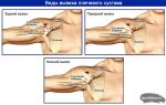
How the human shoulder is arranged, its functions and features. Movement in the shoulder joint is provided by Adduction of the shoulder
There are no special muscles that would cross the sagittal axis of the shoulder joint and be located medially from it, therefore, adduction of the shoulder according to the rule of a parallelogram of forces is carried out with simultaneous contraction of the muscles located in front (pectoralis major muscle) and behind the shoulder joint (latissimus dorsi and teres major). These muscles are helped by:
1) infraspinatus;
2) small round;
3) subscapular;
4) long head of the triceps muscle of the shoulder (see p. 160);
5) coracobrachial muscle (see p. 156).
abdominal muscle(see Fig. 38) is located in the infraspinatus fossa of the scapula, from which it starts. In addition, the place of origin of this muscle is the infraspinatus fascia. Muscle attached to the greater tubercle of the humerus, being partly covered by the prominent trapezium, and partly by the deltoid muscle.
The function of the infraspinatus muscle is to adduct, supine the nation and extend the shoulder in the shoulder joint. Since this muscle is attached to the capsule of the shoulder joint, when the shoulder is supinated, it simultaneously pulls the capsule away, protecting it from infringement.
teres minor muscle(see Fig. 38) is located below the infraspinatus muscle. She starts from the shoulder blade, and attached to the greater tubercle of the humerus and promotes adduction, supination and extension of this bone.
teres major muscle(see fig. 38) starts from the inferior angle of the scapula and attached to the crest of the lesser tubercle of the humerus, often with one tendon from the latissimus dorsi muscle. When contracting, the teres major muscle acts as a rounded elevation when the pronated shoulder is adducted. The function of the muscle is to adduct, pronate and extend the humerus. Subscapularis located on the anterior surface of the scapula, filling the subscapular fossa, from which starts. attached muscle to the lesser tubercle of the humerus. Contracting together with the previous muscles, it produces shoulder adduction; acting in isolation, it is its pronator. Since this muscle is multi-feathered, it has a significant
The special anatomy of the shoulder joint provides high mobility of the arm in all planes, including 360-degree circular movements. But the price for this was the vulnerability and instability of the articulation. Knowledge of anatomy and structural features will help to understand the cause of diseases that affect the shoulder joint.
But before proceeding with a detailed review of all the elements that make up the formation, two concepts should be differentiated: the shoulder and the shoulder joint, which many confuse.
The shoulder is the upper part of the arm from the armpit to the elbow, and the shoulder joint is the structure that connects the arm to the torso.
Structural features
If we consider it as a complex conglomerate, the shoulder joint is formed by bones, cartilage, articular capsule, synovial bags (bursae), muscles and ligaments. In its structure, it is a simple, consisting of 2 bones, a complex articulation of a spherical shape. The components that form it have a different structure and function, but are in strict interaction, designed to protect the joint from injury and ensure its mobility.
Shoulder components:
- scapula
- brachial bone
- articular lip
- joint capsule
- synovial bags
- muscles, including the rotator cuff
- bundles
The shoulder joint is formed by the scapula and humerus, enclosed in a joint capsule.
The rounded head of the humerus is in contact with a fairly flat articular bed of the scapula. In this case, the scapula remains practically motionless and the movement of the hand occurs due to the displacement of the head relative to the articular bed. Moreover, the diameter of the head is 3 times the diameter of the bed.
This discrepancy between shape and size provides a wide range of motion, and the stability of the articulation is achieved due to the muscular corset and ligamentous apparatus. The strength of the joint is also given by the articular lip located in the scapular cavity - cartilage, the curved edges of which extend beyond the bed and cover the head of the humerus, and the elastic rotator cuff surrounding it.
Ligament apparatus
The shoulder joint is surrounded by a dense articular bag (capsule). The fibrous membrane of the capsule is of varying thickness and is attached to the scapula and humerus to form a spacious pouch. It is loosely stretched, which makes it possible to freely move and rotate the arm.
From the inside, the bag is lined with a synovial membrane, the secret of which is the synovial fluid that nourishes the articular cartilage and ensures that there is no friction when they slide. Outside, the articular bag is strengthened by ligaments and muscles.
The ligamentous apparatus performs a fixing function, preventing displacement of the head of the humerus. Ligaments are formed by strong, poorly extensible tissues and are attached to the bones. Poor elasticity is the reason for their damage and rupture. Another factor in the development of pathologies is the insufficient level of blood supply, which is the cause of the development of degenerative processes of the ligamentous apparatus.
Ligaments of the shoulder joint:
- coracohumeral
- upper
- average
- lower
Human anatomy is a complex, interconnected and fully thought out mechanism. Since the shoulder joint is surrounded by a complex ligamentous apparatus, for the latter to slide in the surrounding tissues, mucous synovial bags (burses) are provided that communicate with the joint cavity. They contain synovial fluid, ensure smooth operation of the articulation and protect the capsule from stretching. Their number, shape and size are individual for each person.
Muscular frame
The muscles of the shoulder joint are represented by both large structures and small ones, due to which the rotator cuff of the shoulder is formed. Together they form a strong and elastic frame around the articulation.
Muscles surrounding the shoulder joint:
- Deltoid. It is located above and outside the joint, and is attached to three bones: the humerus, scapula, and collarbone. Although the muscle is not directly connected to the joint capsule, it reliably protects its structures from 3 sides.
- Double-headed (biceps). It is attached to the scapula and humerus and covers the joint from the front.
- Triceps (triceps) and coracoid. Protect the joint from the inside.
The rotator cuff of the shoulder joint provides a wide range of motion and stabilizes the head of the humerus, keeping it in the joint bed.
It is made up of 4 muscles:
- subscapular
- infraspinatus
- supraspinous
- small round
The rotator cuff is located between the head of the shoulder and the acromin, a process of the scapula. If the space between them narrows due to various reasons, the cuff is infringed, resulting in a collision between the head and the acromion, and is accompanied by severe pain.
Doctors gave this condition a name "impingement syndrome". With impingement syndrome, the rotator cuff is injured, leading to its damage and rupture.
blood supply
The blood supply to the structure is carried out with the help of an extensive network of arteries, through which nutrients and oxygen enter the tissues of the joint. Veins are responsible for the removal of metabolic products. In addition to the main blood flow, there are two auxiliary vascular circles: the scapular and acromio-deltoid. The risk of rupture of the large arteries passing near the joint greatly increases the risk of injury.
Elements of the blood supply
- suprascapular
- anterior
- rear
- thoracoacromial
- subscapular
- shoulder
- axillary
innervation
Any damage or pathological processes in the human body are accompanied by pain. Pain can signal the presence of problems or perform security functions.
In the case of joints, tenderness forcibly "deactivates" the diseased joint, preventing its mobility to allow injured or inflamed structures to recover.
Shoulder nerves:
- axillary
- suprascapular
- chest
- ray
- subscapular
- axillary
Development
When a child is born, the shoulder joint is not fully formed, its bones are disconnected. After the birth of the child, the formation and development of the structures of the shoulder continues, which takes about three years. During the first year of life, the cartilaginous plate grows, the articular cavity is formed, the capsule contracts and thickens, the surrounding ligaments strengthen and grow. As a result, the joint is strengthened and fixed, and the risk of injury is reduced.
Over the next two years, the articulation segments increase in size and take their final form. The humerus is the least susceptible to metamorphosis, since even before birth, the head has a rounded shape and is almost completely formed.
Shoulder instability
The bones of the shoulder joint form a mobile joint, the stability of which is provided by muscles and ligaments.
This structure allows for a large range of motion, but at the same time makes the articulation prone to dislocations, sprains and ruptures of the ligaments.
Also, often people are faced with such a diagnosis as joint instability, which is put in the case when, with the movements of the arm, the head of the humerus goes beyond the articular bed. In these cases, we are not talking about trauma, the consequence of which is dislocation, but about the functional inability of the head to remain in the desired position.
There are several types of dislocations depending on the displacement of the head:
- front
- rear
- lower
The structure of the human shoulder joint is such that the scapular bone covers it from behind, and the deltoid muscle from the side and from above. The frontal and internal parts remain insufficiently protected, which leads to the predominance of the anterior dislocation.
Shoulder Functions
The high mobility of the articulation allows for all movements available in 3 planes. Human hands can reach any point of the body, carry weights and perform delicate work that requires high precision.
Movement options:
- abduction
- cast
- rotation
- circular
- bending
- extension
It is possible to perform all the listed movements in full only with the simultaneous and coordinated work of all elements of the shoulder girdle, especially the clavicle and the acromioclavicular joint. With the participation of one shoulder joint, the arms can only be raised to shoulder level.
Knowledge of the anatomy, structural features and functioning of the shoulder joint will help to understand the mechanism of the occurrence of injuries, inflammatory processes and degenerative pathologies. The health of all joints in the human body directly depends on lifestyle.
Excess weight and lack of physical activity harm them and are risk factors for the development of degenerative processes. Careful and attentive attitude to your body will allow all its constituent elements to work for a long time and flawlessly.
The most complete answers to questions on the topic: "the movement in the shoulder joint is provided."
"Muscles of the Upper Limb"
musclesproducingmovementsbelt toplimbs
Schematically, the movements of the belt of the upper limb (scapula and collarbone) are divided into:
Movement forward and backward with abduction of the scapula from the spinal column and adduction to it.
Raising and lowering the scapula and collarbone.
The movement of the scapula around the sagittal axis with the lower angle to the medial and lateral sides.
Circular movement with the lateral end of the clavicle and at the same time with the shoulder blade.
These movements involve six functional muscle groups.
Movementforward
The movement of the girdle of the upper limb forward is produced by muscles that cross the vertical axis of the sternoclavicular joint and are located in front of it. These include:
large pectoral, acting on the belt of the upper limb through the humerus;
small chest;
anterior dentate.
Movementback
Exercise muscles that cross the vertical axis of the sternoclavicular joint and lie behind it. This muscle group includes:
trapezius muscle;
rhomboid muscle, large and small;
latissimus dorsi.
Movementup
Raising the girdle of the upper limb is produced by the following muscles:
1) the upper bundles of the trapezius muscle, which pulls up the lateral end of the clavicle and the acromion of the scapula;
muscle that lifts the scapula;
rhomboid muscles, in the decomposition of the resultant of which there is some component directed upwards;
sternocleidomastoid muscle, which, attaching with one of its heads to the collarbone, pulls it, and, consequently, the scapula up.
Movementdown
Lowering is facilitated by muscles that go from bottom to top, from the chest or spinal column to the bones of the girdle of the upper limb:
pectoralis minor;
subclavian muscle;
lower bundles of the trapezius muscle;
lower teeth of the anterior serratus muscle.
In addition, the muscles that go from the trunk to the shoulder, namely the pectoralis major and the latissimus dorsi, help lowering, mainly with their lower parts.
Rotationshoulder blades(movementlowerangleinsideAndoutside)
The rotation of the scapula inward, with the lower angle to the spinal column, produces a pair of forces formed by:
pectoralis minor muscle
lower part of the rhomboid muscle.
The rotation of the scapula outward, with the lower angle from the spinal column to the lateral side, occurs as a result of the action of a pair of forces formed by the upper and lower parts of the trapezius muscle.
This movement is supported by:
serratus anterior with its lower and middle teeth;
large round muscle with a fixed free upper limb.
Circularmovement
The circular movement of the belt of the upper limb occurs as a result of the alternate contraction of all its muscles.
musclesproducingmovement inshoulderjoint
In the shoulder joint, movements are possible around three mutually perpendicular axes:
abduction and adduction around the anteroposterior axis;
flexion and extension around the transverse axis;
pronation and supination around the vertical axis;
circular motion (circumduction).
These movements are provided by six functional muscle groups.
leadshoulder
The abductors of the shoulder cross the sagittal axis of rotation at the shoulder joint and are located laterally from it. The humerus is abducted by the muscles:
deltoid and
supraspinatus.
Castingshoulder
There are no special muscles that would cross the sagittal axis of the shoulder joint and be located medially from it, therefore, adduction of the shoulder according to the rule of a parallelogram of forces is carried out with simultaneous contraction of the muscles located in front (pectoralis major muscle) and behind the shoulder joint (latissimus dorsi and teres major). These muscles are helped by:
infraspinatus;
small round;
subscapular;
long head of the triceps muscle of the shoulder;
coracobrachial muscles.
All muscles of the upper limb are usually divided into 2 groups: the muscles of the shoulder girdle and the free upper limb, which in turn consist of 3 topographic sections - the muscles of the shoulder, the muscles of the forearm and the hand. Many mistakenly think that the muscles of the shoulder girdle also belong to the muscles of the shoulder, but according to the accepted anatomical classification, this is not the case. The shoulder is a part of the free upper limb, starting from the shoulder joint and ending with the elbow joint.

All muscles of the shoulder anatomical region can be divided into posterior and anterior groups.
Anterior shoulder muscle group
These include:
- biceps brachii,
- coracobrachialis muscle,
- shoulder muscle.
two-headed
It has two heads, from where it got its characteristic name. The long head originates with the help of a tendon from the supraarticular tubercle of the scapula. The tendon passes through the articular cavity of the shoulder joint, lies in the intertubercular groove of the humerus and passes into the muscle tissue. In the intertubercular groove, the tendon is surrounded by a synovial membrane, which connects to the cavity of the shoulder joint.
The short head originates from the top of the coracoid process of the scapula. Both heads merge together and pass into the spindle-shaped muscle tissue. A little above the ulnar fossa, the muscle narrows and passes again into the tendon, which is attached to the tuberosity of the radius of the forearm.

Functions:
- flexion of the upper limb in the shoulder and elbow joints;
- supination of the forearm.
Coracohumeral
The muscle fiber starts from the coracoid process of the scapula, is attached to the humerus approximately in the middle from the inside.
Functions:
- flexion of the shoulder in the shoulder joint;
- bringing the shoulder to the body;
- takes part in turning the shoulder outward;
- pulls the scapula down and forward.

Shoulder
This is a fairly wide muscle that lies directly under the biceps. It starts from the anterior surface of the upper part of the humerus and from the intermuscular septa of the shoulder. Attaches to the tuberosity of the ulna. Function - flexion of the forearm at the elbow joint.

Posterior muscle group
This group includes:
- triceps brachii,
- elbow,
- muscle of the elbow joint.
three-headed
This anatomical formation has three heads, hence the name. The long head originates from the subarticular tubercle of the humerus and below the middle of the humerus passes into the tendon common to the three heads.
The lateral head starts from the posterior surface of the humerus and the lateral intermuscular septum.
The median head starts from the posterior surface of the humerus and both intermuscular septa of the shoulder. It is attached by a powerful tendon to the olecranon of the ulna.
Functions:
- extension of the forearm in the elbow joint;
- adduction and extension of the shoulder due to the long head.

Elbow
It is, as it were, a continuation of the median head of the triceps muscle of the shoulder. It originates from the lateral epicondyle of the humerus, and is attached to the posterior surface of the olecranon of the ulna and to its body (proximal part).
Function - extension of the forearm in the elbow joint.

Elbow muscle
This is a non-permanent anatomical formation. Some experts consider it as part of the fibers of the median head of the triceps muscle, which are attached to the capsule of the elbow joint.
Function - stretches the capsule of the elbow joint, which prevents it from being pinched.
Muscles of the shoulder girdle
It is worth mentioning the muscles of the girdle of the upper limb, which are often considered to be muscle formations of the shoulder:
- deltoid muscle of the shoulder,
- supra- and infraspinatus muscle,
- small and large round
- subscapular.


Both groups of muscles of the shoulder are separated from each other by two connective tissue intermuscular septa, which stretch from the common shoulder fascia (enveloping the entire muscular frame of the shoulder) to the lateral and median edges of the humerus.
Shoulder muscle pain
Pain in the shoulder and shoulder girdle is a common complaint of people of different age groups. Such a symptom may be associated with pathology of the skeleton, joints, ligaments, but most often the cause is hidden in damage to muscle tissue.
Causes
Consider the most common causes of pain in the shoulder region:
- overstrain and sprain of ligaments, tendons, muscles;
- diseases or traumatic injuries of the shoulder joint;
- inflammation of the ligaments and tendons of the muscles (tendinitis);
- rupture of tendons and muscles;
- joint capsulitis (inflammation of the joint capsule);
- inflammation of the periarticular bags - bursitis;
- frozen shoulder syndrome;
- humeroscapular periarthrosis;
- myofascial pain syndrome;
- vertebrogenic causes of pain syndrome (associated with damage to the cervical and thoracic spine);
- impingement syndrome;
- rheumatic polymyalgia;
- myositis of infectious (specific and non-specific) and non-infectious nature (with autoimmune, allergic diseases, ossifying myositis).

Pain in the shoulder area can be associated with both damage to bones, joints, ligaments, and damage to muscle tissue.
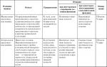
Differential Diagnosis
The following criteria will help distinguish shoulder pain caused by muscle damage from joint diseases.
| sign | Joint diseases | Muscular lesions |
| The nature of the pain syndrome | The pain is constant, does not disappear at rest, slightly increases with movement | Pain occurs or increases significantly with a certain type of motor activity (depending on the damaged muscle) |
| Pain localization | Unlimited, diffuse, spilled | It has a clear localization and certain boundaries, which depends on the localization of the damaged muscle fiber |
| Dependence on passive and active movements | All types of movements are limited due to the development of pain syndrome | Due to pain, the amplitude of active movements decreases, but all passive ones are preserved in full |
| Additional diagnostic features | Change in the shape, contours and size of the joint, its swelling, hyperemia | The joint area is not changed, but there may be swelling in the soft tissue area, slight diffuse redness and an increase in local temperature with inflammatory causes of pain |
What to do?
If you are suffering from shoulder pain, which is associated with damage to muscle tissue, the first thing to do in order to get rid of such an unpleasant symptom is to identify the provoking factor and eliminate it.
If after that the pain still returns, you need to visit a doctor, perhaps the cause of the pain syndrome is completely different. The following tips will help you get rid of pain quickly:
- in case of acute pain, it is necessary to immobilize the sore arm and provide it with complete rest;
- on your own, you can take 1-2 tablets of an over-the-counter pain reliever of a non-steroidal anti-inflammatory drug or apply it to the affected area in the form of an ointment or gel;
- massage can be used only after the elimination of acute pain syndrome, as well as physiotherapy;
- after the pain subsides, it is important to regularly engage in physiotherapy exercises to develop and strengthen the muscles of the shoulder;
- if a person, on duty, is forced to perform daily monotonous hand movements, it is important to take care of protecting the muscles and preventing their damage (wear special bandages, protective and supporting orthoses, perform gymnastics to relax and strengthen, undergo regular therapeutic and preventive massage courses, etc.).
As a rule, the treatment of muscle pain caused by overexertion or mild injury lasts no more than 3-5 days and requires only rest, minimal stress on the hands, correction of the rest and work regimen, massage, and sometimes taking non-steroidal anti-inflammatory drugs. If the pain does not go away or it initially has a high intensity, is accompanied by other alarming signs, it is imperative to visit a doctor for examination and correction of treatment.
This joint is reinforced by the anterior and posterior sternoclavicular, costoclavicular, and interclavicular ligaments. The distal end of the clavicle and the acromion form the acromioclavicular joint, which is reinforced by the coracoclavicular and acromioclavicular ligaments.
The articular cavity of the scapula and the head of the humerus form the shoulder joint. This is a very mobile and therefore rather fragile spherical joint, reinforced by the articular lip, articular capsule and articular-shoulder ligaments.
The movements of the arm in the shoulder joint (Fig. 5.1) occur under the action of many muscles. Flexion is carried out by the clavicular part of the pectoralis major muscle and the anterior part of the deltoid muscle. Extension occurs due to contraction of the latissimus dorsi, teres major, and sternocostal portion of the pectoralis major. Abduction is provided by the deltoid muscle and the muscles of the rotator cuff of the shoulder (subscapular, supraspinatus, infraspinatus and small round muscles). Adduction occurs due to contraction of the pectoralis major (sternocostal part), the latissimus dorsi, and the teres major muscle. The subscapularis and pectoralis major muscles rotate the humerus inward, and the supraspinatus, infraspinatus, and teres minor muscles outward. Horizontal adduction is carried out by simultaneous contraction of the coracobrachial, pectoralis major, and anterior part of the deltoid muscle, and horizontal abduction by contraction of the infraspinatus, teres minor, and posterior part of the deltoid muscle.
Figure 5.1. Range of motion in the shoulder joint. A. Flexion and extension. B. Abduction and adduction. B. Outward and inward rotation. D. Horizontal abduction and adduction.
Figure 5.1 (end). Range of motion of the scapula. D. Raising and lowering. E. Outward and inward rotation. G. Abduction and adduction.
Rotator cuff - muscles directly adjacent to the capsule of the shoulder joint (subscapular, supraspinatus, infraspinatus and small round).
Simultaneously with movements in the shoulder joint, movements of the scapula occur, namely, its abduction, adduction, rotation outward or inward, as well as lifting and lowering. Abduction of the scapula is carried out by the pectoralis minor and anterior serratus muscles, adduction by the rhomboid muscles, rotation by the lower angle outward by the anterior serratus and trapezius muscles, rotation by the lower angle inward by the pectoralis minor and rhomboid muscles, lifting by the muscle that lifts the scapula, and lowering by the pectoralis minor .
Functional muscle groups that produce movements in the shoulder joint
Flexion of the shoulder is carried out:
Anterior deltoid
muscles, this muscle surrounds the shoulder joint in front, outside and
behind, starts from the clavicle, acromial process, spine of the scapula, attaches to the deltoid tuberosity of the humerus:
The pectoralis major muscle, starting from the sternal end of the clavicle, sternum, cartilage of the 2nd-7th ribs and attached to the crest of the greater tubercle of the shoulder
The biceps muscle of the shoulder, which has two heads: short and long; the long head is a biarticular muscle, participates in the flexion of the shoulders, starts from the supraarticular tubercle of the scapula, the short one - from the coracoid process of the scapula, both heads are attached to the tuberosity of the radius.
Shoulder extension, i.e. movement back, carry out:
Latissimus dorsi:
Small round muscle:
Large round muscle;
Long head of the triceps brachii.
Bringing the shoulder produce:
pectoralis major;
latissimus dorsi;
infraspinatus muscle; - small round muscle; - large round muscle;
Subscapularis muscle: - coracobrachialis muscle; - long triceps head
Pronation of the shoulder, turning it inward around the vertical axis, is produced by:
Anterior part of the deltoid muscle;
pectoralis major;
The latissimus dorsi;
teres major muscle:
Shoulder supination, rotation
The back of the deltoid muscle;
Small round muscle.
Flexion of the forearm is produced by:
biceps brachii;
The shoulder muscle, located under the biceps of the shoulder, starting from the anterior surface of the humerus and attached to the coronoid process of the ulna;
Extension of the forearm is performed:
The triceps muscle of the shoulder, which has three heads: long, lateral and medial, the long head starts from the subarticular tubercle of the scapula, and the other two from the posterior surface of the humerus, all heads are attached to the olecranon process of the ulna;
Supination of the forearm is performed:
biceps brachii;
The functional groups of muscles that produce hand movements (flexion and extension, abduction and adduction) include hand flexors, their antagonists - hand extensors, abductor muscles and their antagonists - adductor muscles.
Hand exercises
Hello dear readers, today there will be a rather voluminous, interesting, important topic. I am pleased to see that a healthy lifestyle is gaining more and more popularity in our time. Just the other day, I watched two young girls of 20-25 years old jumping rope merrily near their house, right on the playground!
But, despite the active development of the fitness industry, the promotion of sports and a healthy lifestyle, there are a lot of difficulties and pitfalls on the way of those who train.
Numerous exercises for the hands that both boys and girls like to perform because of superficial knowledge in anatomy, due to the lack of proper exercise technique, due to neglect of safety rules, can cause significant harm to health rather than benefit. This applies not only to exercises on the hands, but also to any other!
In this article, I will talk about the most popular exercises for the muscles of the upper limbs, how to perform them correctly and protect yourself from possible injuries. The muscles of the free upper limb are divided into several groups: flexors and extensors of the shoulder, flexors and extensors of the forearm.
The most important for us are the flexors of the forearm - the biceps of the shoulder, as well as the extensor of the forearm - the triceps of the shoulder. These are the muscles that most of the exercises are aimed at. The muscles of the forearm are involved in holding the projectiles in the hands, in the process of which they receive the load they need.
Functions of the biceps brachii
1. Shoulder flexion.
2. Flexion of the forearm.
3. Supination of the arm (outward rotation of the bones of the upper limb).
Functions of the triceps brachii
1. Extension of the forearm.
2. Extension of the shoulder.
3. Bringing the shoulder in the frontal plane.
4. Shoulder hyperextension (shoulder extension beyond the anatomical position).
Hand exercises
1. Bending the forearms with a barbell while standing;
2. Bending the forearms with dumbbells while sitting on an incline bench;
3. Bending of the forearms with a barbell on the “Scott bench”;
4. "French bench press" with a barbell lying;
5. Bench press, lying with a narrow grip;
6. Extension of the forearms in a block frame while standing;
Standing Barbell Forearm Curl
Working joints: The main work falls on the elbow joint. The shoulder joint deviates forward from the anatomical position by five to ten degrees.
Impact on the main muscle groups: The target muscle in this exercise is the biceps of the shoulder. The muscles of the forearm perform a synergistic function in isometric contraction.
Starting position: Feet stand at the width of the pelvic bones, the feet are parallel. For additional balance, you can take one leg forward a little. Hold the bar with a reverse grip at shoulder width.
Movement: On exhalation, it is necessary to bend the forearms to the maximum contraction of the biceps muscles.
Methodical instructions: The back must be kept straight. A straight back implies the natural curves of the spinal column. The shoulder blades are brought to the spine, the head is in a neutral position.
Forearm curls with dumbbells sitting on an incline bench
Working joints: Elbow joint. The shoulder joint performs an auxiliary function.
Targeting Major Muscle Groups: In this exercise, the agonist (target muscle) is the biceps brachii. The work of the muscles of the forearms is aimed at holding the dumbbells.
Starting position: Sitting on an incline bench (70 - 80 degrees), we hold the dumbbells with a reverse grip in our hands on the sides of the torso. The back should be pressed tightly against the bench, the shoulders should be directed perpendicular to the floor. It is not necessary to block the elbow joint.
Movement: On an exhalation, bend the forearms, contracting the biceps of the shoulder as much as possible. On inspiration, return to the starting position.
Methodical instructions: This exercise is more preferable for those who have diseases of the spine, such as herniated discs and posture disorders. You also need to control the position of the shoulder blades and head.
Forearm Curl with a Barbell on the Scott Bench
In order to perform this exercise, we need a special bench. It is a short and narrow bench on which there is a palm rest. Starting to perform on the bench, you need to put your hands on this support. This is necessary in order to exclude all possible movements in the joints of the limbs. Fixation of the elbow joints entails an increase in the load on the biceps of the shoulder, and the sitting position removes excess weight from the back.
Working joint. Elbow joint:
Effects on muscle groups: Agonist (target muscle) – biceps brachii.
Starting position: Adjust the height of the palm rest so that you are in your comfort zone. When you sit on the bench, keep your back straight and the top of the support not interfere with your breathing. The elbows are on the support, not slipping down from it.
The bar must be taken either by yourself or by asking a training partner. As you exhale, bend your forearms. On inspiration - to the starting position.
Methodical instructions: Performing this exercise, try not to change the angle between the forearm and the hands. The hands should not rotate the bar, this will lead to the fact that the load on the biceps will be lost.
A feature of this exercise is also the fact that in the final phase of the exercise, the angle between the shoulder and forearm should not be less than a straight line (90 degrees). This is due to the fact that we fix the elbows on the stop. With a decrease in this angle, the biceps will simply relax, which will not give the desired effect.
"French bench press" with a barbell lying
Working joints: Elbow joints. The shoulder joints are bent at a right angle (perpendicular to the floor).
Impact on the main muscle groups: Agonist (working muscle) - the triceps muscle of the shoulder, (m. triceps brachii). The biceps and forearm muscles act as stabilizers.
Starting position: Lying on a bench, hold the barbell above you with an overhand grip. Shoulders are perpendicular to the floor. The back of the head, shoulder blades and buttocks must be pressed tightly against the bench. The emphasis of the feet on the floor should be strong enough to ensure maximum balance. As you inhale, lower the barbell towards your forehead. You need to lower it to a right angle between the shoulder and elbow joints. On exhalation, we return to the starting position.
Guidelines: It is strictly forbidden to hold the bar with a reverse grip. This can be very traumatic. To perform an action in this exercise is required only due to flexion and extension in the elbow joint. The shoulder joint must remain motionless.
Close Grip Bench Press
Working joints: Shoulder joint, elbow joint.
Influence of muscle groups: Agonists - triceps muscles of the shoulders. An auxiliary function is performed by the deltoid and pectoral muscles.
Starting position: The exercise is performed on a horizontal bench with racks, hold the bar with a straight closed grip. Shoulders are perpendicular to the floor. Grip width is about two widths of your palm. On inspiration, you need to lower the barbell down to the lower third of the sternum. Touching the chest bar, on the exhale, you must return to the starting position.
Execution Guidelines: At the moment the bar touches the torso, the angle between the torso and shoulder should be approximately 30 degrees.
Extension of the forearms on the block, standing
Working joint: Elbow joint.
Impact on the main muscle groups: Triceps of the shoulder.
Starting position: Stand at the weight-block frame in such a way that your forearms, the handle and the cable of the simulator are in the same plane. Take the handle with a straight closed grip. Leaning slightly and bending your knees, straighten your forearms as much as possible while exhaling. While inhaling, take the starting position.
Guidelines: During this exercise, the back should be in the same position. Keep your elbows as close to your torso as possible. Following these rules will ensure the work of only the triceps muscle of the shoulder. Avoid excess weight in the simulator.
The above exercises on the hands are the most common for a number of reasons. First, they are physiological. This means that a person adapted to perform such movements in the process of his evolutionary development. For example, pull-ups on the bar imitate a person's attempt to climb some obstacle, say, running away from a pack of wild wolves.
The bench press, lying down, is a movement that we perform in order to push a certain heavy object away from us. But bending the dumbbell with an emphasis on the thigh is, of course, a good exercise loved by many athletes, but it has no use in real life.
Secondly, most of these exercises can be done at home with a minimum set of equipment. Fortunately for you and me, exercises for the free upper limb are interchangeable and there will be no serious problems with exercises at home.
So, for example, a barbell bench press, lying with a narrow grip, can be replaced with a “French bench press” with a standing dumbbell. You can perform it with one or two hands, depending on the caliber of dumbbells that you have available.
Another moment! When exercising in the gym, it is better to use a barbell with a straight, rather than a curved neck. This is due to the anatomical structure of the biceps brachii and its function. By bending the forearm, the biceps supinates the arm, that is, rotates it outward. Using a barbell with a curved neck, we do not fully engage the outer bundle of the biceps, because the arm is not fully supinated.
And for a snack, a few words about the most common misconceptions that we are so used to believing in.
1. Exhausting exercises for the biceps day in and day out can make the shoulder immense. Fortunately, this is not the case. Two-thirds of the volume of the shoulder is occupied by triceps. The mark on the centimeter tape when measuring the circumference of the shoulder will creep up if you pay due attention to this particular muscle.
2. After a person stops his workouts in the gym, his muscles turn into fat. In nature, such transformations are not possible. These tissues have completely different chemical composition and metabolic pathways. After you stop training, the body will begin to get rid of muscle tissue first, since four times more energy is spent on its content in the body than on the content of fatty tissue.
3. If you swing according to the Schwarzenegger or Stallone method, you can achieve crazy results in the shortest possible time. This is an erroneous opinion. Whatever incredible exercises you are offered, remember that they are designed for people taking hormonal drugs. The main principle of building a training program is an individual approach to each person.
4. Warm-up - this is for old ladies and newcomers to the gym. Another misconception. People who neglect these exercises are at risk of acquiring serious diseases of the musculoskeletal system. These include osteochondrosis, hernia of the spinal column, inflammation of the articular bags and many other pathologies that significantly reduce the quality of human life.
The treatment of these diseases is often very long, expensive and unpleasant. In a nutshell, we can say that a warm-up before the main work in the gym warms up the joints, enhances the production of intra-articular fluid, increases strength and protects against any possible injuries in the process of exercising in the gym.
"Concentrated" bending of the arms - peak concentration
Agonist: biceps brachii
Synergists: brachial, brachioradial, pronator round.
■ Sitting position. The body is turned half a turn and tilted forward.
■ The shoulder joint of the arm holding the dumbbell is above the thigh
■ The hand holding the dumbbell rests on the outer side of the shoulder
the inner side of the thigh of the leg of the same name.
■ Legs bent at the knees, spread apart.
■ Feet firmly rest on the floor.
■ Raise the dumbbell by bending your forearm.
■ Keep your elbow in place while moving.
FST - Functional Strength Training
Friday, August 3, 2012
Physiology of the shoulder joint
The shoulder joint, or proximal joint of the upper limb, is the most mobile of all joints in the human body.
- The transverse axis, lying in the frontal plane, controls the movements of flexion and extension carried out in the sagittal plane.
- The anteroposterior axis, which lies in the sagittal plane, controls the movements of abduction (movement of the upper limb away from the body) and adduction (movement of the upper limb towards the body), which are realized in the frontal plane.
- The vertical axis, passing through the intersection of the sagittal and frontal planes and corresponding to the third spatial axis, controls the movements of flexion and extension that occur in the horizontal plane when the shoulder is abducted to 90 °, also called horizontal flexion - extesia.
In relation to the longitudinal axis 4, external and internal rotation of the shoulder and the entire upper limb is carried out:
- random rotation, or McConnell replacement rotation, which depends on the presence of a third degree of freedom of movement and can only be carried out in spherical joints with three axes; this movement is provided by contraction of the rotator muscles;
- automatic rotation, or McConnell combined rotation, which occurs without any voluntary action in joints with two and even three axes of movement, if only two axes are used in the latter. We will return to this when we consider the Codman paradox.
In the neutral position, the upper limb hangs freely along the body, so that the longitudinal axis of the shoulder 4 coincides with the vertical axis 3 of the upper limb. The longitudinal axis of the shoulder 4 coincides with the transverse axis 1 at 90° abduction and with the anteroposterior axis 2 at 90° flexion.
- extension: movement with a small amplitude, equal to 45-50 °;
- bending: movement with greater amplitude up to 180°; note that the 180° flexion position can also be considered a 180° abduction position combined with axial rotation (see Codman's Paradox).
The term antepulsion (bringing the organ anteriorly in the frontal plane) and the term retropulsion (retracting the organ posteriorly in the frontal plane) to denote extension are often mistakenly used to denote flexion. These concepts are applicable to the definition of the movement of the shoulder girdle in the horizontal plane and should not be used to describe the movement of the upper limb in general.
- with extension (Fig. 5), while adduction is extremely insignificant;
- with flexion (Fig. 6), while adduction can reach 30-45°.
From a position of abduction of any amount, adduction (also called "relative adduction") is possible in the frontal plane until a neutral position is reached.
lead
- Beyond 90°, the movement of abduction again brings the upper limb closer to the sagittal plane of the body and becomes, strictly speaking, adduction.
- Full 180° abduction can also be achieved through 180° flexion.
As for the muscles and the corresponding movements in the joint, abduction, starting from a neutral position (Fig. 7), passes through three phases:
- abduction from 0 to 60° (Fig. 8), occurring only in the shoulder joint;
- abduction from 60 to 120° (Fig. 9), requiring the connection of the scapular-thoracic "joint";
- abduction from 120 to 180 ° (Fig. 10), requiring the participation of the shoulder joint, the scapular-thoracic "joint" and the tilt of the body in the opposite direction.
Note that pure abduction performed exclusively in the frontal plane, parallel to the plane of back support, is rare. In contrast to this, abduction in combination with flexion, i.e. raising the limb in the plane of the scapula at an angle of 30 ° anterior to the frontal plane, is performed very often, for example, to bring the brush to the mouth or put it on the back of the neck. This position corresponds to the position of balance of the shoulder muscles.
Axial rotation of the upper limb
- forward movement: pectoralis major, difficult minor, serratus anterior;
- backward movement: rhomboid, trapezius (transverse fibers), latissimus dorsi.
rotation movement
- Sagittal plane A, or rather parasagittal, since the true sagittal plane passes through the longitudinal axis of the trunk (this is the flexion-extension plane).
- Frontal, or coronal, plane B, parallel to the plane of the back support (this is the plane of adduction - abduction).
- The transverse plane C, perpendicular to the axis of the body (this is the plane of horizontal flexion - extension), i.e. remaining in the horizontal plane.
Starting from the neutral position (when the arm hangs along the body), shown by the bold dotted line, the arc (for the right upper limb) passes sequentially through the following sectors: (III) - (II) - (VI) - (V) - (IV)
(II) top front left;
(VI) top rear and right;
(V) lower rear and right;
(iv) lower front and right;
(VIII) behind and to the left over a very small extent, since the combined movement of extension and adduction is very limited (in the diagram, sector (VIII), lies below plane C, posterior to sector (III) and to the left of sector (V);
sector (VII) is not visible here, it is located above).
Movement of the shoulder girdle in the horizontal plane
- deltoid (acromial fibers (III), Fig. 101);
- supraspinous;
- trapezoid: upper fibers (acromial and clavicular) and lower fibers (tuberous);
- anterior dentate.
(b) Horizontal flexion (Fig. 17), combined with adduction, has an amplitude of 140° and requires the participation of the following muscles:
- deltoid (anterointernal fibers (I) and anterior external fibers (II) to varying degrees, as well as external fibers (III));
- subscapular;
- large and small chest;
- anterior dentate.
(c) Extension in the horizontal plane (Fig. 19), combined with adduction, is carried out in a limited volume of 30-40 ° and requires the participation of the following muscles:
- deltoid (posterior fibers (IV) and (V), posterior fibers (VI) and (VII) to varying degrees, as well as external fibers (III);
- supraspinous;
- infraspinatus;
- large and small round;
- diamond-shaped;
- trapezoidal (spinous fibers with the addition of two others);
- the latissimus dorsi muscle, acting as an antagonist-synergist with the deltoid muscle, which blocks its important adductor function.
The total amplitude of flexion and extension in the horizontal plane is slightly less than 180°. The movement from the extreme anterior to the extreme posterior position alternately activates various fibers of the deltoid muscle, which is dominant in this function. The sequence of work of different bundles of fibers of the deltoid muscle can be compared to playing scales on the piano.
Decomposition of the movements of the shoulder joint in the coordinate system
Codman's Paradox
- from the starting position (Fig. 26 profile and 27 front), in which the upper limb hangs vertically along the body, the palm is turned inward, and the thumb is anterior Av;
- move it +180° in the frontal plane (Fig. 28);
- proceeding from this position, when the thumb is directed outward, straighten the upper limb by -180 ° in the sagittal plane (Fig. 29);
- in this case, the upper limb will again be located vertically along the body, but the palm will be turned outward, and the thumb - backwards (Fig. 30).
This movement can also be reversed, starting with a 180° flexion followed by a 180° adduction. The limb is rotated outwards by 180°.
- if the arbitrary rotation is zero, then the automatic rotation will be maximum, which leads to Codman's (pseudo)paradox;
- if arbitrary rotation occurs in the same direction as automatic rotation, then the latter is enhanced;
- if voluntary rotation occurs in the opposite direction, then automatic rotation is reduced or even canceled, providing an ergonomic cycle.
Movements to evaluate the overall function of the shoulder joint
- in blue - anterior anti-lateral path C, passes from the opposite side of the joint through the head;
- in green - the anterior homo-lateral path H, passes through the head from the side of the involved joint;
- in red - the posterior path P, directed towards the back from the side of the active joint.
The path described by the fingertips in all these trajectories passes through five different points. The fifth point is common to the three paths (indicated in red in the figure), is located in the region of the opposite scapula and is called the "triple point".
Multi-joint complex of the shoulder girdle
- The shoulder, or scapulohumeral, joint is a truly anatomical joint with two articulating surfaces lined with hyaline cartilage. This is the most important joint in this group.
- Subdeltoid, or "second shoulder" joint. This is not an anatomical, but a physiological joint, consisting of two surfaces sliding on each other. The subdeltoid "joint" is mechanically connected to the shoulder joint, since any movement in the latter causes movement in it.
The second group includes three joints.
- Shoulder-thoracic or scapular-thoracic joint. This is also a physiological, not anatomical joint. It is the most important in this group, although it cannot function without two other joints mechanically connected to it.
- The acromioclavicular joint, which is a true joint, is located at the acromial end of the clavicle.
- The sternoclavicular joint, also a true joint, is located at the sternal end of the clavicle.
In general, the joints of the shoulder girdle can be grouped as follows.
- The first group is represented by the main anatomical joint - the shoulder - mechanically associated with the physiological, associated (not true) subdeltoid joint.
- The second group includes the main physiological (not true) scapular-thoracic "joint", which mechanically connects two associated anatomical joints - the acromioclavicular and sternoclavicular.
In each group, the joints included in it are mechanically interconnected, i.e. operate in partnership. In practice, both of these groups work simultaneously with varying degrees of participation depending on the type of movement being made. It can be said that the five joints of the shoulder girdle complex function simultaneously with varying degrees of participation in different groups.
Articular surfaces of the shoulder joint
Humeral head
- lesser tubercle, oriented anteriorly,
- large tubercle, oriented outwards.
- basal (internal), attached to the edge of the articular cavity,
- external (peripheral), to which the ligaments of the capsule are attached,
- internal (articular), lined with cartilage, which is a continuation of the cartilage of the articular cavity, and in contact with the head of the shoulder.
Simultaneous rotation centers
- from the beginning of the abduction to 50 °, the rotation of the head of the shoulder occurs around a point located somewhere within the circle C 1;
- by the end of the lead (from 50° to 90°), the center of rotation lies within the C2 circle;
- when the shoulder is abducted about 50°, the continuity of motion is broken and the center of rotation now moves upward and medial to the head of the shoulder.
When flexed (Fig. 45, external view), a similar analysis does not show interruption of the OCR path, which runs within a single circle located in the lower part of the humeral head in the middle between its two edges.
Capsule and ligaments of the shoulder joint
- the head of the humerus is surrounded by the cuff of the capsule 1, on which the lower synovial folds 2 lie under the head, are lifted by the return fibers of the capsule;
- the upper strand of the 4th humeroscapular ligament, which strengthens the capsule in its upper part;
- the tendon of the long head of the biceps brachii 3 is dissected;
- the tendon of the subscapularis muscle 5 is dissected near the area of its attachment.
Articular cavity (outside view) (Fig. 48):
- the articular cavity 2 is shown, surrounded by a lip (articular roller), which runs along the edge, a supra-articular notch;
- the tendon of the biceps muscle 3 (dissected here) is attached to the supraarticular tubercle and, dividing into two bundles, forms the articular roller. This tendon is intraarticular;
- articular capsule 8 is strengthened by the following ligaments:
- coraco-humeral 7;
- three strands of the humeroscapular ligament (Fig. 49): upper 9, middle 10 and lower 11;
- the coracoid process is visible in the background, the spine of the scapula is cut off 10 ;
- subscapular tubercle 11 (Fig. 48), from where the tendon of the long head of the triceps muscle of the shoulder originates outside the capsule.
Ligaments of the shoulder joint (Fig. 49, front view):
- the coraco-brachial ligament 3 goes from the coracoid process 2 to the greater tubercle, to which the tendon of the supraspinatus muscle 4 is attached;
- two strands of the coracoscapular ligament diverge above the intertubercular groove at the place where the tendon of the biceps muscle of the shoulder leaves the joint and goes along the groove, which turns into the groove of the biceps muscle, being overlapped from above by the transverse ligament of the shoulder joint 6.
- The shoulder-shoulder ligament consists of three strands: the upper 1 runs from the upper edge of the articular cavity above the head of the humerus, the middle 10 - from the upper edge of the articular cavity and in front of the humerus, and the lower 11 goes through the anterior edge of the articular cavity and below the head of the shoulder.
- These three strands form a Z-like structure in front of the joint capsule. Between them there are two weak points:
- Witbrecht's hole 12, which is the entrance to the subscapular fossa;
- Ruvier's foramen 13, through which the synovial cavity communicates with the beak-shaped bag;
- long head of the triceps brachii 14 .
The back surface of the shoulder joint (Fig. 50).
- deep surface of the middle 2 and lower 3 strands of the humeroscapular ligament;
- at the top are the upper bundles, as well as the coraco-brachial ligament 4, to which the coraco-scapular ligament 5 is attached, which is not essential from the point of view of mechanics;
- intra-articular part of the tendon of the long head of the biceps brachii 6;
- articular cavity 7, strengthened by articular lip 8;
- two ligaments that do not carry mechanical functions, namely suprascapular 9 and osteoscapular 10;
- attachment of three periarticular muscles: supraspinatus 11, infraspinatus 12 and small round 13.
Intra-articular location of the tendon of the biceps brachii
- Irregularities of the articular cavity of the scapula are smoothed out by articular cartilage 1.
- The articular lip deepens the articular cavity. However, the adhesion of the articular surfaces is very small, which leads to a high frequency of dislocations of the shoulder. In its upper part 3, the articular lip is not completely fixed to the bone, its inner edge lies freely in the joint cavity like a meniscus.
- When the joint is in a neutral position, the upper part of the capsule 4 is stretched, and the lower part 5 is relaxed. This relaxed state of the lower part of the capsule and the opening of the synovial folds 6 allow shoulder abduction to occur.
- The tendon of the long head of the biceps brachii muscle 7 begins on the supraarticular tubercle and the upper edge of the articular lip. Coming out of the articular cavity into the intertubercular groove 8, it goes deeper than the capsule 4.
- Within the joint cavity, the tendon of the long head of the biceps is in contact with the synovium in the following three positions:
- it is pressed against the deep surface of the capsule (c) by the synovial lining (s);
- the tendon is covered by the synovial membrane, which forms for it a suspension loop under the capsule or mesotendon;
- the tendon now lies free, but is fully invested by the synovium.
These three parts of the tendon differentiate sequentially along its course from the point of origin, but in all cases the tendon, being inside the capsule, remains extrasynovial.
The role of the glenohumeral ligament
Coracobrachial ligament in flexion and extension
Coaptation of articular surfaces under the action of periarticular muscles
Rear view of the transverse muscles (Fig. 64):
- The supraspinatus muscle 1 originates from the fossa of the scapula and ends on the upper facet of the tubercle of the humerus.
- The infraspinatus muscle 3 is attached to the upper part of the fossa of the scapula and ends on the posterior superior facet of the tubercle of the humerus.
- Small round 4 is attached to the lower part of the fossa of the scapula and ends on the posterior lower part of the facet of the tubercle of the humerus.
On fig. 65 is a front view.
- deltoid muscle 8, consisting of two bundles - lateral 8 and posterior 8 ′, which raises the head of the humerus during abduction;
- the triceps muscle of the shoulder (its long head) 7 attached to the subarticular tubercle of the scapula - presses the head of the humerus against the articular capsule when the elbow joint is extended.
Long muscles - coaptors (Fig. 68, front view), more numerous:
- deltoid muscle 8 with its two bundles (lateral 8 and anterior), clavicular muscle (not shown in the figure);
- the tendon of the long head of the biceps muscle 5, as well as its short head, attached to the coracoid apophysis, near the coracobrachial 6 . This tendon, when flexing the elbow and shoulder, brings the head of the humerus upward;
- the clavicular bundles of the pectoralis major muscle 9 contribute to the anterior cords of the deltoid muscle, but primarily carry out flexion and adduction of the shoulder.
Subdeltoid "joint"
- supraspinatus muscle 3;
- infraspinatus muscle 4;
- teres minor 5 and subscapularis posteriorly, which is not visible in the figure;
- tendon of the long head of the biceps brachii 6, which is visible above and below the notch of the biceps 9 that penetrates the joint.
The dissection of the deltoid muscle allows you to see the serous bag presented in Fig. in section 7 . Further anteriorly, the coraco-brachial tendon is located, which is formed by a common attachment to the coracoid apophysis of the following muscles:
- short bundles of the biceps muscle 13;
- coracobrachial muscle 14, together forming the "front protection" of the joint. Also behind the tendon are long strands of the triceps muscle of the shoulder.
The work of these muscles can be assessed using the following frontal sections of the shoulder girdle:
- the shoulder at rest is located vertically along the body (Fig. 70);
- when abducted, the arm is horizontal (Fig. 71).
In the first case (Fig. 70), both the previously considered muscles and a section of the scapular-shoulder joint 8 with the articular lip and the lower capsular process are visible. Subdeltoid serous bag 7 is located between the deltoid muscle and the upper end of the humerus.
Shoulder-thoracic "joint"
- scapular-dentate space 1 includes the scapula covered with the subscapularis muscle and the large serratus muscle proper;
- dentate-chest space 2 includes the chest wall and the serratus major muscle.
On the right half of the section, which is a functional diagram of the shoulder girdle, the following is shown:
- The scapula is not located in the frontal plane, but is inclined outward and anteriorly, forming an angle of 30 ° with the frontal plane, open anteriorly and outwardly. This angle represents the physiological plane of shoulder abduction.
- The clavicle runs obliquely, in the shape of the letter S in the posterolateral direction and forms an angle of 30 ° with the frontal plane. Anteriorly and medially, the clavicle forms the sternoclavicular joint with the sternum and the acromioclavicular joint with the scapula, following outwards and backwards.
- At rest, the clavicle forms an angle of 60° with the scapula, but this can change due to movements of the shoulder girdle.
When examining the chest from behind (Fig. 73), usually the shoulder blades are presented in the frontal plane. In fact, they should be placed in space at a certain angle due to the curvature of their surface. The scapula in its normal position occupies the space from the second to the seventh rib. In relation to the spinous processes of the vertebrae (midline), its upper-internal angle corresponds to the spinous process of the first thoracic vertebra, the lower angle corresponds to the spinous process of the seventh or eighth thoracic vertebra, the inner tip of the spine of the scapula (i.e., the angle formed by two segments of the inner edge) lies on level of the spinous process of the third thoracic vertebra. The medial or vertebral edge of the scapula is 5-6 cm outward from the spinous processes of the thoracic vertebrae. The lower angle of the scapula is located at a distance of 7 cm from the line of the spinous processes.
Movements in the shoulder girdle
- When the shoulder is abducted backward (retropulsion), the direction of movement of the clavicle becomes somewhat oblique posteriorly and the scapular-sternal angle increases to 70° (right half of the figure).
- When the shoulder is abducted forward (ante-pulsion), the clavicle is located more frontally (the angle is less than 30 °), the plane of the scapula approaches the sagittal direction, the scapular-clavicular angle decreases and approaches 60 °, and the joint tends to the front. In this position, the transverse diameter reaches its largest value.
Between these two opposite positions, the blade plane deviates by an angle of 30° to 40°.
- On the right: turn "down" (the right shoulder blade turns clockwise), the lower corner moves inwards, and the upper outer corner - downwards, the articular cavity looks down.
- Left: turn "up"; this is a movement in the opposite direction, in which the glenoid cavity rotates upward to a greater extent, and the upper outer angle of the scapula moves upward.
The amplitude of this rotation is 45-60 °, the movement of the lower angle of the scapula cm, the movement of the upper outer angle cm, but the most important is the change in the direction of the articular cavity, which is important during the rotational movement of the shoulder girdle.
True movements in the scapular-thoracic "joint"
- It rises by 8-10 cm without any concomitant anterior movement, as previously thought.
- Rotates at an angle of 38°, and this rotation increases almost linearly with increasing abduction from 0 to 145°. Starting from 120° of abduction and further, the magnitude of angular rotation in the shoulder joint and in the scapular-thoracic “joint” is the same.
- Tilts with respect to the transverse axis, passing obliquely from the inside to the outside and from behind to the front, so that the tip of the scapula moves anteriorly and upwards, and its upper part - backwards and downwards. This movement resembles the movement of a person who leans back to look at the upper floors of a skyscraper. The tilt amplitude is 23° with limb abduction ranging from 0 to 145°.
- Rotates around a vertical axis in two-phase mode:
- initially, in abduction from 0 to 90°, the glenoid cavity makes a paradoxical movement at an angle of 10° to rotate posteriorly;
- as abduction moves beyond 90°, the glenoid cavity moves 6° to rotate anteriorly and does not return to its original position in the anteroposterior plane.
In the process of abduction of the upper limb, the glenoid cavity performs a complex series of movements, rising and moving medially to allow the large tubercle of the humerus not to collide with the acromial process in front and slip under the acromial-coracoid ligament.
sternoclavicular joint: articular surfaces
- axis 1 corresponds to the concavity of the clavicular surface and allows movement of the clavicle in the horizontal plane,
- axis 2 corresponds to the concavity of the sternocostal surface and allows movement of the clavicle in the vertical plane.
This joint has two axes and two degrees of freedom, which mechanically corresponds to a universal joint. Nevertheless, some axial rotation is also possible in it.
Sternoclavicular joint: movements
- Costoclavicular ligament 1, attached to the upper surface of the first rib and going up and outward towards the lower surface of the clavicle.
- Very often the two articular surfaces have different radii of curvature, and their congruence is provided by the meniscus 3 like a saddle between horse and rider. This meniscus subdivides the joint into two secondary cavities, which may or may not articulate with each other, depending on the presence or absence of a perforation in the center of the meniscus.
- The sternoclavicular ligament 4, lining the upper part of the joint, is strengthened from above by the interclavicular ligament 5.
- Costoclavicular ligament 1 and subclavian muscle 2.
- X-axis, running horizontally and slightly oblique anteriorly and outwardly, which corresponds to the movements of the clavicle in the vertical plane within the following limits: up 10 cm and down 3 cm.
- The Y-axis, running in a vertical plane obliquely downwards and slightly outwards, crossing the middle part of the costoclavicular ligament and corresponding, according to traditional ideas, to the movements of the clavicle in the horizontal plane. The amplitude of these movements is as follows: the outer end of the clavicle can move 10 cm anteriorly and 3 cm posteriorly. From a purely mechanical point of view, the true axis (Y') is parallel to the axis (Y), but is inside the joint.
In this joint, another, third type of movement is carried out, namely, axial rotation of the clavicle by 30 °. This is possible only when the ligaments are not stressed. Since the sternoclavicular joint is biaxial, with arbitrary rotation around its two axes, automatic (combined) rotation occurs. Observations in practice show that this automatic rotation always accompanies voluntary movements in a given joint.
- The bold line indicates the position of the clavicle at rest.
- Movements are carried out in relation to the point Y′.
- Two crosses show the extreme positions of the clavicular attachment of the costoclavicular ligament.
In the inset, section A is taken at the level of the costoclavicular ligament to demonstrate the tension developing in the ligament in its extreme positions.
- Anterior movement is controlled by the tension of the costoclavicular ligament and the anterior ligament of the capsule 1.
- Posterior movement is limited by the tension of the costoclavicular ligament and the posterior ligament of the capsule 2.
Movement of the clavicle in the frontal plane (Fig. 84, front view). The cross corresponds to the X axis of motion. As the outer end of the clavicle rises (shown in thick line), its inner end slides down and out (red arrow). This movement is controlled by tension in the costoclavicular ligament (shaded line) and tension in the subclavian muscle 2 .
acromioclavicular joint
- the spine of the scapula 1 passes laterally into the acromial process 2 with a flat or slightly convex articular surface 3 on its anterointernal edge; this joint belongs to the planar and is turned anteriorly, inwards and upwards;
- the outer end of the clavicle 4 with a thin lower part due to the bevel of the articular surface 5; this surface is flat or slightly convex and turned downward, backward and outward;
- this joint rises above the glenoid cavity of the scapula 10;
- on the frontal section (plane P), the insert shows that the upper acromioclavicular ligament 12 is not powerful enough;
- the articular surfaces are not sufficiently congruent (as in a third of cases), and the fibrous intraarticular lamina, or meniscus, 11 provides congruence.
In fact, the stability of the joint is provided by two extra-articular ligaments that connect at the base of the coracoid process 6 , attached one to the upper edge of the supraspinatus fossa 9 , the other to the lower surface of the clavicle:
- cone-shaped ligament7, attached on the lower surface of the clavicle to the cone-shaped tubercle at the posterior edge;
- trapezoid ligament8, going obliquely upward and outward to the trapezoid crest of the clavicle; it is a fragment of a triangular shape with an uneven surface, coming from a cone-shaped tubercle anteriorly and outwardly on the lower surface of the clavicle.
On the selected coracoid process (Fig. 86, front view), the cone-shaped 7 and trapezoid ligament 8 are also visible. The cone-shaped ligament is located in the frontal plane, and the trapezius ligament is directed obliquely in such a way that its front surface "looks" forward, inward and upward, thus creating an angle that is open anteriorly and inward.
- the superficial portion of the 11 acromioclavicular ligament is dissected to show its deep fibers reinforcing the 15 capsule;
- cone-shaped ligament 7, trapezoid ligament 8, internal coracoclavicular ligament 12, also called Caldani's bicornuate ligament;
- coracoacromial ligament 13, which does not play a mechanical role in the joint, but is involved in the formation of the infraspinatus fossa. The articulation of the scapula 11 is reminiscent of the proximity of the tendons, the rotator capsule, and the acromiocoracoid ligament.
Superficially (not shown in the diagram) are interwoven fibers of the deltoid and trapezius ligaments, which play a key role in keeping the articular surfaces of the acromioclavicular joint in contact and preventing subluxation.
The role of the coracoclavicular ligaments
- the scapula is shown from above along with the coracoid process 6 and acromion 2;
- the dotted line indicates the contours of the clavicle during the beginning of movement 4 and the end of movement 4′.
With this diagram, it becomes clear that at an open angle between the clavicle and scapula (red arrow), the cone ligament is stretched and controls movement (the two dashed lines represent these positions of the cone ligament).
- the cross symbolizes the center of rotation of the joint;
- the light background shows the initial position of the scapula (the lower half is cut off);
- dark beige background symbolizes the final position of the scapula after rotation in the acromioclavicular joint; this is how the flail rotates in relation to its handle when threshing.
You can also see the tension developed by the cone-shaped (mesh) and trapezoid (dashed) ligaments. This 30° rotation is added to the 30° rotation at the sternoclavicular joint, resulting in a 60° rotation of the scapula.
- the inner end of the clavicle rises by 10°;
- the angle between the scapula and the clavicle increases to 70°;
- clavicle rotates posteriorly up to 45°.
At bending the basic movements are identical, although less pronounced in relation to the increase in the scapular-clavicular angle.
Muscles that carry out movements in the shoulder girdle
- Trapezius muscle, consisting of three parts with different actions.
- The upper acromioclavicular fibers 1 raise the shoulder girdle and do not allow it to sag under the action of the load; they hyperextend the neck and turn the head in the opposite direction with a fixed shoulder joint.
- Intermediate transverse fibers 1' pull the inner edge of the scapula 2-3 cm closer to the spinous processes of the vertebrae and press the scapula against the chest; they move the shoulder joint posteriorly.
- The lower fibers 1 ″, going obliquely downwards and inwards, pull the scapula downwards and inwards.
- Simultaneous contraction of all three beams pulls the scapula inwards and backwards, turning it upwards (20 °), taking an insignificant part in abduction, but playing an important role when carrying weights; it also prevents sagging of the arm and prevents the scapula from moving away from the chest wall.
- Rhomboid muscles2, going obliquely upward and inwards, pull the lower angle of the scapula upwards and medially and thereby raise the scapula and turn it downwards, while the glenoid cavity is oriented downwards; they press the lower angle of the scapula against the ribs. With paralysis of the rhomboid muscles, the scapula moves away from the chest wall.
- The angular muscle (the muscle that lifts the scapula) 3 goes obliquely upward and inwards. Like the rhomboids, it pulls the upper angle of the scapula up and inward by 2 or 3 cm (as happens when we shrug our shoulders). She also takes part in carrying weights. Paralysis of this muscle leads to lowering of the shoulder girdle. She slightly rotates the scapula so that the glenoid cavity "looks" down.
- Serratus Major 4′ (Fig. 94).
- The left half of the circuit is a front view (Fig. 93)
- The pectoralis minor muscle 6 goes obliquely downward, anteriorly and medially. It lowers the shoulder girdle, while the glenoid cavity turns downward (for example, during movements performed on parallel bars). She pulls the scapula outward and anteriorly, while the rear edge moves away from the chest wall.
- Subclavian muscle5 goes obliquely downwards and medially, almost parallel to the clavicle. She lowers the clavicle and with it the shoulder girdle, presses the inner end of the clavicle to the handle of the sternum and thereby aligns the articular surfaces of the sternoclavicular joint.
Schematic representation of the chest in profile (Fig. 94):
- trapezius muscle 1, lifting the shoulder girdle;
- the same as the angular muscle (muscle that lifts the scapula) 3;
- serratus major 4 and 4′, originating from the deep surface of the scapula and following to the posterolateral wall of the sternum with its two portions:
- the upper part goes horizontally and forward 4 . It pulls the shoulder blade forward and outward and does not allow it to move back when we push a heavy object in front of us. In the case of its paralysis, such an effort leads to the fact that the inner edge of the scapula moves away from the chest wall (this is used as a clinical test);
- the lower part goes obliquely anteriorly and downwards 4′. She rotates the scapula upward so that the glenoid cavity turns upward; it is active during flexion and abduction of the upper limb in the shoulder joint, as well as when carrying weights, but only if the arm is already abducted by at least 30 ° (for example, when we carry a bucket of water).
On a horizontal section of the sternum (Fig. 95), you can see the work of the muscles of the shoulder girdle:
- on the right, you can see the action of the serratus 4 and pectoralis minor 5, which abduct the scapula, i.e. move it away from the midline. In addition, the pectoralis minor and subclavian muscles (not shown) lower the shoulder girdle;
- on the left is the action of the trapezius muscle (middle fibers) (not shown in the figure), the rhomboid muscle 1, leading the spinous edge of the scapula to the midline. The rhomboid muscle also raises the scapula.
Supraspinatus and abduction
- behind - the spine of the scapula and the acromial process a;
- in front - coracoid process with;
- from above - coracoid-acromial ligament b. The acromion, ligament, and coracoid process together form a fibrous-osseous arch called the coracoacromial arch.
The supraspinous canal forms a rigid, inelastic ring.
- If the muscle increases in size as a result of cicatricial degeneration or inflammation, it cannot slide inside this channel without getting stuck.
- If, with a nodular thickening, she eventually manages to slip through the canal, the abduction continues with a jump; this phenomenon is known as "jumping shoulder".
- Damage to the rotator cuff by the degenerative process leads to two consequences:
- inability to fully abduct the shoulder (the arm does not reach a completely horizontal position);
- direct contact between the head of the humerus and the coracoacromial arch is responsible for pain associated with abduction of the upper limb in rotator cuff injury syndrome.
It is known that surgical repair of the tendon is quite difficult due to the small size of the supraspinatus canal, which confirms the use of inferior acromioplasty (resection of the lower half of the acromion) and resection of the acromio-coracoid ligament.
- deltoid 1;
- supraspinatus 2 (these two muscles form a pair that initiates shoulder abduction);
- front 3 toothed;
- trapezius 4 (these two muscles form a pair that initiates abduction at the scapular-thoracic “joint”).
Now they began to consider that the subscapular, infraspinatus and small round muscles (not shown in the diagram) also take part in abduction. They pull the head of the humerus downward and inward, thereby forming, together with the deltoid muscle, the second functional pair at the level of the shoulder joint. Finally, the tendon of the biceps brachii is also involved in abduction, since when it is torn, the force of abduction is reduced by 20%.
Physiology of lead
- the anterior (clavicular) bundle contains two components (I, II);
- the middle (acromial) bundle contains one component (III);
- the posterior (awn) bundle contains four components (IV, V, VI, VII).
If we consider the position of each component in relation to the axis of pure abduction (AA′) (Fig. 99, rear view and Fig. 100, front view), then we can see that some components, namely the acromial bundle (III), the most lateral part component II of the clavicular bundle and component IV of the spinal bundle lie outward from the axis of abduction and thus can cause this movement from the very beginning (Fig. 101). Other components (I, V, VI, VII), on the contrary, act as adductors if the upper limb hangs freely along the body. Thus, the last components of the deltoid muscle are antagonists with respect to the first ones and begin to take part in abduction only when, during this movement, they gradually move outward towards the abduction axis (AA'). Thus, their function changes depending on the level from which the lead begins. Note that some components (VI and VII) are always adductors regardless of the size of the lead. Strasser (1917) basically shares this view, but notes that when abduction occurs in the plane of the scapula, i.e. with an accompanying 30° of flexion and with respect to the axis (BB') (Fig. 101) perpendicular to the plane of the scapula, almost the entire clavicular bundle is an abductor from the very beginning.
- acromial bundle III;
- almost immediately followed by components IV and V;
- and finally component II after 20-30° abduction
At abduction combined with 30° flexion:
- components III and II come into action from the very beginning;
- IV, V and I are gradually involved later.
At shoulder external rotation combined with abduction:
- component II is reduced from the very beginning;
- components IV and V do not come into effect even by the end of the lead.
At shoulder internal rotation combined with abduction: The components are involved in reverse order.
The role of the rotator muscles
During abduction (Fig. 102), the force developed by the deltoid muscle D can be translated into a longitudinal component Dr, which will act on the head of the humerus in the form of a force R after subtracting the longitudinal component Pr of the weight of the upper limb P (acting through the center of gravity). This force R can also be decomposed into a force Rc pressing the head of the shoulder to the articular cavity, and a more powerful force RI, which tends to dislocate the head upwards and outwards. If at this moment the rotator muscles (infraspinatus, subscapularis, and teres minor) contract, then their total force Rm directly counteracts the dislocation force RI, thereby preventing external dislocation of the humeral head (see 104). Thus, the force Rm, which does not allow the upper limb to move upward, and the component of the deltoid muscle Dt acting in the opposite direction (upward), constitute a functional pair leading to the abduction of the upper limb. The force generated by the rotator muscles is maximum at 60° abduction. This was confirmed by an electromyographic study of the infraspinatus muscle (Inman).
The role of the supraspinatus muscle
Three phases of abduction
The second phase of abduction (Fig. 106)
- "turn" of the scapula with counterclockwise rotation (for the right scapula), as a result of which the articular cavity turns upward, the amplitude of this movement is 60 °;
- axial rotation at the sternoclavicular and acromioclavicular joints, each adding 30°.
The trapezius 3 and 4 and the serratus anterior 5 muscles are involved in the implementation of the second phase of abduction, which are a pair acting at the level of the scapular-thoracic “joint”.
The third phase of abduction (Fig. 107)
Three phase bending
- anterior, clavicular fibers of the deltoid muscle 1;
- coracobrachial muscle 2;
- superior, clavicular fibers of the pectoralis major muscle 3.
The movement of flexion in the shoulder joint is limited by two factors:
- tension of the coraco-brachial ligament;
- resistance provided by the teres minor, teres major, and infraspinatus.
- by turning the scapula by 60 °, while the articular cavity turns upward and anteriorly;
- axial rotation in the sternoclavicular and acromioclavicular joints, each of which adds 30°.
This movement is provided by the same muscles as the abduction - the trapezius (not shown) and the serratus anterior 6.
The third phase of flexion (Fig. 110)
Muscles - rotators of the shoulder
- Internal rotators (Fig. 112):
- Latissimus dorsi muscle 1.
- teres major muscle 2.
- Subscapularis muscle 3.
- The pectoralis major muscle 4 .
- External rotators (Fig. 113):
- Infraspinatus muscle 5 .
- Small round muscle 6 .
Compared to the numerous and powerful internal rotators, the external rotators are weak. Nevertheless, they are necessary for the proper functioning of the upper limb, since by themselves, without the participation of other muscles, they can move the hand lying in front of the body anteriorly and outward. These medial and lateral movements of the right hand are essential for writing.
- for external rotation (adduction of the scapula) rhomboid and trapezoidal;
- for internal rotation (abduction of the scapula) anterior serratus and thoracic minor.
Adduction and extension
The inset (Fig. 117) schematically shows the action of two muscle pairs, which ensures the reduction of the upper limb.
- A pair formed by the rhomboid muscles 1 and the large round 2.
The synergy of these muscles is absolutely necessary for the implementation of adduction, since if only the teres major muscle contracts and the upper limb resists adduction, the scapula rotates upward around its axis (shown by a cross). Contraction of the rhomboids prevents this rotation of the scapula and allows the teres major to adduct the shoulder.
Contraction of the latissimus dorsi muscle, which is a powerful adductor, tends to displace the head of the humerus downward (black arrow). The long head of the triceps muscle, which is a weak adductor, counteracts this displacement by contracting and elevating the head of the shoulder (white arrow).
Extensor muscles (Fig. 116, rear and outside view):
- extension in the shoulder joint is carried out by the following muscles:
- big round 1 ;
- small round 5;
- posterior deltoid fibers 6 ;
- latissimus dorsi 2;
- extension in the scapular-thoracic "joint" with the reduction of the scapula is carried out:
- rhomboid muscles 4;
- middle transverse fibers of the trapezius muscle 7;
- latissimus dorsi muscle 2.
Method for assessing flexion and abduction according to Hippocrates
- when the fingers are in the mouth (Fig. 119), the flexion of the shoulder joint is 45 °. This movement is necessary for nutrition;
- when the hand is at the top of the skull (Fig. 120), the flexion of the shoulder joint is 120°. This movement is necessary to perform a personal toilet, combing hair, for example.
Concerning extension(Fig. 118): when the hand is located on the iliac crest, the angle of extension of the shoulder joint is 40-45 °.
- when the hand is located on the iliac crest, the humerus is abducted from the body by 45 ° (Fig. 121);
- when the fingers are located on the top of the skull (Fig. 122), the abduction in the shoulder joint is 120 °. This movement is performed during combing, for example.
This method is applicable to study the mobility of almost all joints.