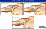
What are the pathways of the spinal cord formed by? Ascending and descending tracts of the spinal cord
Conducting pathways of the CNS are built from functionally homogeneous groups nerve fibers; they represent internal connections between the nuclei and cortical centers located in different parts and departments of the brain, and serve for their functional association (integration). The pathways, as a rule, pass through the white matter of the spinal cord and brain, but can also be localized in the tegmentum of the brainstem, where there are no clear boundaries between the white and gray matter.
The main conducting link in the system of transmitting information from one center of the brain to another is nerve fibers - the axons of neurons that transmit information in the form of a nerve impulse in a strictly defined direction, namely from the cell body. Among the conducting pathways, depending on their structure and functional significance, there are various groups nerve fibers: fibers, bundles, tracts, radiances, adhesions (commissures).
Projection pathways consist of neurons and their fibers that provide connections between the spinal cord and the brain. Projection paths also connect the nuclei of the trunk with the basal nuclei and the cerebral cortex, as well as the nuclei of the trunk with the cortex and nuclei of the cerebellum. Projection paths can be ascending and descending.
Ascending (sensory, sensitive, afferent) projection pathways conduct nerve impulses from extero-, proprio- and interoreceptors (sensitive nerve endings in the skin, organs of the musculoskeletal system, internal organs), as well as from the sense organs in an ascending direction to the brain, mainly to the cerebral cortex, where they mainly end at level IV of the cytoarchitectonic layer.
A distinctive feature of the ascending pathways is the multi-stage, sequential transmission of sensory information to the cerebral cortex through a number of intermediate nerve centers.
In addition to the cerebral cortex, sensory information is also sent to the cerebellum, to midbrain and into the reticular formation.
Descending (efferent or centrifugal) projection pathways conduct nerve impulses from the cerebral cortex, where they originate from the pyramidal neurons of the V cytoarchitectonic layer, to the basal and stem nuclei of the brain and further to the motor nuclei spinal cord and brain stem.
They transmit information related to the programming of body movements in specific situations, therefore they are motor pathways.
A common feature of the descending motor pathways is that they necessarily pass through the internal capsule - a layer of white matter in the cerebral hemispheres that separates the thalamus from the basal ganglia. In the brain stem most of descending pathways heading to the spinal cord and cerebellum go at its base.
35. Pyramidal and extrapyramidal systems
The pyramidal system is a combination of motor centers of the cerebral cortex, motor centers of cranial nerves located in the brain stem, and motor centers in the anterior horns of the spinal cord, as well as efferent projection nerve fibers that connect them together.
Pyramidal pathways provide the conduction of impulses in the process of conscious regulation of movements.
Pyramidal pathways are formed from giant pyramidal neurons (Betz cells), as well as large pyramidal neurons localized in layer V of the cerebral cortex. Approximately 40% of the fibers originate from pyramidal neurons in the precentral gyrus, where the cortical center of the motor analyzer is located; about 20% - from the postcentral gyrus, and the remaining 40% - from the posterior sections of the upper and middle lobar gyrus, and from the supramarginal gyrus of the lower parietal lobule, in which the center of praxia is located, which controls complex coordinated purposeful movements.
Pyramidal pathways are divided into corticospinal and cortical-nuclear. Their common feature is that, starting in the cortex of the right and left hemispheres, they move to the opposite side of the brain (i.e., cross) and ultimately regulate the movements of the contralateral half of the body.
The extrapyramidal system combines phylogenetically more ancient mechanisms for controlling human movements than the pyramidal system. It carries out predominantly involuntary, automatic regulation of complex motor manifestations of emotions. A distinctive feature of the extrapyramidal system is a multi-stage, with many switches, transmission of nerve influences from various parts of the brain to the executive centers - the motor nuclei of the spinal cord and cranial nerves.
Through the extrapyramidal pathways, motor commands are transmitted during protective motor reflexes that occur unconsciously. For example, thanks to the extrapyramidal pathways, information is transmitted when the vertical position of the body is restored as a result of a loss of balance (vestibular reflexes) or during motor reactions to a sudden light or sound effect (protective reflexes that close in the roof of the midbrain), etc.
The extrapyramidal system is formed by the nuclear centers of the hemispheres (basal nuclei: caudate and lenticular), diencephalon(medial nuclei of the thalamus, subthalamic nucleus) and the brain stem (red nucleus, black substance), as well as pathways connecting it with the cerebral cortex, with the cerebellum, with the reticular formation and, finally, with the executive centers lying in motor nuclei cranial nerves and in the anterior horns of the spinal cord.
There is also a somewhat extended interpretation, when to E.S. they include the cerebellum, the nuclei of the quadrigemina of the midbrain, the nuclei of the reticular formation, etc.
Cortical pathways originate from the precentral gyrus, as well as other parts of the cerebral cortex; these pathways project the influence of the cortex to the basal ganglia. The basal nuclei themselves are closely connected with each other by numerous internal connections, as well as with the nuclei of the thalamus and with the red nucleus of the midbrain. The motor commands formed here are transmitted to the executive motor centers of the spinal cord mainly in two ways: through the red nuclear-spinal (rubrospinal) tract and through the nuclei of the reticular formation (reticulospinal tract). Also, through the red nucleus, the influence of the cerebellum on the work of the spinal motor centers is transmitted.
The nerve cell has a large number of processes. The processes removed from the cell body are called nerve fibers. Nerve fibers that do not extend beyond the central nervous system, form conductors of the brain and spinal cord. Fibers that travel outside the central nervous system gather into bundles and form peripheral nerves.
Nerve fibers passing inside the brain and spinal cord have different lengths - some of them come into contact with neurons located close, others with neurons located at a greater distance, and still others are far removed from the body of their cell. In this regard, three types of conductors can be distinguished that carry out the transmission of impulses within the central nervous system.
1. Projection conductors communicate with the overlying sections of the central nervous system with the sections located below. (Fig. 4). Among them, there are two types of paths. Descending conduct impulses from the overlying parts of the brain down and are called centrifugal. They are motor in nature. The paths that direct from the periphery the conductive impulses from the skin, muscles, joints, ligaments, bones to the center have an upward direction and are called centripetal. They are sensitive in nature.
Rice. 4.
I - posterior spinal bundle; II - fibers of the posterior cord; III - spinal tuberous bundle; IV - anterior cortical-spinal bundle; V - lateral cortical-spinal bundle; VI - vestibulo-spinal bundle
2. Commissural, or adhesive, conductors connect the hemispheres of the brain. Examples of such connections are the corpus callosum, which connects the right and left hemisphere, the anterior commissure, the uncinate gyrus commissure, and the gray commissure of the thalamus that connects both halves of the thalamus.
3. Associative, or associative, conductors connect parts of the brain within the same hemisphere. Short fibers connect various convolutions in one or closely spaced lobes, and long ones stretch from one lobe of the hemisphere to another. For example, an arcuate bundle connects the lower and middle departments frontal lobe, the lower longitudinal connects the temporal lobe with the occipital lobe. Allocate the fronto-occipital, frontal-parietal bundles, etc. (Fig. 5).

Rice. 5.
I - upper longitudinal (or arcuate) bundle; II - fronto-occipital bundle; III - lower longitudinal beam; IV - waist bun; V - hook-shaped bundle; VI - arcuate fiber; VII - large commissure (corpus callosum)
Consider the course of the main projection conductors of the brain and spinal cord.
centrifugal ways
The pyramidal path starts from large and giant pyramidal cells (Betz cells) located in the fifth layer of the anterior central gyrus and the paracentral lobule. In the upper sections there are paths for the legs, in the middle sections of the anterior central gyrus - for the trunk, below - for the arms, neck and head. Thus, the projection of human body parts in the brain is presented inverted. From the total amount of fibers a powerful bundle is formed, which passes through the inner bag. Then the pyramidal bundle passes through the base of the brain stem, the pons, entering the medulla oblongata, and then into the spinal cord.
At the level of the pons and medulla, part of the fibers of the pyramidal pathway ends in the nuclei of the cranial nerves (trigeminal, abducens, facial, glossopharyngeal, vagus, accessory, hypoglossal). This short bundle of fibers is called the cortical-bulbar pathway. It starts from the lower sections of the anterior central gyrus. Before entering the nuclei, the nerve fibers of the short pyramidal pathway cross over. Another, longer bundle of pyramidal nerve fibers, starting from the upper sections of the anterior central gyrus, descends down into the spinal cord and is called the cortical-spinal path. The latter, on the border of the medulla oblongata with the spinal cord, forms an incomplete decussation, and most of the nerve fibers (subjected to decussation) continue their way in the lateral columns of the spinal cord, and a smaller part (not crossed) goes as part of the anterior columns of the spinal cord of its side. Both segments end in the motor cells of the anterior horn of the spinal cord.
The pyramidal pathway (cortical-spinal and cortical-bulbar) is the central segment of the path that transmits motor impulses from the cells of the cerebral cortex to the nuclei of the cranial nerves and cells of the spinal cord. It does not go beyond the central nervous system.
From the motor nuclei of the cranial nerves and from the cells of the anterior horns of the spinal cord, the peripheral segment of the path along which the impulse is directed to the muscles begins. Consequently, the transmission of a motor impulse is carried out through two neurons. One conducts impulses from the cells of the cortex of the motor analyzer to the cells of the anterior horns of the spinal cord and to the nuclei of the cranial nerves, the other - to the muscles of the face, neck, trunk and limbs (Fig. 6).
When the pyramidal tract is damaged, movements are disturbed on the side opposite to the lesion, which can be expressed by a complete absence of muscle movements (paralysis) or their partial weakening (paresis). Depending on the location of the lesion, there are central and peripheral paralysis or paresis.

Rice. 6.
I - cortical-spinal bundle; II - cortical-bulbar bundle; III - the crossed part of the cortical-spinal bundle; IV - uncrossed part of the cortical-spinal bundle; V - cross of pyramids; VI - caudate nucleus; VII - hillock; VIII - lentil kernel; IX - pale ball; X - leg of the brain; XI - varolian bridge; XII - medulla oblongata; K. VII - core facial nerve; K. XII - core hypoglossal nerve
The Monakovic bundle begins in the midbrain from the red nuclei. Immediately upon exiting the red nucleus, the fibers cross and, having passed the hindbrain, descend into the spinal cord. In the spinal cord, this bundle of nerve fibers is located in the lateral columns near the bundle of the crossed pyramidal tract and gradually ends, like pyramidal path, in the cells of the anterior horns of the spinal cord.
Monakov's bundle conducts motor impulses that regulate muscle tone.
The roof-spinal bundle connects the anterior colliculus of the midbrain with the anterior and partly lateral columns of the spinal cord. Participates in the implementation of visual and auditory orienting reflexes.
The vestibulospinal bundle originates in the nuclei vestibular apparatus(in the Deiters kernel). The fibers descend into the spinal cord and pass in the anterior and partly lateral columns. The fibers end in the cells of the anterior horns. Since the nucleus of Deiters is connected with the cerebellum, impulses from the vestibular system and the cerebellum to the spinal cord follow this path; participates in the balance function.
The reticular-spinal bundle starts from the reticular formation of the medulla oblongata, passes in different bundles in the anterior and lateral columns of the spinal cord. It ends in the cells of the anterior horn; conducts vital impulses from the coordinating center of the hindbrain.
The posterior longitudinal bundle consists of ascending and descending fibers. It travels through the brainstem to the anterior columns of the spinal cord. This path is followed by impulses from brain stem and segments of the spinal cord, from the vestibular apparatus and nuclei of the eye muscles, as well as from the cerebellum.
To control the work of the whole organism or each a separate body, the motor apparatus, the pathways of the spinal cord are required. Their main task is to deliver impulses sent by the human "computer" to the body and limbs. Any failure in the process of sending or receiving impulses of a reflex or sympathetic nature is fraught with serious pathologies of health and all life activity.
What are pathways in the spinal cord and brain?
The pathways of the brain and spinal cord act as a complex of neural structures. In the course of their work, impulse impulses are sent to specific areas of gray matter. In essence, impulses are signals that prompt the body to act on the call of the brain. Several groups of different according to functional features, are the pathways of the spinal cord. These include:
- projection nerve endings;
- associative paths;
- commissural connecting roots.
In addition, the performance of the spinal conductors necessitates the selection of the following classification, according to which they can be:
- motor;
- sensory.
Sensitive perception and human motor activity
Sensory or sensory pathways of the spinal cord and brain serve as an indispensable element of contact between these two most complex systems in the body. They also send an impulsive message to every organ, muscle fiber, arms and legs. The instantaneous sending of an impulse signal is a fundamental moment in the implementation by a person of coordinated coordinated body movements performed without the application of any conscious effort. The impulses sent by the brain, the nerve fibers can recognize through touch, feeling pain, temperature regime body, musculoskeletal motility.
The motor pathways of the spinal cord predetermine the quality of a person's reflex reaction. Providing the sending of impulse signals from the head to the reflex endings of the ridge and the muscular apparatus, they endow a person with the ability to self-control motor skills - coordination. Also, these pathways are responsible for the transmission of stimulating impulses towards the visual and auditory organs.
Where are the pathways located?
Acquainted with the anatomical hallmarks of the spinal cord, it is necessary to figure out where the very pathways of the spinal cord are located, because this term implies a lot of nerve matter and fibers. They are located in specific living essential substances: gray and white. Connecting the spinal horns and the cortex of the left and right hemispheres, conducting pathways through neural connection provide contact between the two departments.
Functions of conductors of the main human organs consist in the implementation of the intended tasks with the help of specific departments. In particular, the pathways of the spinal cord are located within the upper vertebrae and head, which can be described in more detail as follows:
- Associative connections are a kind of "bridges" that connect the areas between the cortex of the hemispheres and the nuclei of the spinal substance. In their structure there are fibers of various sizes. Relatively short ones do not go beyond the hemisphere or its brain lobe. Longer neurons transmit impulses that travel some distance to the gray matter.
- The commissural tracts are a body with a calloused structure and perform the task of connecting the newly formed sections in the head and spinal cord. The fibers from the main lobe bloom in a ray-like manner, they are placed in the white spinal substance.
- Projection nerve fibers are located directly in the spinal cord. Their performance makes it possible for impulses to arise in the hemispheres in a short time and establish communication with internal organs. The division into ascending and descending pathways of the spinal cord concerns precisely fibers of this type.
System of ascending and descending conductors
The ascending pathways of the spinal cord fulfill the human need for vision, hearing, motor functions and their contact with important systems organism. The receptors for these connections are located in the space between the hypothalamus and the first segments. spinal column. The ascending pathways of the spinal cord are able to receive and send further impulses coming from the surface. upper layers epidermis and mucous membranes, life support organs.

In turn, the descending pathways of the spinal cord include the following elements in their system:
- The neuron is pyramidal (originates in the cortex of the hemispheres, then rushes down, bypassing the brain stem; each of its bundles is located on the spinal horns).
- The neuron is central (it is motor, connecting the anterior horns and the cortex of the hemispheres with the reflex roots; together with the axons, elements of the peripheral nervous system also enter the chain).
- Spinal fibers (conductors lower extremities and column of the spinal cord, including the sphenoid and thin ligaments).
It is rather difficult for an ordinary person who does not specialize in the field of neurosurgery to understand the system represented by the complex pathways of the spinal cord. The anatomy of this department is indeed an intricate structure consisting of neural impulse transmissions. But it is thanks to her that the human body exists as a whole. Due to the double direction in which the conductive pathways of the spinal cord operate, instantaneous transmission of impulses is ensured, which carry information from the controlled organs.
Deep sensory conductors
The structure of the nerve cords, acting in an upward direction, is multi-component. These pathways of the spinal cord are formed by several elements:
- Burdach's bundle and Gaull's bundle (they are paths of deep sensitivity located on the back of the spinal column);
- spinothalamic bundle (located on the side of the spinal column);
- Govers' bundle and Flexig's bundle (cerebellar pathways located on the sides of the column).
Inside the intervertebral nodes are located a deep degree of sensitivity. The processes localized in the peripheral areas end in the most suitable muscle tissue, tendons, bone and cartilage fibers and their receptors.

In turn, the central processes of the cells, located behind, keep the direction towards the spinal cord. Conducting deep sensitivity, the posterior nerve roots do not go deep into the gray matter, forming only the posterior spinal columns.
Where such fibers enter the spinal cord, they are divided into short and long. Further, the pathways of the spinal cord and brain are sent to the hemispheres, where their cardinal redistribution takes place. Their main part remains in the zones of the anterior and posterior central gyri, as well as in the region of the crown.
It follows that these paths conduct sensitivity, thanks to which a person can feel how his muscular-articular apparatus works, feel any vibrational movement or tactile touch. Gaulle's bundle, located right in the center of the spinal cord, distributes sensation from the lower torso. Burdach's bundle is located above and serves as a conductor of the sensitivity of the upper limbs and the corresponding part of the trunk.
How to find out about the degree of sensory?
You can determine the degree of deep sensitivity using several simple tests. For their implementation, the patient's eyes are closed. Its task is to determine the specific direction in which the doctor or researcher makes movements of a passive nature in the joints of the fingers, hands or feet. It is also desirable to describe in detail the posture of the body or the position that its limbs have assumed.
With the help of a tuning fork for vibration sensitivity, it is possible to examine the pathways of the spinal cord. The functions of this device will help to accurately determine the time during which the patient clearly feels the vibration. To do this, take the device and click on it to make a sound. At this point, it is necessary to put on any bony protrusion on the body. In the case when this sensitivity drops out earlier than in other cases, it can be assumed that the posterior pillars are affected.
The test for the sense of localization implies that the patient, by closing his eyes, accurately points to the place where the researcher touched him a few seconds before. A satisfactory indicator is considered if the patient made an error within one centimeter.
Sensory sensitivity of the skin
The structure of the pathways of the spinal cord allows you to determine the degree of skin sensitivity at the peripheral level. The fact is that the nerve processes of the protoneuron are involved in skin receptors. The processes located in the center as part of the posterior processes rush directly to the spinal cord, as a result of which the Lisauer zone is formed there.

Just like the path of deep sensitivity, the skin one consists of several successively combined nerve cells. In comparison with the spinothalamic bundle of nerve fibers, information impulses transmitted from the lower extremities or lower body are slightly higher and in the middle.
Skin sensitivity varies according to criteria based on the nature of the irritant. She happens:
- temperature;
- thermal;
- painful;
- tactile.
Wherein last view skin sensitivity, as a rule, is transmitted by conductors of deep sensitivity.
How to find out about pain threshold and temperature difference?
To determine the level pain, doctors use the injection method. In the most unexpected places for the patient, the doctor inflicts several light injections with a pin. The patient's eyes should be closed, because. he must not see what is happening.
Threshold temperature sensitivity is easy to determine. At normal condition a person experiences different sensations at temperatures, the difference of which was about 1-2 °. To detect a pathological defect in the form of a violation of skin sensitivity, doctors use a special apparatus - a thermoesthesiometer. If not, you can test for warm and hot water.
Pathologies associated with impaired conduction pathways
In the ascending direction, the pathways of the spinal cord are formed in a position due to which a person can feel tactile touch. For the study, it is necessary to take something soft, gentle and in a rhythmic manner conduct a subtle examination to identify the degree of sensitivity, as well as check the reaction of hairs, bristles, etc.

Disorders caused by skin sensitivity today are considered to be the following:
- Anesthesia is the complete loss of sensation of the skin on a specific superficial area of the body. In case of violation pain sensitivity there is analgesia, with temperature - termanesthesia.
- Hyperesthesia is the opposite of anesthesia, a phenomenon that occurs when the threshold of excitation decreases, and when it increases, hypalgesia appears.
- Misperception annoying factors(for example, the patient confuses cold and warm) is called dysesthesia.
- Paresthesia is a violation, the manifestations of which can be a huge variety, ranging from crawling goosebumps, a feeling of electric shock and its passage through the entire body.
- Hyperpathy is the most pronounced. It is also characterized by damage to the thalamus, an increase in the threshold of excitability, the inability to locally determine the stimulus, a severe psycho-emotional coloring of everything that happens, and too sharp a motor reaction.
Features of the structure of descending conductors
The descending pathways of the brain and spinal cord include several ligaments, including:
- pyramidal;
- rubro-spinal;
- vestibulo-spinal;
- reticulo-spinal;
- back longitudinal.
All of the above elements are the motor pathways of the spinal cord, which are components of the nerve cords in a downward direction.
The so-called pyramidal path starts from the largest cells of the same name located in the upper layer of the cerebral hemisphere, mainly in the zone of the central gyrus. Here is the pathway anterior cord spinal cord - this important element system is directed downward and passes through several sections of the posterior femoral capsule. At the point of intersection of the medulla oblongata and spinal cord, an incomplete decussation can be found, forming a straight pyramidal bundle.
In the tegmentum of the midbrain there is a conducting rubro-spinal tract. It starts from the red nuclei. Upon exiting, its fibers cross and pass into the spinal cord through the varoli and medulla oblongata. Rubro-spinal path allows you to conduct impulses from the cerebellum and subcortical nodes.
The pathways of the spinal cord begin in Deiters' nucleus. Located in the brainstem, the vestibulo-spinal path continues in the spinal cord and ends in its anterior horns. The passage of impulses from the vestibular apparatus to the peripheral system depends on this conductor.
In the cells of the reticular formation of the hindbrain, the reticulo-spinal path begins, which is scattered in separate bundles in the white matter of the spinal cord, mainly from the side and front. In fact, this is the main connecting element between the reflex brain center and the musculoskeletal system.
The posterior longitudinal ligament is also involved in connecting motor structures to the brainstem. The work of the oculomotor nuclei and the vestibular apparatus as a whole depends on it. The posterior longitudinal bundle is located in cervical region spine.
Consequences of diseases of the spinal cord
Thus, the pathways of the spinal cord are vital connecting elements that provide a person with the ability to move and feel. The neurophysiology of these pathways is associated with the structural features of the spine. It is known that the structure of the spinal cord, surrounded by muscle fibers, has a cylindrical shape. Within the substances of the spinal cord, associative and motor reflex pathways control the functionality of all body systems.

When there is a disease of the spinal cord, mechanical damage or malformations, the conductivity between the two main centers may be significantly reduced. Violations of the pathways threaten a person complete cessation motor activity and loss of sensory perception.
The main reason for the lack of impulse conduction is the death of nerve endings. The most difficult degree of conduction disturbance between the brain and spinal cord is paralysis and lack of sensation in the limbs. Then there may be performance problems. internal organs associated with the brain damaged neuronal bundle. For example, disorders in the lower part of the spinal cord lead to uncontrolled urination and defecation processes.
Are diseases of the spinal cord and pathways treated?
Just appeared degenerative changes almost instantly affect the conduction activity of the spinal cord. Inhibition of reflexes leads to pronounced pathological changes due to the death of neuronal fibers. It is impossible to completely restore the disturbed conduction areas. The disease comes on rapidly and progresses at lightning speed, so gross conduction disturbances can be avoided only if you start in a timely manner. drug treatment. The sooner this is done, the greater the chances of stopping pathological development.
The impermeability of the passing tracts of the spinal cord needs treatment, the primary task of which will be to stop the processes of the death of nerve endings. This can be achieved only if the factors that influenced the onset of the disease are suppressed. Only after that it is possible to start therapy in order to restore sensitivity and motor functions as much as possible.
Drug treatment is aimed at stopping the process of dying of brain cells. Their task is also to restore the disturbed blood supply to the damaged area of the spinal cord. In the course of treatment, doctors take into account age characteristics, the nature and severity of damage and progression of the disease. In pathway therapy, it is important to maintain constant stimulation of nerve fibers with electrical impulses. This will help maintain satisfactory muscle tone.

Surgical intervention is carried out in order to restore the conductivity of the spinal cord, therefore, it is carried out in two directions:
- Suppression of the causes of paralysis of the activity of neural connections.
- Stimulation of the spinal cord for the speedy acquisition of lost functions.
The operation must be preceded by a complete medical examination the whole organism. This will allow to determine the localization of the processes of degeneration of nerve fibers. In the case of severe spinal injuries, the causes of compression must first be eliminated.
Major pathways of the spinal cord
Without setting ourselves the task of listing all the pathways of the CNS, let us consider the basic principles of organizing these pathways using the most important of them as an example (Fig. 30). The pathways in the CNS are divided into:
ascending- are formed by axons of cells whose bodies are located in the gray matter of the spinal cord. These axons are white matter heading towards upper departments spinal cord, brainstem and cerebral cortex.
descending- are formed by axons of cells whose bodies are located in different nuclei of the brain. These axons descend along the white matter to various spinal segments, enter the gray matter and leave their endings on one or another of its cells.
separate group form propriospinal conducting paths. They can be both ascending and descending, but they do not go beyond the spinal cord. After passing through several segments, they again return to the gray matter of the spinal cord. These paths are located in the deepest part lateral And ventral cords, they connect various nerve centers spinal cord. For example, the centers of the lower and upper limbs.
Ascending pathways.
Tracts of Gaulle (thin bundle) and Burdakh (wedge-shaped bundle). Main ascending paths pass through the dorsal cords of the spinal cord and represent axons of afferent neurons spinal ganglia. They pass throughout the spinal cord and end in the area oblong brain in the nuclei of the dorsal cord, which are called the nuclei of Gaulle and Burdach. That is why they are called Gaull's tract And tract Burdakh.
1. The first link of neurons:
a. The fibers located medially in the cord carry afferent signals to the Gaulle's nucleus from the lower part of the body, mainly from the lower extremities.
b. Lateral fibers go to Burdach's nucleus and transmit afferent signals from receptors in the upper body and forelimbs.
2. The second link of neurons:
In turn, the axons of the cells of the nuclei of Gaulle and Burdach in the brain stem cross and rise in the form of a dense bundle to intermediate brain. This bundle of fibers, already formed by the axons of the cells of the nuclei of Gaulle and Burdach, was called medial loop.
3. The third link of neurons:
The cells of the nuclei of the diencephalon give rise to axons that go to the cerebral cortex.
All other ascending paths do not begin from the neurons of the spinal ganglia, but from neurons located in gray matter of the spinal cord. Therefore, their fibers are fibers not of the first, but of the second order.
1. First link neurons of the spinal ganglia also serve in these pathways, but in the gray matter they leave their endings on the cells, as it were, of the “second link”.
Cells of this "second tier" send their axons to the nuclei of the brain stem and the cerebral cortex. The bulk of the fibers of these pathways runs in the lateral funiculus.
Spinal thalamic pathways (ventral and lateral).
2. The second link of neurons:
It originates at the base of the dorsal horn of the spinal cord. The axons of the neurons forming this path pass to the contralateral (opposite) side, enter the white matter of the opposite lateral or ventral funiculus and rise in it through the entire spinal cord And brain stem down to the core intermediate brain.
2. The third link of neurons:
The neurons of the diencephalon nuclei carry impulses to the cerebral cortex.
All of the above pathways (Gaulle, Burdach, and spinothalamic) connect receptive areas on each side of the body with cortical neurons. opposite hemisphere.
Spinal tracts. Two more pathways passing through the lateral cords connect the spinal cord with cerebellar cortex.
The Flexing Path - located dorsally and contains fibers that do not pass to the opposite side of the brain. This pathway in the spinal cord originates from Clark's nucleus neurons, whose axons reach the medulla oblongata and enter the cerebellum via the inferior cerebellar peduncle.
Gower's Way - located ventral, contains fibers that rise up the lateral funiculus opposite side body, but in the brainstem these fibers cross again and enter the cerebellar cortex from the side on which this path began. In the spinal cord, it starts from the nuclei of the intermediate zone, axons enter the cerebellum through the superior cerebellar peduncle.
If the cerebral cortex is always connected with afferent fibers of the opposite side of the body, then the cerebellar cortex receives fibers mainly from neural structures. of the same name sides.
Descending pathways. Downward fibers are also subdivided into several paths. The names of these pathways are based on the names of those parts of the brain in which they originate.
Cortico-spinal (lateral and ventral) pathways formed by axons pyramidal cells lower layers of the motor cortex of the cerebral hemispheres. These paths are often called pyramidal. The fibers pass through white matter of the cerebral hemispheres, base of midbrain peduncles, by ventral divisions Varolieva bridge And oblong brain in dorsal brain.
o Lateral the path crosses at the bottom of the pyramids of the medulla oblongata and ends at the neurons of the base dorsal horn.
o Ventral the path crosses the pyramids of the medulla oblongata without crossing. Before entering the anterior horn of the gray matter of the corresponding segment of the spinal cord, the fibers of this pathway pass to the opposite side and end on the motor neurons of the anterior horns of the contralateral side.
Thus, one way or another, but the motor area of the cerebral cortex always turns out to be connected with neurons opposite sides of the spinal cord.
Rubro-spinal path - main descending path midbrain, starts at red core. The axons of the neurons of the red nucleus cross immediately below it and, as part of the white matter of the lateral funiculus, descend to the segments of the spinal cord, ending on the cells of the intermediate region of the gray matter. This is due to the fact that the rubrospinal system, along with the pyramidal system, is the main system for controlling the activity of the spinal cord.
Tectospinal path - Originates from neurons quadrigemina of the midbrain and reaches the motor neurons of the anterior horns.
Pathways starting at medulla oblongata:
Vestibulo-spinal- starts from the vestibular nuclei, mainly from the cells of the nucleus of Deiters.
Reticulo-spinal- starts from an extensive accumulation of nerve cells of the reticular formation, occupying central part brain stem. The fibers of each of these pathways end on the neurons of the medial part of the anterior horn of the gray matter of the spinal cord. The main part of the endings are located on the intercalated cells.
Olivo-spinal- formed by the axons of the olive cells of the medulla oblongata, ends on the motor neurons of the anterior horns of the spinal cord.
Section 4
BRAIN
The nerve cell has a large number of processes. The processes removed from the cell body are called nerve fibers. Nerve fibers that do not extend beyond the central nervous system form conductors of the brain and spinal cord. Fibers that travel outside the central nervous system gather into bundles and form peripheral nerves.
Nerve fibers passing inside the brain and spinal cord have different lengths - some of them come into contact with neurons located close, others with neurons located at a greater distance, and still others are far removed from the body of their cell. In this regard, three types of conductors can be distinguished that carry out the transmission of impulses within the central nervous system.
1. Projection conductors communicate with the overlying sections of the central nervous system with the sections located below. Among them, there are two types of paths. Descending conduct impulses from the overlying departments of the go-
TO MOUSE
Rice. 47. Projection fibers of the spinal cord:
1 - posterior spinal bundle; II - fibers of the posterior cord; III - spinal tuberous bundle; IV - anterior cortical-spinal bundle; V - lateral cortical-in-spinal bundle; VI - vestibulo-spinal bundle
Rice. 48. Association Paths:I - upper longitudinal (or arcuate) bundle; II - fronto-occipital bundle; III - lower longitudinal beam; IV - waist bun; V - hook-shaped bundle; VI - arcuate fiber; VII - large commissure (corpus callosum)
brain down and are called centrifugal. They are motor in nature. The paths that direct from the periphery the conductive impulses from the skin, muscles, joints, ligaments, bones to the center have an upward direction and are called centripetal. They are sensitive in nature.
Commissural, or adhesive, conductors connect the hemispheres of the brain. Examples of such connections are the corpus callosum, connecting the right and left hemispheres, the anterior commissure, the commissure of the uncinate gyrus, and the gray commissure of the thalamus, connecting both halves of the thalamus.
Associative, or associative, conductors connect parts of the brain within the same hemisphere. Short fibers connect various convolutions in one or closely spaced lobes, and long ones stretch from one lobe of the hemisphere to another. For example, the arcuate bundle connects the lower and middle sections of the frontal lobe, the lower longitudinal connects the temporal lobe with the occipital lobe. Allocate the fronto-occipital, fronto-parietal bundles, etc. (Fig. 48).
Consider the course of the main projection conductors of the brain and spinal cord.
centrifugal ways
pyramid path begins from large and giant pyramidal cells (Betz cells) located in the fifth layer of the anterior central gyrus and paracentral lobule. In the upper sections there are paths for the legs, in the middle sections of the anterior central gyrus - for the trunk, below - for the arms, neck and head. Thus, the projection of human body parts in the brain is presented inverted. A powerful bundle is formed from the total amount of fibers, which passes through the inner bag (in Fig. 36 - see the knee and the front two-thirds of the back of the thigh). Then the pyramidal bundle passes through the base of the brain stem, the pons, entering the medulla oblongata, and then into the spinal cord.
At the level of the pons and medulla, part of the fibers of the pyramidal pathway ends in the nuclei of the cranial nerves (trigeminal, abducens, facial, glossopharyngeal, vagus, accessory, hypoglossal). This short bundle of fibers is called the cortical-bulbar pathway. It starts from the lower sections of the anterior central gyrus. Before entering the nuclei, the nerve fibers of the short pyramidal pathway cross over. Another, longer bundle of pyramidal nerve fibers, starting from the upper sections of the anterior central gyrus, descends down into the spinal cord and is called the cortical-spinal path. The latter, on the border of the medulla oblongata with the spinal cord, forms an incomplete decussation, and most of the nerve fibers (subjected to decussation) continue their way in the lateral columns of the spinal cord, and a smaller part (not crossed) goes as part of the anterior columns of the spinal cord of its side. Both segments end in the motor cells of the anterior horn of the spinal cord.
The pyramidal pathway (cortical-spinal and cortical-bulbar) is the central segment of the path that transmits motor impulses from the cells of the cerebral cortex to the nuclei of the cranial nerves and cells of the spinal cord. It does not go beyond the central nervous system.
From the motor nuclei of the cranial nerves and from the cells of the anterior horns of the spinal cord, the peripheral segment of the path along which the impulse is directed to the muscles begins. Consequently, the transmission of a motor impulse is carried out through two neurons. One conducts impulses from the cells of the cortex of the motor analyzer to the cells of the anterior horns of the spin
leg brain and to the nuclei of the cranial nerves, the other - to the muscles of the face, neck, trunk and limbs.
When the pyramidal tract is damaged, movements are disturbed on the side opposite to the lesion, which can be expressed by a complete absence of muscle movements (paralysis) or their partial weakening (paresis). Depending on the location of the lesion, central and peripheral paralysis or paresis are distinguished. The characteristics of these violations are given in the corresponding section.
I - cortical-spinal bundle; II - cortical-bulbar bundle; III - the crossed part of the cortical-spinal bundle; IV - uncrossed part of the cortical-spinal bundle; V - cross of pyramids; VI - caudate nucleus; VII - hillock; VIII - lentil kernel; IX - pale ball; X - leg of the brain; XI - varolian bridge; XII - medulla oblongata; K. VII - the nucleus of the facial nerve; K. XII - the nucleus of the hypoglossal nerve
Monaco beam begins in the midbrain from the red nuclei. Immediately upon exiting the red nucleus, the fibers cross and, having passed the hindbrain, descend into the spinal cord. In the spinal cord, this bundle of nerve fibers is located in the lateral columns near the bundle of the crossed pyramidal pathway and gradually ends, like the pyramidal pathway, in the cells of the anterior horns of the spinal cord.Monakov's bundle conducts motor impulses that regulate muscle tone.
Roof-spinal bundle connects the anterior colliculus of the midbrain with the anterior and partly lateral columns of the spinal cord. Participates in the implementation of visual and auditory orienting reflexes.
vestibulo-spinal bundle begins in the nuclei of the vestibular apparatus (in the nucleus of Deiters). The fibers descend into the spinal cord and pass in the anterior and partly lateral columns. The fibers end in the cells of the anterior horns. Since the nucleus of Deiters is connected with the cerebellum, impulses from the vestibular system and the cerebellum to the spinal cord follow this path; participates in the balance function.
Retico-spinal bundle starts from the reticular formation of the medulla oblongata, passes in different bundles in the anterior and lateral columns of the spinal cord. It ends in the cells of the anterior horn; conducts vital impulses from the coordinating center of the hindbrain.
Posterior longitudinal beam consists of ascending and descending fibers. It travels through the brainstem to the anterior columns of the spinal cord. Impulses from the brain stem and segments of the spinal cord, from the vestibular apparatus and nuclei of the eye muscles, as well as from the cerebellum pass along this path.
centripetal paths
Pathway of superficial skin sensitivity carries pain, temperature and, in part, tactile sensations (the main path of touch passes with fibers of deep sensitivity). The path begins in the intervertebral node from cells that have two processes, one of them goes to the periphery to the skin receptors, and the other goes to the spinal cord and ends in the cells of the dorsal horn of the spinal cord. This is the so-called first neuron of the sensory pathway. From the cells of the posterior horn, the second neuron of the skin sensitivity pathway begins. It passes to the opposite side and rises along the lateral columns of the spinal cord, passes through the medulla oblongata, and in the pons varolii and in the region of the midbrain it enters into the medial loop and goes to the outer nucleus of the thalamus. From the thalamus begins the third neuron of the sensory pathway; it passes the internal pouch (at the back of the thigh) and travels to the cerebral cortex. It ends in the region of the posterior central gyrus (parietal lobe).
Path of deep sensitivity It also starts from the nerve cells of the intervertebral node, where impulses are suitable not only from the skin and mucous membranes, but also from muscles, joints, bones, tendons and ligaments. The path of deep sensitivity, carrying irritations from all these formations, enters the spinal cord as part of the posterior columns. Then it rises up along the spinal cord to the oblong, in the nuclei of which the first neuron of this path ends. From the nuclei of the medulla oblongata begins the second neuron of deep sensitivity. Upon exiting the nuclei, the fibers cross, then form a medial loop and go to the lateral nucleus of the visual mound. The third neuron of deep sensitivity begins from the visual hillock, it passes through the internal bag and also ends in the cells of the posterior central gyrus (parietal lobe) (Fig. 50).
I- nuclei of the posterior pillars; II - posterior columns of the spinal cord, III - spinal tuberous bundle; IV - trigeminal nerve: P. - median loop: 3. bug. - visual tubercle: M. t. - corpus callosum; Ch. i. - lentil kernel; V. s. - inner bag
cerebellar conductors, like all ascending conductors, they start from the intervertebral node and go to the gray matter of the spinal cord, where they end in the cells of the posterior horn. From the cells of the posterior horn, the second neuron begins, which is sent in two bundles to the lateral columns of the spinal cord. One bundle, straight, reaches the medulla oblongata, forms the lower cerebellar peduncle and ends in the cells of the cerebellum. Another bundle, crossed, rises up to the midbrain and also enters the cerebellum through the superior cerebellar peduncle.The ascending pathways include sensory pathways that carry olfactory, visual and auditory stimuli. These will be discussed below in the section on cranial nerves.
With the defeat of sensitive conductors, disorders of all types of sensitivity of the corresponding area are observed. So, with the defeat of the corresponding paths of the lateral column, skin (pain and temperature) and partly tactile sensitivity on the opposite side suffers.
In connection with the defeat of the fibers of the cerebellar pathways, disorders of coordination of movements occur. With the defeat of the posterior pillars, deep sensitivity is disturbed - a sense of the position of the organs of movement, localization, a two-dimensional spatial sense. In this regard, the gait is also disturbed, which becomes uncertain, the movements are sweeping, inaccurate.
cranial nerves
The cranial nerves originate in the brainstem, where their nuclei are located. The exceptions are the olfactory, auditory and optic nerves, the first neuron of which is located outside the brain stem.
Most cranial nerves are mixed, i.e. contain both sensory and motor fibers, with sensory predominating in some, and motor in others.
In total there are twelve 12 cranial nerves (Fig. 51).
/ pair - olfactory nerve. It begins in the nasal mucosa in the form of thin nerve threads that pass through ethmoid bone skulls, go to the base of the brain and are collected in the olfactory bulb. From the olfactory bulb comes the secondary olfactory pathway - olfactory tract. The fibers of the olfactory tract partly diverge, forming a triangle. Most of the olfactory fibers end in the central nucleus of the olfactory analyzer, located in the uncinate gyrus on the inner surface of the cortex.
The sense of smell is examined with a set of odorous substances.
The olfactory disorder can be expressed in different ways: in the form total absence perception of odors - anosmia, or a decrease in the perception of odors - hyposmia. Sometimes there is a particularly hypersensitivity to odorous substances - hyperosmia (in childhood almost never seen).
It should be borne in mind that sometimes local damage to the nasal mucosa (for example, with a runny nose) disrupts the perception of odors, which is not at all associated with damage to the olfactory tract itself.
2 pair - optic nerve. The visual path (Fig. 52) begins in the retina. The retina of the eye has a very complex

 nerve fibers approaches the nuclei of the anterior tubercles of the quadrigemina, to the pillow of the thalamus.
From the cells of the external geniculate body, the visual path goes to the cerebral cortex. This segment of the path is called the Graziole bundle.
The visual path ends in the cortex of the occipital lobe, where the central nucleus of the visual analyzer is located.
Visual acuity in children can be checked using a special table. Color perception is checked by a set of color pictures.
structure, it consists of cells called rods and cones. These cells are receptors that perceive various light and color stimuli. In addition to these cells, there are ganglionic nerve cells in the eye, the dendrites of which end in cones and rods, and the axons form the optic nerve. The optic nerves enter the cranial cavity through the bony opening and pass along the bottom of the base of the brain. Based on the brain optic nerves form a half cross - chiasm. Not all nerve fibers are crossed, but only fibers coming from the inner halves of the retina; the fibers coming from the outer halves do not cross.
massive beam neural pathways, which is formed after the intersection of the optic fibers, is called the optic tract. Thus, in the optic tract of each side, nerve fibers pass not from one eye, but from the same halves of the retinas of both eyes. For example, in the left optic tract from both left halves of the retinas, and in the right - from both right halves (Fig. 52).
Most of the nerve fibers of the optic tract go to the external geniculate bodies, a small part
Visual pathway lesion may occur in Fig. 52. Scheme of the visual pathways
1 - „ (according to Bing)
any segment. IN depending on this, a different clinical picture of visual impairment will be observed.
Basically, it is necessary to distinguish three areas of the lesion: before the chiasm, in the region of the chiasm itself (chiasm) and after the optic chiasm. More on this will be discussed below.
L / (oculomotor nerve), IV (trochlear nerve) and VI (abducens nerve) pairs of nerves carry out the movements of the eyeball and are, therefore, oculomotors. These nerves send impulses to the muscles that move eyeball. With the defeat of these nerves, paralysis of the corresponding muscles and restrictions on the movements of the eyeball - strabismus are observed.
In addition, with the defeat of the III pair of cranial nerves, ptosis (drooping of the upper eyelid) and inequality of the pupils are also observed. The latter is also associated with damage to the branch of the sympathetic nerve, which is involved in the innervation of the eye.
V pair - the trigeminal nerve leaves the skull on the front surface, forming three branches: a) orbital, b) zygomatic, c) mandibular.
The first two branches are sensitive. They innervate the skin of the upper facial region, the mucous membranes of the nose, eyelids, as well as the eyeball, upper jaw, gums and teeth. Part of the nerve fibers supplies the meninges.
The third branch of the trigeminal nerve is mixed in terms of fiber composition. Its sensory fibers innervate lower section skin surface of the face, anterior two-thirds of the tongue, oral mucosa, teeth and gums mandible. The motor fibers of this branch innervate the masticatory muscles.
The sympathetic nerve plays an important role in the system of innervation of the trigeminal nerve.
With the defeat of the peripheral branches of the trigeminal nerve, the skin sensitivity of the face is upset. Sometimes there are excruciating attacks of pain (trigeminal neuralgia), caused by an inflammatory process in the nerve. Disorders of the motor portion of the fibers cause paralysis of the masticatory muscles, as a result of which the movements of the lower jaw are sharply limited, which makes it difficult to chew food.
VII pair - the facial nerve (motor) is suitable for all the facial muscles of the face. With a unilateral lesion of the facial nerve, which often occurs as a result of a cold, nerve paralysis develops, in which the following picture is observed: low position eyebrows, palpebral fissure is wider than on the healthy side, the eyelids do not close tightly, the nasolabial fold is smoothed, the corner of the mouth sags, voluntary movements are difficult, it is not possible to frown and lift them up, evenly inflate the cheeks, it is not possible to whistle with lips or pronounce the sound "y". Patients at the same time feel numbness in the affected half of the face, experience pain. Due to the fact that the composition of the facial nerve includes secretory and taste fibers, salivation is disturbed, taste is upset. The fibers of the trigeminal nerve are also involved in the implementation of the function of taste.
VIII pair - auditory nerve begins in the inner ear with two branches. The first - the auditory nerve proper - departs from the spiral ganglion located in the cochlea of the labyrinth. The cells of the spiral ganglion are bipolar, i.e. have two processes, and one group of processes (peripheral) goes to the hair cells of the organ of Corti, the others form the auditory nerve. The second branch of the mixed auditory nerve is called the vestibular nerve, departing from the vestibular apparatus, also located in the inner ear. It consists of three bony tubules and two sacs. A fluid circulates inside the canals - endolymph, in which calcareous pebbles - otoliths float. The inner surface of the sacs and canals is equipped with sensory nerve endings coming from the Scarpov nerve ganglion, which lies at the bottom of the internal auditory canal. The long processes of this node form the vestibular nerve branch. When exiting inner ear auditory and vestibular branches join.
Having entered the cavity of the medulla oblongata, these nerves approach the nuclei lying here, after which they are again disconnected, each following its own direction.
From the nuclei of the medulla oblongata, the auditory nerve goes already under the name of the auditory pathway. Moreover, part of the fibers crosses at the level of the bridge and passes to the other side. The other part goes along its side, including neurons from some nuclear formations (trapezoid body, etc.). This segment of the auditory pathway is called the lateral loop; it ends in the posterior tubercles of the quadrigemina and the internal geniculate bodies. The crossed auditory pathway also fits here. From the internal geniculate bodies, the third segment of the auditory pathway begins, which passes through the internal bag and approaches temporal lobe where the central nucleus of the auditory analyzer is located.
With unilateral damage to the auditory nerve and its nuclei, deafness develops in the ear of the same name. With unilateral injury auditory tract(in particular, the lateral loop), as well as the cortical auditory zone, there are no pronounced auditory disorders, there is some hearing loss in the opposite ear (due to double innervation). Complete cortical deafness is possible only with bilateral foci in the corresponding auditory zones.
The vestibular nerve, starting from the Scarp's node and having traveled some distance together with the auditory branch, enters the cavity of the medulla oblongata and approaches the angular nucleus. The angular nucleus consists of the lateral nucleus of Deiters, the superior nucleus of Bekhterev and inner core. From the angular nucleus, the conductors go to the cerebellar vermis (dentate and roofing nuclei), to the spinal cord along the fibers of the vestibulo-spinal and posterior longitudinal bundle. Through the latter, a connection is made with the oculomotor nuclei of the midbrain. There is a connection with the thalamus.
With the defeat of the vestibular apparatus, as well as the vestibular nerve and its nuclei, the balance is upset, dizziness, nausea, and vomiting appear.
IX pair - glossopharyngeal nerve includes sensory, motor, and secretory fibers. The glossopharyngeal nerve originates from four nuclei located in the medulla oblongata, some nuclei are common with the vagus nerve. This pair of nerves is closely related to the X pair (vagus nerve). The glossopharyngeal nerve supplies sensory (gustatory) fibers to the posterior third of the tongue and palate, and together with the vagus nerve innervates the middle ear and pharynx. The motor fibers of this nerve, together with the branches of the vagus nerve, supply the muscles of the pharynx. Secretory fibers innervate the parotid salivary gland.
With the defeat of the glossopharyngeal nerve, a number of disorders are observed, for example, taste disorders, a decrease in sensitivity in the pharynx, as well as the presence of mild spasms of the pharyngeal muscles. In some cases, salivation may be impaired.
X pair - the vagus nerve departs from the nuclei located in the medulla oblongata, some of the nuclei are common with the IX pair. The vagus nerve performs a number of complex functions of a sensitive, motor and secretory nature. So, it supplies motor and sensory fibers to the muscles of the pharynx (together with the IX pair), soft palate, larynx, epiglottis, vocal cords. Unlike other cranial nerves, this nerve extends far beyond the skull and innervates the trachea, bronchi, lungs, heart, gastrointestinal tract and some other internal organs, as well as blood vessels. Thus, the further course of its fibers takes part in the autonomic innervation, forming the parasympathetic nervous system.
In violation of the function of the vagus nerve, especially with bilateral partial damage, a number of severe disorders can occur, such as swallowing disorders, voice changes (nasality, dysphonia, aphonia); there is a series severe violations from the cardiovascular and respiratory systems. With full you-
If the function of the vagus nerve is turned off, death may occur due to paralysis of the heart and respiratory activity.
XI pair - accessory nerve, is a motor nerve. Its nuclei are located in the spinal cord and medulla oblongata. The fibers of this nerve innervate the muscles of the neck and shoulder girdle, in connection with which such movements as turning the head, raising the shoulders, bringing the shoulder blades to the spine are carried out.
With damage to the accessory nerve, atrophic paralysis of these muscles develops, as a result of which it is difficult to turn the head, the shoulder is lowered. Nerve irritation can cause tonic convulsions. neck muscles, causing the head to be forcibly tilted to the side (torticollis). Clonic spasm in these muscles (bilateral) causes violent nodding movements.
XII pair - hypoglossal nerve. These are the motor nerves of the tongue. The fibers start from the nucleus located at the bottom of the rhomboid fossa. Fibers of the XII pair innervate the muscles of the tongue, giving it maximum flexibility and mobility. With damage to the hypoglossal nerve, atrophic phenomena can develop in the muscles of the tongue, its ability to move is weakened, which is necessary to perform the speech function and the function of eating. In such cases, speech becomes unclear, it becomes impossible to pronounce complex words. With bilateral damage to the hypoglossal nerve, anarthria develops. A typical picture of speech and phonation disorders is observed with a combined lesion of the IX, X and XII pairs of nerves, known as bulbar palsy. In these cases, the nuclei of the medulla oblongata or the roots and nerves extending from them are affected. There is paralysis of the tongue, severe speech disorders, as well as swallowing disorders, choking, liquid food pours out through the nose, the voice becomes nasal. Such paralysis is accompanied by muscle atrophy and bears all the signs of peripheral paralysis. More often there are cases of lesions of the central pathway (cortical-bulbar). In childhood, with bilateral lesions of the cortico-bulbar tract, for example, after suffering parainfectious encephalitis, phenomena develop that are outwardly similar to bulbar palsy, but differ in the nature of localization. Since this paralysis is central, there is no muscle atrophy. This type of disorder is known as pseudobulbar palsy.