The structure of the outer middle and inner ear of a person. The structure of the human ear
The human ear is an organ responsible not only for the ability to perceive the sounds of the surrounding world, but also for the sense of body position in space, which is necessary for proper coordination of movements and balance.
All parts of the ear (outer, middle, inner) function in direct proportion to each other, and diseases that affect one of the parts can completely disrupt the functions of the others.
Let us consider in more detail the anatomy and structure of the human ear, as well as diseases that can affect the hearing organs.
outer ear
The outer ear of a person consists of the auricle and the external auditory canal, which is limited from the middle ear by the tympanic membrane.
Diseases:
- labyrinthitis - inflammation of the mucous membranes lining the inner surface of the cochlea and canals. Most often it develops after incompletely cured otitis media, craniocerebral injuries and infectious diseases. It is manifested by severe dizziness, reaching nausea and vomiting, periodic disturbances in coordination of movements, chaotic movements of the eyeballs that occur from several times a day, to hourly attacks.
Important: it should be remembered that the clinical picture of labyrinthitis and brain diseases is largely similar, and with the symptoms listed, in no case can one expect an independent solution to the problem. Consult a doctor: in some cases, only special diagnostic methods can help identify the cause of dizziness and incoordination.
To begin with, let's deal with the structure of the outer ear: it is supplied with blood through the branches of the external carotid artery. In innervation, in addition to the branches of the trigeminal nerve, the ear branch of the vagus nerve often takes part, which, in turn, branches in the posterior wall of the auditory canal. There is mechanical irritation of this wall and they often contribute to the appearance of the so-called reflex cough.
The structure of our outer ear is as follows, the outflow of lymph from the walls of the auditory canal enters the nearest lymph nodes, which are located in front of the auricle, on the mastoid process itself and under the lower wall of the auditory canal. Educational processes that occur in the external auditory canal are often accompanied by a significant increase and the appearance of pain in the area of the following lymph nodes.
Let's look at the eardrum from the side of our ear canal, we can see a certain concavity in its center, which resembles a funnel. The deepest place in this concavity is the navel. In front and behind it is the handle of the malleus, which is fused with the fibrous layer of the tympanic membrane. At the very top, the handle ends with a small, pin-like elevation, which is a short process. And from it, the front and back folds already diverge to the front and back. They separate the relaxed part of the eardrum from the stretched one.
The structure and anatomy of the middle ear in humans

If we talk about the anatomy of the human middle ear, then here we see the tympanic cavity, the mastoid process and the Eustachian tube, they are interconnected. The tympanic cavity is a small space inside the temporal bone, between the inner ear and the eardrum. The middle ear, its structure has the following feature: in front, the tympanic cavity communicates with the cavity of the nasopharynx through the Eustachian tube, and behind - through the entrance to the cave with the cave itself, as well as the cells of the mastoid process. Also in this cavity is air, which enters it through the Eustachian tube.
The anatomy of the middle ear in infants under three years of age differs from the anatomy of the adult ear: in newborns, the bony auditory canal is absent, as well as the mastoid process. They have only one bone ring, in the inner edge of which there is a so-called bone groove. It is in it that the eardrum is inserted. The ring is absent in the upper sections and there the tympanic membrane is attached directly to the lower edge of the scale of the temporal bone, which is called the rivinium notch. When a child is three years old, his external auditory canal is fully formed.
There is nothing surprising in the fact that a person is considered to be the most perfect sensory organ of the hearing aid. It contains the highest concentration of nerve cells (over 30,000 sensors).
Human hearing aid
The structure of this apparatus is very complex. People understand the mechanism by which the perception of sounds is carried out, but scientists are not yet fully aware of the sensation of hearing, the essence of signal transformation.
In the structure of the ear, the following main parts are distinguished:
- outdoor;
- average;
- internal.
Each of the above areas is responsible for performing specific work. The outer part is considered a receiver that perceives sounds from the external environment, the middle part is an amplifier, and the inner part is a transmitter.
The structure of the human ear
The main components of this part:
- ear canal;
- auricle.
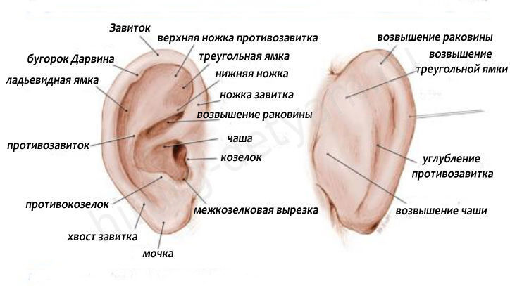
The auricle consists of cartilage (it is characterized by elasticity, elasticity). From above it is covered with integuments. Below is the lobe. This area has no cartilage. It includes adipose tissue, skin. The auricle is considered a rather sensitive organ.
Anatomy
Smaller elements of the auricle are:
- curl;
- tragus;
- antihelix;
- curl legs;
- antitragus.
Koshcha is a specific coating lining the ear canal. Inside it contains glands that are considered to be vital. They secrete a secret that protects against many agents (mechanical, thermal, infectious).
The end of the passage is represented by a kind of dead end. This specific barrier (tympanic membrane) is required to separate the outer, middle ear. It begins to oscillate when sound waves hit it. After the sound wave hits the wall, the signal is transmitted further, towards the middle part of the ear.
Blood to this site goes through two branches of arteries. The outflow of blood is carried out through the veins (v. auricularis posterior, v. retromandibularis). localized in front, behind the auricle. They also carry out the removal of lymph.
In the photo, the structure of the outer ear
Functions
Let us indicate the significant functions that are assigned to the outer part of the ear. She is capable of:
- receive sounds;
- transmit sounds to the middle part of the ear;
- direct the wave of sound towards the inside of the ear.
Possible pathologies, diseases, injuries
Let's note the most common diseases:
Average
The middle ear plays a huge role in signal amplification. Amplification is possible due to the auditory ossicles.
Structure
We indicate the main components of the middle ear:
- tympanic cavity;
- auditory (Eustachian) tube.
The first component (tympanic membrane) contains a chain inside, which includes small bones. The smallest bones play an important role in the transmission of sound vibrations. The eardrum consists of 6 walls. Its cavity contains 3 auditory ossicles:
- hammer. Such a bone is endowed with a rounded head. This is how it is connected to the handle;
- anvil. It includes the body, processes (2 pieces) of different lengths. With the stirrup, its connection is made by means of a slight oval thickening, which is located at the end of a long process;
- stirrup. In its structure, a small head is distinguished, bearing an articular surface, an anvil, legs (2 pcs.).
Arteries go to the tympanic cavity from a. carotis externa, being its branches. Lymphatic vessels are directed to the nodes located on the lateral wall of the pharynx, as well as to those nodes that are localized behind the ear shell.
The structure of the middle ear

Functions
Bones from the chain are needed for:
- Conducting sound.
- Transmission of vibrations.
The muscles located in the middle ear area are specialized for various functions:
- protective. Muscle fibers protect the inner ear from sound irritations;
- tonic. Muscle fibers are necessary to maintain the chain of auditory ossicles, the tone of the tympanic membrane;
- accommodative. The sound-conducting apparatus adapts to sounds endowed with different characteristics (strength, height).
Pathologies and diseases, injuries
Among the popular diseases of the middle ear, we note:
- (perforative, non-perforative, );
- catarrh of the middle ear.
Acute inflammation can appear with injuries:
- otitis, mastoiditis;
- otitis, mastoiditis;
- , mastoiditis, manifested by injuries of the temporal bone.
It can be complicated, uncomplicated. Among the specific inflammations, we indicate:
- syphilis;
- tuberculosis;
- exotic diseases.
Anatomy of the outer, middle, inner ear in our video:
Let us indicate the weighty importance of the vestibular analyzer. It is necessary to regulate the position of the body in space, as well as to regulate our movements.
Anatomy
The periphery of the vestibular analyzer is considered to be part of the inner ear. In its composition, we highlight:
- semicircular canals (these parts are located in 3 planes);
- statocyst organs (they are represented by sacs: oval, round).
The planes are called: horizontal, frontal, sagittal. The two sacs represent the vestibule. The round pouch is located near the curl. The oval sac is located closer to the semicircular canals.
Functions
Initially, the analyzer is excited. Then, thanks to the vestibulo-spinal nerve connections, somatic reactions occur. Such reactions are needed to redistribute muscle tone, maintain body balance in space.
The connection between the vestibular nuclei, the cerebellum determines the mobile reactions, as well as all the reactions for the coordination of movements that appear during the performance of sports, labor exercises. To maintain balance, vision and musculo-articular innervation are very important.

Pathologies, diseases, injuries
Violations that may be present in the work of the vestibular apparatus are manifested in.
There are a lot of diseases that signal their development with pain in the ears. To determine what specific disease affected the organ of hearing, you need to understand how the human ear is arranged.
Diagram of the auditory organ
First of all, let's understand what an ear is. This is an auditory-vestibular paired organ that performs only 2 functions: the perception of sound impulses and responsibility for the position of the human body in space, as well as for maintaining balance. If you look at the human ear from the inside, its structure suggests the presence of 3 parts:
- external (external);
- average;
- internal.
Each of them has its own no less intricate device. Connecting, they are a long pipe penetrating into the depths of the head. Let us consider the structure and functions of the ear in more detail (the diagram of the human ear demonstrates them best).
What is the outer ear
The structure of the human ear (its outer part) is represented by 2 components:
- ear shell;
- external ear canal.

The shell is an elastic cartilage that completely covers the skin. It has a complex shape. In its lower segment there is a lobe - this is a small skin fold filled inside with a fatty layer. By the way, it is the outer part that has the highest sensitivity to various kinds of injuries. For example, for fighters in the ring, it often has a form that is very far from its original form.
The auricle serves as a kind of receiver for sound waves, which, falling into it, penetrate deep into the organ of hearing. Since it has a folded structure, the sound enters the passage with little distortion. The degree of error depends, in particular, on the place where the sound comes from. Its location is horizontal or vertical.
It turns out that more accurate information about where the sound source is located enters the brain. So, it can be argued that the main function of the shell is to catch sounds that should enter the human ear.
If you look a little deeper, you can see that the shell extends the cartilage of the external ear canal. Its length is 25-30 mm. Next, the cartilage zone is replaced by bone. The outer ear completely lines the skin, which contains 2 types of glands:
- sulfuric;
- greasy.
The outer ear, the device of which we have already described, is separated from the middle part of the hearing organ by a membrane (it is also called the tympanic membrane).
How is the middle ear
If we consider the middle ear, its anatomy is:
- tympanic cavity;
- eustachian tube;
- mastoid process.
All of them are interconnected. The tympanic cavity is a space outlined by the membrane and the region of the inner ear. Its location is the temporal bone. The structure of the ear here looks like this: in the anterior part, there is a union of the tympanic cavity with the nasopharynx (the function of the connector is performed by the Eustachian tube), and in its posterior part, with the mastoid process through the entrance to its cavity. Air is present in the tympanic cavity, which enters there through the Eustachian tube.

The anatomy of the ear of a person (child) up to 3 years old has a significant difference from how the ear of an adult is arranged. Babies do not have a bone passage, and the mastoid process has not yet grown. The children's middle ear is represented by only one bone ring. Its inner edge has the shape of a groove. It just houses the tympanic membrane. In the upper zones of the middle ear (where there is no this ring), the membrane is connected to the lower edge of the scales of the temporal bone.
When the baby reaches the age of 3, the formation of his ear canal is completed - the structure of the ear becomes the same as in adults.
Anatomical features of the internal department
 The inner ear is the most difficult part of it. The anatomy in this part is very complex, so she was given a second name - "membranous labyrinth of the ear." It is located in the stony zone of the temporal bone. It is attached to the middle ear with windows - round and oval. Comprises:
- vestibule;
- snails with the organ of Corti;
- semicircular canals (filled with fluid).
In addition, the inner ear, the structure of which provides for the presence of the vestibular system (apparatus), is responsible for constantly keeping the body in a state of balance by a person, as well as for the possibility of accelerating in space. The vibrations that occur in the oval window are transmitted to the fluid that fills the semicircular canals. The latter serves as an irritant for the receptors located in the cochlea, and this already becomes the cause of the launch of nerve impulses.
It should be noted that the vestibular apparatus has receptors in the form of hairs (stereocilia and kinocilia), which are located on special elevations - maculae. These hairs are located one opposite the other. By shifting, stereocilia provoke the occurrence of excitation, and kinocilia help inhibition.
Summing up
In order to more accurately imagine the structure of the human ear, the diagram of the organ of hearing should be in front of the eyes. It usually depicts a detailed structure of the human ear.
 Obviously, the human ear is a rather complex system, consisting of many different formations, each of which performs a number of important and truly irreplaceable functions. The diagram of the ear demonstrates this clearly.
Regarding the structure of the outer part of the ear, it should be noted that each person has individual genetically determined features that in no way affect the main function of the hearing organ.
Ears need regular hygienic care. If you neglect this need, you can partially or completely lose your hearing. Also, lack of hygiene can lead to the development of diseases affecting all parts of the ear.
The ear contains two sensory organs with different functions (hearing and balance), which, nevertheless, anatomically form a single whole.
The ear is located in the stony part of the temporal bone (the stony part is sometimes simply called the stony bone) or the so-called pyramid, and consists of the cochlea and the vestibular apparatus (labyrinth), which includes two fluid-filled sacs and three semicircular canals, also filled with fluid. The organ of hearing, unlike the vestibular apparatus, has auxiliary structures that ensure the conduction of sound waves: the outer ear and the middle ear.
The outer ear is Auricle, external auditory meatus about 3 cm long and eardrum. The auricle consists mainly of elastic cartilage, which enters the external opening of the external auditory canal. Further, the external auditory meatus is a bone canal with a slight S-shaped bend. In its cartilaginous part are numerous ceruminous glands that secrete ear wax. The tympanic membrane is stretched across the inner end of the bony canal and is the boundary of the middle ear.
Middle ear
The middle ear contains tympanic cavity, lined with a mucous membrane and containing auditory ossicles - hammer, anvil and stapes, eustachian tube, which is a continuation of the tympanic cavity forward into the pharynx, as well as numerous cavities in the mastoid process of the temporal bone, lined with a mucous membrane.
.png)
The tympanic membrane is almost round, 1 cm in diameter; it forms the outer wall of the tympanic cavity. The eardrum is made up of three layers. The predominantly rigid connective tissue base of the tympanic membrane is devoid of tension only in a small area near its upper end. Its inner surface is lined with a mucous membrane, and the outer one with skin. The long handle of the malleus attached to the tympanic membrane causes it to curve inward like a funnel. The auditory ossicles together with the tympanic membrane make up the sound-conducting apparatus. Hammer, anvil and stapes form an unbroken chain connecting eardrum and foramen ovale, into which the base of the stirrup is embedded.
The ossicles conduct vibrations generated by sound waves in the tympanic membrane to the oval window of the inner ear. The oval window together with the first coil of the cochlea forms the inner bony border of the tympanic cavity. The base of the stirrup in the oval window transmits vibrations to the fluid that fills the inner ear. The hammer and stirrup are additionally fixed by two muscles, on which the intensity of sound transmission depends.
inner ear
The inner ear is surrounded by a hard bone capsule and consists of systems of ducts and cavities (bone labyrinth) filled with perilymph.
Inside the bony labyrinth is a membranous labyrinth filled with endolymph. Perilymph and endolymph differ mainly in their content of sodium and potassium. The membranous labyrinth contains the organs of hearing and balance. Bone spiral (cochlea) of the inner ear, about 3 cm long, forms a canal, which in humans makes approximately 2.5 turns around the bony central rod - the columella. On the transverse section of the cochlea, three separate cavities are visible: in the middle is the cochlear canal. The cochlear canal is also often called the middle scala, below it is the tympanic and vestibular scalas, which are connected at the top of the cochlea through a hole - the helicotrema.
These cavities are filled with perilymph and end with a round cochlear window and an oval window of the vestibule, respectively. The cochlear duct is filled with endolymph and is separated from the scala tympani by the main (basilar) membrane, and from the vestibular scala by the Reissner (vestibular) membrane.
Organ of Corti (spiral organ) is located on the main membrane. It contains about 15,000 auditory sensory cells arranged in rows (inner and outer hair cells), as well as many supporting cells. The hairs of sensory cells are attached to the gelatinous integumentary (tentorial) membrane located above them.
auditory pathway
Hair cells form synapses with neurons whose cell bodies lie in the spiral ganglion of the cochlea in the central shaft. From here, the central branches of their axons go as part of the cochlear and vestibular nerves of the cranial nerve VIII (vestibulocochlear nerve) to the brain stem. There, the axons of the cochlear nerve terminate in the cochlear nuclei, and the axons of the vestibular nerve terminate in the vestibular nuclei.
On its way to the auditory region in the anterior transverse gyrus of the temporal lobe, the auditory pathway passes through several synaptic switches, including in the medial geniculate body of the diencephalon.


