What parts of the brain are responsible for what. What part of the brain is responsible for memory. Parts of the brain and their functions: cortex
The brain is the main regulator of the functions of any living organism, one of the elements Until now, medical scientists have been studying the features of the brain and discovering new incredible possibilities. This is a very complex organ that connects our body with the external environment. The parts of the brain and their functions regulate all life processes. External receptors catch signals and inform any part of the brain about incoming stimuli (light, sound, tactile, and many others). The response is immediate. Let's take a closer look at how our head "processor" works.
General description of the brain
The parts of the brain and their functions completely control our life processes. Consists human brain out of 25 billion neurons. This incredible number of cells forms the gray matter. The brain covers several layers:
- soft;
- hard;
- arachnoid (cerebrospinal fluid circulates here).
Liquor is a cerebrospinal fluid, in the brain it plays the role of a shock absorber, a protector from any impact force.
In both men and women, the brain is developed in exactly the same way, although its weight is different. More recently, the debate has subsided that the weight of the brain plays some role in mental development and intellectual abilities. The conclusion is unambiguous - it is not. The weight of the brain is approximately 2% of the total mass of a person. In men, its average weight is 1,370 g, and in women - 1,240 g. The functions of the parts of the human brain are developed in a standard way, vital activity depends on them. Mental abilities depend on the quantitative connections created in the brain. Each brain cell is a neuron that generates and transmits impulses.
The cavities inside the brain are called ventricles. AT different departments craniocerebral paired nerves leave.
Functions of the brain regions (table)
Every part of the brain has a job to do. The table below clearly demonstrates this. The brain, like a computer, clearly performs its tasks, receiving commands from the outside world.
The functions of the brain regions, the table reveals schematically and succinctly.

Let's take a closer look at the parts of the brain below.
Structure
The picture shows how the brain works. Despite this, the most significant part is occupied by all parts of the brain and their functions play a huge role in the functioning of the body. There are five main divisions:
- final (of the total mass is 80%);
- posterior (bridge and cerebellum);
- intermediate;
- oblong;
- average.
At the same time, the brain is divided into three main parts: the brain stem, the cerebellum, and the two cerebral hemispheres.

telencephalon
It is impossible to briefly describe the structure of the brain. To understand the parts of the brain and their functions, it is necessary to study their structure closely.
The telencephalon stretches from the frontal to the occipital bone. Two cerebral hemispheres are considered here: left and right. This department differs from others in the largest number of furrows and convolutions. The development and structure of the brain are closely linked. Experts have identified three types of bark:
- ancient (with olfactory tubercle, anterior perforated substance, semilunar subcallosal and lateral subcallosal gyrus);
- old (with dentate gyrus - fascia and hippocampus);
- new (represents the rest of the cortex).
The hemispheres are separated by a longitudinal groove, in its depths there is a vault and a corpus callosum, which connect the hemispheres. The corpus callosum itself is lined and belongs to the neocortex. The structure of the hemispheres is quite complex and resembles a multi-level system. Here, the frontal, temporal, parietal and occipital lobes, subcortex and cortex are distinguished. performed by the large hemispheres great amount functions. It is worth noting that the left hemisphere commands right side body, and the right on the contrary - the left.

Bark
The surface layer of the brain is the cortex, it has a thickness of 3 mm, covers the hemispheres. The structure consists of vertical nerve cells with processes. The cortex also contains efferent and afferent nerve fibers and neuroglia. The parts of the brain and their functions are discussed in the table, but what is the cortex? Its complex structure has horizontal layering. The building has six layers:
- external pyramidal;
- external granular;
- internal granular;
- molecular;
- internal pyramidal;
- with spindle cells.
Each has a different width, density, shape of neurons. Vertical bundles of nerve fibers give the cortex a vertical striation. The area of the cortex is approximately 2,200 square centimeters, the number of neurons here reaches ten billion.

Parts of the brain and their functions: cortex
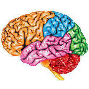
The cortex controls several specific bodily functions. Each share is responsible for its own parameters. Let's take a closer look at the functions associated with hotels:
- temporal - controls the sense of smell and hearing;
- parietal - responsible for taste and touch;
- occipital - vision;
- frontal - complex thinking, movement and speech.
Each neuron contacts other neurons, there are up to ten thousand contacts (gray matter). Nerve fibers are white matter. Some part unites the hemispheres of the brain. White matter includes three types of fibers:
- association links connect different cortical areas in one hemisphere;
- commissural connect the hemispheres to each other;
- projection ones communicate with lower formations, have paths of analyzers.
Considering the structure and functions of the parts of the brain, it is necessary to emphasize the role of the gray and hemispheres inside have (gray matter), their main function is the transmission of information. The white matter is located between the cerebral cortex and the basal ganglia. There are four parts here:
- between furrows in convolutions;
- in the outer places of the hemispheres;
- included in the inner capsule;
- located in the corpus callosum.
The white matter located here is formed by nerve fibers and connects the cortex of the convolutions with the underlying sections. form the subcortex of the brain.
The telencephalon - manages all the vital functions of the body, as well as the intellectual abilities of a person.
diencephalon
Brain regions and their functions (table above) include diencephalon. If you look in more detail, it is worth saying that it consists of ventral and dorsal parts. The hypothalamus belongs to the ventral, and the thalamus, metathalamus, and epithalamus to the dorsal.
The thalamus is a mediator that directs the received irritations to the hemispheres. It is often referred to as the "optic tubercle". It helps the body quickly adapt to changes in the external environment. The thalamus is connected to the cerebellum via the limbic system.
The hypothalamus controls autonomic functions. The influence goes through the nervous system, and, of course, the glands internal secretion. Regulates work endocrine glands controls metabolism. The pituitary gland is located directly below it. Body temperature, cardiovascular and digestive systems are regulated. The hypothalamus also controls our eating and drinking behavior, regulates wakefulness and sleep.
Rear
The hindbrain includes the pons located in front and the cerebellum, which is located behind. Studying the structure and functions of the brain regions, let's take a closer look at the structure of the bridge: the dorsal surface is covered by the cerebellum, the ventral one is represented by a fibrous structure. The fibers are directed transversely in this section. On each side of the bridge, they depart to the cerebellar middle peduncle. In appearance, the bridge resembles a thickened white roller located above the medulla oblongata. The nerve roots exit into the bulbar pontine groove.
The structure of the posterior bridge: on the frontal section, it can be seen that the department of the anterior (large ventral) and posterior (small dorsal) parts consists. Between them, the trapezoid body serves as a boundary, the transverse thick fibers of which are classified as auditory pathway. Conductor function is completely dependent on the hindbrain.
Cerebellum (small brain)
The table "Department of the brain, structure, functions" indicates that the cerebellum is responsible for the coordination and movement of the body. This department is located behind the bridge. The cerebellum is often referred to as the "small brain". It occupies the posterior cranial fossa, covers the rhomboid. The mass of the cerebellum ranges from 130 to 160 g. Above are the large hemispheres, which are separated by a transverse fissure. bottom the cerebellum is adjacent to the medulla oblongata.
Two hemispheres are distinguished here, the lower, upper surface and the worm. The boundary between them is called a horizontal deep slit. A lot of cracks cut the surface of the cerebellum, between them there are thin convolutions (rollers). Between the grooves there are groups of convolutions, divided into lobules, they represent the lobes of the cerebellum (posterior, flocculent-nodular, anterior).
The cerebellum contains both gray and white matter. Gray is located on the periphery, forms a cortex with molecular and pear-shaped neurons, and a granular layer. Under the cortex there is a white substance that penetrates into the gyrus. In the white matter there are blotches of gray (its nuclei). In cross section, this ratio is similar to a tree. Those who know the structure of the human brain, the functions of its departments, will easily answer that the cerebellum is the regulator of the coordination of the movements of our body.

midbrain
The midbrain is located in the region of the anterior pons and goes to the papillary bodies, as well as to the optic tracts. Here clusters of nuclei are distinguished, which are called tubercles of the quadrigemina. The structure and functions of the brain regions (table) indicate that this department is responsible for latent vision, the orienting reflex, gives orientation to reflexes to visual and sound stimuli, and also maintains muscle tone human body.
medulla oblongata: brainstem
The medulla oblongata is a natural extension of the spinal cord. That is why the structure has a lot in common. This becomes especially clear if we examine the white matter in detail. It is represented by short and long nerve fibers. In the form of nuclei, gray matter is represented here. Parts of the brain and their functions (the table is presented above) indicates that the medulla oblongata controls our balance, coordination, regulates metabolism, controls breathing and blood circulation. Also responsible for these important reflexes our body like sneezing and coughing, vomiting.
The brain stem is divided into the hindbrain and midbrain. The trunk is called the middle, oblong, bridge and diencephalon. Its structure is descending and ascending paths connecting the trunk with the spinal cord and brain. In this part, control over the heartbeat, breathing, articulate speech is carried out.
The brain is the main controlling organ of the central nervous system(CNS), has been working on the study of its structure and functions for more than 100 years a large number of specialists in various fields, such as psychiatry, medicine, psychology and neurophysiology. Despite a good study of its structure and components, there are still many questions about the work and processes that take place every second.
The brain belongs to the central nervous system and is located in the cranial cavity. Outside, it is reliably protected by the bones of the skull, and inside it is enclosed in 3 shells: soft, cobweb and hard. Between these membranes circulates cerebrospinal fluid - cerebrospinal fluid, which serves as a shock absorber and prevents concussion of this organ in case of minor injuries.
The human brain is a system consisting of interconnected departments, each part of which is responsible for performing specific tasks.
To understand the functioning, it is not enough to briefly describe the brain, therefore, in order to understand how it works, you first need to study its structure in detail.
What is the brain responsible for

This body, like spinal cord, belong to the central nervous system and plays the role of an intermediary between the environment and the human body. With its help, self-control, reproduction and memorization of information, figurative and associative thinking, and other cognitive psychological processes are carried out.
According to the teachings of Academician Pavlov, the formation of thought is a function of the brain, namely the cerebral cortex, which are the highest organs nervous activity. Per different types The cerebellum, the limbic system, and some areas of the cerebral cortex are responsible for memory, but since memory is different, it is impossible to single out any specific area responsible for this function.
It is responsible for managing the vegetative vital functions of the body: respiration, digestion, endocrine and excretory systems, body temperature control.
To answer the question of what function the brain performs, first you should conditionally divide it into sections.
Experts distinguish 3 main parts of the brain: anterior, middle and rhomboid (posterior) section.
- The anterior one performs higher psychiatric functions, such as the ability to know, the emotional component of a person’s character, his temperament and complex reflex processes.
- The middle one is responsible for sensory functions and processing of information received from the organs of hearing, vision and touch. The centers located in it are able to regulate the degree pain, since the gray matter, under certain conditions, is capable of producing endogenous opiates that increase or decrease pain threshold. It also plays the role of a conductor between the cortex and the underlying sections. This part controls the body through various innate reflexes.
- Rhomboid or posterior section, responsible for muscle tone, coordination of the body in space. Through it, purposeful movement of various muscle groups is carried out.
The structure of the brain cannot be simply briefly described, since each of its parts includes several departments, each of which performs certain functions.
What does the human brain look like

Brain anatomy is a relatively young science, since long time was banned due to laws prohibiting the autopsy and examination of organs and the head of a person.
The study topographic anatomy brain region in the head area, it is necessary for accurate diagnosis and successful therapy various topographic anatomical disorders, for example: skull injuries, vascular and oncological diseases. To imagine what a human GM looks like, first you need to study them appearance.
In appearance, GM is a gelatinous mass of yellowish color, enclosed in a protective shell, like all organs. human body They are 80% water.
The large hemispheres occupy practically the volume of this organ. They are covered with gray matter or bark - the highest organ of human neuropsychic activity, and inside - with white matter, consisting of processes of nerve endings. The surface of the hemispheres has a complex pattern, due to the convolutions and ridges going in different directions between them. According to these convolutions, it is customary to divide them into several departments. It is known that each of the parts performs certain tasks.
In order to understand what the human brain looks like, it is not enough to examine their appearance. There are several study methods that help to study the inside of the brain in a section.
- Sagittal section. It is a longitudinal section that passes through the center of the human head and divides it into 2 parts. It is the most informative method research that diagnoses various diseases this organ.
- The frontal section of the brain looks like a cross section of large lobes and allows you to see the fornix, hippocampus and corpus callosum, as well as the hypothalamus and thalamus, which control the vital functions of the body.
- Horizontal cut. Allows you to consider the structure of this organ in a horizontal plane.
The anatomy of the brain, as well as the anatomy of the human head and neck, is a rather difficult subject to study for a number of reasons, including the fact that their description requires studying a large amount of material and having a good clinical background.
How the human brain works
Scientists around the world are studying the brain, its structure and functions that it performs. Over the past few years, many important discoveries have been made, however, this part of the body remains not fully understood. This phenomenon is explained by the complexity of studying the structure and functions of the brain separately from the cranium.
In turn, the structure of the brain structures determines the functions performed by its departments.
It is known that this organ consists of nerve cells (neurons) interconnected by bundles of filamentous processes, but it is still not clear how their interaction as a single system occurs simultaneously as a single system.
The diagram of the structure of the brain, based on the study of the sagittal section of the cranium, will help to explore the sections and membranes. In this figure, you can see the cortex, the medial surface of the cerebral hemispheres, the structure of the trunk, cerebellum and the corpus callosum, which consists of a roller, trunk, knee and beak.
The GM is reliably protected from the outside by the bones of the skull, and inside by 3 meninges: hard arachnoid and soft. Each of them has its own device and performs certain tasks.
- The deep soft shell covers both the spinal cord and the brain, while entering all the cracks and grooves of the cerebral hemispheres, and in its thickness there are blood vessels that feed this organ.
- The arachnoid membrane is separated from the first by a subarachnoid space filled with liquor (cerebrospinal fluid), it also contains blood vessels. This sheath consists of connective tissue, from which filiform branched processes (strands) depart, they are woven into a soft sheath and with age their number increases, thereby strengthening the connection. Between them. The villous outgrowths of the arachnoid bulge into the lumen of the sinuses of the dura mater.
- The hard shell or pachymeninx consists of a connective tissue substance and has 2 surfaces: the upper one, saturated with blood vessels, and the inner one, which is smooth and shiny. With this side, the pachymeninx is adjacent to the medulla, and the outer side is adjacent to the cranium. Between the hard and arachnoid there is a narrow space filled with a small amount of liquid.
in the brain healthy person circulates about 20% of the total volume of blood that enters through the posterior cerebral arteries.
The brain can be visually divided into 3 main parts: 2 cerebral hemispheres, brainstem and cerebellum.
Gray matter forms the cortex and covers the surface of the cerebral hemispheres, and its a small amount of in the form of nuclei is located in the medulla oblongata.
In all brain regions there are ventricles, in the cavity of which the cerebrospinal fluid, which is formed in them, moves. In this case, the fluid from the 4th ventricle enters the subarachnoid space and washes it.
The development of the brain begins even during the intrauterine presence of the fetus, and it is finally formed by the age of 25.
Main parts of the brain

picture is clickable
What is the brain composed of and study the composition of the brain ordinary person can be from the pictures. The structure of the human brain can be viewed in several ways.
The first divides it into components that make up the brain:
- Final, represented by 2 cerebral hemispheres, united by the corpus callosum;
- intermediate;
- average;
- oblong;
- the posterior borders on the medulla oblongata, the cerebellum and the bridge depart from it.
It is also possible to single out the main composition of the human brain, namely, it includes 3 large structures that begin to develop even during embryonic development:
- diamond-shaped;
- average;
- anterior brain.
In some textbooks, the cerebral cortex is usually divided into sections, so that each of them plays a specific role in the higher nervous system. Accordingly, allocate following departments forebrain: frontal, temporal, parietal and occipital zone.
Large hemispheres
First, consider the structure of the cerebral hemispheres.
The end brain of a person controls all vital important processes and is divided by a central sulcus into 2 cerebral hemispheres, covered on the outside with a bark or gray matter, and inside they consist of white matter. Between themselves, in the depths of the central gyrus, they are united by the corpus callosum, which serves as a link connecting and transmitting information between other departments.
The structure of the gray matter is complex and, depending on the site, consists of 3 or 6 layers of cells.
Each lobe is responsible for performing certain functions and coordinates the movement of the limbs on its part, for example, right part processes non-verbal information and is responsible for spatial orientation, while the left one specializes in mental activity.
In each of the hemispheres, specialists distinguish 4 zones: frontal, occipital, parietal and temporal, they perform certain tasks. In particular, the parietal part of the cerebral cortex is responsible for visual function.
The science that studies the detailed structure of the cerebral cortex is called architectonics.
Medulla
This section is part of the brain stem and serves as a link between the dorsal and the bridge of the terminal section. Since it is a transitional element, it combines the features of the spinal and structural features of the brain. The white matter of this section is represented by nerve fibers, and the gray matter is in the form of nuclei:
- The nucleus of the olive, is a complementary element of the cerebellum, is responsible for balance;
- The reticular formation connects all the sense organs with the medulla oblongata, is partially responsible for the work of some parts of the nervous system;
- The nuclei of the nerves of the skull, these include: glossopharyngeal, vagus, accessory, hypoglossal nerves;
- The nuclei of respiration and circulation, which are connected with the nuclei of the vagus nerve.
This internal structure is due to the functions of the brain stem.
He is responsible for defensive reactions body and regulates vital processes such as heartbeat and blood circulation, so damage to this component leads to instant death.
Pons
The structure of the brain includes the pons, it serves as a link between the cerebral cortex, the cerebellum and the spinal cord. Consists of nerve fibers and gray matter, in addition, the bridge serves as a conductor main artery nourishing the brain.
midbrain
This part has a complex structure and consists of a roof, a midbrain part of a tire, a Sylviian aqueduct and legs. In the lower part it borders on the posterior region, namely the pons and the cerebellum, and at the top of it is the diencephalon connected to the terminal.
The roof consists of 4 hills, inside which the nuclei are located, they serve as centers for the perception of information received from the eyes and hearing organs. Thus, this part is included in the zone responsible for receiving information, and refers to the ancient structures that make up the structure of the human brain.
Cerebellum
The cerebellum occupies almost the entire back part and repeats the basic principles of the structure of the human brain, that is, it consists of 2 hemispheres and an unpaired formation connecting them. The surface of the cerebellar lobules is covered with gray matter, and inside they consist of white, in addition, the gray matter in the thickness of the hemispheres forms 2 nuclei. The white matter connects the cerebellum to the brainstem and spinal cord with three pairs of legs.
This brain center is responsible for coordinating and regulating motor activity human muscles. It also helps to maintain a certain posture in the surrounding space. Responsible for muscle memory.
Bark
The structure of the cerebral cortex is quite well studied. So, it is a complex layered structure 3-5 mm thick, which covers the white matter of the cerebral hemispheres.
The cortex is formed by neurons with bundles of filiform processes, afferent and efferent nerve fibers, glia (provide the transmission of impulses). It has 6 layers, different in structure:
- grainy;
- molecular;
- external pyramidal;
- internal granular;
- internal pyramidal;
- the last layer consists of spindle-shaped cells.
It occupies about half the volume of the hemispheres, and its area in a healthy person is about 2200 square meters. see. The surface of the bark is dotted with furrows, in the depths of which one third of its entire area lies. The size and shape of the furrows of both hemispheres is strictly individual.
The cortex was formed relatively recently, but is the center of the entire higher nervous system. Experts distinguish several parts in its composition:
- neocortex (new) main part covers more than 95%;
- archicortex (old) - about 2%;
- paleocortex (ancient) - 0.6%;
- intermediate cortex, occupies 1.6% of the total cortex.
It is known that the localization of functions in the cortex depends on the location of the nerve cells that pick up one of the types of signals. Therefore, there are 3 main areas of perception:
- Touch.
- Motor.
- Associative.
The last region occupies more than 70% of the crust, and its central purpose is to coordinate the activity of the first two zones. It is also responsible for receiving and processing data from the sensory zone, and the goal-directed behavior caused by this information.
Between the cerebral cortex and the medulla oblongata is the subcortex or, in other words, the subcortical structures. It consists of visual tubercles, hypothalamus, limbic system and other nerve nodes.
The main functions of the brain regions
The main functions of the brain are to process data received from the environment, as well as control the movements of the human body and its mental activity. Each part of the brain is responsible for performing specific tasks.
The medulla oblongata controls the body's defense functions such as blinking, sneezing, coughing, and vomiting. It also controls other reflex vital processes - respiration, saliva secretion and gastric juice, swallowing.
With the help of the Varoliyev bridge, the coordinated movement of the eyes and facial wrinkles is carried out.
The cerebellum controls the motor and coordination activity of the body.
The midbrain is represented by the stalk and the quadrigemina (two auditory and two visual hillocks). With its help, orientation in space, hearing and clarity of vision is carried out, it is responsible for the muscles of the eyes. Responsible for the reflex turn of the head towards the stimulus.
The diencephalon consists of several parts:
- The thalamus is responsible for the formation of feelings, such as pain or taste. In addition, he manages tactile, auditory, olfactory sensations and rhythms of human life;
- The epithalamus consists of the pineal gland, which controls the daily biological rhythms, dividing the daylight hours into the time of wakefulness and the time of healthy sleep. Has the ability to detect light waves through the bones of the skull, depending on their intensity, produces the appropriate hormones and controls metabolic processes in the human body;
- The hypothalamus is responsible for the work of the heart muscles, the normalization of body temperature and blood pressure. With its help, a signal is given to select stress hormones. Responsible for feelings of hunger, thirst, pleasure and sexuality.
The posterior pituitary gland is located in the hypothalamus and is responsible for the production of hormones that affect puberty and functioning of the human reproductive system.
Each hemisphere is responsible for its own specific tasks. For example, the right cerebral hemisphere accumulates data about the environment and the experience of communicating with it. Controls the movement of the limbs on the right side.
In the left cerebral hemisphere there is a speech center responsible for human speech, it also controls analytical and computational activities, and abstract thinking is formed in its cortex. Similarly, the right side controls the movement of the limbs on its side.
The structure and function of the cerebral cortex directly depend on each other, so the gyrus conditionally divides it into several parts, each of which performs certain operations:
- temporal lobe, controls hearing and charm;
- the occipital part regulates vision;
- in the parietal, touch and taste are formed;
- the frontal parts are responsible for speech, movement and complex thought processes.
The limbic system consists of the olfactory centers and the hippocampus, which is responsible for adapting the body to change and regulating the emotional component of the body. It creates lasting memories by associating sounds and smells with certain period the time during which sensory upheavals occurred.
In addition, it controls restful sleep, data retention in short-term and long-term memory, intellectual activity, control of the endocrine and autonomic nervous system, and participates in the formation of the reproductive instinct.
How the human brain works

The work of the human brain does not stop even in a dream, it is known that some departments also function in people who are in a coma, as evidenced by their stories.
The main work of this body is carried out with the help of the cerebral hemispheres, each of which is responsible for a certain ability. It is noted that the hemispheres are not the same in size and function - the right side is responsible for visualization and creative thinking, usually more than the left side, which is responsible for logic and technical thinking.
It is known that men have a larger brain mass than women, but this feature does not affect mental abilities. For example, this figure for Einstein was below average, but his parietal zone, which is responsible for cognition and the creation of images, was large, which allowed the scientist to develop the theory of relativity.
Some people are endowed with super abilities, this is also the merit of this body. These features are manifested in a high speed of writing or reading, photographic memory and other anomalies.
One way or another, the activity of this body is of great importance in conscious management human body, and the presence of the bark distinguishes humans from other mammals.
What, according to scientists, constantly occurs in the human brain
Specialists who study the psychological capabilities of the brain believe that the performance of cognitive and mental functions occurs as a result of biochemical currents, however, this theory is based on this moment is questioned, because this organ is a biological object and the principle of mechanical action does not allow to know its nature completely.
The brain is a kind of steering wheel of the whole organism, performing a huge number of tasks every day.
The anatomical and physiological features of the structure of the brain have been the subject of study for many decades. This organ is known to be special place in the structure of the central nervous system (central nervous system) of a person, and its characteristics are different for each person, therefore it is impossible to find 2 absolutely identically thinking people.
Video
Scientists consider the cortex of the frontal region as a set of formations that show with early age pronounced personality in anatomical structure. Among these formations there are those that are new, " human» fields that are developing in more late age. These include field 46.
Field 46 is a "human field", because it is an evolutionary neoplasm that differentiates late. Field 46 is the last to mature and reaches 630% of its original size. Because this field is inhibitory, you can see that the children do not control their movements and grab everything that lies badly. This behavior is typical of monkeys.
General
It is impossible to specifically develop the frontal lobes of the brain in children. In society, there is an incorrect opinion that physical activity promotes increased blood circulation in the brain, thereby developing all parts of the brain. Physical activity fills the motor-motor centers of the brain, while the rest of the brain ‘ rest‘because when performing different tasks, the brain uses certain centers, and not the whole brain.
Based on the foregoing, in order to determine the exercises for the development of the frontal lobes, you need to find out what functions the frontal lobes are responsible for, during which we will be able to develop the frontal lobes.
The frontal lobe, like the others, consists of substances.
Location
The frontal lobe is located in the anterior parts of the hemispheres. The frontal lobe is separated from the parietal lobe by the central sulcus, and from the temporal lobe by the lateral sulcus. Anatomically, it consists of four convolutions - vertical and three horizontal. The convolutions are separated by furrows. The frontal lobe makes up a third of the mass of the cortex.
Assigned functions
Evolutionarily it happened that active development frontal lobes is not associated with mental and intellectual activity. frontal lobes evolved in humans. How more people could share food in his community, the more likely the community could survive. In women, the frontal lobes arose for the specific purpose of sharing food. The men got this area as a gift. Not having those assigned tasks that lie on the shoulders of a woman, men began to use the frontal lobes in a variety of ways (think, build, etc.) to manifest Dominance.
Essentially, the frontal lobes are brake centers. Also, many people ask what the left or right frontal lobe of the brain is responsible for. The question was posed incorrectly, because in the left and right frontal lobes there are corresponding fields, which are responsible for specific functions. Roughly stated, the frontal lobes are responsible for:
- thinking
- movement coordination
- conscious control of behavior
- centers of memory and speech
- display of emotions
What fields are included
Fields and subfields are responsible for specific functions that are generalized under the frontal lobes. Because The polymorphism of the brain is huge, the combination of the sizes of different fields makes up the individuality of a person. Why is it said that over time a person changes. Throughout life, neurons die, and the remaining ones form new connections. This introduces an imbalance in the quantitative ratio of links between different fields that are responsible for different functions.
Not only that, different people The margin sizes are different, so some people may or may not have these margins at all. Polymorphism was identified by Soviet researchers S.A. Sarkisov, I.N. Filimonov, Yu.G. Shevchenko. They showed that the individual ways of building the cerebral cortex within one ethnic group are so large that no common signs can be seen.
- Field 8 - located in the posterior sections of the middle and superior frontal gyri. Has a center of voluntary eye movements
- Field 9 - dorsolateral prefrontal cortex
- Field 10 - Anterior prefrontal cortex
- Field 11 - olfactory area
- Box 12 - control of the basal ganglia
- Field 32 - Receptor area of emotional experiences
- Field 44 - Broca's Center (processing information about the location of the body relative to other bodies)
- Field 45 - music and motor center
- Field 46 - motor analyzer of head and eye rotation
- Field 47 - nuclear zone of singing, speech motor component
- Subfield 47.1
- Subfield 47.2
- Subfield 47.3
- Subfield 47.4
- Subfield 47.5
Damage symptoms
Symptoms of the lesion are revealed in such a way that the allocated functions cease to be adequately performed. The main thing is not to confuse some of the symptoms with laziness or imposed thoughts about it, although this is part of the diseases of the frontal lobes.
- Uncontrolled grasping reflexes (Schuster reflex)
- Uncontrolled grasping reflexes when the skin of the hand is irritated at the base of the fingers (Reflex Yanishevsky-Bekhterev)
- Extension of the toes with irritation of the skin of the foot (Hermann's symptom)
- Maintaining an uncomfortable hand position (Barré sign)
- Constant rubbing of the nose (Duff's symptom)
- Speech disorder
- Loss of motivation
- Inability to concentrate
- memory impairment
Such symptoms can cause the following injuries and illnesses:
- Alzheimer's disease
- Frontotemporal dementia
- Traumatic brain injury
- Strokes
- Oncological diseases
With such diseases and symptoms, a person can not be recognized. A person may lose motivation, his feelings of defining personal boundaries are blurred. Possible impulsive behavior associated with the satisfaction of biological needs. Because damage to the frontal (inhibitory) lobes opens up the boundaries of biological behavior that is controlled by the limbic system.
Answers to popular questions
- Where is the speech center located in the brain?
- It is located in the center of Broca, namely in the posterior part of the inferior frontal gyrus
- Where is the memory center in the brain?
- Memory is different (auditory, visual, gustatory, etc.). Depending on which center processes certain sensors, information from this sensor is stored in those centers
Biological Memory- this is the ability of living organisms to perceive information about irritation, fix and store it, and subsequently use the amount of stored information to organize behavior.
Distinguish between genetic and acquired memory. genetic memory-information received from parents through germ cells. The carriers of genetic memory are nucleic acids. Information about the structure of a particular organism and its functioning is recorded on DNA molecules in the form of a genetic code. Acquired (individual) memory- arises in ontogenesis on the basis of life experience and is associated with the properties of the nervous system. There are four types of conscious memory: motor associated with the memorization and reproduction of movements; figurative, the basis of which is the memorization of objects and their properties; verbal-logical associated with memorization, recognition and reproduction of thoughts, concepts; emotional memory responsible for the memorization and reproduction of sensory perceptions together with the objects that cause them.
Short-term memory - memory to recent events. (memory lasts 0.5 hours).
Long term memory- the main type of memory of a person, thanks to which he can exist as an individual. This memory stores all, without exception, images, events, knowledge, skills, abilities. This memory is the basis of human conditioned reflex activity.
Age features
A distinctive characteristic of the memory of preschoolers is the predominance of figurative memory, especially visual, over verbal. From the age of 4, the skills of arbitrary memory begin to appear, expressed in the acceptance of the “remember” task. Arbitrary memory is especially successful in a game form. Repetition is the main way of remembering. At the age of 6, children already have ideas about arbitrary ways of remembering in everyday life, but they are not transferred to the learning situation. As the general mental development, there are fundamental changes in memory. In the course of assimilation of educational material, younger students widely use judgments and conclusions, although at the same time they try to accurately imitate the model of the teacher. The visual-figurative nature of memory and the focus on the exact assimilation of what the teacher offers lead to such a feature of memory as literalness, which is manifested in the reproduction of texts. With age, they do not necessarily become wiser, but often lose self-confidence. We begin to worry about forgetfulness over trifles that we previously did not attach importance to, such as the fact that we keep losing our keys or forgetting where we parked the car. This kind of forgetfulness happens to anyone at any age. But at 20, she doesn’t bother a bit, and at 40, we are already thinking: “What is happening to me? Or am I already approaching the sunset of life?
Areas of the brain responsible for memory. the left hemisphere is predominantly responsible, while the right hemisphere dominates in involuntary forms of memory. Trauma to the occipital region can lead to defects in visual memory, and disturbances in the parietal region can affect tactile memory. Malfunctions in the motor area of the brain can lead to impaired motor memory.
Sleep, sleep phases, hypnogenic brain zones.
Sleep is a special physiological state of a person.
Currently, there are 2 main phases of sleep:
1. REM sleep - duration REM sleep 20-30 min. At this time, a person has dreams. There is an increase in the tone of the limbs, twitching of the limbs, rotation eyeballs breathing and heart rate increase. If a person wakes up in REM sleep, then he is able to remember dreams.
2. The phase of slow sleep - lasts about 1.5-2 hours. It is characterized by complete relaxation of the body, slowing of breathing and heartbeat. Dreams do not dream.
Normal duration sleep for an adult is 8 hours. During this time, the phases of sleep repeatedly change places (about 4 times). During the night, a person has at least 4 dreams.
Hypnogenic areas of the brain include:
1) Visual tubercles;
2) Reticular formation;
3) Frontal lobes of the brain.
If the temporal lobe is damaged on one side of the brain, memory processes may still proceed, albeit with some impairment. But with bilateral damage, the ability of consciousness to record and store information completely disappears. This occurs as a result of physical injury or due to a deficiency of neurochemical elements, as, for example, in Alzheimer's disease.
The work of memory is due to the activity of nerve cells - neurons. Signals from one neuron to another are transmitted by so-called neurotransmitters - special substances (acetylcholine), which are found in large quantities in the hippocampus. With a lack of acetylcholine, the ability to assimilate knowledge disappears and only spontaneous memory functions, based on the sensory reactions of the body.
The metabolic processes of the body include the oxidation of glucose and fats for energy, part of which is spent on the synthesis of acetylcholip in the brain. With harmonious aging of the body, the amount of synthesized acetylcholip decreases, but remains sufficient to think normally. One of the possible consequences of a lack of acetylcholine and other neurotransmitters may be the inhibition of thought processes, which damages memory: a person has a somewhat slow reaction to external signals both during the observation and recording of information, and during its retrieval from memory. In order not to lose the ability to function normally with aging, it is wise to always remain calm (it is known that a person’s memory weakens in proportion to the growth of his anxiety). If a person becomes nervous about short-term delays in the work of his memory, then he only worsens the situation. To compensate for the decline in mental activity, you need to learn new thinking strategies that make it easier and faster to retrieve information from memory, then its normal work will be ensured until old age.
2.1. What determines the quality of memory?
With age, memory weakens, but the effectiveness of its work is not the same in older people, as it is not the same in children. The most homogeneous in this regard are middle-aged people. Children and the elderly experience many of the same difficulties with respect to memory activities. In particular, they have a shorter than usual period of concentration. They have difficulty in analyzing information and are not capable of spontaneous organization of the thought process. They do not know how to accurately assess for themselves the meaning of perceived information and have difficulty forming associations related to information that needs to be remembered. Both those and others do not fix information in memory well. The main difference between children and old people is that children remember recent events better, while old people remember events that are more distant in time (because they do not process new impressions efficiently).
In general, memory adapts to living conditions and functions normally up to old age, but only if the person constantly uses it. With insufficient motivation, she weakens, often switches to work in other areas.
The quality of human memory is influenced by many factors. The main causes of poor memory performance are psychological in nature (with the exception of pathological cases).
The mind of such a person is occupied exclusively with negative thoughts, and there is no room in it for anything else that could stimulate memory. In the mind of an upset person, the thought of the trouble that has befallen him entails a long chain of memories of past troubles. Such a painful condition is aggravated by obsessive thoughts, when a person is struggling and cannot remember a fact that is completely irrelevant to the essence of the matter. Nervous tension finally blocks memory
If you are faced with a difficult question and you cannot immediately retrieve the necessary information from memory, simply ignore it and continue the conversation on the same topic. Thus, you will be able to cope with the excitement and not lose the thread of the conversation. In addition, this gains the time required to restore the memory of the forgotten. Memory rarely returns instantly, and the more factors make it difficult to work, the more time it takes for the subconscious to search for the necessary information.
Forgetting a word, a person begins to worry, gets worried, not realizing that by doing so he only worsens his situation. Memory has a paradoxical feature: the longer and harder we try to remember the word that is "spinning on the tongue", the more time it takes for us to consciously retrieve it from memory. The fact is that when we try to speed up the process of remembering, we begin to get nervous and this makes it difficult for the brain to work. Only by switching our attention to another subject, we allow our subconscious mind to search for the necessary information at a speed convenient for it.
All chemicals and medications have a detrimental effect on memory function. condition causing drowsiness. The list of them is quite long. These are sedatives, antidepressants, antihistamines, and many antiepileptics.
One of the main causes of memory problems is the abuse of sleeping pills, because they are used more often and more regularly than other drugs. Sleeping pills cause drowsiness and lethargy, dulling vigilance and attention. A similar effect is caused by some cardiac drugs. Memory impairment is noticeable in alcoholics of any age. Alcohol reduces the ability to learn and slows down thought processes, resulting in poor recording and storage of information. Just a few sips of alcohol is enough to disrupt short-term memory. Even moderate doses of alcohol adversely affect the cognitive processes of the brain (abstract thinking, information processing, memorization).
Effects alcohol intoxication take a long time to affect the functioning of the brain.
Excess caffeine in the blood causes nervousness, excitability, palpitations, incompatible with attention. Ideally, for the normal functioning of memory, the brain should be both alert and relaxed. The abuse of tobacco and coffee deprives a person of the opportunity to relax.
There are many other physical disorders that are bad for memory function: increased arterial pressure, diabetes mellitus (even in mild forms), thyroid disease, effects of anesthesia, hearing and vision loss, pesticide poisoning, beriberi (especially alcohol).
Memory problems occur with various brain tumors, although the latter provoke mainly epilepsy and impaired motor function of the body.
Memory, types of memory. Areas of the brain responsible for memory. Age features
Biological Memory is the ability of living organisms to perceive information about irritation, fix and store it, and subsequently use the amount of stored information to organize behavior.
Distinguish between genetic and acquired memory. genetic memory-information received from parents through germ cells. The carriers of genetic memory are nucleic acids. Information about the structure of a particular organism and its functioning is recorded on DNA molecules in the form of a genetic code. Acquired (individual) memory- arises in ontogenesis on the basis of life experience and is associated with the properties of the nervous system. There are four types of conscious memory: motor associated with the memorization and reproduction of movements; figurative, the basis of which is the memorization of objects and their properties; verbal-logical associated with memorization, recognition and reproduction of thoughts, concepts; emotional memory responsible for the memorization and reproduction of sensory perceptions together with the objects that cause them.
Short-term memory is memory for events that have just taken place. (memory lasts 0.5 hours).
Long-term memory is the main type of human memory, thanks to which it can exist as an individual. This memory stores all, without exception, images, events, knowledge, skills, abilities. This memory is the basis of human conditioned reflex activity.
A distinctive characteristic of the memory of preschoolers is the predominance of figurative memory, especially visual, over verbal. From the age of 4, the skills of arbitrary memory begin to appear, expressed in the acceptance of the “remember” task. Arbitrary memory is especially successful in a game form. Repetition is the main way of remembering. At the age of 6, children already have ideas about arbitrary ways of remembering in everyday life, but they are not transferred to the learning situation. As the overall mental development, there are fundamental changes in memory. In the course of assimilation of educational material, younger students widely use judgments and conclusions, although at the same time they try to accurately imitate the model of the teacher. The visual-figurative nature of memory and the focus on the exact assimilation of what the teacher offers lead to such a feature of memory as literalness, which is manifested in the reproduction of texts. With age, they do not necessarily become wiser, but often lose self-confidence. We begin to worry about forgetfulness over trifles that we previously did not attach importance to, such as the fact that we keep losing our keys or forgetting where we parked the car. This kind of forgetfulness happens to anyone at any age. But at 20, she doesn’t bother a bit, and at 40, we are already thinking: “What is happening to me? Or am I already approaching the sunset of life?
The parts of the brain responsible for memory are predominantly in the left hemisphere, while the right hemisphere dominates in involuntary forms of memory. Trauma to the occipital region can lead to defects in visual memory, and disturbances in the parietal region can affect tactile memory. Malfunctions in the motor area of the brain can lead to impaired motor memory.
Sleep, sleep phases, hypnogenic brain zones.
Sleep is a special physiological state of a person.
Currently, there are 2 main phases of sleep:
1. REM sleep - the duration of REM sleep. At this time, a person has dreams. There is an increase in the tone of the limbs, twitching of the limbs, rotation of the eyeballs, breathing and heartbeat become more frequent. If a person wakes up in REM sleep, then he is able to remember dreams.
2. The phase of slow sleep - lasts about 1.5-2 hours. It is characterized by complete relaxation of the body, slowing of breathing and heartbeat. Dreams do not dream.
The normal sleep duration for an adult is 8 hours. During this time, the phases of sleep repeatedly change places (about 4 times). During the night, a person has at least 4 dreams.
What part of the brain is responsible for memory?
Working memory, that is, a constant one that increases as an individual grows older, I suppose is located in the cerebral cortex, but the most important thing is that the pattern of the surface of the cerebral cortex is the recorded one, choppedquot ;, genetic innate memory on two disks hemispheres of the brain. The dark matter of the brain is a jelly in which electro-chemical processes take place, and as in any jelly, there are clots, so in the brain there are these clotsquot ;, which are centers, i.e. neurons that interact with each other. During the lifetime of the individual jelly the brain thickens and is located on inner surface the cerebral cortex, so to speak waste materialquot ;, which blocks the interaction of the main neural nodes located in dark matter of the brain with the cortex (with a pattern of the cortex), i.e. genetic form memory. In this case, a pseudo-memory appears - but there is a subconsciousness that works distortedly. due to the fact that the cell resource has already been worked out. That's why we forget, we don't remember everything from birth.
Maybe this is nonsense. But how do you know :-)
There are several types of memory - auditory memory, visual, tactile, olfactory and gustatory. As far as I understand, Hippocampusquot ;, which is located in Forebrainquot ;, is responsible for the functioning of memory in the brain.
Memory is the ability to reproduce and, importantly, retain the memorized and processed material for a long, unlimited time. There are: short-term memory, this is the most used memory of a person, as a type of short-term - operational - used by mnemonists and card sharpers at the card table. Long-term memory is a closed type of memory that holds 75 percent of the information of the individual. As well as visual, auditory and tactile, the latter develops with blindness. Not subject to self extraction, only under hypnotic sleep. In general, the entire central nervous system of the individual is responsible for the thought process, and memory, including, with persistent and irreversible amnesia syndrome, the individual is subject to observation by a psychiatrist in the PNI.
There is no clear localization. Long-term memory is neural connections cerebral cortex. Part of the brain, namely the hippocampus, is located deep in the medial temporal regions of the cerebral hemispheres, at the base of the skull. Responsible only for the transfer of information from one type - short-term to another type - long-term memory.
brain anatomy
The human brain is still a mystery to scientists. It is not only one of the most important organs of the human body, but also the most complex and poorly understood. Learn more about the most mysterious organ of the human body by reading this article.
"Brain Introduction" - cerebral cortex
In this article, you will learn about the main components of the brain, as well as how the brain works. This is by no means an in-depth overview of all research on the features of the brain, because such information would take up entire stacks of books. The main purpose of this review is to familiarize you with the main components of the brain and the functions that they perform.
The cerebral cortex is the component that makes the human being unique. The cerebral cortex is responsible for all the traits inherent exclusively in man, including a more perfect mental development, speech, consciousness, as well as the ability to think, reason and imagine, since all these processes take place in it.
The cerebral cortex is exactly what we see when we look at the brain. it outer part brain, which can be divided into four lobes. Each bulge on the surface of the brain is known as a gyrus, and each indentation is known as a sulcus.
Four lobes of the brain
The cerebral cortex can be divided into four sections, which are known as lobes (see image above). Each of the lobes, namely the frontal, parietal, occipital and temporal, is responsible for certain functions, ranging from the ability to reason to auditory perception.
- The frontal lobe is located in the front of the brain and is responsible for the ability to reason, motor skills, cognitive abilities and speech. At the back of the frontal lobe, next to the central sulcus, lies the motor cortex. This area receives impulses from different parts of the brain and uses this information to set parts of the body in motion. Damage to the frontal lobe of the brain can lead to sexual disorders, problems with social adaptation, reduce concentration, or increase the risk of such consequences.
- The parietal lobe is located in the middle part of the brain and is responsible for processing tactile and sensory impulses. These include pressure, touch, and pain. The part of the brain known as the somatosensory cortex is located in this lobe and has great importance to perceive sensations. Damage to the parietal lobe can lead to problems with verbal memory, impaired eye control, and speech problems.
- The temporal lobe is located in the lower part of the brain. This lobe also houses the primary auditory cortex needed to interpret the sounds and speech we hear. The hippocampus is also located in the temporal lobe, which is why this part of the brain is associated with memory formation. Damage to the temporal lobe can lead to problems with memory, language skills, and speech perception.
- The occipital lobe is located at the back of the brain and is responsible for interpreting visual information. The primary visual cortex, which receives and processes information from the retina, is located in the occipital lobe. Damage to this lobe can cause vision problems such as difficulty recognizing objects, texts, and colors.
brain stem
The brain stem consists of the so-called hindbrain and midbrain. The hindbrain, in turn, consists of medulla oblongata, pons and reticular formation.
Hind brain
The hindbrain is the structure that connects the spinal cord to the brain.
- The medulla oblongata is located just above the spinal cord and controls many of the vital functions of the autonomic nervous system, including heart rate, respiration, and blood pressure.
- The pons connects the medulla oblongata to the cerebellum and helps in coordinating the movement of all parts of the body.
- The reticular formation is a neural network located in the medulla oblongata that helps control functions such as sleep and attention.
midbrain
The midbrain is the smallest area of the brain that acts as a kind of relay station for auditory and visual information.
The midbrain controls many important functions, including the visual and auditory systems, as well as eye movement. Parts of the midbrain, referred to as the "red nucleus" and "black matter", are involved in the control of body movement. The substantia nigra contains a large number of dopamine-producing neurons located in it. Degeneration of neurons in the substantia nigra can lead to Parkinson's disease.
Cerebellum
The cerebellum, also sometimes referred to as the "little brain", lies on top of the pons, behind the brainstem. The cerebellum consists of small lobes and receives impulses from the vestibular apparatus, afferent (sensory) nerves, auditory and visual systems. It is involved in the coordination of movement, and is also responsible for memory and learning ability.
thalamus
Located above the brainstem, the thalamus processes and transmits motor and sensory impulses. In essence, the thalamus is a relay station that receives sensory impulses and transmits them to the cerebral cortex. The cerebral cortex, in turn, also sends impulses to the thalamus, which then sends them to other systems.
Hypothalamus
The hypothalamus is a group of nuclei located along the base of the brain next to the pituitary gland. The hypothalamus connects to many other areas of the brain and is responsible for controlling hunger, thirst, emotions, regulating body temperature, and circadian (circadian) rhythms. The hypothalamus also controls the pituitary gland by secreting hormones that allow the hypothalamus to exercise control over many bodily functions.
limbic system
The limbic system is made up of four major elements, namely the amygdala, the hippocampus, the limbic cortex, and the septal area of the brain. These elements form connections between the limbic system and the hypothalamus, thalamus, and cerebral cortex. The hippocampus plays an important role in memory and learning ability, while the limbic system itself is central to the control of emotional responses.
Basal ganglia
The basal ganglia are a group of large nuclei partially surrounding the thalamus. These nuclei play an important role in the control of movement. The red nucleus and the substantia nigra of the midbrain are also associated with the basal ganglia.
Brain Overview
The cerebral cortex (see top picture). This part of the brain, which in turn is divided into: the occipital lobe, temporal lobe, parietal lobe and frontal lobe. Here are the areas responsible for the activity of such body functions as vision, speech, hearing, etc. Some of these areas are responsible for several functions at once. And now let's take a closer look at the main parts of the brain (see the bottom figure):
1) The forebrain is associated with the most important mental processes, such as thinking, planning and making any decisions. The hippocampus is responsible for the functioning of memory. The thalamus also serves as a relay for all information entering the brain. Well, the nerve cells located in the hypothalamus process information coming from the autonomic nervous system (thus, serving as a conductor for the regulatory systems of the body) and then give the body signals for some action.
2) In the midbrain there are two small hills - in other words, collicles. Collicules are clusters of cells that transmit information from the senses to the brain.
3) The hindbrain consists of the pons and the medulla oblongata, which control the process of breathing and heartbeat; and the cerebellum, which is responsible for movement and cognitive processes associated with precise time control.
Annual expenses for the treatment of diseases of the nervous system and brain (the survey was conducted among US residents):
In our country, unfortunately, these diseases are not given due attention and such statistics are not available, but it is obvious that they exist and it is necessary to deal with these issues.
The neuron is the main “working force” of the human brain. The primary function of neurons is to transmit information to other nerve cells, muscles, or glandular cells. Many interconnected neurons form the very structure of the brain. On average, the human brain contains from one to one hundred billion nerve cells (this figure can vary depending on many factors).
A neuron consists of: a cell body, dendrites, and an axon. The cell body consists of a nucleus and cytoplasm. The axon, having received an electrical impulse, breaks out of the cell body and in most cases establishes a relationship with nerve endings. Dendrites also go beyond the cell body, after which they receive information from other nerve cells. Synapse - the area of contact of nerve cells with each other or with the tissues they innervate. Formed from the remnants of axons received from other nerve cells, the synapse completely covers the cell body and dendrites. A neural signal is the transmission of electrical impulses by the axon, whose length can vary from a couple of centimeters to one meter or more. Many axons are also sheathed in myelin, which serves as a catalyst for the transmission of information. The composition of this shell can vary depending on the location of the nerve cell itself: for example, in the brain this shell is made up of the so-called oligodendrocytes, and in the peripheral nervous system - Schwann cells (or neurolemmocytes). Also nerve impulses entail cyclical opening and closing of ion channels (permeable water-filled formations), due to which ions (charged atoms) and smaller particles can move not only within the cell, but also go beyond it. And then the flow of ions creates a small flow of electricity, which causes minor changes in the cell membrane.
Neurons can generate electricity mainly due to the fact that their inner and outer parts have different polarities. When an electrical impulse occurs, the change in polarity from negative to positive entails the accumulation of an electrical charge in the cell membrane. This phenomenon has already entered the science under the name "action potential". Then, the accumulated impulse at a speed of about kilometers per hour passes through the membrane.
After passing through the membrane and reaching the border of the axon, the electrical charge stimulates the release of neurotransmitters (substances produced by the body that are indispensable in most life processes). Neurotransmitters are usually released around the nerve endings. Then they cling to the surface of a cell so that they can move with it. Most often, they choose a nerve cell as their “victim”, but it also happens that it turns out to be a glandular cell or part of muscle tissue. Cell receptors serve as a kind of "switch". Each of them has its own clearly marked area of the brain, which can respond to receptors in completely different ways, depending on which of the neurotransmitters they carry. The way neurotransmitters get to this very site can be compared to how a key opens a lock. When the transmitter is finally in place, it immediately causes a response, which can be different: the accumulation of an action potential, the contraction of a certain muscle or group of muscles, the stimulation of enzyme production, or the temporary blocking of the release of neurotransmitters.
In general, the concept of "neurotransmitters" and how they appear and what functions they perform in our body is one of the main and most carefully studied sections of neurology.
The behavior of neurotransmitters is mainly studied in animals, but scientists are confident that the discoveries made in this area can be applied to humans, for example, help to identify (and further eliminate) the causes of Alzheimer's disease or Parkinson's disease. By studying the circulation of various chemical substances in the body, you can learn and understand a lot: how our memory works, why we have such a high sexual need, how mental diseases or disorders manifest themselves in the body, etc.
neurotransmitters and neuromodulators.
ACh is formed at the axon terminals (also called "axon terminals"). When the action potential (impulse described above) reaches the nerve endings, there is a massive release of charged calcium ions, after which acetylcholine first passes through the synapse and then attaches to the cell's receptors. Being in muscle tissues, ACh stimulates sodium circulation, which causes muscle contraction. Acetylcholine is then broken down by another substance called Acetylcholinesterase (AChE) and then resynthesized again. There are also antibodies that block cellular receptors to which ACh attaches. These antibodies have been shown to cause bulbospinal palsy, a disease characterized by fatigue and muscle weakness.
Much less studied is the circulation of acetylcholine in the brain. But, as recent studies in this area have shown, acetylcholine is an integral part of such phenomena as memory, attention and sleep. The primary goal of scientists at the moment is to find ways to regenerate the nerve cells that control the release of acetylcholine (namely, the absence of these cells leads to Alzheimer's disease). Medicines used in medicine to treat Alzheimer's disease interfere with the action of acetylcholinesterase and thus prevent a decrease in the level of acetylcholine in the body.
Amino acids are the building blocks found throughout the body, including the brain. Certain types of amino acids can also function as neurotransmitters.
Transmitters glycine and gamma-aminobutyric acid prevent the death of nerve cells. The effect of gamma-aminobutyric acid can be enhanced with benzodiazepines or anticonvulsants. In the course of Huntington's disease, the concentration of gamma-aminobutyric acid in the body decreases, which, in turn, worsens coordination of movements.
Glutamate and aspartate in the body act as pathogens. They activate various receptors, including N-methyl-D-aspartic (NMDA) receptors, which are responsible for many processes in the body - from learning and memory development to the development of the nervous system as a whole. Stimulation of NDMA receptors causes significant changes in the brain, however, excessive stimulation can cause irreparable harm to the body - up to the destruction of nerve cells.
NDMA receptors, their functioning, structure, location in the body - all this is actively studied by scientists to this day. For the treatment of various disorders, both neurological and psychiatric, are already being developed. medications that can stimulate or, conversely, block the work of NDMA receptors.
Catecholamines. Dopamine and norepinephrine are integral components of both the brain and the peripheral nervous system. Dopamine is mainly found in three areas of the brain: in the area that controls the movement of the body, in the causing external manifestations symptoms mental illness site and in the area controlling the hormonal response. The first of these sections is directly related to the occurrence various kinds diseases, as shown by recent scientific studies. The symptoms of Parkinson's disease (muscle trembling, loss of flexibility, difficult movements) are manifested precisely due to a lack of dopamine in the brain. Medical scientists have made a discovery that exposure to levodopa (that is, the substance that makes up dopamine) has a beneficial effect on those suffering from Parkinson's disease, giving patients the opportunity to move and walk more freely.
The second of the above areas (causing external manifestations of symptoms of mental illness) plays, among other things, a huge role in the work of consciousness and the manifestation of emotions. It has been scientifically proven that schizophrenia is directly related to the disruption of this area. Although drugs that block excessive production of dopamine are quite successful in their task - to eliminate the symptoms of mental illness - it is better to study the problem "from the inside". A detailed study of dopamine helps scientists better understand the very nature of mental illness.
And finally, dopamine, contained in the third part of the brain (controlling the hormonal response), controls the functioning of the endocrine system. Thanks to him, hormones are produced in the hypothalamus and then accumulate in the pituitary gland in order to be released into the blood as needed.
Nerve fibers containing norepinephrine are located outside the brain. Insufficient or excessive concentration of this substance, in addition to Alzheimer's and Parkinson's diseases, also leads to Korsakoff's syndrome (also called "Korsakoff's dysnoia") - a disease that carries the same symptoms as chronic alcoholism. According to scientists, norepinephrine can also affect learning and memory. Also, with the help of norepinephrine, the sympathetic nervous system regulates the heartbeat and blood pressure. During severe stress the organs of the sympathetic system and the adrenal glands are immediately activated, starting to produce this hormone.
Serotonin. This neurotransmitter is found not only in the brain, but also outside it - mainly in platelets and in gastrointestinal tract. Located in the brain, serotonin is responsible for processes and feelings such as sleep, mood, fears and depression. Scientists have found that substances similar in structure to serotonin (for example, fluoxetine) can, like him, relieve symptoms of depression and constant nervous tension.
Peptides. Peptides are chains of amino acids linked together. They should not be confused with proteins - proteins have a larger and more complex structure.
In 1973, scientists discovered an area of the brain that produces opiates. This led to the conclusion that the human brain can produce substances that have about the same effect as opium. Some time later, in the course of a scientific study, an opiate was discovered that resembles in its structure morphine (a kind of opium used earlier in medicine as a pain reliever). This substance was called "enkephalin" (the name literally translates as "in the head"). A little later, endorphins were discovered - another type of opiate peptides (the word "endorphin" is derived from "endogenous morphine"). Like morphine, endorphins relieve pain and make you sleepy.
It is not yet known exactly what purpose opiate peptides serve in our bodies. Presumably, they are produced by brain cells during times of high stress to relieve pain and help adapt to a stressful situation in order to overcome it as quickly as possible. If this hypothesis is correct, then it explains why injuries received during stress or, for example, fights, are sometimes noticed by us only after a few hours - nerve cells under the influence of endorphins do not perceive pain signals received from the senses.
Opiates are inextricably linked to areas of the brain that are activated by incoming signals of pain or physical injury. Pain signals are transmitted to the central nervous system (brain and spinal cord) using myelinated fibers, mainly class "C" (myelated fibers are divided into several classes depending on the functions performed; in addition to C-fibers, there are also A?-fibers, A? -fibers, etc.). As recent discoveries of scientists have shown, C-fibers contain the so-called "substance P" - it is because of it that we feel burning pain during injury or during illness. Substance P is produced in the body under the influence of capsacin (which, by the way, is part of hot pepper Chile).
trophic factors. In the course of scientific research, scientists discovered microscopic proteins, which, as it turned out, are very important for the development and functioning of certain groups of neurons. These proteins are produced in the brain and never leave it. Scientists have also discovered a genetic code that affects which of the nerve cells these proteins can attach to and which they cannot. This discovery allowed science to take a huge step towards understanding what trophic factors are. Also thanks to this discovery in the future it will be possible to develop new methods of treatment of various abnormalities in the brain and diseases such as Alzheimer's disease and Parkinson's disease.
Hormones. The endocrine system, like the nervous system, also serves as the body's communication system. Hormones perform approximately the same function in the endocrine system as neurotransmitters perform in the nervous system. There are many sources of hormones in our body: pancreas, kidney, heart, adrenal glands, gonads, thyroid and parathyroid glands, thymus etc. But the main role in the endocrine system is played by the pituitary gland, which directs the flow of hormones into the blood. Endorphins released into the blood by the pituitary gland can also function as hormones. The endocrine system is responsible for many natural processes and the needs of the human body: sex, emotions, response to stress, as well as growth, reproduction, metabolism, etc. Thanks to hormones, our brain becomes "plastic", i.e. can quickly respond to any external stimuli.
There are two groups of hormones: thyroid and steroid. Steroid hormones, in turn, are divided into six types - androgens, estrogens, progestins, glucocorticoids, mineralocorticoids and vitamin D. Hormone receptors are located in many organs of the human body, but most of them are in the brain. Both thyroid and steroid hormones are able to bind to proteins, which, in turn, bind to DNA and affect the gene structure of the body. Changes in the gene structure entail changes in the cellular structure of the body and affect many processes occurring in it.
In general, the head is influenced not only by those hormones that were described above. Along with these are metabolic hormones such as insulin (also known as "growth hormone"), ghrelin and leptin. This type of hormones affects the activity of the nervous system, as well as its structure.
In moments of stress or disruption of our “internal clock”, hormones immediately enter the bloodstream, and then are already distributed throughout the body. Once in the brain, hormones stimulate the production of gene products that can, firstly, serve as synaptic neurotransmitters, and secondly, affect the structure of brain cells.
As a result, the structure of the brain itself is also changing - as they say, "slowly but surely." Also, our brain adapts to the constantly changing environment around us. Hormones are indispensable during this adaptation, as well as protection from possible stressors. However, stress hormones such as the glucocorticoid cortisol can also significantly affect fundamental brain processes, including learning. Severe and prolonged stress can cause irreversible damage to the brain.
Let's take the process of reproduction in women as an example to show how hormones circulate through our body and what results this leads to. The nerve cells of the hypothalamus produce gonadoliberin, a peptide that acts on the cells of the pituitary gland. Then, both in the feminine and in male body two hormones are produced: follicle-stimulating hormone (also called "prolan A" or "FSH") and luteinizing hormone ("prolan B", "LH"). Further, in the male body, these two hormones circulate to the testicles, where they release the male hormone testosterone (androgen) into the bloodstream. AT female body FSH and LH act on the ovaries to release the female hormones estrogen and progesterone. Testosterone, estrogen and progesterone are often referred to as "sex hormones".
Increased levels of testosterone in men or estrogen and progesterone in women also entail changes in cellular structure, causing higher sexual activity. Sex hormones also affect many functions of our body: attention, mood, memory, pain, etc. The “gender identity” of the brain is determined by which hormones affected it to a greater extent in the prenatal and postnatal period of its development, although recent scientific research has also revealed a dependence on the number of genes in the Y chromosome. However, scientists have found many significant physical differences between the male brain and the female brain. For example, they have a different structure and size of the neuronal connections of the hypothalamus, as well as the cortex and hippocampus.
Gender is not only sexual behavior and differences in the process of reproduction. It affects many areas of the brain and most of its functions, from the way we perceive pain and respond to stress to the development of strategies for solving any cognitive task. But while there are differences, it is still fair to say that there are more similarities than differences between the brains of men and women.
Also, research in the field of anatomy has revealed that there are differences between the brains of people of traditional sexual orientation and non-traditional. Based on this, we can conclude that hormones and genes that affect the human body at the very beginning of its development also form sexual orientation and in general everything that can be generalized by the word “sexual”, but it is too early to judge this: scientists still trying to find the last missing pieces in this puzzle.
Gases. It has been proven that gases can also serve as neurotransmitters. However, these two gases, nitric oxide and carbon monoxide (carbon monoxide), do not function in exactly the same way as neurotransmitters. Due to their structure, they do not accumulate in any particular area of the body. They are produced with the help of enzymes, which are produced as needed by nerve cells. Gases do not activate receptors as normal neurotransmitters do. They simply penetrate into neighboring cells and, already being in them, act on their various parts or on the enzymes contained in them.
Although the role of carbon monoxide in the body is not yet fully understood, it has already been scientifically confirmed that nitric oxide performs several functions at once. For example, due to the circulation of nitric oxide, men can experience an erection. Located in the nerve endings of the intestine, it regulates the process of digestion. Being in the brain, it controls the work of cyclic guanosine mophosphate. The damage done to nerve cells during severe stress due to excess levels of glutamate produced may also be related to nitric oxide.
Adenosine triphosphate (ATP), a chemical energy source of cells, which is present in all cells of the body, is directly involved in the activation of Tuesday messengers. ATP is usually located in the cytoplasm.
It would be nice to give an example here. Let's build the sequence of events:
1) norepinephrine joins the neuron;
2) the activated receptor of the neuron, in turn, involves the G-protein in the cell membrane;
3) already inside cell membrane, G-protein causes the enzyme adenylate cyclase to transform ATP into cyclic adenosine monophosphate (cAMP);
4) the second messenger cAMP affects many intracellular processes: from changes in the operation of ion channels to changes in the structure of genes in a protein (naturally, it continues to play its role as a transmitter).
It is also believed, though not proven, that second messengers also play a role in the production and subsequent release of neurotransmitters, as well as in intercellular circulations of various kinds.
It is also worth adding the participation of second messengers in the process of brain metabolism and in such processes as the growth and development of the body. It is also worth noting that the influence of messengers on the gene structure of cells can lead to long-term changes. cell structure and, as a result, the behavior of the organism itself as a whole.
- brain health
- Basic concepts
- Food
- Physical activity
- mental exercises
- Socialization
- Spirituality
- About the brain
- Stress
- Think new
- Make Music
- News
- Brain Overview
- About company
- About company
- News


