General characteristics of the urinary system
URINARY SYSTEM
1. Short theoretical course
2. General characteristics of the urinary system
3. The structure of the kidneys
4.Ureters, bladder, urethra
Short theoretical course
General characteristics of the urinary system
The urinary system consists of the kidneys, ureters, bladder, urethra, urogenital sinus (in females) or urogenital canal (in males). The organs of urinary excretion carry out the production, temporary storage and excretion from the body of liquid end products of metabolism - urine. They perform an excretory function, extracting from the blood and removing from the body harmful products of nitrogen metabolism (urea, uric acid, ammonia, creatine, creatinine), foreign substances (paints, drugs, etc.), some hormones (prolane, androsterone, etc.). Removing excess water, minerals and acidic products, the kidneys regulate the water-salt metabolism and maintain the relative constancy of the osmotic pressure and the active reaction of the blood. The kidneys synthesize hormones (renin, angiotensin) involved in the regulation of blood pressure and diuresis (urination).
Brief data on the development of the urinary system. In the most primitively organized multicellular animals (hydra), the excretory function is carried out diffusely over the entire surface of the body without any structural adaptations. However, most asexual (flatworms) and primary cavity invertebrates in the body parenchyma have a system of primary excretory tubules - protonephridia. This is a system of very thin tubules that run inside long cells. One end of the tubule sometimes opens on the surface of the body, the other is closed by special process cells. From the surrounding tissues, the cells absorb liquid metabolic products and move them along the tubules with the help of flagella lowered into the tubule. The actual excretory function here is inherent in the cells. The tubules are only excretory pathways.
With the advent of the coelom, a secondary body cavity (in larvae of annelids), the protonephridial system is morphologically associated with it. The walls of the tubules protrude somewhat as a whole, washed with interstitial fluid. The function of selective absorption in the excretion of metabolic products passes to them. Process cells are reduced. They retain ciliated flagella that promote fluid through the tubule. Subsequently, the closed end of the tubule breaks through an opening into the secondary cavity of the body. A flickering funnel is formed. The tubules themselves thicken, lengthen, bend, continuing from one segment of the coelom to another (the whole is segmented). These modified tubules are called nephridia. The latter are metamerically located on two sides of the body and are connected to each other by their end sections. This leads to the formation of a longitudinal duct on each side of the body - a primitive ureter, into which all segmental nephridia are torn off along the way of its course. The primitive ureter opens outward either as an independent opening or into the cloaca. In the body cavity, next to the nephridia, blood vessels form a dense network of capillaries in the form of glomeruli. A similar structure has an excretory system in primitive chordates - lancelet, cyclostomes, fish larvae. It is located in front of the body of the animal and is called the pronephros, or head kidney.
The further course of changes in the excretory system is characterized by a gradual shift of its elements in the caudal direction with a simultaneous complication of structures and formation into a compact organ. A pelvic, or definitive kidney, and a trunk, or intermediate kidney appear. The intermediate kidney functions throughout life in fish and amphibians, and during the embryonic period of development in reptiles, birds and mammals. Definitive kidney or metanephros develops only in reptiles, birds and mammals. It develops from two rudiments: urination and urination. The urinary part is formed by nephrons - complex convoluted urinary tubes that carry a capsule at the end into which the vascular glomerulus protrudes. The nephrons differ from the tubules of the trunk kidney in greater length, tortuosity, and a large number of capillaries in the vascular glomerulus. The nephrons and the blood vessels surrounding them are united by connective tissue into a compact organ. The urinary part develops from the posterior end of the duct of the intermediate kidney and is called definitive ureter. Growing to a compact mass of nephrogenic tissue, the ureter forms the renal pelvis, stalks and calyces and comes into contact with the urinary tubules of the kidney. At the other end, the definitive ureter unites with the genital canal into the urogenital canal and, in reptiles, birds, and monotremes, opens into the cloaca. In placental mammals, it opens with an independent opening of the urogenital canal (sinus). The intermediate section of the outlet pathways between the ureter and the genitourinary canal forms a bag-like extension - the bladder. It is formed in placental mammals from sections of the walls of the allantois and cloaca at the point of their contact.
During ontogeny in mammals, nephrogenic tissue differentiates in the area of the segmental legs of the mesoderm of all somites sequentially, starting with the head and ending with the pelvic. At the same time, during the intrauterine development of an individual, first the head kidney is laid, then the trunk kidney, and finally the pelvic kidney with their characteristic structures. The pronephros is formed at an early stage of embryonic development in the region of the first 2–10 somites from the material of the segmental pedicles; it exists for several tens of hours and does not function as a urinary organ. In the process of differentiation, the material of the segmental pedicles is laced off from the somites, stretched towards the ectoderm in the form of tubules that remain in contact with the cell. This is the tubule of the pronephros with the infundibulum facing as a whole. The opposite ends of the tubules merge and form tubular ducts running caudally. Soon, the protuberance is reduced. Oviducts are formed on the basis of its ducts. After the laying of the pronephros, the nephrogenic tissue of the next 10–29 segments begins to differentiate with the formation of an intermediate (trunk) kidney. The intermediate kidney functions as an excretory organ. Excretion products (urea, uric acid, etc.) flow down the duct of the intermediate kidney into the cloaca, and from there into the allantois, where they accumulate.
By the end of the embryonic period, there is a rapid growth and differentiation of the nephrogenic tissue of the posterior segments - the pelvic kidney. The function of the mesonephros at the same time fades. Nephrons begin to form from the 3rd month, and their neoplasm continues not only during uterine development, but also after birth (in a horse up to 8 years, in a pig up to 1.5 years). The differentiation of the nephron begins with the laying of the renal corpuscle. Then the tubule of the nephron develops and, finally, the collecting duct. During the fetal period, the weight of the kidneys increases 94 times, from birth to adulthood - 10 times. The relative weight of the kidneys is reduced from 0.4 to 0.2%. Simultaneously with the laying of the definitive kidney, a diverticulum grows from the duct of the intermediate kidney - the rudiment of the ureter. Growing into the nephrogenic bud, it forms the pelvis and renal calyces. The main mass of nephrons develops in the peripheral parts of the kidney - in the cortex. The cortical substance at the beginning of the fetal period grows very intensively. Then, in terms of growth rate, it is overtaken by the medulla - the central parts of the organ, where the structures that drain urine are concentrated. In newborn animals, compared with adults, the cortical layer is poorly developed. Its growth and differentiation of nephrons are active in the first year of life and continue, albeit with less intensity, until puberty. In old animals, the processes of cell renewal in the kidney are disturbed, the ability of the renal epithelium to reabsorb substances decreases.
Types of kidneys. In the process of phylogenesis of animals of different families and genera, several types of the definitive kidney were formed, depending on the degree of fusion of its sections:
1. multiple
2. striated multipapillary
3. smooth multipapillary
4. smooth single papillary
Multiple kidney the most fragmented. It consists, as it were, of individual kidneys (up to 100 or more), united by layers of connective tissue and a capsule into a single compact organ. Each kidney consists of cortex and medulla and is connected to its own calyx. A stem emerges from each cup. The stalks combine to form the ureter, which drains urine from the kidney. A multiple kidney is inherent in a bear, an otter, cetaceans.
In a furrowed multipapillary kidney individual kidneys - the lobules of the kidney are connected to each other by middle sections. The cortical substance of the lobules is delimited by furrows from each other, and the medulla forms a large number of papillae, each of which is lowered into its own calyx. Such kidneys in cattle.
AT smooth multipapillary kidneys the cortical substance of the renal lobules has merged, and the medulla forms separate papillae. Such kidneys in a pig, a person.
AT smooth single papillary kidneys merged not only the cortical, but also the medulla with the formation of one large roller-shaped papilla. Such kidneys are found in most mammals, and among domestic animals in horses, small cattle, and dogs.
The structure of the kidneys
Bud- hep - in most cases, bean-shaped, brown-red. On the kidney, dorsal and ventral surfaces, lateral and medial edges, cranial and caudal ends are distinguished. On the medial edge there is a depression - kidney gate leading to the renal fossa sinus. Arteries enter the gate of the kidney, veins and the ureter exit. The pelvis and other branches of the ureter are located in the sinus. From above, the kidney is covered with a fibrous capsule, which grows tightly only in the region of the gate. A large amount of adipose tissue accumulates over the capsule and in the sinus of the kidney, forming the fatty capsule of the kidney. The ventral surface of the kidney is covered with a serous membrane. On a longitudinal section in the kidney, 3 zones are visible: cortical, cerebral and intermediate. Cortical zone lies on the periphery, brown-red in color and is urinary, as it mainly consists of nephrons. brain zone lies in the central parts of the organ, brownish-yellowish in color and is urinal. border zone located between the cortical and cerebral zones, dark red, contains a large number of large vessels.
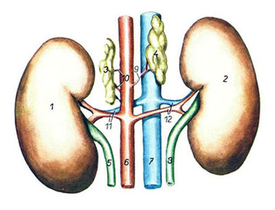
Kidneys and adrenal glands of cattle from the ventral surface
1 - right adrenal gland; 2 - left adrenal gland; 3 - right kidney; 4 - left kidney; 5 - caudal vena cava; 6 - abdominal aorta; 7 - right ureter; 8 - left ureter; 9 - right renal artery and vein; 10 - left renal artery and vein; 11 - caudal adrenal branch of the right renal artery; 12 - caudal adrenal branch of the left renal artery.
The kidneys of cattle are oval, belong to the type of striated multi-papillary. The fibrous capsule of the kidney goes deep into the furrows. The cranial end of the kidney is already caudal. The hilum of the kidney is wide. The left kidney is twisted along the longitudinal axis, hanging on the mesentery, which allows it to move behind the right kidney when the scar is filled. The mass of each kidney is 500–700 g, and the relative mass is 0.2–0.3%. The cortical urinary zone of the kidney is divided into lobes. The border zone is well defined. The cerebral zone in each lobe has the shape of a pyramid, with the base directed towards the cortical zone, and the apex, called papilla, - in a cup. In the kidney of cattle, there are 16–35 renal pyramids. The tops of the renal papillae are dotted with papillary openings through which urine flows into the renal calyces - the terminal branches of the ureter. From the cups, urine flows down the stalks into two ducts, which in the region of the gate are combined into one ureter. The right kidney is in contact with the liver, lies at the level from the 12th rib to the 2nd-3rd lumbar vertebra, the left kidney - from the 2nd to the 5th lumbar vertebra. Innervated by vagus and sympathetic nerves. Vascularized by the renal artery.
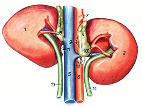
Kidneys and adrenal glands of a pig from the dorsal surface
1 - left kidney; 2 - right kidney; 3 - left adrenal gland; 4 - right adrenal gland; 5 - left ureter; 6 - abdominal aorta; 7 - caudal vena cava; 8 - right ureter; 9 - right middle adrenal artery; 10 - left middle adrenal arteries; 11 - left renal artery and vein; 12 - right renal artery and vein.
In a pig, the kidneys are smooth multi-pointed, bean-shaped, flattened dorsoventrally. Pyramids 10–12, the same number of papillae. Some papillae may merge. Calyxes approach the papillae, opening directly into the renal pelvis, located in the sinus of the kidney. Both kidneys lie in the lumbar region at the level of 1–4 lumbar vertebrae.
The horse's kidneys are smooth, single-papillary. The right kidney is heart-shaped, the left kidney is bean-shaped. The border zone is wide and well defined. The number of renal pyramids reaches 40–64. The papillae are fused into one directed to the renal pelvis. The right kidney lies almost entirely in the hypochondrium, at the level from the 16th (14–15th) rib to the 1st lumbar vertebra. The left kidney lies at the level of 1-3 lumbar vertebrae rarely enters the hypochondrium.
Horse kidneys from the ventral surface
1 - right kidney; 2 - left kidney; 3 - right adrenal gland; 4 - left adrenal gland; 5 - caudal vena cava; 6 - abdominal aorta; 7 - celiac artery; 8 - right renal artery and vein; 9 - cranial mesenteric artery; 10 - left renal artery and vein; 11, 12 - renal lymph nodes; 13 - right ureter; 14 - left ureter.
Histological structure. The kidney is a compact organ. The stroma forms a capsule and the thinnest layers inside the organ, which go mainly along the course of the vessels. The parenchyma is formed by the epithelium, the structures of which can only function in close contact with the circulatory system. The kidneys of all types are divided into lobes. The lobe is a renal pyramid with a portion of the cortex covering it. The lobes are separated from each other by renal columns - areas of cortical substance penetrating between the pyramids. The lobes consist of lobules that do not have clear boundaries. A lobule is a group of nephrons that flow into one collecting duct, which runs through the center of the lobule and is called the brain ray, as it descends into the medulla. In addition to the branching collecting duct, the cerebral ray contains straight tubules (loops) of the nephron.
Nephron- the main structural and functional unit of the kidney. There are up to 8 million nephrons in the kidneys of cattle. 80% of them are in the cortical substance - these are cortical nephrons. 20% are located in the medulla and are called juxtamedullary. The length of one nephron is from 2 to 5 cm. The nephron is formed by a single-layer epithelium and consists of capsules of the nephron, proximal, nephron loop (Henle) and distal. The nephron capsule looks like a double-walled bowl, its inner wall (inner leaf) is closely connected with the blood capillaries. The outer leaf of the capsule is built by a single-layer squamous epithelium. Between the leaves of the capsule there is a slit-like cavity of the capsule. The capillaries anastomose with each other, forming a vascular glomerulus of 50≈100 loops. Blood flows to the vascular glomerulus through the afferent arteriole. The glomerular capillaries unite to form the efferent arteriole. The arrangement of capillaries between two arterioles is called miraculous arterial system kidneys.
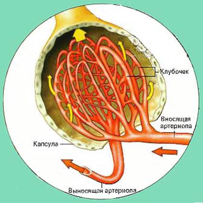
Renal Filtration System
The nephron capsule together with the vascular glomerulus is called renal corpuscle. All renal corpuscles are located in the renal cortex. In the renal corpuscle, primary urine is formed - glomerular filtrate, by filtering the components of blood plasma. This becomes possible due to the structural features of the renal corpuscle. The afferent arteriole has a larger lumen than the efferent arteriole. This creates increased pressure in the capillaries of the vascular glomerulus. In the endothelium of the capillaries there are gaps and numerous fenestra - the likeness of very small pores, which contributes to the leakage of plasma. The epithelium of the inner leaf of the capsule is closely adjacent to the endothelium of the capillaries, repeating all their curves, being separated only by the basement membrane. It is formed by peculiar flat process cells with a diameter of 20–30 microns - podocytes. Each podocyte has several large processes - cytotrabeculae, from which numerous small processes extend - cytopodia, attached to the basement membrane. There are gaps between the cytopodia. As a result, a biological kidney filter is formed, which has a selective ability. Normally, blood cells and large protein molecules do not pass through it. The remaining parts of the plasma can be part of the primary urine, which therefore differs little from blood plasma. The amount of primary urine - glomerular filtrate in large animals is several hundred liters per day. The glomerular filtrate enters the lumen of the renal corpuscle capsule, and from there into the nephron tubule. It undergoes reverse selective absorption into the bloodstream - reabsorption components of the glomerular filtrate, so that the secondary urine removed from the body is only 1–2% by volume of the primary urine and does not correspond at all to it in chemical composition. Secondary urine contains 90 times less water, sodium, 50 times less chlorides, 70 times more urea, 30 times more phosphates, 25 times more uric acid. Sugar and protein are normally absent. Reabsorption begins and proceeds most actively in the proximal nephron.
Part proximal The nephron includes the proximal convoluted tubule and the straight tubule, which at the same time is part of the nephron loop. The lumen of the capsule of the renal corpuscle passes into the lumen of the proximal convoluted tubule. Its walls are formed by a single layer of cuboidal epithelium, which is a continuation of the epithelium of the outer layer of the nephron capsule. The proximal convoluted tubules are about 60 µm in diameter, lie in the cortex, curving in close proximity to the renal corpuscle. The cells of the proximal convoluted tubule at the apical pole, facing the lumen of the tubule, bear a large number of microvilli that form a brush border - an adaptation for active absorption of substances. The round nucleus is shifted to the basal pole. The plasmalemma of the basal pole forms deep invaginations in the form of folds inside the cell. Elongated mitochondria lie in rows between these folds. At the light level, these structures look like basal striations. Cells actively absorb glucose, amino acids, water and salts and have a cloudy, oxyphilic cytoplasm. Throughout the proximal section, the entire amount of sugar, amino acids and small protein molecules that have entered the glomerular filtrate, 85% of water and sodium, is reabsorbed.
The proximal convoluted tubule passes into nephron loop (Henle). This is a straight tubule that enters the medulla at different depths. The nephron loop is divided into descending and ascending parts. The descending part is first formed by cuboidal epithelium, the same in structure and function as in the proximal convoluted tubule, and therefore this area is also referred to the proximal nephron as its direct tubule. The lower section of the descending part of the nephron loop has a diameter of 15 microns, is formed by a squamous epithelium, the nuclei of which protrude into the lumen of the tubule and is called a thin tubule. Its cells have light cytoplasm, few organelles, single microvilli, and basal striation. The thin tubule of the nephron loop continues into its ascending part. It absorbs salts and removes them into the tissue fluid. In the upper section, the epithelium becomes cubic and passes into the distal convoluted tubule with a diameter of up to 50 microns. The thickness of its walls is less, and the lumen is larger than in the proximal convoluted tubule.
Walls distal convoluted tubule formed by a cubic epithelium with a light cytoplasm without a brush border, but with basal striation. It reabsorbs water and salts. The distal convoluted tubule is located in the cortical substance and in one of its areas is in contact with the renal corpuscle between the afferent and efferent arterioles. In this place called dense spot, the cells of the distal convoluted tubule are tall and narrow. It is believed that they detect changes in the sodium content in the urine. During normal kidney function, 30–50% of nephrons are actively functioning. With the introduction of diuretics - 95-100%.
Juxtamedullary nephrons differ in structure and function from cortical nephrons. Their renal bodies are larger, lie in deep areas of the cortical substance. The afferent and efferent arterioles have the same diameter. The nephron loop, especially its thin tubule, is much longer, reaching deep layers of the medulla. In the area of the dense spot there is a juxtaglomerular (periglomerular) apparatus - an accumulation of several types of cells, in total forming endocrine complex of the kidneys regulating renal blood flow and urination. It is involved in the synthesis of renin, a hormone that stimulates the production of vasoconstrictor substances (angiotensins) in the body, and also stimulates the production of the hormone aldosterone in the adrenal glands. From the distal nephron, urine enters the collecting duct.
collecting ducts are not part of the nephrons. These are the terminal branches of the ureter that penetrate the kidney parenchyma and fuse with the ends of the nephrons. The areas of the collecting ducts lying in the cortical substance are formed by a cubic epithelium with a very light cytoplasm, in the medulla - by a cylindrical epithelium. Some absorption of water continues in the collecting ducts due to the hypertonicity of the surrounding tissue fluid. As a result, urine becomes even more concentrated. The collecting ducts form an extensive system. They pass in the center of the brain rays of the cortex and in the medulla and are combined into papillary ducts, opening with holes at the top of the papillae.
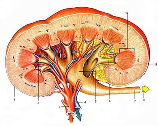
Diagram of the structure of the kidney
1 - kidney capsule; 2 - arcuate artery; 3 - renal artery; 4 - renal vein; 5 - renal pelvis; 6 - renal calyx; 7 - ureter; 8 - urine; 9 - cortical substance; 10 - brain zone.
Blood supply to the kidney It is carried out by a large paired renal artery, which enters the kidney in the region of the gate and branches into interlobar arteries. In the border zone of the kidney, they pass into the arcuate arteries. A large number of interlobular arteries depart from them into the cortical substance. These arteries branch into intralobular arteries, from which the afferent arterioles depart, branching into the capillaries of the vascular glomerulus. The capillaries gather into the efferent arteriole. Here we see wonderful arterial system of the kidney capillaries between two arteries. In these capillaries, blood is filtered with the formation of primary urine. The efferent arteriole branches again into the capillaries surrounding the tubules of the nephron. Reabsorbed substances enter these capillaries from the tubules of the nephron. The capillaries unite to form veins that carry blood out of the kidney.