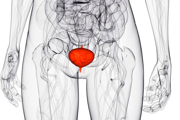
Distinctive features of a bladder tumor in women
Pathological neoplasms of the bladder are diagnosed in 20% of patients with problems with this organ. Of this number, 25% refers to malignant formations.
In women, this pathology is diagnosed 3 times less than in men. Such statistics are explained by the fact that more men smoke and work in hazardous industries.
Two types of tumors can develop in the bladder: benign and malignant. Benign tumors are found in most cases. They are characterized by slow growth and a rare degeneration into a malignant tumor.

epithelial
This type of tumor includes benign formations, localized only within the tissues of the bladder. These include:
- polyps. They are papillary-type formations with a fibrovascular wide base. The polyp has an elongated stalk covered with urothelium. The formation is surrounded by modified villi, in which the width is greater than the length.
Papillomas. In their structure, papillomas are similar to polyps. They also have a wide base and stem. Only unlike polyps, they tend to branch out.
The leg of the papilloma consists of fibrous tissue in the middle, which contains the blood vessels. The formation is covered by several epithelial layers. They are distinguished by a high degree of insemination and recurrence.
Non-epithelial
Non-epithelial species include formations that affect all tissues of the bladder. There are several types of non-epithelial tumors:
- Fibroids. Localized in the connective tissue of the organ cavity, and is an oval or round tumor on a stalk, with clearly defined boundaries. As a rule, fibroma does not grow more than 3 cm in diameter. This type of tumor is prone to slow growth, with damage to the wall of the bladder.
- Leiomyomas. This is a hormone-dependent formation that occurs in the connective and muscle tissues of the bladder cavity. In appearance, it resembles a rounded knot, ranging in size from a few mm to 3 cm in diameter.
- Rhabdomyomas. It is formed only in the deep layers of the striated muscle. It is a dense formation with a homogeneous structure. This species is characterized by growth throughout the affected muscle, not covering adjacent tissues.
- Hemangiomas. Localized in the vascular tissue of the organ, and consists of independently developing cells of the endothelial type. Usually diagnosed in children. The tumor has a limited growth period, which is about 12 months, after which the hemangioma stops growing or regresses on its own.
- Neurinomas. They are formed on the sheath of the nerve fibers of the organ, as a result of the growth of its cells. It is characterized by rapid growth and severe pain symptoms. The tumor has the shape of a flat oval or cobweb.
- Fibromyxomas. They belong to embryonic fibromas with multicentric growth, localized in the area of connective tissue. Differs in multiple formation of knots. As a rule, there is one large central node and several small nodules located nearby.
Types
Tumors of the bladder are divided into types according to the area of the lesion and the degree of involvement of adjacent tissues in the pathological process. According to these characteristics, 2 types are distinguished: invasive and superficial.
invasive
An invasive tumor is affecting all layers of the bladder and adjacent tissues regardless of primary location. These tumors are characterized by rapid growth and severe symptoms. As it develops, it affects nearby organs, leading to their dysfunction.
Surface
With superficial tumors, only the epithelial layer of the organ cavity is involved in the pathological process. The formation is located both in the epithelium and on its surface in the form of a polyp or papilloma.
Penetration to other structures of the bladder does not occur. These pathologies have a smoothed symptomatology, which manifests itself as the formation grows. They are of particular danger with extensive growth, as they can provoke the overlap of the cavity lumen.
stages
Tumors of a malignant nature that affect the bladder go through several stages of their development:
- 1 stage. It is the beginning of a disease in which a small tumor with limited edges is formed in the tissues of the organ, localized in the epithelium. At this stage, the formation can increase in diameter without growing into muscle tissue.
- 2 stage. It is characterized by damage to the muscle tissues of the organ.
- 3 stage. It is characterized by extensive growth of the tumor, which covers most of the organ. At the same time, its germination outside the bladder is observed, which leads to adhesion with adjacent tissues and organs. This stage marks the beginning of the metastasis process. Secondary tumors are found in regional lymph nodes.
- 4 stage. In the last stage of development, cancer affects the entire organ, including the ureters, which leads to their narrowing and overlap. The presence of distant and adjacent metastases is noted.
Symptoms
This pathology is characterized by certain symptoms:
- Blood in the urine. This sign is one of the first signs of the onset of the development of the disease. As a rule, blood appears slightly in the form of scarlet droplets or streaks. The appearance of blood may be rare or isolated. But as the formation grows, the frequency of manifestations increases.
- Urinary incontinence. This symptom is typical only for women. Basically, it manifests itself during physical exertion.
- Frequent urge to urinate. Occur as a result of irritation and overstretching of the epithelium.
- Pain in the lower abdomen, extending to the frontal region. At first, the pain is strictly localized and appears quite rarely. Then, it becomes more intense and extends to the lumbar region.
- Difficulty urinating. In most cases, it is a late symptom of the disease. Caused by narrowing of the lumen of the ureter.
The reasons
As the reasons provoking the development of pathological formations in the bladder, indicate the following:
- professional activity, associated with hazardous production, where aromatic amines, derivatives of heavy metals are used;
- smoking;
- chronic diseases of the bladder, in the absence of treatment;
- presence in the body human papillomavirus;
- radiation or chemotherapy.
Diagnostics
A number of standard methods are used to diagnose tumors:
- ultrasound. Allows you to consider the structure of the organ, the shape of the tumor and determine the degree of its growth;
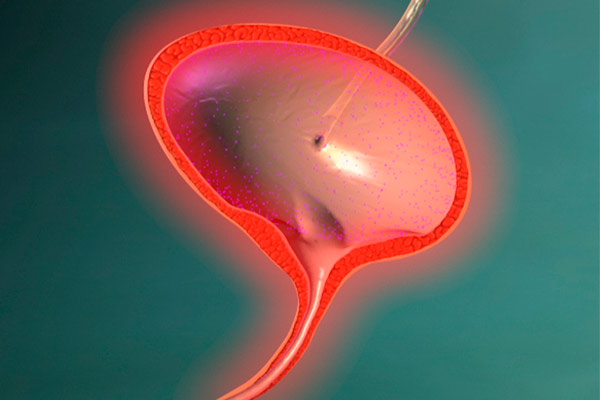
- cystoscopy. It is a study of the cavity of the body, by introducing into it through the urethra, a cystoscope;
- endoscopic biopsy with a morphological study of the biopsy. It is carried out simultaneously with cystoscopy, allows you to determine the presence of malignant cells;
- cystography. It is an x-ray study in which an image of the bladder is obtained by filling it with a substance of a radiopaque type. It makes it possible to identify additional formations, even if they are small in size;
- CT. Allows layer-by-layer examination of the affected and healthy tissue at the cellular level.
Treatment
Treatment is prescribed depending on the quality of the pathological formation. As a rule, with small benign tumors, expectant tactics are followed, since when irritating factors are eliminated, they can regress on their own.
They do not apply any treatment, watching the growth. Therapy is started only when severe negative symptoms are detected, or the growth of formations. The main treatment is removal of the tumor. For this, several methods are used:
- Transvesical electroexcision. It is applied at extensive or numerous growth. It is the removal of the formation through the opening of the bladder by dissecting the tissues.
- Partial cystectomy. It is the most traumatic method of treatment, which involves the partial removal of the affected organ. It is used when most of the bladder is affected. As a rule, adjacent tissues involved in the malignant process are also removed from women.
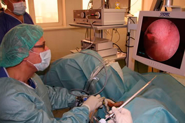
- Transurethral resection. With this type of operation, a special device is used, which is inserted through the ureter. Using a loop, the tumor is removed from the organ, and then a catheter is inserted into the canal to remove urine.
Cystoscopy with transurethral electroresection, electrocoagulation. With this method, removal is carried out using a resectoscope, which is inserted through the urethra and ureter.
This device cuts the tumor and simultaneously cauterizes the operated tissue, which ensures the absence of blood loss and the rapid recovery of blood vessels and epithelium.
Rehabilitation
The rehabilitation period will depend on the method used for treatment. With sparing techniques, the recovery time takes 5 to 7 days. After traumatic treatment, this period increases to 2 weeks or more.
In the early days, the woman will be uncomfortable with the catheter, which removed 2-5 days after surgery. Until the tissues are completely healed, it is necessary to adhere to a certain diet, in which all products that irritate the mucous membrane are excluded.
An appointment is scheduled for the entire period of rehabilitation antibiotics and interferon. Within a month after the operation, physical activity is prohibited, which will provoke tissue damage, which will be reflected in the appearance of blood in the urine.
Forecasts
In the presence of benign formations in the bladder, a constant examination by a doctor is required, since there is a risk of their degeneration. According to statistics, this happens in 30% of cases. Most often, the cause is a weakening of the immune system and the constant action of an irritating factor.
Despite the fact that removal is the most preferred technique, it still does not give a 100% guarantee of a positive result and relapse occurs in 25% of cases. Therefore, it is recommended to combine it with chemotherapy.
After minimally invasive treatment of all patients more than 95% survive. The radical method has less positive results. In this case, only half of the treated patients survive.
In this video, a specialist talks about the disease and the prognosis of a cure: