Kidney pyelectasis in adults and children: treatment, signs, causes
Pathology, which is characterized by anatomical enlargement of the renal pelvis, is called pyelectasis of the kidney. The pelvis is a place in which urine accumulates from the kidneys, subsequently sent to the ureters. Pyeloctasia of the kidneys is a non-independent disease, pathology speaks of disturbances in the activity of organs that are involved in the outflow of urine.
Classification of pathology by severity in adults
Why does an increase in the renal pelvis develop in adults? In the renal calyx, the fluid that enters the body accumulates and is processed, then it enters the pelvis, where it turns into urine. Due to certain processes, urine cannot move in full to the ureter, which is why the renal pelvis becomes distended (normally slit-like). The condition rarely goes away on its own. The expansion of the pelvis is usually divided into the following degrees:
- mild (does not require therapy, a systematic visit to a specialist is sufficient);
- medium (involves systematic monitoring of the organ using ultrasound and drug treatment);
- severe (requires the use of surgery to prevent the cessation of kidney function).
Forms of development of pathology
Pathology is divided, based on what factors provoked the expansion of the renal pelvis, into the following types:

Bilateral and unilateral pathology
The expanded pelvis of the kidney is divided depending on the degree of damage to the parties:
- Bilateral pyelectasis. The expansion occurred immediately in two pelvis. Bilateral pyelectasis is most often observed in children.
- Unilateral (right-sided pyelectasis, left-sided and pyelectasis of a single kidney). The expansion is observed in one pelvis.
Causes of pyelectasis of the kidney
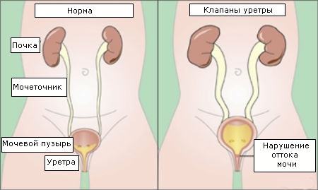 Urethral valves are a congenital pathology of the mucous membrane of the urethra.
 There are the following causes of pyeloectasia:
- Innate dynamic:
- narrowing of the lumen of the urethra;
- phimosis (impossibility of exposing the head of the penis);
- valves in the urethra;
- neurological pathologies that provoked violations of the urinary process.
- Acquired dynamic:
- hormonal disorders;
- diseases that provoke an increase in the volume of urine;
- inflammatory processes in the kidneys;
- infections accompanied by poisoning of the body;
- neoplasms in the urethra and prostate;
- narrowing of the urethra due to trauma or inflammatory diseases;
- neoplasms in the prostate of a benign nature.
- Congenital Organic:
- pathologies in the structure of the kidneys, which provoked pressure on the ureter;
- pathology of the upper urinary tract;
- pathology of the structure of the ureter.
- Purchased Organic:
- inflammatory processes of the ureter and neighboring organs;
- neoplasms in the urinary system;
- neoplasms of any nature in nearby organs;
- displacement of the kidneys;
- urolithiasis disease.
Symptoms of pyelectasis
 The disease has no characteristic symptoms, therefore, at the slightest malfunction of the urinary system, it is necessary to undergo a diagnosis.
 Expansion of the renal pelvis occurs without its own symptoms. More often, pathology for a long period does not make itself felt and does not cause any inconvenience. Pyelectasis in adults in most cases is diagnosed during examinations conducted to determine other diseases. With pyeloectasia, the following symptoms are observed:
- Narrowing of the mouth of the ureter, due to which a spherical and cystic protrusion of the intravesical ureter is formed.
- The confluence of the ureter into the urethra (in men) and into the vagina (in women).
- The return flow of urine from the urinary cavity back to the kidney through the ureter.
- Expansion of the ureter, which is accompanied by failures in urination.
Pyelectasis in children
 In most cases, boys are more prone to the appearance of pathology.
 Experts are convinced that moderate pyelectasis of the right kidney is more common in children than pyelectasis of both kidneys and pyelectasis on the left. Often, pathology is diagnosed in male children. If we talk about newborns, then pyeloectasia in them is more often a congenital pathology and is caused by anomalies in the structure of the ureter and other organs of the urinary system. It often happens that the pathology went away on its own up to 2 years, however, if after growing up the pyelectasis does not go away, the child should be systematically taken to an ultrasound scan, which shows an echo picture of the extensions.
Factors affecting the development of pyeloectasia in children:
- pathologies in the development of the fetus, which provoke the appearance of a valve in the urethra;
- weakened muscle tone (in cases of prematurity);
- squeezing of the ureter;
- violation of the active bladder due to neurogenic factors (for example, overcrowding of the urinary cavity).
Pyelectasis during pregnancy
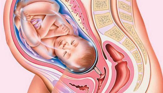 In pregnant women, pressure on the ureter causes the uterus to enlarge.
 A condition when the pelvis of the kidney is enlarged in pregnant women provokes pressure on the ureter, an enlarged uterus (the renal calyces may also be affected). However, this is not the only reason; pyeloectasia can also develop due to hormonal disorders. Pyelectasis of the left kidney is diagnosed during pregnancy several times less than the right one. They call the pathology "passing" because it can disappear on its own without the use of medical manipulations. This happens after a woman gives birth.
It should be noted that when diagnosing pyelectasis during pregnancy, it is important to establish exactly whether the anomaly has developed due to the position or whether it began a little earlier than pregnancy. In case of pathology, they do not resort to termination of pregnancy, however, if pyelectasis is chronic, this can seriously affect further childbirth. Due to this factor, the admissibility of pregnancy in chronic pathology can be determined only after a proper examination has been carried out and the condition of the kidney has been studied.
Is pathology dangerous?
Pyelectasis of the kidneys in adults is dangerous because of the factors that provoke it. The disturbed exit of urine from the kidneys with untimely treatment provokes squeezing, and then atrophy of the tissues of the organ. Because of this, the kidney begins to function worse over time, which often leads to its complete failure. Pathology can provoke the development of chronic and acute pyelonephritis (inflammation of the kidneys and calyces), which adversely affects the organ. That is why, if you suspect pyelectasis, you should not delay contacting a doctor and go through all the necessary studies in order to find out exactly what caused the expansion of the pelvis, you need to undergo an examination and start treating the problem as soon as possible.
Diagnostics
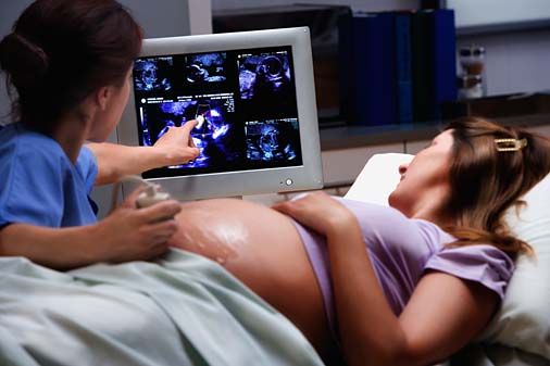 The volume of the renal pelvis can be studied using ultrasound diagnostics.
 Conditions when the pelvis is enlarged in an adult are determined by ultrasound, during which specialists study the volume of the renal pelvis during and after the urination process. Additionally, an echo picture and a significant size of the pelvis (the norm is 6 mm or more) and their changes over the next year, if any, are examined. When the size has increased, this means that the pyelectasis is progressing. Then the patient will have to pass a general urine test. If there is little data obtained, they resort to the help of additional examination methods, including urography (an X-ray method for examining the urinary tract, which is based on the ability of the kidney to secrete certain radiopaque substances previously introduced into the body) and cystography (an X-ray examination method, the purpose of which is to obtain an image of the urinary cavity by filling it with a contrast agent).