What is a bladder tumor
The role played by the bladder in the human body is to accumulate liquid waste products (urine) and then remove them from the body with the help of the contractile function of the muscles, which are its main structural part.
Bladder tumor is the most common oncological disease of the urinary tract and accounts for more than 60% of all cases of neoplasms of the urinary system and more than 2% of oncological pathologies of the whole body.
What is a bladder tumor
A neoplasm in the tissues of the bladder can be represented by many types and have different activity in terms of the formation of metastases. The victims of the disease are mostly people over 55 years of age, often living in environmentally unfavorable areas or working in hazardous industries. Partly, negative changes in the environmental situation, social factors and smoking are to blame for the increase in the frequency of diagnosing a tumor.
A tumor of the bladder in men occurs 6 times more often than in women, which is associated with some anatomical and physiological features of the male body:
- genetic predisposition, increasing after 50 years;
- hormonal disorders that stimulate tumor development;
- development of prostate adenoma.
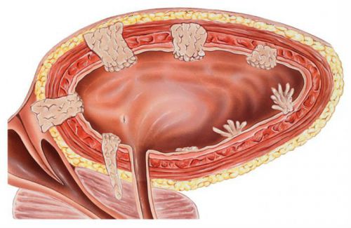
The two most common types of tumors are:
- benign tumor of the bladder (highly differentiated), located on the surface of the mucosa;
- a malignant tumor (poorly differentiated) that affects muscle tissue.
In the first case, the growth of the tumor occurs in the direction of the cavity of the bladder, forming a connection in the form of a leg with the mucous surface. As a rule, a tumor that has this growth pattern is called exophytic or papillary.
The second type is characterized by intensive ingrowth into the tissue structure with the rapid formation of metastases. This type of tumor is called endophytic or invasive.

Smoking has been found to be one of the leading risk factors for bladder cancer.
The reasons
Carcinogenic substances, entering the body, go through all stages of metabolism and are partially excreted by the kidneys. It is they who are the root cause of the development of neoplasms, since they are in direct contact with the mucous membrane of the bladder. Exposure to carcinogens can disrupt the work of the genetic apparatus of cells, thereby interfering with the reproduction of cells characteristic of this organ and provoking the growth of cancer cells.

Carcinogenic substances, in various forms, affect a person throughout his life.
The main external causes that provoke the development of bladder cancer:
- daily contact with volatile chemicals generated during the production of plastic, rubber and other products or paint and varnish industries;
- smoking;
- exposure to ionizing radiation;
- viral diseases (mainly human papillomavirus disease, which has highly oncogenic properties);
- systematic untimely or incomplete emptying of the bladder in prostate adenoma;
- chronic inflammatory diseases of the bladder.
An increase in the size of the prostate gland with the prostate provokes the development of stagnant processes in the bladder and disrupts the elasticity of the muscle layer. The impossibility of complete emptying causes deformation of the walls of the bladder, they stretch, creating a "reservoir", where urine accumulates and the negative effect on the mucous membrane increases.

Chronic inflammatory diseases are also risk factors, since the process of tissue regeneration occurring during inflammatory processes, under the influence of carcinogens or due to genetic disorders, can acquire the character of metaplasia, that is, there will be a replacement of damaged cells with modified cells or cells that do not belong to this organ. Often this process is characterized as leukoplakia of the walls of the bladder and refers to precancerous conditions.
Important: The increase in the incidence of bladder tumors in age groups over 60 is due to an increase in the duration of exposure to negative factors, as well as hormonal changes in the body and a decrease in the activity of the immune system.
WHO classification
Almost all pathological neoplasms of the bladder are the result of the development of transitional cell carcinoma. However, there is a possibility of development of other forms:
- squamous cell carcinoma;
- glandular cancer (adenocarcinoma);
- undifferentiated tumor;
- lymphoma.
The classification approved by the World Health Organization (WHO) is based on the results of histological studies of bladder cancer and includes:
Neoplasms of the epithelium:
- some types of papillomas (transitional cell and squamous);
- transitional cell carcinoma;
- combination of transitional cell carcinoma with tissue metaplasia;
- glandular cancer;
- undifferentiated cancer;
- squamous cell carcinoma.
Non-epithelial neoplasms:
- highly differentiated neoplasms (benign tumor of the bladder);
- poorly differentiated neoplasms (rhabdomyosarcoma).
Neoplasms with metastatic spread:
- glandular metaplasia;
- polypoid cystitis;
- squamous metaplasia.
Bladder papilloma refers to benign neoplasms, but under certain conditions it can turn into a malignant form.
Extensive tumor spreads:
- multiple cystic formations;
- follicular cystitis;
- small coplakia (extensive neoplasms in the form of multiple plaques).
Important: Neoplasms of a non-epithelial nature, especially malignant ones, are quite rare and mainly in young people. They are distinguished by rapid growth and extensive spread of metastases.
Characteristics of neoplasms
Transitional cell papilloma has a similar structure to papillomas that form on the surface of the skin, it is abundantly covered with villi and freely located in the cavity of the bladder. Papilloma is abundantly supplied with blood vessels and is attached to the mucous surface with a thick base. The mucosa around the papilloma may be swollen and deformed.
Transitional cell carcinoma has some similarities with papilloma, but unlike it, it develops from the lower layers of the epithelium, has a larger size and is accompanied by strong necrotic changes in nearby tissues.
Important: The final diagnosis of transitional cell carcinoma is possible only with a histological examination, due to the strong similarity of its manifestations with bladder papilloma.
Glandular cancer has predominantly malignant characteristics and is the result of metaplasia of glandular tissue. The following types of glandular cancer are usually distinguished:
- originally formed on the wall of the bladder;
- developed from the urinary duct;
- which is a consequence of metastases of malignant neoplasms of other organs (prostate, uterus, rectum).
Undifferentiated cancer is significantly different from previous forms of tumors, as it has a structure of tuberous compaction, alternating with areas of tissue affected by necrosis and ulceration. Histological examination of the structure of the tumor is characterized by heterogeneity and disordered arrangement of cells, the presence of a large number of metaplastic processes.

Bladder cancer x-ray
Symptoms
The most common symptom of a bladder tumor is intravesical bleeding, which is accompanied by the appearance of blood in the urine. A similar phenomenon is called hematuria, and it has nothing to do with the size, type and stage of development of the tumor. The main role is played by the location of the neoplasm and the possibility of its infringement or deformation during urination.
Due to the fact that the tumor, as a rule, has a developed vascular system and has a good blood supply, being located in the region of the bladder neck, it is subjected to a systematic traumatic effect due to the contraction of the muscle layer, and, as a rule, is accompanied by bleeding.
Along with hematuria, other symptoms of a bladder tumor may be observed:
- obstruction of the outflow of urine due to the overlap of the mouth of the bladder with clotted blood or the location of the tumor in the urethra;
- pain when urinating;
- persistent pain in the lower abdomen, radiating to the lower back or rectum, acquiring an acute form when urinating;
- the development of inflammatory diseases of the kidneys, due to deformation of the mouths of the ureters and impaired outflow of urine from the kidneys;
- the appearance of sediment and an uncharacteristic odor in the urine due to the presence of tissue particles affected by necrosis.
The location of the tumor of the bladder in the region of the bladder triangle
Dysuretic signs associated with loss of elasticity and a decrease in the volume of the bladder determine the size, location, stage of development and degree of differentiation of the tumor.
Important: Neoplasms that are localized on the lateral or anterior wall of the bladder may not cause symptoms for a long time, thereby making timely diagnosis difficult.
Diagnostics
Tumor diagnosis is the second stage of the standard scheme, consisting of symptom detection, diagnosis and treatment. The purpose of diagnostics is:
- confirmation of the presence of a neoplasm;
- determination of tumor localization;
- determination of the histological characteristics of the tumor;
- determination of the degree of spread and the presence of metastases;
- assessment of the condition of the kidneys;
- assessment of the degree of risk during surgery.
At the first stages of diagnosis, a thorough analysis of the existing symptoms is carried out:
- find out their duration;
- intensity;
- the presence of concomitant symptoms (loss of appetite, weight loss, weakness).
If there is at least one sign that causes suspicion of the presence of a neoplasm, it is necessary to conduct a set of special studies, which include:
- bimanual palpation;
- laboratory analysis of urine;
- magnetic resonance imaging (MRI);
- ultrasound examination (ultrasound);
- cystoscopy.
Bimanual palpation is performed through the vagina when diagnosing a bladder tumor in women and through the rectum in men. This method is rather auxiliary, since it can be used to estimate the size of a large tumor located on the body of the bladder. Tumors of small sizes, located in the region of the vesical triangle, cannot be determined by palpation.
The results obtained from a cytological examination of urine are also not decisive in making a diagnosis, since false positive results may occur in the presence of concomitant diseases, for example, in chronic cystitis.
MRI is a highly informative diagnostic method. It can be used to determine:
- the presence of a neoplasm;
- degree of distribution;
- depth of penetration into tissues;
- condition of nearby and distant organs and tissues.
A three-dimensional image of any organ, obtained during MRI diagnostics, allows you to identify any changes in its structure.
Ultrasound is also a fairly informative method and more affordable, unlike MRI. The percentage of neoplasms detected in the bladder using ultrasound is more than 80%. To eliminate errors in diagnosis, the procedure is carried out at its maximum filling.

When examining the bladder using ultrasound, it is advisable to use a transrectal probe.
Excretory urography is an X-ray method for diagnosing pathologies of the bladder and urinary system. A radiopaque substance is injected into the blood, and after a certain amount of time, several pictures are taken at a certain time interval. The contrast agent, deposited in the kidneys and urinary tract, allows you to get highly informative pictures about the state of the entire urinary system.
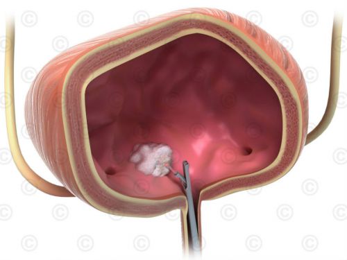
Cystoscopy is a priority diagnostic method that allows diagnosing a tumor with 98% accuracy. The examination is performed using a flexible cystoscope equipped with fiber optics, inserted through the urethra into the bladder. If the preliminary diagnosis is confirmed, then in some cases, simultaneously with cystoscopy, tissue is taken for histological examination or a transurethral resection (TUR) of the tumor is performed.
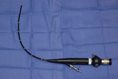
With the help of a fiber optic cystoscope, it is possible to carry out diagnostic and surgical operations.
Treatment
The methods used in the treatment of bladder tumors depend on the nature of the neoplasm (malignant, benign forms) and location (invasive or papillary type). In almost all cases, surgical intervention takes place, which can be divided into the following types:
- TUR of bladder tumor.
- Open surgery to remove a fragment of the affected tissue.
- Electrocoagulation of a non-invasive tumor.
- radical cystectomy.
- Chemo-and radiation therapy.
Transurethral resection is performed in case of diagnosing a benign tumor of the bladder. The neoplasm is removed along with adjacent tissues to visually healthy boundaries. At the same time, tissues are taken for histological examination.
Important: During TUR of the bladder, the probability of recurrence of the disease is more than 45%.

Electrocoagulation is performed in case of diagnosing a benign form of the tumor.
In the case of diagnosing a poorly differentiated invasive form of a tumor, removal of the bladder with external diversion of urine or the creation of a container for accumulating fluid from a fragment of the rectum is indicated. Due to the fact that it is quite difficult to treat the tumor, plastic surgery is usually postponed, giving preference to the creation of percutaneous urinary diversion routes.
If the tumor has grown into nearby tissues in the images, they must also be removed, followed by a course of chemotherapy. Thus, the treatment of a tumor in the bladder of men is often accompanied by the removal of the prostate gland, and in women of the urethra and uterus. A course of chemotherapy and radiation therapy can be carried out before, after and instead of surgery, using intravesical or intravenous administration of drugs.
Despite the large selection of diagnostic methods, the prognosis in the treatment of bladder tumors can hardly be called optimistic. Even with early detection of neoplasms and timely treatment, the risk of recurrence is very high. Therefore, for the purpose of prevention, it is necessary to undergo regular examinations, especially for people working in hazardous industries, suffering from chronic inflammatory diseases of the urinary tract and undergoing surgery to remove bladder neoplasms.