Bladder in women in men
Urine, constantly filtered by the kidneys from the blood plasma, flows down the ureters into the bladder. Here it accumulates up to a certain volume and then is excreted through the urethra from the body. The process of urination, or micturition, is a complex of complex and sequential actions that the organ performs together with the urethra up to 10 times a day, under the control of the spinal nerves and the cerebral cortex. Let us consider in more detail how this happens, where the bladder is located, whether there are differences in its structure and functions in men, women and children of different ages, what is the view of its activity in oriental medicine.
How does the bladder work
This unpaired spherical organ is designed to serve as an excellent container for urine flowing through the ureters. It can stretch and increase its volume if necessary, but up to certain values. Depending on what kind of person has height and weight, the size of the organ also differs. The average bladder capacity is 500-700 ml, but there are significant individual fluctuations.
Thus, the volume of the bladder in men is slightly larger than in women and children, and varies from 350 to 750 ml. The female organ holds 250-550 ml of urine; the norm of volume in children, given their constant growth, also gradually increases. So, at the age of one year it is 50 ml, at 3 years old - 100 ml, and at 11-14 years old it can reach up to 400 ml. In some conditions, when it is impossible to empty the bladder in time, its walls stretch significantly, and the capacity in adulthood reaches 1000 ml (1 liter) of urine.
The size of the organ has individual characteristics in terms of gender or age, but it can also be influenced by various pathological or physiological conditions. For example, some diseases or degenerative processes.
All these factors can be represented as follows:
- surgical correction that reduces the size of the organ;
- long-term chronic diseases leading to "wrinkling";
- neoplasms that reduce the volume of internal space;
- influence from other internal organs (for example, squeezing the bladder in women with a growing uterus during pregnancy);
- neurological diseases;
- degenerative-dystrophic processes in old age, leading to loss of normal tone of the detrusor or sphincters.
The brain is actively involved in urination
The inner surface of the body has special baroreceptors that respond to an increase in pressure in it. As soon as approximately 200 ml of urine accumulates, the pressure in the cavity increases, and signals about this go to the cerebral cortex, to those parts of it that are responsible for the act of urination. From this point on, a feeling of urge is formed, and the person knows that he will soon need to go to the toilet.
As urine accumulates, the urge to urinate increases, but the sphincter of the bladder is in a compressed state, preventing involuntary leakage of fluid. With the help of the sphincters of the organ and the urethra, a person can retain urine from 2 to 5 hours. The micturition process itself is regulated both by the cerebral cortex and by nerve branches extending from the spinal cord, and occurs as a result of contraction of the muscle layer and relaxation of the sphincters.
In children, the process of forming a normal urination process is quite long and takes 3-4 years (although if parents try, you can teach a child to ask for a potty at 1.5-2 years). From an unconditioned spinal reflex, it becomes an arbitrarily reflex. This involves the cerebral cortex, subcortical centers, spinal zones (sections of the spinal cord), and the peripheral nervous system.
There are many different congenital and acquired diseases in which the process of urination is disturbed. The reasons may lie in organic, or somatic, pathologies of the organ that affect the normal structure of tissues (infectious diseases, neoplasms, effects of neighboring organs) or in violation of nervous regulation.
Structure
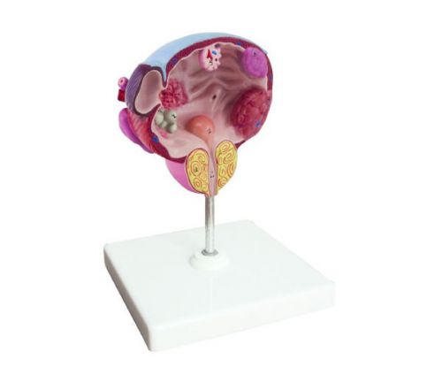
The anatomy of the bladder includes its localization in the human body, interaction with surrounding structures, macroscopic (conditional division into parts) and microscopic structure (from which tissues). This organ looks like a small rounded sac, and is located in the pelvic cavity. If it is in an empty state, it occupies a small volume and is completely hidden by the pubic articulation. It adjoins this bone formation with its anterior surface. As it fills up, its size also increases, the walls of the organ straighten out, and it gradually begins to rise above the pubic joint. In this state, it can be palpated (palpated) during a medical examination, an ultrasound scan can be performed, and a puncture can be performed through the anterior abdominal wall.
The walls of the organ can be affected by various diseases, and the urethra can be squeezed by the enlarged prostate gland
The back surface of the bladder in women is in contact with the organs of the reproductive system: the vagina, uterus and ovaries. Further behind is the final segment of the intestine, the rectum. The urinary bladder in men is separated from the intestines by seminal vesicles and a segment of the vas deferens. The upper part of the body borders on the loops of the small intestine. In newborn babies, it is higher than in adults, above the pubic joint. Only after a few months the tip is hidden behind the bone formation.
The human bladder can be divided into several components:
- walls - front, side, back;
- body;
- bladder neck.
The anterior wall of the organ borders on the anterior abdominal wall and the pubic articulation, separated from them by a layer of loose adipose tissue, which fills the prevesical space. The back and side walls are also separated from neighboring structures by fiber and the visceral sheet of the peritoneum (a special tissue layer covering all organs). The upper part of the organ is more mobile and able to stretch significantly, since it is not fixed by the ligamentous apparatus. With a large stretching, the wall thickness can be only 2-3 mm, with an empty organ it reaches 15 mm.
On the back wall, in its middle part, the bubble has two holes. These are the mouths of the ureters, symmetrically located, and they flow into the organ cavity at a certain angle. This fact is extremely important, since this forms a kind of "closing" mechanism that prevents the ingress of urine during detrusor contraction and urination back into the ureters. When this mechanism is violated, vesicoureteral reflux is formed, which can be called both an independent disease and a complication of other pathologies of the urinary system.
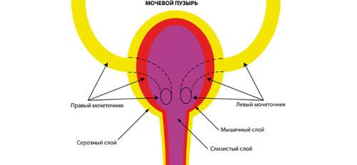
The oblique confluence of the ureters is very important for the formation of a special valvular mechanism.
The upper part of the hollow organ is conditionally divided into the top and bottom. The bottom part is behind and turned down, and the top is directed to the anterior abdominal wall and passes into the umbilical ligament. The bottom of the bladder, when filled with urine, rises above the pubic joint, so the tip begins to fit snugly against the anterior abdominal wall. Between the bottom and the top is the body of the organ.
The lower part gradually narrows and forms the neck of the bladder, which, through the sphincter apparatus, passes into the urethra. In a man, the upper part of the urethra and the neck of the bladder are covered by the tissue of the prostate gland, which, with the development of pathological processes in it, has a huge impact on the process of urination. The bladder in women in its lower part borders directly on the muscles of the pelvic diaphragm.
The wall of the organ is three-layered and consists of the following structures:
- mucous membrane and submucosal layer;
- detrusor, or muscle layer;
- outer shell covered by the visceral layer of the peritoneum.
Histological examination (examination of tissues under a microscope) reveals that the mucosa consists of an outer epithelial layer and a submucosal plate below it, formed by loose connective tissue. It is thanks to the submucosal layer that, with an unfilled cavity, the mucous membrane forms a large number of folds, which straighten out when the organ is stretched. But the submucosal layer is not present everywhere. It is absent in the region of the so-called bladder triangle, the tops of which are the openings of the ureters and the mouth of the urethra. In this zone, the mucous membrane is adjacent directly to the muscle layer.
The urothelium, or epithelial layer of the mucous membrane, has several rows of cells. Each of them performs a specific task. So, the outermost layer consists of rounded cells, which, when stretched, the walls of the organ become flat, thereby ensuring the integrity of the structure.
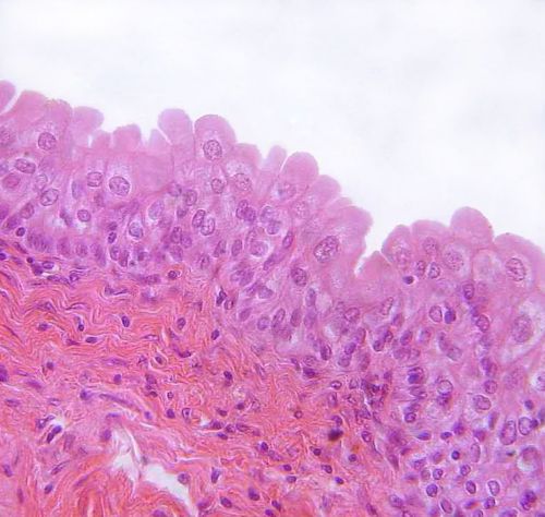
The transitional epithelium of the mucous membrane consists of several rows of cells of various shapes and purposes.
The muscle layer is made of three types of fibers, the functionality of which ensures the operation of the entire organ: longitudinal, transverse, circular. Circular muscle fibers are especially developed around the ureters flowing into the organ and the mouth of the urethra. In these places they form muscular sphincters, or sphincters. With cystoscopy, on the resulting photo of the bladder from the inside, the ureteral sphincters look like small depressions, and the more developed sphincter in the lower part of the organ looks like a crescent-shaped platform with a pink tint.
Functions
The most important task of the body is to accumulate a certain amount of urine, keep it for a certain time and regularly remove it from the body. These tasks are performed in the prescribed mode, if the mucous membrane is not affected by an inflammatory or tumor process, the size of the organ is within the normal range, and all the sphincters and detrusor, regulated by the nervous system, function like a “clock”.
As soon as there is a violation of even one of these mechanisms, the functionality of the organ is disturbed, which is expressed by various dysuric symptoms. So, with a neurogenic disorder, the normal regulation of the muscle layer and sphincters from the nervous system “breaks down”. This occurs with congenital or acquired neurological diseases, and hypo- or hyperreflexia is diagnosed, which is expressed either by incontinence or urinary retention (when the patient cannot urinate regularly). In another pathology, vesicoureteral reflux, which is formed in the absence or underdevelopment of the valvular and sphincter mechanisms of the ureters, there is a reverse flow of urine. This can lead to undesirable consequences in the form of pyelonephritis and other kidney diseases.

Oriental medicine specialists have a completely different view of health and disease
What is the urinary meridian and canal
From the point of view of oriental medicine, each internal human organ has special channels, or meridians, through which it receives energy. These meridians, including the bladder canal, intertwine and connect with each other, exit one from the other, forming a single whole. It is the interaction of the channels of the internal organs and the energy flow flowing through them that explains both the health of people and their various diseases.
The meridian of the bladder not only regulates the formation of urine in the kidneys, its accumulation and removal during urination, it is through it that all toxins and toxins are removed from the body. It is quite long and branched, due to which it can influence the activities of other organs. The bladder canal starts from the eyes, passes through the parietal part of the head, then between the shoulder blades runs along the spine and at the sacrum enters the inside of the body, reaching the kidneys and ending in a hollow organ. Its branches cover the head, body, descend to the feet.
This meridian is paired and symmetrical, belongs to the Yang type; energy moves along it in a centrifugal direction. If it is excessive, then the following symptoms are formed: pain in the abdomen and back, increased urination, spastic contraction of the calf muscles, pain in the eyes, lacrimation, and there may be nosebleeds. With a lack of urination energy, urination becomes rare, swelling, pain in the spine, weakness in the legs, and hemorrhoids appear.
The minimum energy activity of the channel is observed at night, between 3 and 5 o'clock, at this time it is not allowed to influence the meridian. The most convenient time for influencing the channel is the interval between 15 and 17 hours. It was then that the specialists of oriental medicine seek to treat the patient by influencing the organs through the meridian of the bladder.