Radial bone fracture treatment and rehabilitation. How to recognize a fracture of the radius bone of the arm? Diagnosis of a radius fracture
Fractures of the radius are caused by a traumatic factor and the individual characteristics of the patient’s body. However, rehabilitation measures for various injuries in this anatomical area are approximately the same.
What is a fracture, types of fractures

A fracture is a violation of the integrity of the bone caused by external mechanical action with deformation of the surrounding tissues and disruption of the function of the damaged section. Fractures happen:
- open if the integrity of the skin is damaged;
- closed;
- splintered, including fragmented when a large number of fragments are formed;
- no splinters.
In addition, fractures are classified in relation to the axis:
- transverse,
- longitudinal,
- oblique,
- fragmented,
- from bending,
- driven in,
- helical.
According to the nature of the mixing of fragments:
- in width,
- by lenght,
- at an angle
- on the periphery.
Fractures caused by a tumor, osteomyelitis and other diseases are called pathological.
According to localization they distinguish:
- metaphyseal,
- diaphyseal,
- epiphyseal,
- intra-articular.
Fractures are classified based on the number of bones affected:
- isolated - one bone is injured;
- multiple - several bones are affected;
- combined - bones and internal organs are damaged.
Fractures of the radius sharply reduce the ability of patients to work and are manifested by severe pain in the forearm and swelling. Depending on the type of fracture, the symptoms may be supplemented by the presence of a hematoma, tissue rupture with bone coming out into the wound, the presence of deformation in the area of the fracture with intact skin, etc.
The diagnosis is made on the basis of a survey, examination, palpation, the presence of pathological syndromes (crepitus, pathological mobility), as well as a set of instrumental diagnostic results.
Principles of treatment of radial fractures

The goal of treatment is to restore the anatomical integrity of the bone and the function of the damaged section.
There are two types of treatment for fractures: operative and conservative. They try to resort to surgical interventions in extreme cases and in the presence of certain indications for this method of treatment.
Fractures of the radius are classified depending on the traumatic factor and the individual characteristics of the patient’s body.
Let's look at some of them below.
A fracture without displacement of fragments is most favorable for the patient, does not require surgical intervention and allows the patient to recover quickly. Occurs at different heights of the radius. With an isolated fracture (with the integrity of the ulna), its diagnosis can be difficult. Treatment consists of fixing the fracture site with a two-split plaster cast and then replacing it with a circular plaster cast.
A fracture with displacement of fragments in certain cases requires osteosynthesis (external, transosseous or intraosseous) with plates, screws, screws or wire sutures.
In the presence of extra-articular non-comminuted fractures, manual reposition of the fragments is performed under local anesthesia and a two-split plaster cast is applied. After the swelling subsides, it is changed to a circular plaster cast until the end of the immobilization period.
In some situations, fractures of the radius are combined with dislocation of the head of the ulna. In this case, in addition to repositioning the fragments, it is necessary to realign the head of the ulna.
Immobilization: fixation with a plaster cast from the base of the fingers to the upper third of the shoulder in a physiological position.
Fractures of the radius in the neck and head are of the following types:
- without displacement of bone fragments;
- with displacement of bone fragments;
- comminuted fracture with displacement;
- intra-articular fracture.
First of all, it is necessary to diagnose a fracture and find out whether there is displacement of bone fragments. After this, treatment tactics are developed. If there is no displacement of the fragments, conservative treatment is prescribed, consisting of anesthesia and application of a plaster cast. If there is displacement of fragments or fragmentation of the bone head, surgical treatment is necessary, consisting of osteosynthesis.
If the head of the radial bone is crushed or comminuted, it may be removed. However, such measures are not practiced in children, so as not to affect the bone growth area.
One of the most common injuries to the forearm is a fracture of the radius in a typical location. Then the fracture area is localized in the lower part of the beam. This injury occurs as a result of a fall on an outstretched arm with the wrist joint bent or extended.
Immobilization: from the metacarpophalangeal joint to the upper third of the forearm. Duration: from 1 month (fracture without displacement of bone fragments) to 1.5-2 months (with displacement of bone fragments).
Therapeutic gymnastics: breathing exercises, gymnastics complexes for joints free from plaster casts with the obligatory involvement of the fingers.
Post-immobilization period: exercises are performed in front of a table with a smooth surface to facilitate sliding of the hand. Exercises in warm water, as well as everyday activities, in particular self-care, are useful. It is necessary to avoid carrying heavy objects and hanging. Massage of the affected limb is very useful.
Very often, a fracture of the radius in a typical location is combined with avulsion of the styloid process. The diagnosis is made based on a survey, examination, palpation (fragment crepitation syndrome), as well as the results of an X-ray examination.
Displacement of the styloid process during a fracture can be not only in the dorsal or palmar region, but also at various angles. Treatment tactics are selected strictly individually in each specific case after an X-ray examination, and in some cases, computed tomography.
One of the types of treatment for this fracture is manual reposition of the fragments under local anesthesia, followed by plaster immobilization of the limb. However, this approach may result in secondary displacement of bone fragments, which will complicate further treatment of the fracture.
General methods of rehabilitation after a fracture of the radius
Rehabilitation of forearm bone fractures for different types of fractures in a given anatomical region varies slightly. It is important to know the general directions of restoration measures and vary techniques depending on the characteristics of a particular fracture.
First period: immobilization
In case of a fracture of the radius, after comparing the bone fragments, a plaster cast is applied from the base of the fingers to the upper third of the shoulder. The arm should be bent at the elbow joint at an angle of 90 degrees and supported by a scarf. Immobilization time: for an isolated fracture of the radius - 1 month, for multiple fractures (radius and ulna) - 2 months.
During this period, therapeutic gymnastics exercises are performed for joints free from plaster casts: active, passive and static, as well as imaginary movements (ideomotor) in the elbow joint.
1.5 weeks after the fracture, magnetic stimulation of the muscles and affected nerves, pulsed UHF EP (exposure directly through the plaster cast) or red (holes for the emitter are cut out in the plaster) are used.
Massage of the collar area, general ultraviolet irradiation.
Second period: removable orthosis
After the plaster cast has been replaced with a removable plaster orthosis, gymnastics should be aimed at preventing the occurrence of contracture in the joints: all joints are worked out sequentially from the fingers to the shoulder. Occupational therapy is added: restoration of self-care skills. During this period, the following are very useful: thermal physiotherapy, therapeutic exercises in warm water (hydrokinesitherapy), mechanotherapy.
The thermal regime when exercising in water should be soft. Water temperature: from 34 to 36 °C. Gymnastics is performed with the arm (forearm, hand) completely immersed in water. Hydrokinesitherapy is prescribed after removal of the plaster cast.
Attention is paid to all joints from fingers to elbows. In the initial stages, the patient helps himself to do exercises with his healthy arm. All movements should be performed before the pain syndrome, and not through it.
The exercises begin with flexion and extension of the joints, then adduction and abduction, pronation and supination are done.
It is quite possible to supplement exercises in water with exercises with soft sponges and balls; subsequently, the size of the objects should decrease. To train fine motor skills, buttons are lowered into the water, which the patient must grab and fish out.
Physical factors used in the post-immobilization period: lidase, potassium, lidase ultraphonophoresis, salt baths.
Third period: no fixation

At the third stage, when fixation is not required, the load on the affected limb is not limited. When performing a complex of physical therapy, additional equipment for weights is used, as well as hanging and resistance exercises. During this period, the emphasis is on complete restoration of the limb and elimination of residual effects of the fracture.
Therapeutic physical education includes complexes of gymnastics, mechanotherapy and hydrokinesitherapy.
Hydrokinesitherapy: the lesson is carried out as in the previous stage, but is supplemented by performing household manipulations. They are designed to increase the range of motion in the joints and allow the patient to expand the scope of exercises: imitation of washing hands and dishes, washing and squeezing, etc.
Therapeutic physical education is supplemented with occupational therapy (restoration of everyday skills and self-care functions).
Complete recovery of the limb occurs after 4-5 months for an isolated fracture and after 6-7 months for a multiple fracture.
Shock wave therapy
For poorly healing fractures and the formation of false joints, it is prescribed. This method is based on the targeted impact of an ultrasonic wave on the fracture area to stimulate tissue regeneration processes and accelerate the formation of callus. This type of therapy allows you to speed up rehabilitation time and in certain cases is an excellent alternative to surgical treatment.
Complications
Complications after fractures of the radius can be triggered by the nature of the fracture, incorrect treatment tactics, or the actions of the patient. They are divided into early and late.
Early complications:
- Attachment of infection with the development of a purulent process in an open fracture.
- Sudeck's syndrome.
- Circulatory disorders.
- Secondary displacement of bone fragments due to incorrect application of a plaster cast or incorrect reposition of fragments.
- Damage to tendons, ligaments with the formation of diastasis between bones or adhesions between tendons (the cause of stiffness in the joints).
- Turner's neuritis.
Late complications:
- trophic disorders;
- ischemic contracture;
- improper healing of the fracture.
Fractures in the area of the radius vary in severity. In this regard, their treatment will differ. But the rehabilitation procedures are the same. The attending physician can combine restorative techniques depending on the patient’s condition and the characteristics of his fracture.
Video on the topic “physical therapy after a broken arm”:
TV channel “Belarus-1”, program “Health” on the topic “Fracture of the radius and other injuries of the hand: can rehabilitation be neglected?”:
Contents of the article: classList.toggle()">toggle
A fracture of the radius of the arm is considered one of the most common injuries.
It accounts for almost 16% of all injuries sustained at home. It is especially common in women during menopause.
The first mentions of fracture can be found in ancient medical treatises of Egypt and China. Even then, ancient healers paid attention to this type of injury and made recommendations for the treatment and rehabilitation of victims.
Fracture of the radius in a typical location
Traumatologists have such a concept as “fracture of the ray in a typical place.” This is because the vast majority of fractures (almost 75%) occur in the distal part of the bone (closer to the hand).
Fracture of the middle and proximal (located closer to the elbow) part of the radius occurs in only 5% of cases.
There are two types:
- Smith, or flexor. It happens when a person falls on a hand bent towards the back of the forearm. As a result, the bone fragment of the radius is displaced to the outer surface of the forearm;
- Wheels, or extensor. Occurs when the victim falls on the palmar surface of the hand. As a result, hyperextension occurs in the wrist joint, and the bone fragment is displaced towards the dorsum of the forearm.
As can be seen from the description, Smith's fracture and Wheel's are mirror images of each other.
Classification of injury
Depending on the nature of occurrence:
- Pathological - occur not so much under the influence of mechanical force, but as a result of a decrease in bone mineral density. The disease, a clear manifestation of which is pathological fractures, is called osteoporosis;
- Traumatic. They arise as a result of the impact of any mechanical factor on the bone: impact, fall, twisting, excessive physical activity, etc.
Depending on the violation of the integrity of the skin:
- Closed fracture of the radius of the arm, when the skin over the injury site is not damaged;
- Open. In this case, the integrity of the skin is broken, and bone fragments come out.
Depending on the fault line:

Any type of fracture can be with or without displacement of bone fragments.
There is also an anatomical classification:
- Fracture of the diaphysis (body) of the bone;
- Intra-articular fracture of the head and neck of the radius;
- Fracture of the styloid process.
Symptoms
The injury is accompanied by a fairly clear clinical picture. The main signs and symptoms of a broken arm are as follows:

First aid for a fracture of the radius of the arm
 There are three fundamental steps that must be carried out when providing first aid. These include:
- Early immobilization (immobilization) of the injured limb;
- Adequate pain relief;
- Local exposure to cold;
Immobilization of an injured limb is the first step in first aid. Correct fixation of a limb performs several tasks at once:
- Minimizes additional bone displacement;
- Reduces the risk of soft tissue damage from fragments;
- Reduces pain.
Before immobilization, it is important to free your hand from rings, watches, bracelets, etc. Otherwise, they can cause compression of blood vessels and nerves. To give a fixed limb a physiological position, it must be bent at the elbow joint at an angle of 90 degrees and brought to the body, turning the hand upward.
To minimize pain, you can use drugs from the NSAID group.(non-steroidal anti-inflammatory drugs). These include diclofenac, ibuprofen, ketonal, dexalgin, Celebrex, etc. The listed drugs can be taken in tablet form or as intravenous and intramuscular injections.
Local application of cold also reduces pain. In addition, under the influence of low temperature, vasoconstriction occurs and tissue swelling decreases.
Use cold for pain relief with caution so as not to cause frostbite. To do this, wrap heating pads or ice packs in a towel before use.
Diagnostics
 Radiation diagnostic methods are the “gold standard” in the diagnosis of fractures. Most often in routine practice, radiography of the limb in two projections is used.
An X-ray will show not only the presence of a fracture, but also its nature, the presence of fragments, the type of displacement, etc. These data play a key role in choosing treatment tactics.
Sometimes traumatologists use computed tomography to diagnose complex injuries.
Treatment of radius fractures
Treatment tactics directly depend on the nature of the damage and are selected individually in each specific case.
In the case of a bone fracture in a typical location, treatment consists of closed reduction (“reassembly”) of the bone fragments and application of a plaster cast to prevent displacement. Typically the plaster cast covers the hand, forearm and lower third of the shoulder.
How long to wear a cast for a fracture of the radius of the arm? Immobilization lasts on average 4-5 weeks. Before removing the plaster cast, a control x-ray is required. This is necessary to assess the fusion of inert fragments.

Sometimes it may not be possible to treat an injury with a cast alone. Then resort to the following methods:
- Percutaneous fixation of fragments with knitting needles. The advantage of the method is its speed and low trauma. However, with this treatment it is impossible to begin early development of the wrist joint;
- Open reduction of bone fragments using metal structures. In this case, the surgeon makes an incision in the soft tissue, compares bone fragments and fixes them with a metal plate and screws.
Unfortunately, surgical methods have a number of negative aspects. First of all, there is the risk of wound infection. Therefore, after the operation it is necessary to take a course of broad-spectrum antibiotics. The second disadvantage of surgical treatment of fractures is the long rehabilitation period.
Recovery time
The duration of the recovery period depends on the complexity of the injury and is, on average, 6-8 weeks. The duration of recovery is influenced by factors such as the scale of the operation, the speed of wound healing, the state of the immune system, the presence of bone diseases, etc.
Often, the recovery process after a fracture of the radius is delayed due to the fact that patients neglect doctors’ recommendations, in particular, they independently remove plaster casts ahead of schedule. This is fraught with a number of complications, which will be discussed below.
If, after removing the cast, your arm swells, this is a normal process; you can find out how to get rid of swelling after a broken arm.
Rehabilitation and how to develop an arm after a fracture of the radius
Rehabilitation after a fracture should be carried out comprehensively and include massage, physiotherapy, as well as physical therapy. The success of treatment largely depends on how responsibly a person approaches each of the listed activities.
Massage
 You can begin the restoration of a limb with a massage. Correctly performed massage after a fracture of the radius has an analgesic effect, improves recovery processes, and also prevents muscle wasting.
They start with a shoulder massage, then work with the elbow joint, and only after that they move on to massage the areas around the injury. Finally, a hand massage is performed. The duration of the massage session is about 15 minutes.
Physiotherapy methods
Physiotherapy plays an important role in rehabilitation. The following procedures are used:
- Electrophoresis with calcium preparations. The essence of electrophoresis is the slow directed movement of drug particles deep into the tissue. Calcium increases bone mineral density and accelerates the healing of bone fragments;
- Low frequency magnetic therapy. Has an analgesic and anti-inflammatory effect;
- UHF method. This technique is aimed at warming up soft tissues. As a result, local metabolism improves, which accelerates regeneration;
- Ultraviolet radiation. Under the influence of ultraviolet radiation, vitamin D is produced, which is necessary for better absorption of calcium.
Exercise therapy classes
As a result of prolonged immobilization, muscles lose tone, which is fraught with the development of malnutrition. This is why timely initiation of exercise therapy for a fracture of the radius is so important. Classes should begin with the simplest exercises, for example, with alternate bending of fingers. The doctor will write out an exercise regimen on how to develop your arm after a fracture of the radius.
Exercises after a fracture of the radius should be performed carefully, without sudden movements.
It is important to carry out exercise therapy under the guidance of a specialist who will select a set of exercises in accordance with the patient’s physical capabilities and ensure that they are performed correctly.
Complications and possible consequences
They can be divided into two groups: immediate complications of injury and its long-term consequences.
Immediate complications of injury include:
- Damage to the nerve bundle (for example, rupture). It entails a violation of sensitivity (thermal, tactile, motor, etc.);
- Damage to the finger tendons, as a result of which the function of flexion or extension of the hand may be impaired;
- Damage to blood vessels with the formation of a hematoma;
- Partial or complete muscle rupture;
- Infectious complications (for example, infection joining the wound surface).
Long-term complications are not as common. These include osteomyelitis (purulent melting of bone), limb deformation due to improper fusion of bone fragments, and the formation of contractures.
Features of a fracture of the radius in a child
The structure of a child's bones is different from that of an adult. This is due to the presence of bone growth zones, better blood supply, as well as the characteristics of the periosteum - the membrane that covers the bones from the outside.
The formation of “green branch” type fractures is very common in childhood., or subperiosteal fracture. Due to the fact that the periosteum in children is very flexible, it does not lose its integrity during injury.
When a bone falls or is hit, it bends, the convex side breaks, and the concave side remains intact. Thus, the fracture is incomplete and heals much faster.
Despite these features, fractures in children should be taken seriously. There are often cases when improper fusion of bones in childhood leaves an imprint in the form of impaired hand function for life.
When falling, a person instinctively stretches their arms forward to reduce damage. An unsuccessful landing on a hard surface can result in a fracture of the radius.
Factors provoking the occurrence of a fracture
- Malignant formations of bone tissue lead to a decrease in their strength. Metastases often involve the bone marrow. Loss of strength may be associated with the development of osteoporosis.
- Bone structures become thinner due to age-related changes.
- A fracture of the radius can occur after being involved in an accident.
- Athletes are at risk of injuring their bones during training.
- People get similar injuries when working with various mechanisms. It is necessary to observe safety precautions at work.
Symptoms
A fracture of the radius can be identified by several signs:
- The injured hand swells.
- The victim complains of severe pain, which deprives him of peace.
- In an open fracture, bone fragments tear the skin and soft tissue.
- The broken radius bone is displaced. Because of this, the length of the injured arm becomes shorter.
- During the examination, the doctor palpates the injured limb. The appearance of a crunch indicates a fracture of the radius. The reason for this sound is that friction occurs between the sharp edges of the bone fragments.
- After receiving a strong blow, large vessels are damaged. A large hematoma forms on the arm in the area of the fracture.
- The victim cannot move his broken hand.
Types of Fractures
Depending on the mechanism of occurrence, there are several types of injuries:- A characteristic sign of an oblique fracture is the intersection of the damaged bone at an angle. In most cases, people receive such an injury after an unsuccessful fall on a bent palm.
- The fracture line for a transverse fracture runs at a right angle. Injury can occur after receiving a blow from a blunt object.
- A distinctive feature of a comminuted fracture of the radius is the formation of a large number of bone fragments. In this case, there is no break line.
- The radius can be damaged when performing a rotational movement. Most of the victims are women over 45 years old. The changes that occur at this age have a negative impact on bone strength.
- An open fracture causes damage to the skin and muscles. Sharp bone fragments simply tear tissue. A closed fracture of the radius with displacement is more difficult to diagnose, since wounds do not form on the human body. Such an injury can be identified by a large hematoma that forms after damage to blood vessels.

First you need to immobilize the injured arm. For this purpose, you can use a tire. The sore arm should be bent at a right angle and placed in a scarf. Severe pain can be relieved by taking painkillers. An ice compress is applied to the area of the radius fracture. Due to the low temperature, blood vessels narrow. This allows you to reduce the area of the hematoma and prevent the spread of edema. In case of an open fracture, measures must be taken to stop the bleeding. A tourniquet is applied to the damaged artery and the wound is disinfected. We have listed in more detail the rules for providing first aid for fractures in the corresponding article.
Diagnostics

To distinguish a fracture from a bruise, we recommend reading the corresponding article. The condition of the blood vessels in case of a fracture of the radius can be determined by external signs. When blood circulation is impaired, the skin becomes pale and dry. A bluish tint indicates that insufficient oxygen is reaching the tissues. The absence of a pulse confirms that a fracture of the radius has occurred. To confirm the diagnosis, the patient undergoes radiography. In the process of short-term exposure to X-rays, doctors receive information about the condition of bone tissue. If additional questions arise, the victim is referred for a computed tomography scan. In the picture you can see the smallest defects of various fabrics.
Drug treatment
Often, a displaced fracture of the radius results in minor damage. In this case, you can use conservative treatment methods. First of all, it is necessary to achieve the correct position of the bone fragments. After this, a plaster cast is applied to the injured arm. Severe pain is relieved by administering painkillers. To prevent inflammation of the open wound, the patient is prescribed antibiotics. With an open fracture of the radius, the skin breaks. Along with the soil, pathogens of various diseases can enter the wound. One of the most common is tetanus. To avoid infection, anti-tetanus serum is used.Improper care after a fracture can lead to Sudeck syndrome. The victim's muscles atrophy. To avoid this, it is enough to carry out all procedures in accordance with the rules.
Complications
- Sharp edges of bone fragments can damage tendons. The victim completely loses the ability to move his fingers.
- The patient experiences swelling of Turner's hand. The complication leads to severe pain when moving the fingers. The disease becomes more complicated with the development of osteoporosis. The victim is unable to move the affected arm and may be unable to work for a long time. Injury poses a serious challenge for people who engage in physical labor.
- Infection during a fracture of the radius becomes the cause of acute osteomyelitis. The patient's temperature rises and the body becomes intoxicated.
Surgical methods

Surgical intervention is prescribed to patients in cases where it is impossible to achieve positive results using conservative methods. To ensure proper fusion of the radius, the following methods are used:
- A radius fracture can be repaired by open reduction. During the operation, the surgeon makes a small incision in the patient's skin. Then the doctor repositions the bone fragments. Broken bones are held in position using plates. Recovery after surgery is quite slow. In addition, the possibility of pathogenic microorganisms entering the wound should not be ruled out.
- Successful treatment of a radius fracture can be carried out using special devices. They are used to fix broken bones. In this case, soft tissues are not damaged and blood circulation in the damaged area is not disrupted. However, such devices are quite expensive. It is inconvenient for doctors to treat the wound and bandage it.
Physiotherapy

The duration of fusion of the arm takes about 2 months. During the UHF process, damaged tissues are heated. This improves blood circulation in the fracture area. Nutrients begin to flow into the cells. Patients note a decrease in pain. Exposure to ultraviolet rays serves to increase the production of vitamin D. This vitamin is involved in the absorption of calcium in the digestive system. The healing of broken bones can be accelerated by electrophoresis with calcium. An electromagnetic field is applied to the patient's body. This facilitates the penetration of calcium into bone tissue located deep under the skin.
What determines the duration of wearing a plaster cast?
To prevent compression of injured tissues, the bandage is not applied very tightly. After the swelling subsides, the bandage is tightened more tightly. The duration of treatment for a radius fracture is influenced by the location of the injury. If the head and neck of the radius are damaged, healing occurs within 20 days. In case of injury to the central region, a plaster cast will have to be worn for about 70 days. This deprives a person of the ability to move the affected arm for a long time. During this time, the muscles lose efficiency. Radiocarpal diarthrosis needs to be treated for about 56 days. The restoration of bone tissue is accompanied by the formation of bone callus. Doctors evaluate the correct fusion of bone fragments during x-rays. If healing is successful, the doctor removes the plaster cast. We discussed the issue of when to wear a plaster cast in more detail in the article: “How long to wear a plaster cast for a fracture of the radius.”Physiotherapy
Developing the muscles of the injured arm begins on the 3rd day after the injury. To begin with, it is better to focus on passive movements so as not to displace bone fragments. Try to bend the injured finger in all phalanges. After a week, the load can be increased. During this period, the swelling in the arm should decrease. Be sure to redistribute the load to avoid pain. Severe pain when massaging the fingers indicates that the plaster cast does not fix the bone fragments tightly enough. Therapeutic gymnastics includes exercises that help develop shoulder muscles. During training, use your elbow joint. Bend your injured arm at the elbow and lift it up. Slowly lower your sore arm down. The exercise will help you regain the functionality of your arm. Regular exercise can improve muscle tone.
You need to train 2 times a day. As the hand heals, the load can be increased. You can restore the functionality of your arm muscles by kneading a piece of plasticine. After removing the cast, you can begin exercises with an expander.A long period of treatment leads to impaired hand motor skills. You can give a few simple exercises to restore the functionality of the limb. To develop joints, you can use regular cereal. When sorting grains, coordination improves and hand strength increases. We wrote in more detail about how rehabilitation should proceed after a fracture of the radius in the corresponding article.
Fracture of the radius. VIDEO
Content
If we take the statistics of forearm fractures for study, the radius bone (Latin name radius), with almost the same anatomy and structure, breaks much more often than the ulna. This is due to the psychological peculiarity of a person, when falling, to put his hands in front of the body, then the most powerful blow falls on the part of the surface where the bone comes out. Although it does not serve as a support for the body, like the lower limbs, the ability to move the arms depends on its proper functioning. In the event of an injury, it is important to quickly seek medical advice.
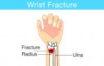
What is the radius bone
The forearm (the area of the arm from the elbow to the beginning of the hand) consists of two bones of similar structure (in Latin, ulna - ulna, radius - radius). The bones of the human forearm often become a buffer during a blow or fall, so the likelihood of injury is very high. As practice shows, due to less dense bone tissue, women suffer from fractures in this area more often than men. Risk groups include menopausal women (over 50 years old) and children (under 10 years old).
Concomitant injuries in case of injury to the radius:
- dislocations of adjacent bones;
- ligament ruptures;
- injuries to the ulna.
Where is the radius bone located?
In the forearm area, radius is the closest “neighbor” of the ulna. Therefore, they are interconnected and dependent on each other. If the palm is turned back when the arm is raised, they are both parallel, but when the palm is turned in the other direction, the bones “cross”. The beam partially rotates around the ulna, which provides rotation ability (pronation) and rotation ability (supination). In addition, where the radius bone is located in position can be determined by the thumb.
The structure of the radius
The radius consists of a long body (diaphysis) and two ends - distal and proximal. The distal epiphysis is more massive; it contains the articular surface of the wrist and the styloid process, which connects to the hand. The anatomy of the proximal end of the radius is as follows: it consists of a head and an articular circle, with the help of which the radius is connected to the bones of the shoulder. Below the head is the neck of the radius, even lower is the tuberosity, to which the biceps brachii muscle is attached. The development of the radius occurs due to the appearance of ossification points.
There are three types of edges:
- front (edge rounded);
- back (edge rounded);
- lateral (the edge is pointed, the edge is directed towards the ulna).
Radius fracture
Any injury to the forearm does not pose a serious threat to the patient’s life, but can cause unpleasant consequences due to disruptions in the functioning of the nervous and vascular systems. Fractures of the radius are painful, and the functionality of the upper extremities is often impaired. With correct diagnosis and thoughtful treatment, the patient fully recovers within a quarter of a year. Depending on the method of injury, pathological and traumatic fractures are distinguished, and according to the degree of damage to the skin, closed or open are determined.
Consequences of radius damage:
- damage to blood vessels and nerve endings of the hand;
- poor circulation and the onset of tissue necrosis due to pinching;
- loss of motor ability of the hand (complete or partial);
- infection of connective tissues and epithelium, ulcers and other foci of inflammation, the wound heals slowly;
- development of osteoporosis due to infection in an open fracture.

Common types of fractures are listed in the table:
In a typical place
Often the bone is susceptible to fractures at its thinnest point, so such injuries are referred to as a fracture of the radius in a typical place. This type of injury to the forearm is very common, accounting for 15% of all injuries to the human skeleton. Typical fractures occur approximately 3 cm from the wrist and are called the distal metaepiphysis. Statistics show that the left hand is more often broken than the right. Typical radial fractures in international practice are assigned ICD code S52.5.
Types of typical radial fracture:
- Colles (flexion, the fragment is displaced towards the dorsal surface);
- Smith (extensor, the fragment is displaced towards the palmar surface).
With offset
A situation in which fragments of the epimetaphysis, leaving their usual place, are displaced to the side is a displacement. With such damage, the hand hurts greatly, swelling increases, and even external signs show that the bones are placed incorrectly. A displaced fracture of the radius bone of the arm requires reposition and application of a splint, and in complex cases, surgery. For proper fusion, it is necessary to apply plaster for up to a month. It is better to get information on how to relieve swelling after a fracture of the radius from a doctor; self-medication can harm yourself.
Symptoms of a displaced fracture:
- sharp severe pain;
- crunching sound when trying to move your arm;
- external signs of an irregularly shaped hand;
- severe swelling that does not subside;
- the appearance of a hematoma is quite possible;
- impaired finger mobility.

Fracture of the styloid process
This type of injury is more common during the fall and winter months due to frequent falls on ice. There are 2 types of fractures of the styloid process of the radius - compression (a small crack appears, no displacement occurs) and avulsion (during a fall in the hand, the articular surface is dislocated inward, a tear occurs). The latter type is less common, but it is more painful and requires urgent reduction. Remember how long a cast is worn for this type of radius fracture. It will take at least 30 days from the date of application.
Impacted fracture
In a situation where a broken bone is forced into another, an impacted fracture of the radius is diagnosed. In practice, it occurs less frequently than other types of damage. If the radial joint is injured due to an impacted fracture, the arm often loses functionality. The hand heals slowly and requires constant monitoring. To apply the correct treatment methods, the doctor needs to have as much information as possible about the nature of the injury.
Treatment of a radius fracture
Restoring the functionality of the hand after an injury mainly depends on the choice of the correct method of combating the disease and the qualifications of the traumatologist. Treatment of a fracture of the radius is often carried out conservatively (application of an immobilization bandage) and surgically (for a displaced or impacted fracture) in ways. To achieve a good effect in case of a fragment fracture, open (manual reduction of fragments) or closed (skin incision at the site of impact) reduction is performed, and osteosynthesis methods are also used.
Osteosynthesis techniques:
- knitting needles;
- plates;
- distraction devices.
Rehabilitation after a fracture of the radius
The doctor conducts an examination, removes the plaster and sends you for a control x-ray. If everything is in order, you need to start rehabilitation after a fracture of the radius:
- To quickly restore performance, various expanders are used, and physical therapy is recommended, especially exercises for the fingers and hands.
- Physiotherapy procedures, massage and proper nutrition are of great importance for the healing process, especially in combination with exercise therapy.
- Based on the patient's medical history, oral restorative medications are prescribed.

There are the following causes of fractures:
- falling forward;
- osteoporosis (especially in people aged 60+);
- falling from a bicycle, moped, motorcycle;
- negligent attitude towards safety at work.
Video: beam fracture in a typical location
Attention! The information presented in the article is for informational purposes only. The materials in the article do not encourage self-treatment. Only a qualified doctor can make a diagnosis and give treatment recommendations based on the individual characteristics of a particular patient.
Found an error in the text? Select it, press Ctrl + Enter and we will fix everything!A common injury to the forearm is a fracture of the radius. Diagnosed in 16% of cases of all bone lesions or in 40% of arm fractures. The radius is the most mobile part of the upper limb and is very thin, so it is easy to break. Damage to the area located near the hand (distal metaepiphysis) often occurs. In medical circles, such an injury is diagnosed as a fracture in a typical location.
Anatomical certificate
One of the two bones that make up the human forearm is called the radius. The ulna is located on the side of the little finger, and the radius is located on the outside of the arm, in front of the ulna. In its structure, it can be distinguished: the epiphyses (upper and lower), the bone body itself, which has a triangular shape. The surfaces are conventionally divided into posterior, anterior, lateral (side), and its edges are classified as interosseous, posterior and anterior.
The multifaceted motor function of the hand is possible thanks to the coordinated work of the joints. The forearm is crowned with joints at both ends. Where the radius and ulna meet together is the elbow joint. It is responsible for the process of extension and flexion of the arm, turning the forearm down and up. Where the bones adjoin the wrist, there is another joint - the wrist.
The bones of the proximal (remote from the body) row of the wrist (triquetral, lunate and scaphoid), as well as the radius, participate in the formation of this joint, and the ulna does not reach it, being supplemented by the articular disc. In its shape, it resembles an ellipse and provides extension and flexion of the hand, abduction and adduction. Rotational movements occur in conjunction with the bones of the forearm.
Causes leading to injuries
Due to the influence of external or internal factors, a fracture of the radius bone of the arm occurs with or without displacement. The most common causes leading to injury are:
- work injury;
- traffic accident;
- sports injury;
- falling from a height onto an outstretched arm;
- osteoporosis.
Partial or complete disruption of the integrity of the bone is called a fracture. If the impact force exceeds its strength, the structure is damaged. This is due to excessive stress, a blow, a fall, or human diseases, due to which the bones become brittle or thin.
Important! Trauma to the beam also provokes a fracture of the wrist joint with or without displacement.
Classification of fractures
Like any other injuries, these fractures are classified depending on the degree of damage, the nature of the injury and its location.
There are closed fractures of the radius (in which the skin retains its integrity) and open (when, along with the bone structure, soft tissues are also damaged, and fragments come out).
If the injury did not cause displacement of the fragments, the fracture is classified as “without displacement.” When, under the influence of the force of an impact, the fragments separate, forming a gap of more than two millimeters between themselves, it is called a displaced fracture of the radius. The broken fragment will move under the influence of the muscles.
Based on the position of the injured person’s hand, fractures of the ray in the wrist joint can be:
- extensor, which are also called Wheel fractures, when bone fragments are displaced towards the beam and to the rear;
- flexion, better known as Smith fractures, when the blow falls on the bent hand, its back side, and the fragments move away towards the surface of the palm.
Often this injury is defined as intra-articular and is complicated by the separation of the styloid process (in more than half of the cases), which often entails a fracture of the wrist bone. In the case where the joint remains intact, they speak of extra-articular injuries.
A bone fracture occurs in a transverse or oblique direction. If there is a direct injury to the limb, then, most likely, a transverse injury will appear; in rare cases, a comminuted fracture may occur, in which more than three splinter fragments are obtained.
If the arm is compressed on two different sides, it is called a compression fracture. Under varied, strong pressure, the radius bone breaks into small fragments that affect the soft tissue around it. This type of damage has recently become more common. This is primarily due to technological progress, the emergence of vehicles, and automation of production.
A rare type of injury in this area is an impacted fracture, when one part of a bone fragment, under the force of impact, enters another fragment.
Main symptoms of injury
You can determine the presence of a fracture by knowing the main symptoms:
- a characteristic crunching sound of bone fragments is heard (crepitus);
- sharp pain upon injury and intense painful sensations that persist for a long time;
- hematoma due to disruption of the integrity of blood vessels;
- hyperthermia (increased temperature) of the affected area;
- swelling;
- if the bone fragments have shifted significantly, a bump or dent is visible in the wrist area;
- redness of the skin at the site of injury;
- in the case where the nerve endings have been affected, there is a loss of sensitivity in the fingers (numbness, tingling, feeling of cold) and their mobility;
- increased pain with any attempt to move the arm or hand.
It is important to remember that even if after some time the painful sensations dull or disappear altogether, this does not mean that the damage is not serious. Do not forget that a displaced fracture of the forearm is a serious injury, and the treatment and recovery processes can take a long time, regardless of its severity.
First aid and diagnostics
In case of any damage, it is necessary to obtain qualified assistance from medical personnel. Injuries are not always as simple and insignificant as they seem at first glance. The victim must be taken to the nearest emergency room, and in difficult situations it is better to call an ambulance to the scene of the incident.
First you need to examine the injured limb. If clothing prevents you from doing this, you should not take it off. Any movements will provoke an attack of pain and can lead to displacement of bone fragments. It is better to carefully roll up or cut the sleeve. If there is damage to the skin, the wound is washed and treated with an antiseptic. A three percent solution of hydrogen peroxide will help stop the bleeding. The wound must be covered with a sterile bandage, which is applied very carefully and not too tightly.
A cold compress will help reduce pain and swelling of the injured limb. It is best to use ice. For convenience, it is first poured into a bag, and the bag is wrapped in a cloth or towel. Do not allow bare skin to come into contact with ice, as this will lead to negative consequences. If you don't have ice on hand, any food from the freezer or refrigerator will do. You can pour chilled water into a bottle and apply it to the damaged area. You should not keep the lotion for too long; after fifteen minutes you need to remove it for a while and after a while use the cold again.
Before transporting the victim to the hospital, it is necessary to fix the limb, immobilizing it as much as possible. Immobilization is carried out using a special transport ladder splint. If you don’t have one at hand, use suitable materials at hand: sticks, boards, pipes, thick strips of cardboard. You can attach the injured limb to an improvised splint using bandages, belts, or strips of fabric.
If the victim complains of unbearable pain, give him any non-narcotic painkiller (Ketanov, Tempalgin, Celebrex, Analgin, Brustan). These simple steps are enough to provide first aid; further treatment is carried out in the trauma department.
Diagnostics
The correct diagnosis can only be made by a traumatologist based on a thorough examination. First, an anamnesis is collected, which shows the mechanism of injury and the patient’s complaints about general health. Then the doctor examines the injured limb and checks its functional abilities by palpation. An important point in diagnosis is an x-ray examination, without which it is impossible to make an accurate diagnosis.
The image is taken in two projections for detailed visualization. Additionally, computed tomography or magnetic resonance imaging is prescribed. If neurological symptoms are detected, a neurologist (neurosurgeon or vascular surgeon) is invited for consultation, who will treat along with a traumatologist.
What you need to know about treatment
There are several directions in the treatment of radial bone fractures: conservative and surgical. The advisability of this or that therapy is determined only by the doctor based on the examination results, the nature of the injury and the individual characteristics of the patient (age, concomitant diseases).
Typically, non-displaced fractures are treated conservatively by applying a bandage (polymer or plaster). Fixation is carried out until the bone has completely fused to prevent displacement of the broken fragments.
If a displaced fracture is diagnosed, all fragments must be returned to their natural physiological position (reduced). Only after this the limb is fixed with plaster. Reposition is carried out under local anesthesia manually or using special devices (Sokolovsky, Edelstein and the like). The plaster cast is removed after about a month to a month and a half; during the entire period of treatment, several radiographic studies are performed to monitor.
In the event that it is not possible to carry out a closed reduction, the displacement of bone fragments is unstable and critical, or has occurred repeatedly, it is recommended to carry out surgical intervention, which is a closed reduction using special metal pins, or osteosynthesis. In modern medicine, there are two ways of performing osteosynthesis of the wrist joint:
- transosseous - using a rod apparatus or an Ilizarov apparatus;
- bone - when there is a need to use plates with angular stability.
External fixation devices (screws, plates) are rarely used. When treating children, doctors prefer the conservative method and resort to surgery as a last resort.
Rehabilitation
At the final stage of therapy, it is necessary to perform a number of rehabilitation procedures. If necessary, the attending physician prescribes a course of therapeutic massage, physiotherapy, physical therapy, water therapy or the use of an orthosis.
We should not forget about the correct diet. For a speedy recovery, the patient needs to include foods rich in vitamins and calcium in his diet. These include: dairy products, fresh fruits and vegetables, fish, honey, nuts.
Complete recovery after a displaced fracture of the radius occurs after at least two months in the case when healing proceeded without medical errors (incorrect or incomplete alignment of fragments, improper immobilization of the arm, lack of control over the recovery process) and complications.
Negative consequences of a fracture
A lack of calcium or other substances in the body will cause poor bone tissue regeneration. Prolonged inactivity of a fixed limb will cause muscle flaccidity, especially if the patient did not pay attention to physical training before surgery.
May be observed:
- repeated displacement of broken fragments under plaster;
- bone deformation;
- neurotrophic abnormalities of the limb;
- development of purulent-inflammatory processes (typical of open fractures);
- disorder of innervation (supply of nerve cells) in the affected area;
- vascular disorders under plaster.
Rotting of tissues in the area of installed metal structures is rarely observed. The plaster cast deserves special attention, as it should not dangle and at the same time compress soft tissues.
Following your doctor's instructions and taking a course of rehabilitation measures will help you recover faster and return to your normal pace of life.