Preparing for an ultrasound of the bladder. Ultrasound of the bladder, preparation. Ultrasound of the bladder: what can be found out
When conducting an ultrasound of the bladder, preparation for the study plays a huge role, since without this it will be quite difficult to make a correct diagnosis.
Patients going for an ultrasound of the pelvic organs must know all the rules for preparing for this examination, since the reliability of the results depends on this.
Features of ultrasound examination of the bladder
There can be many indications for such useful research. Ultrasound is prescribed for those patients who have certain symptoms, but it is impossible to make an accurate diagnosis without additional examination.
The doctor sends the patient for an ultrasound if he presents with complaints such as frequent urination, pain in the kidneys and bladder, urine mixed with blood, difficulty in emptying and other signs of kidney stones, vesicoureteral reflux, and so on.
In addition, an ultrasound examination may be prescribed to assess kidney function. It is possible that problems with the bladder are a consequence of cystitis in acute or chronic form or pyelonephritis. Representatives of the stronger sex may additionally be prescribed a prostate examination to exclude the possibility of developing adenoma and inflammatory process in this area.
Ultrasound examination of the bladder in women is often accompanied by additional diagnostic procedures in the pelvic area. This makes it possible to make sure that the problem with the bladder is not related to the appendages and uterus.
How to prepare for an ultrasound
In order to get an accurate result during the examination, the patient must know how to prepare for an ultrasound of the bladder. For a quality examination of the bladder, it is necessary to pre-fill it, for which you need to drink about 2 liters of water 2 hours before the start of the procedure. It is worth considering that we are talking exclusively about non-carbonated liquid, preferably without artificial colors and preservatives. You can drink plain water, tea or homemade compote.
If the bladder is full, the doctor will be able to see not only the contours of the organ and its shape, but also the thickness of the walls. In the case where the female or male genital organs are additionally examined, a filled bladder is also very important, as it makes it possible to obtain a clearer picture.
As for how to prepare for an ultrasound of the bladder, for women it is first of all important to refrain from urinating 5-6 hours before the examination. But if you can’t bear it, you can partially empty your bladder, and after that you need to immediately drink a lot of liquid so that it fills up again.

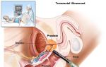
If the doctor has prescribed a transrectal procedure rather than a regular one, you need to prepare more carefully; for this you need to do an enema a few hours before the ultrasound of the bladder. A similar procedure is performed not only when examining the bladder, but also the prostate.
How is an ultrasound of the bladder done?
The transabdominal method is most often used for ultrasound scanning of internal organs. This suggests that the required image can be obtained through the abdominal wall. However, this approach is not possible with every patient. For example, if the patient is obese or the doctor suspects a prostate tumor, an ultrasound examination will be performed through the rectum. In the case when we are talking about representatives of the fair sex, instead of the transabdominal method of examination, ultrasound of the bladder in women can be performed using the transvaginal method.
This procedure is very simple and does not last long. In most cases, 15 minutes is enough for a specialist to get an accurate picture of what is happening. During the ultrasound scan, the patient must lie still on his back. A special gel is applied to the area where the transmitter will operate, after which the desired area can be examined.

In some cases, additional actions are possible in the process of preparing for an ultrasound of the bladder and the examination itself. It all depends on the specific case, so the doctor may prescribe additional procedures, the purpose of which will be to obtain the most accurate and correct picture. Before sending a patient for an ultrasound of the bladder, the specialist must explain all the nuances of this procedure.
What does the study show?
Ultrasound scanning of the body is an extremely important diagnostic procedure. With its help, the doctor can make the correct diagnosis in a controversial situation, as he will receive accurate confirmation of his guesses.
During the ultrasound, the specialist has the opportunity to examine the shape of the bladder. This is very important, because if there is a tumor, this organ will become asymmetrical. Doctors also pay attention to the size of the bladder. If the organ is too small, this may indicate cystitis or fibrous walls. An enlarged bladder is a sign of prostate adenoma, kidney and urethral stones and some other serious ailments.
In addition, during an ultrasound examination, the doctor examines the external and internal contours of the organ, checks it for the presence of foreign bodies and neoplasms, and also excludes or confirms injury to the walls, which can lead to a violation of their integrity.
In addition to examining the bladder, a urine test is also performed using ultrasound scanning. This is necessary in order to determine the state of the contents of the organ being examined. It is quite possible that blood or other impurities will be found in the urine, which indicates the development of a number of ailments.
Ultrasound of the bladder makes it possible to determine cystitis by signs such as thickened walls and uneven contours of the organ. In addition, the device will show stones, if any. In this case, you can determine not only their presence, but also their size and shape.

When examining a patient for prostate adenoma, the doctor may ask you to empty your bladder and then continue scanning again. This way you can see the residual amount of urine. If there is a lot of it, then we are talking about an adenoma, since the inflammatory process does not allow the fluid to leave the body normally.
Based on examination of the inner walls of the bladder, many problems can be identified. The image transmitted by the ultrasound machine allows you to see protrusions, neoplasms, polyps, cysts, and so on. In addition, the study makes it possible to detect changes in the ureter area that can occur due to tumors and inflammatory processes.
Indications for ultrasound scanning of the bladder and kidneys
Ultrasound is a simple and almost completely safe procedure, but proper preparation for an ultrasound of the kidneys and bladder must be done. Most often, a pelvic examination is performed if the patient presents with lower back pain - indirect evidence that the kidneys are developing abnormally or that an inflammatory process is occurring in them. This procedure is useful for diagnosing tumors, urolithiasis and prolonged hyperthermia. The latter is especially true for young patients.
In modern medical diagnostics, the main place is occupied by the method of ultrasound scanning - ultrasound. The method is especially in demand in urology and andrology, since it allows one to recognize the slightest structural changes in the pelvic organs, diagnose and classify all pathological disorders in the genitourinary system.
How to prepare for an ultrasound?
Today, ultrasound is a common way to examine the prostate gland (prostate), bladder and kidneys. To successfully undergo an ultrasound scan of the pelvic and genitourinary system, preliminary preparation of the patient is necessary:
- the presence of open wounds and sterile dressings makes ultrasound impossible;
- colonoscopic, gastroscopic, X-ray examinations of the digestive system and reproductive organs for some period of time exclude ultrasound examination in general and TRUS in particular;
- for three days before the examination of the prostate gland and kidneys, you should not smoke or drink alcohol;
- in order to avoid disorders of the digestive system such as increased gas formation and bloating, it is recommended to follow a special diet and the procedure is carried out on an empty stomach;
- the patient must warn the ultrasound specialist about the medications he is taking.
However, scanning the bladder, prostate and kidneys separately requires special preparation. The person being examined must take all preparatory details responsibly, as this is the key to correct and reliable diagnosis of possible diseases of the pelvic and genitourinary organs, including the prostate and kidneys, and the prescription of timely and adequate therapy.
Ultrasound scanning of the kidneys - preparing correctly
Only urgent ultrasound of the kidneys in emergency situations can be performed without prior preparation. In the case of a routine kidney examination, you need to prepare for the procedure carefully, following all the doctor’s recommendations.
The main requirement for the patient is to adhere to a diet that will eliminate temporary disturbances in the digestive system and help clearly visualize the structural state of the kidneys. You need to follow a diet for three days before the procedure. It is necessary to exclude from the diet all foods that can cause increased gas formation. Do not use:
- fresh flour products;
- lactic acid products;
- sweet sparkling water;
- raw fruits and vegetables;
- fried, smoked, fatty meat and fish.
Before an ultrasound scan of the kidneys, you can eat porridge, lean boiled white meat, bread crumbs, and low-fat cheese. The diet rules include taking sorbents, such as activated carbon, Espumisan, after each meal.
Patients who are overweight or have a digestive problem such as constipation are advised to take laxatives on the eve of the study.
Ultrasound examination of the pelvic organs is always done on an empty stomach, preferably in the morning, otherwise it is recommended not to eat for 8 hours before the procedure. In 30-40 minutes. Before the ultrasound, you should drink about half a liter of still water.
Preparing for an ultrasound scan of the bladder
Preliminary preparation for an ultrasound of the pelvic organs is not required only in urgent situations. The only requirement is a full bladder.
Often performed in conjunction with a transabdominal prostate examination. The person takes a lying position. A product is applied to the skin in the abdominal area to improve contact with the sensor. The prostate and bladder scanning procedure lasts no more than 15 minutes.
During a routine ultrasound examination of the bladder and prostate gland, it is necessary not to go to the toilet 2 hours before the start of the diagnosis, restrain the urge to urinate, and during the last hour drink at least one liter of still water in portions. In case of an unbearable desire to urinate, partial emptying of the bladder is allowed. Then you need to drink additional fluid. Ultrasound of the prostate gland and bladder, with its absolute filling, allows you to obtain clear data, reliable structural and dimensional characteristics.

 A detailed examination of the characteristics and quality of the pelvic organs is possible only with a full bladder. Therefore, preparation for the study also includes taking a large amount of liquid about half an hour before the ultrasound.
 How to prepare for a transrectal prostate scan?
TRUS of the prostate gland requires emptying the lower intestine of feces. This can be done using:
- microenemas;
- glycerin suppositories;
- natural laxatives.
Carrying out a cleansing procedure for TRUS of the prostate using a microenema involves introducing special solutions into the rectum. You can use chamomile decoction, saline solution with vegetable oil, a mixture of milk and butter (100 mm: 20 g). During use, the selected microenema product should have a temperature of 36 - 39 degrees. You can also use Microlax microenema purchased at a pharmacy. The medicine is produced in bottles with a special tip.
The use of glycerin-based suppositories in preparation for TRUS of the prostate is widespread. This method causes a mild laxative effect, acting on the rectal mucosa. The suppository is inserted into the rectum through the anus 2-3 hours before TRUS.
It is necessary to carefully study the instructions for using glycerin suppositories. Before TRUS of the prostate gland, medicinal laxatives are often prescribed. Usually these are: “Fitolax”, “Senadexin”, “Mukofalk”. The listed laxatives must be taken at least 10–11 hours before TRUS of the prostate. If the study requires urgent implementation, use quick laxatives. The result should be expected after 20 minutes. after reception.
Ultrasound is of great importance in diagnosing bladder diseases, but you need to understand that you need to prepare for this procedure in advance.
Preparing for an ultrasound of the bladder includes several important points, which we will look at later.
Indications for the study procedure
Reasons for performing ultrasound are:
- Difficulty urinating.
- Signs of kidney stones.
- Urine with blood.
Ultrasound examination is necessary to study kidney function. In men, it is also performed for prostate adenoma or signs of inflammation.
Women need a procedure to fully study the functioning of the genitourinary system and a detailed study of the organs.
What will it show?
This process can reveal:
- The shape and size of the organ. Its decrease indicates cystitis, and its increase indicates narrowing of the urethra.
- The presence of neoplasms is established.
- Organ contents. We are talking about blood, pus.
- Foreign bodies.
- Contours.
- Violation of integrity. Diagnostics helps determine the type of damage.
- Increased tone.
- Inflammation.
- Organ prolapse.
- Prostate pathologies.
- Ovarian diseases.
Bladder capacity can also be determined by ultrasound. Modern devices automatically calculate this indicator.
Determination of residual urine in the bladder
Residual urine is an indicator that determines the presence or absence of diseases in the urinary tract.
Normally, urine remains in the organ cavity should not be more than 10% from the total volume of urine. The calculation of this indicator is of great diagnostic importance and helps to establish or exclude the presence of pathology.
 To determine this indicator, the study is carried out before and after urination. Having examined the organ twice, in a filled state and without liquid, the specialist can tell about the amount of residual urine. Images of the organ are evaluated. The length of its ultrasound shadow is determined using formulas.
Suspension, sediment and flakes in urine during ultrasound indicate impaired metabolism, injury, the presence of kidney stones, and hormonal imbalance. This main signs of malfunction genitourinary system and metabolic processes.
In this situation, an ultrasound scan of a full bladder is performed. This allows you to visualize sediment, contours, and possible changes in the walls of the organ. The study makes it possible to determine not only the presence of flakes and sediment, but also studies in detail its distribution and quantity.
Normal findings on ultrasound of the bladder
Normal indicators are:
- Shape: if the organ is healthy, the shape is clearly visible. On transverse photographs it is a round organ, and on longitudinal photographs it is ovoid.
- volume: for women 200-500 ml, for men 300-700 ml.
- Structure: normally echo-negative.
- Residual urine: maximum 50 ml.
- The walls of the organ: must be of the same thickness, from 2 to 4 mm.
Features of preparation
The process differs depending on the gender and age of the patient.
In women and men
Men need to drink 2 hours before the procedure 1-1.5 liters of water. Water must be filled in the bladder; emptying is strictly prohibited.
Women need to drink 2 hours before the procedure 0.8-1 l of water. A woman’s body is different from a man’s, so she needs less fluid to fill the organ being examined. It is forbidden to have bowel movements before the procedure.
Menstruation is not a reason to cancel the procedure. Diagnosis can be carried out during menstruation. You should also prepare.
The day before the ultrasound, it is prohibited to drink alcoholic and sugary drinks. They can influence the outcome.
In children
 Practically no different from the algorithm of adults. need a drink from 0.5 to 0.7 l of liquid. A child’s body is smaller than an adult’s, so this amount of water is quite sufficient for the procedure. The child should be given liquid 1.5 hours before the test.
If the baby wants to empty his bladder, you need to try to explain to him that this cannot be done. If, nevertheless, the baby could not resist and emptied his bladder, he must be given water again to compensate for the deficiency.
It is better not to give sweet carbonated drinks and juices to your child the day before the procedure.
In pregnant women
Two days before the procedure, you need to exclude spicy, fatty and fried foods from your diet. You need to eat only healthy food.
In the first and second trimester, you need to drink 2 hours before the procedure at least 0.5 liters of liquid. In the third trimester, you do not need to drink water first.
A woman should know that the procedure is performed on an empty stomach. It is better not to eat in the morning before the examination. This will make it possible to obtain accurate ultrasound results.
There is no special method for quickly filling an organ with urine. It is recommended to drink 2 hours before the test still water. It may be mineral. Not only is it more convenient to drink water from a bottle, it is easier to calculate the amount of water you drink.
How is it carried out?
There is nothing complicated or dangerous in this procedure. A man enters the office and lies down on the bed. The lower abdomen is lubricated with a special gel. Then, using a special apparatus, the organ is studied. The gel facilitates the sliding of the device, which is passed over the patient’s abdomen. He doesn't feel any pain.
Usually The procedure lasts 10 minutes. Then the person gets up, wipes the gel off his stomach, and can go to the toilet to relieve himself.
Results are obtained immediately or the next day after the procedure. It depends on the doctor's workload. The image shows the absence or presence of organ diseases. Decryption is carried out only by a specialist. He can tell what condition the organ is in.
– one of the important factors influencing the accuracy of the research results and the correct diagnosis. The measures that a person must take before the examination require a clear statement by the specialist and mandatory implementation on the part of the patient. Ultrasound examination is distinguished by its simplicity and quick results, has no contraindications, and the procedure can be performed even on small children.
Indications for the procedure
Ultrasound of the bladder is indicated for patients who show signs of any pathologies of the urinary system. Research is carried out in the following cases:
- pain in the pubic area;
- increased urge to urinate;
- presence of blood in urine;
- uncontrolled urination;
- suspicion of cystitis;
- suspicion of ;
- cysts in the bladder.

In addition, it is possible to carry out a procedure to assess the functionality of the kidneys, the degree of damage to the bladder due to cystitis (it will help determine the form of the disease), to control the course.
For differential diagnosis, echography of the prostate in men and the internal genital organs in women can be performed simultaneously.
Nutritional considerations before the study
After the doctor has determined the indications for diagnosis, you need to clarify how to prepare for an ultrasound of the bladder. Preparatory activities begin three days before the examination and consist of following a certain diet.

It involves consuming foods that do not cause increased gas formation. The diet should include the following dishes:
- enveloping porridges on the water;
- boiled lean meat (rabbit, chicken, beef);
- yesterday's white bread;
- boiled or baked lean fish;
- hard boiled eggs.

You should completely avoid:
- dairy products;
- legumes;
- raw vegetables and fruits;
- fatty meats and fish;
- smoked meats;
- desserts and sweet carbonated drinks;
- beer.

Also, 2 days before the planned study you need to take sorbents (Smecta, activated carbon).
These rules apply when the patient does not have a tendency to flatulence or other digestive tract problems. Otherwise, before the ultrasound, the diet should be followed for 1 week.
Also, 24 hours before the procedure, you should completely abstain from any alcoholic beverages and do not smoke for several hours.
Daily routine before the procedure
 If the ultrasound procedure is planned before lunch, then it is better to come to see a specialist on an empty stomach. The last dinner the day before should be no later than 18.00. When the procedure is scheduled after lunch, in this case you can have a light breakfast in the morning. After 1 hour you need to drink activated carbon. It is advisable not to drink water, since an hour before the procedure you will need to fill your bladder as much as possible, drinking at least one liter of water. You should monitor your feelings: immediately before the test there should be no strong urge to urinate, as the doctor will press on the stomach during the diagnosis.
If an ultrasound scan of the bladder is necessary, preparation also includes bowel movement.
If an unplanned procedure is being carried out and the person has not restricted himself in food, then before the ultrasound diagnosis you need to give an enema or drink laxatives.
Before the procedure, the patient must inform the doctor if he is taking any medications.
The importance of drinking regime
The main point of diagnosis is maximum filling of the bladder with water. Otherwise, it will be difficult for the doctor to examine the presence of pathologies, and erroneous formations may be visualized.

This is explained by the fact that the empty organ has many folds with similar echogenicity inherent in possible pathologies. When it is filled, all folds are straightened, and the risk of incorrect diagnosis is reduced.
What you need to take with you
Preparation for research is not only about the physiological readiness of the body. You need to have some things with you:
- medical record with the results of all studies;
- referral from the attending physician;
- diaper or towel;
- wipes to remove gel from the stomach;
- money;
- a bottle of water to fill your bladder if you are in line.

It will be difficult to perform an ultrasound examination on patients who suffer from urinary incontinence. Also, people who are overweight will need to move part of their abdomen away from the sensor to get as accurate an image of the bladder as possible.