Lesson on the topic "composition and structure of bones." Structure and classification of bones Structure and growth of bones human skeleton
Each human bone is a complex organ: it occupies a certain position in the body, has its own shape and structure, and performs its own function. All types of tissues take part in bone formation, but bone tissue predominates.
General characteristics of human bones
Cartilage covers only the articular surfaces of the bone, the outside of the bone is covered with periosteum, and the bone marrow is located inside. Bone contains fatty tissue, blood and lymphatic vessels, and nerves.
Bone has high mechanical qualities, its strength can be compared with the strength of metal. The chemical composition of living human bone contains: 50% water, 12.5% organic substances of a protein nature (ossein), 21.8% inorganic substances (mainly calcium phosphate) and 15.7% fat.
Types of bones by shape divided into:
- Tubular (long - humeral, femoral, etc.; short - phalanges of the fingers);
- flat (frontal, parietal, scapula, etc.);
- spongy (ribs, vertebrae);
- mixed (sphenoid, zygomatic, lower jaw).
The structure of human bones
The basic structure of the unit of bone tissue is osteon, which is visible through a microscope at low magnification. Each osteon includes from 5 to 20 concentrically located bone plates. They resemble cylinders inserted into each other. Each plate consists of intercellular substance and cells (osteoblasts, osteocytes, osteoclasts). In the center of the osteon there is a canal - the osteon canal; vessels pass through it. Intercalated bone plates are located between adjacent osteons.

Bone tissue is formed by osteoblasts, secreting the intercellular substance and immuring itself in it, they turn into osteocytes - process-shaped cells, incapable of mitosis, with poorly defined organelles. Accordingly, the formed bone contains mainly osteocytes, and osteoblasts are found only in areas of growth and regeneration of bone tissue.
The largest number of osteoblasts is located in the periosteum - a thin but dense connective tissue plate containing many blood vessels, nerve and lymphatic endings. The periosteum ensures bone growth in thickness and nutrition of the bone.
Osteoclasts contain a large number of lysosomes and are capable of secreting enzymes, which can explain their dissolution of bone matter. These cells take part in the destruction of bone. In pathological conditions in bone tissue, their number increases sharply.
Osteoclasts are also important in the process of bone development: in the process of building the final shape of the bone, they destroy calcified cartilage and even newly formed bone, “correcting” its primary shape.
Bone structure: compact and spongy
On cuts and sections of bone, two of its structures are distinguished - compact substance(bone plates are located densely and orderly), located superficially, and spongy substance(bone elements are loosely located), lying inside the bone.

This bone structure fully complies with the basic principle of structural mechanics - to ensure maximum strength of the structure with the least amount of material and great lightness. This is also confirmed by the fact that the location of the tubular systems and the main bone beams corresponds to the direction of action of the compressive, tensile and torsional forces.
Bone structure is a dynamic reactive system that changes throughout a person's life. It is known that in people engaged in heavy physical labor, the compact layer of bone reaches a relatively large development. Depending on changes in the load on individual parts of the body, the location of the bone beams and the structure of the bone as a whole may change.
Connection of human bones
All bone connections can be divided into two groups:
- Continuous connections, earlier in development in phylogeny, immobile or sedentary in function;
- discontinuous connections, later in development and more mobile in function.
There is a transition between these forms - from continuous to discontinuous or vice versa - semi-joint.

The continuous connection of bones is carried out through connective tissue, cartilage and bone tissue (the bones of the skull itself). A discontinuous bone connection, or joint, is a younger formation of a bone connection. All joints have a general structural plan, including the articular cavity, articular capsule and articular surfaces.
Articular cavity stands out conditionally, since normally there is no void between the articular capsule and the articular ends of the bones, but there is liquid.
Bursa covers the articular surfaces of the bones, forming a hermetic capsule. The joint capsule consists of two layers, the outer layer of which passes into the periosteum. The inner layer releases fluid into the joint cavity, which acts as a lubricant, ensuring free sliding of the articular surfaces.
Types of joints
The articular surfaces of articulating bones are covered with articular cartilage. The smooth surface of articular cartilage promotes movement in the joints. Articular surfaces are very diverse in shape and size; they are usually compared to geometric figures. Hence name of joints based on shape: spherical (humeral), ellipsoidal (radio-carpal), cylindrical (radio-ulnar), etc.
Since the movements of the articulated links occur around one, two or many axes, joints are also usually divided according to the number of axes of rotation into multiaxial (spherical), biaxial (ellipsoidal, saddle-shaped) and uniaxial (cylindrical, block-shaped).
Depending on the number of articulating bones joints are divided into simple, in which two bones are connected, and complex, in which more than two bones are articulated.
Skeletal functions
In the life of the human body, the skeleton performs a number of important functions:
- 1. Support function : the skeleton serves as a support for muscles and internal organs, which, fixed to the bones by ligaments, are held in their position.
- 2. Locomotor (motor) function: The bones that make up the skeleton are levers that are driven by muscles and participate in motor acts.
- 3. Spring function: the ability to soften shocks from collisions with solid objects when moving, thereby reducing the shaking of vital organs. This happens due to the arched structure of the foot, ligaments and cartilaginous pads inside the joints (connections between bones), the curvature of the spine, etc.
- 4. Protective function : the bones of the skeleton form the walls of cavities (thoracic cavity, cranial cavity, pelvis, spinal canal), protecting the vital organs located there.
- 5. Participation of skeletal bones in metabolism, primarily in mineral metabolism: bones are a depot of mineral salts (mainly calcium and phosphorus) necessary both for the formation of bone tissue and for the functioning of the nervous system, muscles, blood coagulation system and other body systems. Bones contain about 99% of all calcium; when there is a lack of calcium for the body's activities, calcium is released from the bone tissue.
- 6. Participation of skeletal bones in hematopoiesis: red bone marrow, located in the bones, produces red blood cells, granular forms of white blood cells and platelets.
Structure and classification of bones
Bone - a living organ consisting of various tissues (bone, cartilage, connective tissue and blood vessels). Bones make up about 20% of the total body mass. The surface of the bone is uneven, it contains bulges, depressions, grooves, holes, roughnesses to which muscles, tendons, fascia and ligaments are attached. Vessels and nerves are located in the grooves, canals and slits, or notches. On the surface of each bone there are holes that go inward (the so-called nutrient foramina).
The structure of bones includes organic (ossein and osseomucoid) and inorganic (mainly calcium salts) substances. Organic substances provide bone elasticity, and inorganic substances provide its hardness. The child's bones contain more ossein, which provides higher elasticity, which to a certain extent prevents fractures. In old age and old age, the amount of organic substances decreases and the amount of mineral salts increases, which makes bones more fragile.
Classification of bones by shape. Tubular bones have the shape of a tube with a bone marrow canal inside. The body of the bone, or its middle part, is called the diaphysis, and the expanding ends are called epiphyses; the outer surfaces of the epiphyses are covered with cartilage and enter the joints, i.e. serve for connection with neighboring bones (Fig. 3.2). The area between the diaphyses and epiphyses, consisting mainly of cartilaginous tissue, is called the metaphysis, thanks to which bones grow in length (bone growth zone). The diaphyses are built of dense, and the epiphyses are built of spongy bone substance, covered with a dense layer on top. Tubular bones are located in the skeleton of the limbs and are divided into long (femur, tibia, humerus, ulna) and short (located in the metacarpus, metatarsus, phalanges of the fingers). Spongy bones consist of spongy bone tissue covered with a thin layer of dense bone tissue. There are long (ribs and sternum), short (carpal, tarsal bones), sesamoid (patella, pisiform) spongy bones. Sesamoid bones are small bones located in the thickness of the tendons and strengthen them in places of high load and high mobility. Flat Bones perform a protective function and support function (skull, scapula, pelvic bones). mixed bones, forming the base of the skull, are represented by a fixed connection of bones of different shapes and structures. IN air bones contains a cavity with air, lined with mucous membrane (frontal, sphenoid, ethmoid bones and upper jaw).
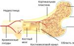
Rice. 3.2. :
1 – osteon (Haversian system); 2 – compact substance; 3 – spongy substance; 4 - Bone marrow; 5 – blood vessels that deliver nutrients and oxygen to bone cells; 6 – central medullary cavity; 7– bone head
The surface of the bone is covered periosteum, and the articular surfaces do not have periosteum and are covered with articular cartilage. The periosteum is a thin white-pink film, its color is due to the large number of blood vessels that pass from the periosteum into the bone through special openings and participate in the nutrition of the bone. It consists of two layers: fibrous (fibrous surface layer) and osteofibrous (inner bone-forming layer containing osteoblasts - special “growth” cells). The mechanism of bone growth varies: flat bones grow due to the periosteum and connective tissue of the sutures; tubular bones thicken due to the periosteum, and grow in length due to the cartilaginous plate located between the epiphysis and diaphysis (bone growth zone).
The bone canals and the space between the bone plates are filled bone marrow which performs the function of hematopoiesis and is involved in the formation of immunity. There are red bone marrow (a reticular mass of red color, in the loops of which there are hematopoietic stem cells and bone-forming cells), penetrated by blood vessels that give it a red color, and nerves, and yellow bone marrow, which arises as a result of the replacement of hematopoietic cells with fatty ones during ontogenesis. The younger the child, the more intense his hematopoiesis processes are and the more red bone marrow is contained in the bone cavities; in an adult, it is stored only in the sternum, wings of the ilium and the epiphyses of the tubular bones.
Skeletal bone connections divided into synarthrosis (continuous in structure and immobile in function) and joints, or diarthrosis (intermittent and ensuring mobility of the musculoskeletal system). There is also a transitional form of the compound - symphysis (half-joint), which has minimal mobility (Fig. 3.3).

Rice. 3.3. :
A -
joint, or diarthrosis (discontinuous connection):
B, V –
various types of synarthrosis (continuous joints):
B –
fibrous junction; IN
– synchondrosis (cartilaginous junction); G
– symphysis (hemiarthrosis or semi-joint): 1 –
periosteum; 2 –
bone; 3
– fibrous connective tissue; 4 –
cartilage; 5 – synovial membrane; 6
– fibrous membrane; 7 – articular cartilage; 8 –
articular cavity; 9 –
a gap in the interpubic disc; 10 –
interpubic disc
Joints provide the ability to move body parts relative to each other. Based on the number of articular surfaces in a joint, a simple joint is distinguished (it includes two articular surfaces - for example, the interphalangeal joint), a complex joint (has two or more pairs of articular surfaces - for example, the elbow joint), a complex joint (contains intra-articular cartilage that divides the joint two chambers - for example, the knee joint), combined (several isolated joints, rigidly linked and functioning together - for example, the temporomandibular joint).
According to the number of possible axes of movement, joints are distinguished uniaxial (flexion and extension – radial, ulnar, interphalangeal), biaxial (flexion and extension, abduction and adduction - wrist and knee) and multi-axis (perform all the listed movements and, in addition, a circular movement - the shoulder joint, the joints between the processes of the thoracic vertebrae).
The structure of the joints, regardless of the functions performed, is similar (Fig. 3.4 - using the example of the knee joint). It includes the epiphyses of bones, covered with hyaline or fibrous articular cartilage 0.2–0.5 mm thick, which facilitates the sliding of articular surfaces and serves as a buffer and shock absorber. The articular surface of the epiphysis of one bone is convex (has an articular head), the other is concave (glenoid cavity). The articular cavity is hermetically surrounded by an articular capsule, which is tightly attached to the bones included in the joint, and consists of an outer fibrous layer, which performs a protective function, and an inner synovial layer. The cells of the synovial layer secrete a thick transparent substance into the joint cavity synovial fluid, reducing friction of articular surfaces, participating in metabolism, softening compression and shock of articular surfaces.

Rice. 3.4.
On the outside, ligaments and muscle tendons are attached to the joint capsule, further strengthening the joint. Ligaments connect the two bones that make up the joint, secure the bones in a certain position, and, due to their low extensibility, keep the bones from moving during movement. Ligaments are also involved in fixing internal organs, leaving them with little possibility of displacement, which is necessary, for example, during pregnancy and digestion. Ligaments consist of collagen and a small amount of elastic fibers. At the points of attachment to the bone, the fibers of the ligaments penetrate the periosteum. Such a close connection between them leads to the fact that damage to the ligaments leads to damage to the periosteum. In large joints (hip, knee, elbow), parts of the joint capsule are thickened for greater strength and are called the peri-marsal ligament. In addition, there are ligaments inside and outside the joint capsule that limit and inhibit specific types of movement. They are called external, or accessory, ligaments.
Chemical composition of bones
Bones are composed of organic, inorganic (mineral) substances and water. In childhood and adolescence, the content of organic substances in the bones exceeds the amount of mineral substances; in old age, the amount of organic substances decreases. Bones contain the bulk of the minerals found in the body. Their excess is deposited in the skeleton. When there is a lack of minerals, the body replenishes them from the bones. Consequently, the skeleton is involved in the metabolism of minerals occurring in the human body.
Bones are strong and elastic. The elasticity of bones depends on the amount of organic matter. Therefore, it is greater in children and young people than in old age. If you decalcify a bone by keeping it in an acid solution for some time, all the minerals are removed. This bone can be tied into a knot.
The strength of the bones is very high. It is 5 times higher than that of reinforced concrete. If you heat a bone over a fire, all organic substances will be destroyed, but the mineral substances will remain. Such a bone retains its shape and arrangement of bone plates, but loses elasticity and becomes fragile. Minerals give strength to bones. As people age, their bones become brittle and their elasticity decreases. Therefore, they are more susceptible to fractures.
Bone growth
In the early stages of development of the human embryo, its skeleton consists of connective tissue. Then it becomes cartilaginous. The skeleton of a newborn does not consist entirely of bone tissue. As the child grows, skeletal cartilage is replaced by bone tissue and the bones grow in length and thickness. Some bones do not go through the cartilaginous stage, such as the bones of the skull.
The growth of bone thickness occurs due to the bone-forming cells of the periosteum. At the same time, the bone tissue on the inner surface of the compact substance is absorbed and the volume of the bone cavity increases. The bone grows in length due to cartilaginous growth plates located between the body and the epiphyses of the bone. The cells of the cartilaginous growth plates form bone tissue and the body of the bone lengthens.
Some bones are formed in the human embryo from several parts, subsequently forming one bone. Thus, complete ossification of the pelvic bone occurs by 14-16 years, and tubular bones - at 18-25 years. Skeletal development and growth stop in men at 20-25 years of age, and in women at 18-21 years of age. During the development of the human skeleton, not all cartilage is replaced by bone tissue. In an adult, the ends of the ribs and part of the skeleton of the nose remain cartilaginous. The surfaces of the epiphyses of bones are covered with cartilage.
“Human Anatomy and Physiology”, M.S.Milovzorova
The system of organs of support and movement - the musculoskeletal system - is a skeleton consisting of bones and their joints, and muscles. Muscles are an active part of the musculoskeletal system. Muscle contractions move the bones of the skeleton. With the help of muscles, a person can remain motionless for a long time, often holding very complex choreographic poses. The total number of muscles in humans is approximately 600. They...
Bones are made up of hard bone tissue. Bone cells are located at a distance from one another and are connected by numerous processes. The bulk of bone tissue is made up of intercellular substance. It consists of osteons and intercalated plates located between them. Between the bone plates are bone cells. The intercellular substance contains organic substances and is impregnated with mineral salts, which give it strength. Bone tissue belongs...
Bone composition. Bones are very strong. The human tibia, in an upright position, can withstand a load of 1500 kg (Fig. 38).
The greater strength of bones depends on their composition. They are formed by both organic and inorganic compounds. The meaning of these substances can be easily determined by performing a simple experiment. If you bake a bone for a long time, water is removed from it, and organic compounds burn. When this is done carefully, the bone does not lose its shape, but becomes so brittle that when touched it immediately crumbles into small but very hard particles consisting of inorganic substances.
It is not difficult to remove inorganic compounds - mineral salts - from bone. Among them we name calcium carbonate and calcium phosphate. To do this, the bone is kept for 24 hours in a 10% HCl solution. The inorganic compounds gradually dissolve and the bone becomes so flexible and stretchable that it can be coiled. But as soon as you let go of the ends of this spiral, it unwinds and returns to its previous position. Organic compounds give bones flexibility and elasticity.
The combination of the hardness of inorganic compounds with the elasticity of organic compounds provides greater bone strength. The bones of an adult, but not an old person, are the strongest.
Bone structure. The strength of bones is determined not only by their composition, but also by their structure.
Long bones, such as the bones of the shoulder, forearm, thigh, and lower leg, are hollow in the middle part. These are tubular bones. At their ends there are thickened heads in which there is no cavity. The tubular structure of long bones ensures their strength and lightness at the same time. After all, it is known that a metal or plastic tube is almost as strong as a solid rod of the same material, equal in length and diameter. Therefore, in engineering, strong and lightweight structures are often made from pipes. In the cavities of the tubular bones there is connective tissue rich in fat - yellow bone marrow.

The heads of the tubular bones are formed by spongy substance (Fig. 39), which consists of many intersecting bone plates. They are located in those directions in which the bones experience the greatest tension or compression. This structure ensures the strength and lightness of the bones. Many light and strong structures, such as bridges and radio masts, are built from intersecting metal beams (Fig. 40).

Short bones, such as the carpal bones, tarsal bones, and vertebrae, are also formed mainly by spongy substance. Flat bones have the same structure, such as the shoulder blades, ribs, pelvic bones and the roof of the skull. The spaces between the bone plates are filled with red bone marrow, which is formed by connective tissue.
The surface of the bones is covered with periosteum (Fig. 41, 1). This is a thin but dense layer of connective tissue fused to the bone. The periosteum contains blood vessels and nerves. The heads of long bones, covered with cartilage (2), do not have a layer of periosteum.

Bone growth. During embryonic development of a person, the skeleton is gradually formed. At first it consists of soft connective tissue, which is then replaced by cartilage. In a newborn, most of the cartilage tissue has already been replaced by bone, but this replacement is completed only by the age of 22-25. During skeletal ossification in some bones, soft connective tissue is directly replaced by bone, bypassing the cartilage stage. During childhood and adolescence, people's bones grow in length and thickness. In adults, bone matter is constantly renewed.
To study the growth and renewal of bone matter, experiments were carried out on animals.
A special non-poisonous dye was added to the calf's food. They took breaks in feeding such food: ten days they gave food with paint, the next ten days without it, and so on several times. From the intestines, the paint was carried by the blood to all organs. After the bull was slaughtered, one of its long tubular bones was sawed crosswise. The cut revealed colored and white layers alternating in the form of concentric rings. It became clear that the bone had grown in thickness and during growth it was covered on the outside with new layers. Another experience has shown that this is indeed the case. The skin of a young dog's thigh was cut, the muscles were pulled apart, and a wire was tied around the femur. Years have passed. After the animal died, it was opened up. There was no wire ring on the surface of the femur. It was found in the internal cavity of the bone.
What explains the growth of bone thickness? Cells on the inner surface of the periosteum rapidly divide and deposit new layers of bone cells on the surface of the bone. An intercellular substance is formed around these cells.
In adults, bones do not lengthen or thicken. But the replacement of old bone substance with new continues throughout life. How does this happen? It was found that there are special cells in the bones that destroy old bone matter. Now it’s clear how the wire ring placed on the dog’s femur got into the internal cavity. The old bone substance was destroyed from the inside, and a new one was formed from the surface.
■ Long bones. Short bones. Flat bones. Periosteum.
? 1. What substances make up bone? 2. What structure do bones have? 3. Does the strength and lightness of the skeletal bones depend on it? 4. What causes bones to grow in thickness?
▲ Roll out two identical sheets of paper into a hollow tube and a solid stick. Place each of them horizontally on two stands and, hanging gradually increasing weights from the middle of them, determine which of them bends under less and which under greater load. Think about what feature of bone structure you discovered through this experiment.
Lesson No. 2. Connecting bones. Human skeleton
The purpose of the lesson:
formation of a holistic system of knowledge about the structure of the human skeleton;
expanding knowledge about the diversity of skeletal functions;
to introduce the structural features of the human support system in connection with upright walking.
know the general structure of the skeleton, types of bone connections in various sections;
be able to navigate in determining the location of certain sections and joints of bones in the skeleton;
apply knowledge to prevent spinal diseases.
Lesson type: combined.
Type: modular lesson.
Technological map (module) of the lesson
Human skeleton. Types of bone connections.
Read the purpose of the lesson carefully.
Goal: to determine the initial level of mastery of material on the topic “Musculoskeletal system.”
Write the topic of the lesson in your notebook.
Complete the assignment by carefully reading each question and answer options.
Test task.
I.Choose the correct answer.
1. The skeleton performs the following functions:
B) protection;
B) movements;
D) all named functions.
2. Bone hardness is given by:
A) calcium and phosphorus salts;
B) calcium and potassium salts;
C) potassium and phosphorus salts;
D) there is no correct answer.
3. The elasticity of bones is due to the presence in them of:
A) minerals;
B) organic substances;
B) organic and mineral;
D) there is no correct answer.
4.Red bone marrow is located:
A) in spongy substance;
B) in a compact substance;
B) in the periosteum;
D) there is no correct answer.
5. Label the main parts of the tubular bone.
6. In which case are the bone types correctly indicated?
A) tubular, spongy, flat;
B) tubular, flat, elongated;
B) tubular, spongy, compact;
D) tubular, compact, elongated.
7. The bone grows in thickness due to:
A) bone heads;
B) periosteum;
B) red bone marrow;
D) yellow bone marrow.
8.*With an average human weight of 70 kg, the mass of the skeleton is only 8-9 kg, that is, the skeleton is relatively light. It is also known to be highly durable. What explains the strength and lightness of the bones of the skeleton?
We work independently
Each correct answer is worth 1 point.
Complete the task within 10 minutes.
Be careful when performing work. If a correction is made, make a note accordingly.
The answer is worth 3 points
The maximum number of points is 10.
Goal: while watching a video, gain an idea of the general structure of the human skeleton and its main components.
Carefully read the questions you must answer after watching the video.
What are the main sections of the human skeleton?
What makes up the main skeleton?
What is the appendicular skeleton?
Why are the pelvic bones of the lower extremity girdle so wide?
What shape does the spine have?
What is this form associated with?
What sections is the skull divided into?
Examine and study the structure of the skull.
Participate in discussion with the class.
Take a good look
Video fragment
Find the answer in paragraph 11, pp. 52-53.
Purpose: to study the structure of the main skeleton.
After watching a fragment of the video and working with the textbook text, answer the questions:
What makes up the spine?
How many vertebrae form it?
What structure do the vertebrae have?
Why don't the vertebrae rub against each other?
What curves does the spine have?
Make a diagram of the structure of the spine in your notebook:
department is formed by what.
Check your understanding of the material
Fill in the missing words.
The human skeleton consists of ____ And _____ . The girdle of the upper and lower limbs and the limbs themselves form ___ ____.
The axial skeleton is formed by _____ and ______.
The spine is divided into sections: ______, _____, _____, ______,____.
The cervical region consists of ___
The thoracic region is formed by ____ pairs ____, 12_____ and ______. Together they form ______ ______.
The lumbar and _____ regions consist of____
vertebrae The last section of the spine is ______, it consists of __ ______ vertebrae.
Work individually.
Text pp. 53-54, fig. 22.
Discuss the results of your work in pairs.
For each correctly inserted word you get 1 point.
Max score – 20.
Time 10 min.
Purpose: to study the appendicular skeleton and the types of bone connections in it.
Read the questions carefully. To which you should receive an answer.
What parts does the skeleton of the upper limbs consist of? Make a diagram
What parts does the upper limb belt consist of? Give me the answer reference diagram.
List the bones that make up the upper limb.
Draw a diagram of the structure of the skeleton of the lower extremities.
List the bones that make up the lower limb
Test your knowledge.
Test your knowledge. A.
1. Label the bones with numbers and write their names in your notebook. What is shown under the letters A, B.
work with paragraph 12, read the section carefully.
Work individually.
Work independently.
For each correct answer 1 point.
Mach -14 points.
Time 10 min.
Purpose: to study the types of connections.
After watching the video, answer the questions:
Movable articulation of bones - ______.
Fixed articulation of bones - _____.
Semi-movable articulation of bones -_____.
Watch the video fragment carefully.
Hand in your work to your teacher.
Purpose: summing up.
Read the purpose of the lesson. Have you achieved it?
Discuss with the class. In what degree. 55-47 points - high level of assimilation. Rating: “5”.
46-35 – good level of assimilation, grade – “4”.
34-28 – average, “3”, below 28 – “2”.
Homework. *11,12, finish work
Individually. Calculate your points by comparing your answers with the test scale.
Answer card.
UE-0
1-g; 2-a; 3-b; 4-a; 5: 1-bone head; 2- spongy substance; 3 - dense (compact) substance; 4-periosteum; 5 – yellow bone marrow; 6 – diaphysis;
6 –a;
7-b.
8- bone composition, bone tissue structure.
UE-3. Answers are written out sequentially.
Main and additional; skeleton of the upper limbs; spine and skull; cervical, thoracic, lumbar, sacral, coccygeal regions; 7 vertebrae, 12 pairs of ribs, 12 pairs of ribs and sternum (chest bone), Thorax, sacrum, 5; coccygeal, 5-4.
UE-4
A – free upper limb.
1-clavicle;
2-blade;
B – free hind limb.
1 – pelvic bone, 2 – femur; 3 – tibia; 4 – fibula;
5-tarsus, calcaneus.
UE-5
1 – joint;
2 – seam;
3- symphysis.