Human structure: musculoskeletal system. Human musculoskeletal system. ODS hygiene. Types of bone connections
In the process of evolution, animals mastered more and more new territories, types of food, and adapted to changing living conditions. Evolution gradually changed the appearance of animals. In order to survive, it was necessary to search for food more actively, hide better or defend against enemies, and move faster. Changing along with the body, the musculoskeletal system had to ensure all these evolutionary changes. The most primitive protozoa have no supporting structures, move slowly, flowing with the help of pseudopods and constantly changing shape.
The first support structure to appear is cell membrane. It not only separated the organism from the external environment, but also made it possible to increase the speed of movement due to flagella and cilia. Multicellular animals have a wide variety of support structures and devices for movement. Appearance exoskeleton increased the speed of movement due to the development of specialized muscle groups. Internal skeleton grows with the animal and allows it to reach record speeds. All chordates have an internal skeleton. Despite significant differences in the structure of musculoskeletal structures in different animals, their skeletons perform similar functions: support, protection of internal organs, movement of the body in space. The movements of vertebrates are carried out due to the muscles of the limbs, which carry out such types of movement as running, jumping, swimming, flying, climbing, etc.
Skeleton and muscles
The musculoskeletal system is represented by bones, muscles, tendons, ligaments and other connective tissue elements. The skeleton determines the shape of the body and, together with the muscles, protects the internal organs from all kinds of damage. Thanks to joints, bones can move relative to each other. The movement of bones occurs as a result of contraction of the muscles that are attached to them. In this case, the skeleton is a passive part of the motor apparatus that performs a mechanical function. The skeleton consists of dense tissues and protects internal organs and the brain, forming natural bone containers for them.
In addition to mechanical functions, the skeletal system performs a number of biological functions. Bones contain the main supply of minerals that are used by the body as needed. The bones contain red bone marrow, which produces blood cells.
The human skeleton includes a total of 206 bones - 85 paired and 36 unpaired.

Bone structure
Chemical composition of bones
All bones consist of organic and inorganic (mineral) substances and water, the mass of which reaches 20% of the mass of the bones. Organic matter of bones - ossein- has elastic properties and gives elasticity to bones. Minerals - salts of carbon dioxide and calcium phosphate - give bones hardness. High bone strength is ensured by a combination of the elasticity of ossein and the hardness of the mineral substance of bone tissue.

Macroscopic bone structure
On the outside, all bones are covered with a thin and dense film of connective tissue - periosteum. Only the heads of long bones do not have periosteum, but they are covered with cartilage. The periosteum contains many blood vessels and nerves. It provides nutrition to bone tissue and takes part in the growth of bone thickness. Thanks to the periosteum, broken bones heal.
Different bones have different structures. A long bone looks like a tube, the walls of which consist of a dense substance. This tubular structure long bones gives them strength and lightness. In the cavities of the tubular bones there is yellow bone marrow- loose connective tissue rich in fat.

The ends of the long bones contain cancellous bone substance. It also consists of bony plates that form many intersecting septa. In places where the bone is subject to the greatest mechanical load, the number of these partitions is highest. The spongy substance contains red bone marrow, the cells of which give rise to blood cells. Short and flat bones also have a spongy structure, only on the outside they are covered with a layer of damlike substance. The spongy structure gives bones strength and lightness.
Microscopic structure of bone
Bone tissue belongs to the connective tissue and has a lot of intercellular substance, consisting of ossein and mineral salts.

This substance forms bone plates arranged concentrically around microscopic tubules that run along the bone and contain blood vessels and nerves. Bone cells, and therefore bone, are living tissue; it receives nutrients from the blood, metabolism occurs in it, and structural changes can occur.

Types of bones
The structure of bones is determined by the process of long historical development, during which the body of our ancestors changed under the influence of the environment and adapted through natural selection to the conditions of existence.
Depending on the shape, there are tubular, spongy, flat and mixed bones.
Tubular bones are located in organs that make rapid and extensive movements. Among the tubular bones there are long bones (humerus, femur) and short bones (phalanxes of the fingers).
Tubular bones have a middle part - the body and two ends - the heads. Inside the long tubular bones there is a cavity filled with yellow bone marrow. The tubular structure determines the bone strength required by the body while requiring the least amount of material. During the period of bone growth, between the body and the head of the tubular bones there is cartilage, due to which the bone grows in length.
Flat Bones They limit cavities within which organs are placed (skull bones) or serve as surfaces for muscle attachment (scapula). Flat bones, like short tubular bones, are predominantly composed of spongy substance. The ends of long tubular bones, as well as short tubular and flat bones, do not have cavities.

Spongy bones built primarily of spongy substance covered with a thin layer of compact. Among them, there are long spongy bones (sternum, ribs) and short ones (vertebrae, carpus, tarsus).
TO mixed bones These include bones that are made up of several parts that have different structures and functions (temporal bone).
Protrusions, ridges, and roughness on the bone are places where muscles are attached to the bones. The better they are expressed, the more developed the muscles attached to the bones are.
Human skeleton.
The human skeleton and most mammals have the same type of structure, consisting of the same sections and bones. But man differs from all animals in his ability to work and intelligence. This left a significant imprint on the structure of the skeleton. In particular, the volume of the human cranial cavity is much larger than that of any animal that has a body of the same size. The size of the facial part of the human skull is smaller than the brain, but in animals, on the contrary, it is much larger. This is due to the fact that in animals the jaws are an organ of defense and acquisition of food and are therefore well developed, and the volume of the brain is less than in humans.
The curves of the spine, associated with the movement of the center of gravity due to the vertical position of the body, help a person maintain balance and soften shocks. Animals do not have such bends.
The human chest is compressed from front to back and close to the spine. In animals it is compressed from the sides and extended towards the bottom.
The wide and massive human pelvic girdle has the shape of a bowl, supports the abdominal organs and transfers body weight to the lower limbs. In animals, body weight is evenly distributed between the four limbs and the pelvic girdle is long and narrow.
The bones of the lower limbs of humans are noticeably thicker than the upper ones. In animals there is no significant difference in the structure of the bones of the fore and hind limbs. Greater mobility of the forelimbs, especially the fingers, allows a person to perform a variety of movements and types of work with his hands.
Skeleton of the torso axial skeleton
Skeleton of the torso includes a spine consisting of five sections, and the thoracic vertebrae, ribs and sternum form chest(see table).

Scull
The skull is divided into the brain and facial sections. IN brain The section of the skull - the cranium - contains the brain, it protects the brain from blows, etc. The skull consists of fixedly connected flat bones: the frontal, two parietals, two temporal, occipital and sphenoid. The occipital bone is connected to the first vertebra of the spine using an ellipsoidal joint, which allows the head to tilt forward and to the side. The head rotates along with the first cervical vertebra due to the connection between the first and second cervical vertebrae. There is a hole in the occipital bone through which the brain connects to the spinal cord. The floor of the skull is formed by the main bone with numerous openings for nerves and blood vessels.

Facial the skull section forms six paired bones - the upper jaw, zygomatic, nasal, palatine, inferior nasal concha, as well as three unpaired bones - the lower jaw, vomer and hyoid bone. The mandibular bone is the only bone of the skull that is movably connected to the temporal bones. All bones of the skull (with the exception of the lower jaw) are connected motionlessly, which is due to their protective function.
The structure of the human facial skull is determined by the process of “humanization” of the monkey, i.e. the leading role of labor, the partial transfer of grasping function from the jaws to the hands, which have become organs of labor, the development of articulate speech, the consumption of artificially prepared food, which facilitates the work of the masticatory apparatus. The cranium develops in parallel with the development of the brain and sensory organs. Due to the increase in brain volume, the volume of the cranium has increased: in humans it is about 1500 cm 2.
Skeleton of the torso
The skeleton of the body consists of the spine and rib cage. Spine- the basis of the skeleton. It consists of 33–34 vertebrae, between which there are cartilage pads - discs, which gives the spine flexibility.
The human spinal column forms four curves. In the cervical and lumbar spine they are convexly facing forward, in the thoracic and sacral spine - backward. In the individual development of a person, bends appear gradually; in a newborn, the spine is almost straight. First, the cervical curve forms (when the child begins to hold his head straight), then the thoracic curve (when the child begins to sit). The appearance of lumbar and sacral curves is associated with maintaining balance in an upright position of the body (when the child begins to stand and walk). These bends have important physiological significance - they increase the size of the thoracic and pelvic cavities; make it easier for the body to maintain balance; soften shocks when walking, jumping, running.

With the help of intervertebral cartilage and ligaments, the spine forms a flexible and elastic column with mobility. It is not the same in different parts of the spine. The cervical and lumbar spine have greater mobility; the thoracic spine is less mobile, as it is connected to the ribs. The sacrum is completely motionless.

There are five sections in the spine (see diagram “Divisions of the spine”). The size of the vertebral bodies increases from the cervical to the lumbar due to the greater load on the underlying vertebrae. Each vertebrae consists of a body, a bony arch and several processes to which muscles are attached. There is an opening between the vertebral body and the arch. The foramina of all vertebrae form spinal canal where the spinal cord is located.
Rib cage formed by the sternum, twelve pairs of ribs and thoracic vertebrae. It serves as a container for important internal organs: heart, lungs, trachea, esophagus, large vessels and nerves. Takes part in respiratory movements due to the rhythmic raising and lowering of the ribs.
In humans, in connection with the transition to upright walking, the hand is freed from the function of movement and becomes an organ of labor, as a result of which the chest experiences a pull from the attached muscles of the upper limbs; the insides do not press on the front wall, but on the lower one, formed by the diaphragm. This causes the chest to become flat and wide.
Skeleton of the upper limb
Skeleton of the upper limbs consists of the shoulder girdle (scapula and collarbone) and the free upper limb. The scapula is a flat, triangular bone adjacent to the back of the rib cage. The collarbone has a curved shape, reminiscent of the Latin letter S. Its significance in the human body is that it sets the shoulder joint some distance from the chest, providing greater freedom of movement of the limb.
The bones of the free upper limb include the humerus, the bones of the forearm (radius and ulna) and the bones of the hand (bones of the wrist, bones of the metacarpus and phalanges of the fingers).

The forearm is represented by two bones - the ulna and the radius. Due to this, it is capable of not only flexion and extension, but also pronation - turning inward and outward. The ulna at the top of the forearm has a notch that connects to the trochlea of the humerus. The radius bone connects to the head of the humerus. In the lower part, the radius has the most massive end. It is she who, with the help of the articular surface, together with the bones of the wrist, takes part in the formation of the wrist joint. On the contrary, the end of the ulna here is thin, it has a lateral articular surface, with the help of which it connects to the radius and can rotate around it.

The hand is the distal part of the upper limb, the skeleton of which is made up of the bones of the wrist, metacarpus and phalanges. The carpus consists of eight short spongy bones arranged in two rows, four in each row.
Skeleton hand
Hand- the upper or forelimb of humans and monkeys, for which the ability to oppose the thumb to all the others was previously considered a characteristic feature.
The anatomical structure of the hand is quite simple. The arm is attached to the body through the bones of the shoulder girdle, joints and muscles. Consists of 3 parts: shoulder, forearm and hand. The shoulder girdle is the most powerful. Bending your arms at the elbow gives your arms greater mobility, increasing their amplitude and functionality. The hand consists of many movable joints, it is thanks to them that a person can click on the keyboard of a computer or mobile phone, point a finger in the desired direction, carry a bag, draw, etc.

The shoulders and hands are connected through the humerus, ulna and radius. All three bones are connected to each other using joints. At the elbow joint, the arm can be bent and extended. Both bones of the forearm are connected movably, so during movement in the joints, the radius rotates around the ulna. The brush can be rotated 180 degrees.
Skeleton of the lower limbs
Skeleton of the lower limb consists of the pelvic girdle and the free lower limb. The pelvic girdle consists of two pelvic bones, articulated at the back with the sacrum. The pelvic bone is formed by the fusion of three bones: the ilium, the ischium and the pubis. The complex structure of this bone is due to a number of functions it performs. Connecting to the thigh and sacrum, transferring the weight of the body to the lower limbs, it performs the function of movement and support, as well as a protective function. Due to the vertical position of the human body, the pelvic skeleton is relatively wider and more massive than that of animals, since it supports the organs lying above it.
The bones of the free lower limb include the femur, tibia (tibia and fibula) and foot.

The skeleton of the foot is formed by the bones of the tarsus, metatarsus and phalanges of the fingers. The human foot differs from the animal foot in its arched shape. The arch softens the shocks the body receives when walking. The toes in the foot are poorly developed, with the exception of the big one, as it has lost its grasping function. The tarsus, on the contrary, is highly developed, the calcaneus is especially large in it. All these features of the foot are closely related to the vertical position of the human body.

Human upright walking has led to the fact that the difference in the structure of the upper and lower limbs has become significantly greater. Human legs are much longer than arms, and their bones are more massive.
Bone connections
There are three types of bone connections in the human skeleton: fixed, semi-movable and mobile. Fixed type of connection is a connection due to fusion of bones (pelvic bones) or the formation of sutures (skull bones). This fusion is an adaptation to bear the heavy load experienced by the human sacrum due to the vertical position of the torso.
Semi-movable the connection is made using cartilage. The vertebral bodies are connected to each other in this way, which contributes to the tilt of the spine in different directions; ribs with the sternum, which allows the chest to move during breathing.
Movable connection, or joint, is the most common and at the same time complex form of bone connection. The end of one of the bones that forms the joint is convex (the head of the joint), and the end of the other is concave (the glenoid cavity). The shape of the head and socket correspond to each other and the movements carried out in the joint.

Articular surface The articulating bones are covered with white shiny articular cartilage. The smooth surface of articular cartilage facilitates movement, and its elasticity softens the shock and shock experienced by the joint. Typically, the articular surface of one bone forming a joint is convex and is called the head, while the other is concave and is called the socket. Thanks to this, the connecting bones fit tightly to each other.
Bursa stretched between the articulating bones, forming a hermetically sealed joint cavity. The joint capsule consists of two layers. The outer layer passes into the periosteum, the inner layer releases fluid into the joint cavity, which acts as a lubricant, ensuring free sliding of the articular surfaces.
Features of the human skeleton associated with work and upright posture
Labor activity
The body of a modern person is well adapted to work and walking upright. Upright walking is an adaptation to the most important feature of human life - work. It is he who draws a sharp line between man and higher animals. Labor had a direct impact on the structure and function of the hand, which began to influence the rest of the body. The initial development of upright walking and the emergence of labor activity entailed further changes in the entire human body. The leading role of labor was facilitated by the partial transfer of the grasping function from the jaws to the hands (which later became organs of labor), the development of human speech, and the consumption of artificially prepared food (facilitates the work of the masticatory apparatus). The cerebral part of the skull develops in parallel with the development of the brain and sensory organs. In this regard, the volume of the cranium increases (in humans - 1,500 cm 3, in apes - 400–500 cm 3).
Upright walking
A significant part of the characteristics inherent in the human skeleton is associated with the development of bipedal gait:
- supporting foot with a highly developed, powerful big toe;
- hand with a very developed thumb;
- the shape of the spine with its four curves.
The shape of the spine was developed thanks to a springy adaptation to walking on two legs, which ensures smooth movements of the torso and protects it from damage during sudden movements and jumps. The body in the thoracic region is flattened, which leads to compression of the chest from front to back. The lower limbs also underwent changes in connection with upright walking - widely spaced hip joints give stability to the body. During evolution, a redistribution of body gravity occurred: the center of gravity moved down and took a position at the level of 2–3 sacral vertebrae. A person has a very wide pelvis, and his legs are widely spaced, this allows the body to be stable when moving and standing.
In addition to the curved spine, the five vertebrae of the sacrum, and the compressed chest, one can note the elongation of the scapula and the expanded pelvis. All this entailed:
- strong development of the pelvis in width;
- fastening the pelvis to the sacrum;
- powerful development and a special way to strengthen the muscles and ligaments in the hip area.
The transition of human ancestors to upright walking entailed the development of the proportions of the human body, distinguishing it from monkeys. Thus, humans are characterized by shorter upper limbs.
Upright walking and work led to the formation of asymmetry in the human body. The right and left halves of the human body are not symmetrical in shape and structure. A striking example of this is the human hand. Most people are right-handed, and about 2–5% are left-handed.
The development of upright walking, which accompanied the transition of our ancestors to living in open areas, led to significant changes in the skeleton and the entire body as a whole.
Man is a vertebrate whose closest relative is the monkey. The life activity systems of these two biological species are very similar; however, as a result of the acquisition of new evolutionary skills, which include upright walking, the human body has acquired only its own characteristics.
In particular, this affected the musculoskeletal system (MS): the human chest is flatter, the pelvis has become wider, the length of the lower extremities has exceeded the length of the upper, the volume of the head part of the skull has increased, and the facial part has decreased.
Structure and functions of the musculoskeletal system
The musculoskeletal system consists of movable and fixed bone joints, muscles, fascia, ligaments, tendons and other connective tissues necessary to perform locomotor (motor), support and protective functions.
It includes over 200 bones, about 640 muscles and many tendons.
The central nervous system (CNS) regulates the activity of the central nervous system.
Vital organs are protected by bone structures. The most protected organ, the brain, is located in a “box” sealed from the outside - the skull. The spinal canal protects the spinal cord, the chest protects the respiratory organs.
Functions of ODS
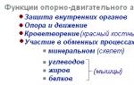
Supportive, protective and motor - these are the three most important functions of the musculoskeletal system that form the body of any vertebrate, without which it cannot exist.
But besides them, the musculoskeletal system also performs the following functions:
- softening, spring during sudden movements and vibrations;
- hematopoietic;
- metabolic (metabolic) - exchange of calcium, iron, phosphorus, copper, important mineral elements;
- biological - ensuring important life processes (blood circulation, hematopoiesis and metabolism).
The versatility of the ODS is caused by the complex structure and composition of bones, their strength, and at the same time lightness and elasticity, the presence of various types of connections between bones (articular, cartilaginous and rigid).
Bone is the cornerstone element of the musculoskeletal system
Bone is a solid living organ in which continuous processes occur:
- bone formation and resorption (destruction of bone tissue);
- production of red and white blood cells;
- accumulation of minerals, salts, water, organic compounds.
Bone has the ability to grow, change and regenerate. So, a small, newly born child has over 270 bones, and an adult has about 206. This is due to the fact that as they grow, many bones lose cartilage and fuse together.
Bone composition
The bones of the musculoskeletal system include the following elements:
- periosteum - outer film of connective tissue;
- endosteum - an internal connective tissue layer that forms the medullary canal inside the tubular bones;
- bone marrow is the soft tissue substance inside the bone;
- nerves and blood vessels;
- cartilage.
All bones are composed of organic (mainly collagen) and inorganic elements. The younger the body, the more organic compounds there are in the bones. In an adult, the collagen content in bones drops to 30%.
Bone structure
The structure of bone under a microscope looks like a set of concentric layers - plates inserted into each other, consisting of protein, mineral substance (hydroxyopatite) and collagen. This structural unit is called an osteon. The inner plate forms the so-called Haversian canal - a conductor for nerves and blood vessels. In total, an osteon can contain up to 20 similar plates, between which there are star-like bone cells. There are also insert plates between the osteons themselves. The lamellar structure, penetrated by the neurovascular Haversian canals, is characteristic of all bone surfaces, both external and internal, except for spongy bones. The presence of channels promotes the active participation of bones in mineral and bone metabolism and hematopoiesis (blood formation).
Cellular structure of bones
There are three types of cells in bones:
- Osteoblasts are immature young bone cells that synthesize the matrix - intercellular substance. They form on the surface of growing bones, as well as in places of bone damage. Over time, osteoblasts become cemented in the matrix and transform into osteocytes. These are the main participants in osteogenesis (bone synthesis).
- Osteocytes are mature, non-dividing, almost non-matrix-producing cells that communicate with each other through the channels of the cavities (lacunae) in which they are located. Tissue fluid circulates between the processes of osteocytes, its movement occurs due to the vibration of osteocytes. Osteocytes are living cells - thanks to them, metabolism is carried out and the mineral and organic balance in the bones is maintained.
- Osteoclasts are huge multinucleated cells that destroy old bone tissue. They, too, like osteoblasts, are important participants in bone formation. A balance must be maintained between osteoblasts and osteoclasts: if there are more osteoclasts than osteoblasts, osteoporosis begins in the bones.
Most bones develop from cartilaginous tissue, except for the bones of the skull, lower jaw and, presumably, the collarbone - they are formed from connective tissue.

Types of bones
The human musculoskeletal system is represented by bones of various types - long, flat, short, mixed, sesamoid.
- Long tubular bones have a rounded, hollow shape when cut. The middle elongated part of the bone (diaphysis) is filled inside with yellow bone marrow. At both ends of the tubular bone there is a head (epiphysis), covered on top with hyaline cartilage, and inside consisting of a spongy substance that contains red bone marrow. The growing part of the bone (metaphysis) is the area between the epiphysis and diaphysis. In children and adolescents, the metaphysis consists of cartilage, which is replaced by bone at the end of growth. The long tubular bones include the bones of the limbs, in particular the longest one, the femur.
- Flat bones are non-hollow, have a thin cut and consist of a spongy substance, covered on top with a compact smooth layer. The scapula, pelvic bones, and ribs have this structure.
- Short bones have a tubular or flattened structure, but there is no single cavity inside them. Cells with bone red marrow are separated by partitions. The short bones include the phalanges of the fingers, the carpus, the metacarpus, the tarsus, and the metatarsus.
- Mixed bones can combine elements of flat and short bones. Mixed bones include the vertebrae, occipital and temporal bones of the skull.
- Sesamoid bones are located deep in the tendon, at the point where it passes through the joint (knee, wrist, foot, etc.), they usually lie on the surface of another bone. Their task is to protect the tendon and strengthen the muscle by increasing the power arm.
All bones have irregularities in the form of protrusions, tubercles, depressions, and grooves. This is necessary for connecting bones and attaching muscle tendons.
A few notes about bone marrow
The bone marrow, unlike the brain and spinal marrow, has nothing to do with the central nervous system; it does not have neurons. This is a hematopoietic organ consisting of myeloid two-component tissue (stroma + hemal component).
In the growing bones of the skull and facial bones, mucous bone marrow is formed - a gelatinous consistency depleted of cells.
Main components of the human skeleton
The skeleton is the static basis of the human musculoskeletal system. The construction of the whole body begins with it. Skeletal anatomy must be adapted to each organ individually and to the entire set of vital systems, providing all the necessary functions of the musculoskeletal system.
Human skull
Let's start with the part that crowns the skeleton - the skull.
Humans are the highest mammals in the evolutionary chain, and this is reflected in our skull. The volume of the adult human brain is about 1500 cubic centimeters, so the brain portion of the human skull is relatively larger than that of animals. Relatively - this is in comparison with the front part. The human lifestyle inevitably led to the fact that in the process of evolution, people's brains grew and their jaws became smaller, because man, having learned to use tools, abandoned raw food.
The brain part of the skull consists of four unpaired and two paired bones fused together:
- unpaired - frontal, sphenoid, ethmoid and occipital;
- paired - two temporal and two parietal.
All the bones of the brain part of the adult skull are connected motionlessly, but in a newborn the sutures remain uncovered for a long time, connecting to each other through “fontanelles” - soft cartilaginous tissue - this is how nature took care of the growth of the skull.
In the occipital part of the skull there is an opening connecting the brain and the spinal cord; arteries supplying the brain with blood also pass through it. The skull is attached to the spine using an elliptical joint. Mobility is provided by the first two cervical vertebrae, called the atlas and epistrophy.
The facial part includes the following bones:
- paired bones: facial jaw, cheekbones, nasal bones, nasal cavity bones, palate;
- unpaired bones: lower jaw, hyoid bone, vomer.
The lower jaw is the only movable articular joint of the skull, and where there is a joint, there are diseases such as arthritis, dislocation, osteonecrosis, etc.
The spine is the basis of the ODS
The spine is the axial rod of the human motor system. Unlike animals, it has a vertical position, which is also reflected in its structure: in profile, the spine in humans looks like the Latin letter S. These natural curves of the spine are designed to counteract the compressive forces to which the vertebrae are continuously exposed. They play the role of shock absorbers and balance the spine when dynamic load increases.

If there were no bends, our spine could break during a normal jump and it would be difficult to maintain balance.
In total, the spine has five vertebral sections and up to 34 vertebrae (maybe a couple less due to the different number of vertebrae in different people in the rudiment of the tail - the coccyx).
- the cervical spine has 7 vertebrae;
- chest - 12;
- lumbar and sacral - five vertebrae each;
- coccygeal - from 3 to 5.
Distribution of curves in the spine
The curvatures of the spine in adjacent sections are oppositely directed:
- cervical spine - the bend is directed forward, it is called lordosis.
- thoracic region - the bend is directed backwards, this is kyphosis. Exceeding the norm is called stooping.
- lumbar region - lordosis;
- sacral region - kyphosis.
Excessive bending in the lumbosacral region can lead to displacement of the vertebrae (spondylolisthesis), hernia, and destabilization of the spinal column.
The flexibility of the spinal column is also controlled by the vertebrae, which are semi-movably connected to each other using cartilaginous plates - intervertebral discs. Dystrophic changes in the discs lead to a catastrophic disease - osteochondrosis, from which all other orthopedic pathologies originate.
Let us now consider the remaining large elements included in the ODS.
The musculoskeletal system includes such important parts of the skeleton as the chest, shoulder girdle, upper and lower limbs, and pelvic girdle.
Rib cage
The chest is the repository of the organs of the chest cavity (heart, trachea, lungs). It is reinforced with a rib frame of 12 pairs of ribs:
- The first 7 pairs in front are semi-movably attached to the sternum;
- The 8th, 9th and 10th pairs of ribs are connected to each other by cartilage;
- the last two pairs are free.
At the back, all the ribs and vertebrae articulate, forming the costoarticular joint.
The thoracic region is inactive, so osteochondrosis in the chest is quite rare, but joint blockage, arthrosis, and intercostal neuralgia can be frequent sources of pain here.
Shoulder girdle
The shoulder girdle consists of two wedge-shaped shoulder blades and two curved clavicular bones, connecting in front to the sternum and behind to the shoulder blades. The upper limb is tied to the shoulder girdle. The shoulder joint is the loosest joint in the human body - this determines multidimensional free movement of the arm, but at the same time it threatens with problems such as shoulder dislocation, glenohumeral periarthritis, etc.
Upper limbs
Everyone seems to know what the upper limbs are made of, but anatomical terms do not always coincide with people’s definitions: many people call the collarbone the shoulder, and the upper arm the forearm. In fact, the hand consists of:
- from the humerus (the upper part of the arm that fits into the shoulder joint);
- the forearm, which includes two bones - the ulna and radius;
- carpal bone.
The hand has a lot of small bones:
- the wrist consists of eight bones, seven of which are arranged in two rows;
- metacarpus - made of 5 bones;
- fingers - from phalanges (two in the thumbs, three in the rest).
Such a terrible disease as rheumatoid arthritis begins precisely in the small wrist joints, so they can be a good indicator of this pathology.
Pelvic girdle
Located approximately in the middle of the body skeleton, the pelvic girdle plays an important role in distributing all loads on the spine (the center of gravity of the body is located just above it) and in balancing the spine. In addition, the pelvis protects important organs of the genitourinary system. Through the caudal foramen at the bottom, the hip and pelvis joint is attached to the spine.
The pelvic girdle consists of fused paired bones - the ilium, ischium and pubis. The hip joint (HJ) is made up of the acetabulum (the socket in the ilium) and the head of the femur.
Problems with the hip joint that lead to disability are coxarthrosis and hip dislocation. In addition, there are congenital anomalies associated with displacements and underdevelopment of the pelvic bones, leading to severe forms of scoliosis.
Lower limbs
The lower limbs include the femur and tibia (tibia and fibula) and feet, connected by the knee joints.
Foot composition:
- seven bones of the forearm, of which the calcaneus is the largest;
- five metacarpal bones;
- 14 phalanges of fingers (two in the big ones, three in all the others).
The knee joint, as well as the ankle, are the most loaded joints in the human body, so arthrosis, tendonitis, heel spurs, sprains and ligament tears make up the lion's share of problems with the lower extremities.
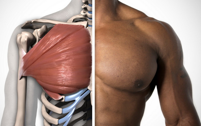
Muscle structure of the ODS
The musculoskeletal system also includes muscles: they are inextricably linked with the skeleton, without them it would simply collapse into a pile of bones. They are also not only a holding force, but also an active driving force.
Muscles consist of elastic tissue, microscopically represented by muscle cells - myocytes.
Muscle types
There are three types of muscles:
- skeletal or striated;
- smooth;
- cardiac.
The movement of absolutely all parts of our skeleton, including facial expressions, is carried out precisely by striated muscles. Skeletal muscles make up the majority of all muscles - there are more than 600 of them, and the total relative weight in the human body is about 40%. The smoothness and coordination of all movements is created due to the presence of agonist and antagonist muscles, which create two multidirectional efforts: the agonists perform the movement, the antagonists resist it.

The motor function of skeletal muscles is caused by their ability to contract in response to a signal from a nerve impulse coming from the central nervous system. The work of the muscles of this group is completely subject to the control of the human brain.
Striated muscles are 70-80% water, and the remaining 20% are proteins, glycogen, phosphoglycerides, cholesterol and other substances.
The most-most muscles of the body:
- The calf and chewing muscles are recognized as the strongest.
- The largest is the gluteal;
- The smallest are the ears;
- The longest muscle is the sartorius muscle, stretching from the ilium to the tibia.
Smooth muscle is a tissue that is part of all internal organs, skin and blood vessels. Spindle-shaped muscle cells make slow movements, not subject to human will and control - they are controlled only by the autonomic nervous system (ANS). Without smooth muscles, digestion, blood circulation, bladder function and other vital processes are impossible.
The cardiac muscle is included in a separate group, since it is striated, and at the same time it is not subordinate to human consciousness, but is subordinate only to the ANS. Also unique is the ability of the muscle to contract when removed from the chest cavity.
Muscle classification
There are a lot of muscles in the human body. They can be combined into separate groups according to their functions, the direction of the fibers, their relationship to the joints and their shape. Let's summarize the classification in a table:
| Classification type | Muscle names |
| By function: | Flexors, extensors, adductors, abductors, rotators, erectors, elevators, depressors, sphincters and dilators, synergists and antagonists |
| By fiber direction: | Rectus, transverse, teres, oblique (unipennate, bipennate, multipennate, semitendinous, semimembranosus) |
| In relation to joints: | One-piece, two-piece, multi-piece |
| By form: | Simple:
|
The human musculoskeletal system is a complex symbiosis of different systems: skeletal, muscular, nervous, and autonomic. It is inextricably linked with a person; any life process depends on it. It is designed simply beautifully, developing with us. There is nothing superfluous in it, so damage to a single part of it can destabilize the entire SDS and cause a number of subsequent diseases.
An abbreviation is a unit of speech that is formed by reducing several words to one or two or three letters each. In Russian speech they can be represented by a variant of a compound word or an initial type. In the first case, a combination of morphemes is assumed, in the second, only the initial letters are taken. What does the abbreviation “ODS” mean? Its decoding for different areas of activity is given below.
The role of abbreviations and classification
So, as already indicated above, the abbreviation can be represented by two main types, and it is also necessary to indicate a third, special case. Thus, we get the classification:
- initial options;
- compound words;
- acronyms.
The latter type is a letter combination that is read and pronounced as a continuous word, and not letter by letter, unlike the initial version. Examples of such abbreviations: NATO (alliance), NASA (space agency), RAS (academy), ABVA (a group from Sweden), VUZ (educational institution) - all these words have long been perceived not as abbreviations, but as commonly used ones.
Examples of words obtained by the complex abbreviation method are: maternity hospital, terrorist attack, collective farm, party committee, regional committee, Komsomol (as we see, during the time of socialist rule, such abbreviations became firmly established in the life of society).
Initial forms, read letter by letter: FBI, FMS, KGB. As a separate form, we can also distinguish such abbreviations that were developed to define an already existing specific single concept, for example, comprehensive automobile insurance except liability (CASCO).
Abbreviations make people's lives easier by shortening complex and long words and phrases, while saving human effort and time.
Reduction of ODS
Speaking about various types of abbreviations, it should be noted that in different areas of human activity the same combinations of letters can have completely different meanings and be in no way connected in meaning.
Thus, the abbreviation JV can be interpreted as a “joint venture” when it comes to economic relations, and as a “north pole” in the context of geography.
The abbreviation ODS implies completely different interpretations, depending on what field of activity we are talking about. Medicine, biology, and construction perceive the reduction of the general labor force differently. The decoding will be different for each industry. Next, we will consider in detail each of the interpretation options.
Medicine
So, reduction of the ODS. The decoding in medicine is simple: the human musculoskeletal system.
This organ system is represented by the bone skeleton and the muscular component, its main functions are:

- Support. The skeleton itself is the main frame of the body, and together with the muscles it literally “holds” the body in the required position, determines the location of the internal organs and fixes them.
- Motor. Thanks to the movable articulation of the vertebrae and joints, as well as through the contraction and work of muscles, the musculoskeletal system provides movement in space.
- Protective. The most important organs - the brain and bone marrow - are under the powerful protection of bone (the skull in the first case and the spine itself in the second). All other organs of the human body are protected in one way or another either by bone (the organs of the chest are protected by the ribs) or by muscles (the abs protect the organs of the abdominal cavity).
Thus, we found out what ODS means in medicine. Decoding in this area reveals the physiological meaning of the concept.
Biology
Let us consider biology as the next branch of knowledge and human activity. The definition says that it is the science of nature, of all living things and of the patterns inherent in organic life. This knowledge system also uses the abbreviation ODS. Biology gives exactly the same interpretation as medicine and interprets the abbreviation as “musculoskeletal system.”
The only difference can be considered that the concept of the musculoskeletal system in biology is a little broader, but in medicine it is specified by the fact that we are talking specifically about a person. In biology, the musculoskeletal system is considered as a set of organs not only of humans, but also of any other animal.

ODS: decoding in construction
What is the interpretation of the abbreviation in the construction industry? When we are talking about the construction of a residential building with an elevator, an industrial building, or a transport facility, the abbreviation ODS is also used. Explanation in this case: combined Represents a set of links for the management or operation of equipment, production, and a transport enterprise. Refers to safety and security systems.

ODS TsUKS - abbreviation decoding
Another area of activity where the ODS reduction is used is saving people. More precisely, the abbreviation is used by people serving in the Ministry of Emergency Situations and in the services under its control. ODS is interpreted in this area as a service.
Often these three letters stand next to others, namely, TsUKS. If we are talking about ODS TsUKS, then we mean the operational duty service of the crisis management center. This unit is engaged in minimizing the consequences of natural disasters and eliminating fires.

Abbreviations occupy a special place in the Russian language. Abbreviations simplify speaking and writing and save time. The same combination of letters can be deciphered differently in different fields of activity, and the abbreviation ODS is proof of this.
The musculoskeletal system ensures movement and preservation of the animal’s body position in space, forms the external shape of the body and participates in metabolic processes. It accounts for about 60% of the body weight of an adult animal.
Conventionally, the musculoskeletal system is divided into passive and active parts. The passive part includes bones and their connections, on which the nature of the mobility of bone levers and links of the animal’s body depends (15%). The active part consists of skeletal muscles and their auxiliary devices, thanks to the contractions of which the bones of the skeleton are set in motion (45%). Both active and passive parts have a common origin (mesoderm) and are closely interconnected.
Functions of the movement apparatus:
1) Motor activity is a manifestation of the vital activity of the organism; it is what distinguishes animal organisms from plant organisms and determines the emergence of a wide variety of modes of movement (walking, running, climbing, swimming, flying).
2) The musculoskeletal system forms the body shape - the exterior of the animal, since its formation took place under the influence of the Earth’s gravitational field, its size and shape in vertebrates are characterized by significant diversity, which is explained by the different conditions of their habitat (terrestrial, ground-woody, air , water).
3) In addition, the movement apparatus provides a number of vital functions of the body: searching and capturing food; attack and active defense; carries out the respiratory function of the lungs (respiratory motility); Helps the heart move blood and lymph through the vessels (“peripheral heart”).
4) In warm-blooded animals (birds and mammals), the movement apparatus ensures the maintenance of a constant body temperature;
The functions of the movement apparatus are provided by the nervous and cardiovascular systems, respiratory, digestive and urinary organs, skin, and endocrine glands. Since the development of the movement apparatus is inextricably linked with the development of the nervous system, when these connections are disrupted, first paresis occurs, and then paralysis of the movement apparatus (the animal cannot move).
The basis of the passive part of the movement apparatus is the skeleton. The skeleton is the bones connected in a certain order that form a solid frame (skeleton) of the animal’s body. The skeleton includes about 200-300 bones (Horse -207), which are connected to each other using connective, cartilage or bone tissue. The skeletal mass of an adult animal is 15%. All functions of the skeleton can be divided into two large groups: mechanical and biological. Mechanical functions include: protective, support, locomotor, spring, anti-gravity, and biological functions include metabolism and hematopoiesis (hemocytopoiesis).
15. Bone structure.
Bone has a complex structure and chemical composition. In a living organism, bone contains 50% water, 28.15% organic substances, including 15.75% fat, and 21.85% inorganic substances, represented by compounds of calcium, phosphorus, magnesium and other elements. Defatted, bleached and dried bone (macerated) consists of 1/3 of organic substances called “ossein” and 2/3 of inorganic substances.
Every bone (Latin Os - bone) is an independent body. It has a certain shape, size, structure. Bone as an organ in an adult animal consists of the following components closely related to each other:
1) Periosteum - periosteum, is located on the surface of the bone and consists of two layers. The outer (fibrous) layer is made of dense connective tissue and performs a protective function, strengthens the bone and increases its elastic properties. The inner (osteogenic) layer of the periosteum is made of loose connective tissue, which contains nerves, blood vessels and a significant number of osteoblasts (osteoforming cells). Due to this layer, development, growth in thickness and regeneration of bones occur after damage. The periosteum firmly fuses with the bone with the help of connective tissue perforating (Sharpey's) fibers that penetrate deep into the bone. Thus, the periosteum performs protective, trophic and osteoforming functions.
A bone without periosteum, like a tree without bark, cannot exist. The periosteum, with the bone carefully removed from it, can again form bone due to the intact cells of its inner layer.
2) Compact (dense) bone substance - substantiacompacta - is located behind the periosteum and is built from lamellar bone tissue, which forms bone crossbars (beams). A distinctive feature of the compact substance is dense arrangement of bone bars. The strength of the compacta is ensured by its layered structure and channels, inside of which there are blood-carrying vessels. In terms of strength, the compact substance is equal to cast iron or granite.
3) Spongy bone - substantiaspongiosa - is located under the compact substance inside the bone and is also built from lamellar bone tissue. A distinctive feature of the spongy substance is that the bone crossbars are loosely arranged and form cells, so the spongy substance really resembles a sponge in structure. Compared to compact bone, it has much more pronounced deformation properties and is formed precisely in those places where compression and tension forces act on the bone. The direction of the bone beams of the cancellous substance corresponds to the main stress lines. Elastic deformations in the spongy substance are much more pronounced (4-6 times). The distribution of compact and spongy substances depends on the functional conditions of the bone. The compact substance is found in those bones and in those parts of them that perform the functions of support and movement (for example, in the diaphysis of tubular bones). In places where, with a large volume, it is necessary to maintain lightness and at the same time strength, spongy substance is formed (for example, in the epiphyses of tubular bones).
4) Inside the bone there is a bone marrow cavity - cavummedullae, the walls of which from the inside, as well as the surface of the bone beams, are covered with a thin fibrous connective tissue membrane - endoosteum. Like the periosteum, the endosteum contains osteoblasts, due to which the bone grows from the inside and is restored during fractures.
5) In the cells of the spongy substance and the bone marrow cavity there is red bone marrow - medullaossium rubra, in which hematopoiesis processes take place. In fetuses and newborns, all bones form hematopoiesis, but with age, gradually, myeloid (hematopoietic) tissue is replaced by adipose and red bone marrow turns yellow - medullaossiumflava - and loses its hematopoietic function (in domestic animals this process begins from the second month after birth) . The ratio between red and yellow bone marrow in one-month-old calves is 9:1, and in adults it is 1:1. Red bone marrow is stored longest in the spongy substance of the vertebrae and sternum.
6) Articular cartilage - cartilagoarticularis - covers the articular surfaces of the bone and is built of hyaline cartilaginous tissue. The thickness of cartilage varies greatly. As a rule, it is thinner in the proximal part of the bone than in the distal part. Articular cartilage does not have a perichondrium and never undergoes ossification. With a large static load, it becomes thinner.
In addition to the 6 components mentioned above, a growing bone also has others that form bone growth zones. In such a bone there is also metaphyseal cartilage, which separates the body of the bone (diaphysis) from its ends (epiphyses), and three types of specially constructed bone tissue in contact with this cartilage and called subchondral bone.
In order to intensify the activity of students in the lesson, a frontal survey is conducted, which helps the children remember previously learned concepts and aims them at further learning new material. At the beginning of the lesson, a problem arises that needs to be solved, which allows students to develop logical thinking and attention. In this lesson, the bulk of the material being studied is written down in the form of diagrams that the teacher builds during the lesson together with the students. The quality of the material being studied is checked in the form of a frontal survey. The lesson is designed for both auditory and visual children.
Lesson methods: problem-search, reproductive, verbal
Forms of work in the lesson: frontal survey, work in pairs, individual work.
Lesson plan:
- Org. moment.
- Updating knowledge – frontal survey.
- Formulation of the problem.
- ODS value.
- Chemical composition of bones.
- Macro- and microscopic structure of bones.
- Construction of cause-and-effect relationships.
- Types of bones.
- Bone growth.
- Consolidation.
- Homework.
Tasks: give an idea of the relationship between the skeleton and muscles, the meaning of the ODS; introduce the classification of bones, show, using the example of the structure of a tubular bone, the connection between the macro- and microscopic structure of bone matter, introduce the chemical composition of bones and identify cause-and-effect relationships.
Equipment: tables “Human skeleton”, “Structure of bones”.
During the classes
I. Organizational moment.
II. Updating knowledge during a frontal survey.
– What is fabric?
Tissue is a group of cells and intercellular substance, similar in structure and origin, that perform common functions.
– What types of fabrics do you know?
There are 4 types of tissues: epithelial, connective, muscle, nervous.
– Give characteristics of connective tissue and its classification.
Connective tissue cells have a well-developed intercellular substance, which determines the mechanical properties of the tissue. This includes supporting tissue - cartilage and bone, liquid - blood, adipose tissue.
– What are organ systems?
An organ system is a group of organs that perform common physical functions.
III. Learning new material.
“Movement is life,” said Voltaire.. Indeed, man is adapted, and perhaps condemned by nature, to movement. People cannot help but move and begin to do this consciously already at 4 months after birth - reaching, grabbing various objects.
– Thanks to what do we move in space, run, walk, jump, crawl, swim, and perform many thousands of different straightening, bending, turning every day?
All this is provided by the musculoskeletal system, or musculoskeletal system.
Therefore, the topic of today's lesson...(the students formulate it themselves and write it down in a notebook, and the teacher writes it down on the board).
– What organs are included in the system of support and movement? (Skeleton and muscles)
1. The meaning of ODS: support and preservation of body shape; movement; protection of organs from injury; hematopoietic. (studies are written down in a notebook)
2. Chemical composition of bones. (A story with elements of a conversation and drawing a diagram)
Conclusion: Based on knowledge of the chemical composition of bones, cause-and-effect relationships can be identified: hardness of inorganic substances + flexibility and elasticity of organic substances = bone strength.
Macro- and microscopic structure of tubular bones. (Story, working with a table).
Working with Fig. 48 on page 46 during the teacher’s story about the macroscopic structure of bone: periosteum, compact substance → spongy substance, medullary cavity, red and yellow bone marrow (their composition, function, location).
Working with Fig. 19 on page 49 of the textbook during the teacher’s story: rounded holes (cylinders - 1), surrounded by concentric rows of bone plates (2 and B); sections of the canals through which blood vessels (3) and nerves pass. Thus, the compact substance consists of numerous tubes, in the walls of which there are bone cells in the form of plates → in the human body, lightness, strength, “saving of material.”
Answer the questions:
– Why is bone tissue a type of connective tissue? (In bone tissue cells, the intercellular substance is well developed, it is hard and durable, in cartilage tissue it is strong and elastic).
– What determines the hardness and elasticity of bones that determine their strength? (From the ratio of organic and inorganic substances).
– Why do the bones of children become more easily deformed, while the bones of old people break more often? (Children have more organic matter in their bones, while old people have more inorganic matter in their bones).
Types of bones, bone growth (Story with elements of conversation, drawing up a diagram)
Bone growth in length due to the cartilaginous tissue at the end parts of the bones, in thickness due to the periosteum.
IV. Fastening:
- Why do the skeleton and muscles belong to a single organ system? (They perform the same functions).
- What are the supporting, protective and motor functions of the skeleton and muscles? (Support and preservation of body shape, movement and protection of organs from injury).
- What is the chemical composition of bones? (organic and inorganic substances).
- At what age are bones strongest? (20 to 40 years old).
- What types of bones do you know and what function do they perform? (Tubular - moving and lifting weights, spongy - supporting, flat - protective).
V. Homework:
§ 10, questions at the end of the paragraph.
VI. Summing up the lesson and grading.
Resources used:
- Kolesov D.V. and others. Biology. Man: Textbook. For 8th grade. general education textbook establishments. – M.: Bustard, 2009.
- Biology. 8th grade. Lesson plans based on the textbook by D.V. Kolesova, R.D. Mash, I.N. Belyaev “Biology. Human. 8th grade.”Part 1/ Comp. I.F. Ishkin - Volgograd: Teacher - AST, 2003.
- Kolesov D.V. Biology. Man, 8th grade: Thematic and lesson planning for the textbook by D.V. Kolesova and others. “Biology. Human. 8th grade" 2nd edition, stereotypical - M.: Bustard, 2003.
- Lesson developments for educational kits “Biology. Man", 8(9) grade, D.V. Kolesova, R.D. Masha, I.N. Belyaeva; A.S. Batueva and others; A.G. Dragomilova, R.D. Masha. – M.: VAKO, 2005.