How to develop a finger after a fracture - tips and exercises. Fractured finger
Fracture is a violation of the linear integrity of the bone under the influence of a force exceeding the tensile strength of the bone. The main cause of fractures in the world is trauma. It ranks third in disease statistics.
Finger fracture is a serious pathology, despite the small size of this part of the body. According to statistics, finger fractures account for 5% of all fractures. Finger fractures are considered severe injuries to the hand, as they significantly reduce its functionality.
Diagnosis of a broken finger, as a rule, does not cause difficulties, but with treatment the situation is different. To completely restore the shape and function of the bone, you must strictly follow all recommendations for the treatment of this pathology. Deviation from treatment requirements leads to serious complications and even disability.
Anatomy of the hand
 The human hand is an extremely complex formation, from an evolutionary point of view. It consists of 30 - 32 bones of various shapes and functions with the help of numerous tendons and muscles arranged in layers. The complex organization of the hand allows movements around all three axes.
 The fingers are topographically related to the hand and significantly increase its functional load. Despite the fact that their bony skeleton allows movements only in one plane, and the radius of movement does not exceed 180 degrees, thanks to the articulation with the hand, the fingers also acquire the ability to make adducting and abducting movements. This organization of the hand significantly increases the range of movements and their accuracy.
Bones and joints of the hand
Topographically, the boundaries of the hand extend from the line connecting the styloid processes of the ulna and radius. Visually, this line crosses the far part of the forearm at the point where a small bony tubercle protrudes on its dorsal surface.The brush consists of three sections:
- wrist;
- metacarpus;
- fingers of the hand.
The wrist normally consists of 8 bones arranged in 2 rows. Proximal ( near) row consists of four bones, forming a kind of semicircle, which is the articular fossa for articulation with the bones of the forearm. These bones include the scaphoid, lunate, triquetrum, and pisiform. The second row also consists of 4 bones, which articulate with the bones of the first row on the proximal side, and on the distal side ( distant) – with metacarpal bones. Among the bones of the second row, there are the trapezoid, trapezoid, capitate and hamate bones. Rarely, x-rays will reveal an additional ninth bone, called the central bone.
Pastern
The metacarpus consists of five tubular bones, slightly curved with a convex outward. All these bones have an oblong triangular body ( diaphysis) and two epiphyses ( end). The proximal epiphyses are thicker than the distal ones and form articular fossae for articulation with the distal row of carpal bones. The distal epiphyses are thinner and form articular heads for articulation with the proximal phalanges of the fingers. On the sides of both the proximal and distal epiphyses there are articular surfaces for connecting the metacarpal bones to each other.
Fingers of the hand
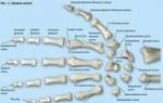
All fingers, with the exception of the thumb, consist of three phalanges - proximal, middle and distal. The thumb lacks the middle phalanx. Each phalanx is a small tubular bone with a body and two ends. Unlike the bones of the metacarpus, the phalanges have only one true epiphysis - the proximal one, and the distal end of the bone does not form an epiphysis. The epiphysis of the proximal phalanges is concave and articulates with the heads of the metacarpal bones. The epiphyses of the middle and distal phalanges are two articular fossae separated by a ridge. The distal ends of all phalanges are flattened and form block-shaped articular heads for articulation with the articular surfaces of the proximal epiphyses of the phalanges. This shape of the joint eliminates lateral movements of the fingers and allows only flexion and extension of the fingers. The distal phalanx gradually narrows and ends with a tuberosity for the attachment of muscle tendons.
In cross-section, the phalanx of the finger is an oblong bone with a canal in the center containing the bone marrow. There is a thin layer of spongy substance around the canal. The spongy substance, in turn, is surrounded by a dense compact substance, which gives the bone density. The diaphysis of the bone is covered with periosteum, rich in blood vessels and nerves. The periosteum is responsible for the growth of bone in width. The ends of the bones are covered with a layer of hyaline cartilage, which has less friction compared to the periosteum and performs a shock-absorbing function ( those. shock mitigation). The small strip of bone tissue located between the epiphyses and diaphysis is called the metaphysis. It, in turn, corresponds to the growth zone responsible for bone growth in length.
Ligamentous apparatus, muscles and their innervation
Due to the fact that there are at least 20 names of hand ligaments, it would be most logical to cover only those ligaments and tendons that are directly related to the work of the fingers.Among the ligaments of the fingers, it is necessary to distinguish only the collateral ones. At one end they are attached to the lateral surfaces of the heads of the metacarpal bones, and at the other to the lateral sides of the proximal phalanges. The interphalangeal joints, like the metacarpophalangeal joints, have their own collateral ligaments, which, like the first ones, are attached to the sides of the articular surfaces above and below the phalanges. The main function of these ligaments is to strengthen the joint capsule and ensure movement in the joint only within permitted physiological limits. Thus, the collateral ligaments prevent dislocation of the metacarpophalangeal and interphalangeal joints during pathological lateral flexion of the finger.
The muscular system of the hand is responsible for the movements of the fingers. It is conventionally divided into the muscles of the palmar and dorsal surface. The muscles of the palmar surface, in turn, are divided into 3 groups - the muscles of the eminence of the thumb, the muscles of the eminence of the little finger and the middle group of muscles. A description of the shape of the muscles, their locations and places of attachment will be omitted due to the complexity and high specificity of this material. If desired, this information can be found in any anatomical atlas. The main emphasis will be on the function of each muscle, since the absence of certain movements during a fracture of the fingers can be used to judge the nerve that is damaged. Also, only those hand muscles that are directly responsible for finger movements will be listed. The remaining muscles of the hand will be lowered.
The following muscles of the eminence of the thumb are distinguished:
- abductor pollicis brevis;
- muscle that opposes the thumb;
- flexor pollicis brevis;
- adductor pollicis muscle.
This muscle performs abduction, slight opposition of the thumb ( movement towards the little finger), and also partially flexes the thumb. This muscle is innervated by the median nerve.
Muscle that opposes the thumb to the hand
The muscle moves the thumb towards the little finger. This muscle is innervated by the median nerve.
Flexor pollicis brevis
The muscle produces flexion of the proximal phalanx of the thumb. Its innervation is carried out partially by the median and ulnar nerve.
Adductor pollicis muscle
The function of this muscle is to move the thumb towards the proximal phalanx of the index finger ( casting) and partial flexion of the proximal phalanx of the thumb. The muscle is innervated by the ulnar nerve.
The following muscles of the eminence of the little finger are distinguished:
- abductor digiti minimi muscle;
- flexor digitorum brevis;
- muscle opposite the little finger.
The muscle produces movement of the little finger to the ulnar side, as well as flexion of its proximal phalanx. Its innervation is carried out by the ulnar nerve.
Flexor digiti brevis
The muscle flexes the little finger and is partially involved in its adduction. Innervation is carried out by the ulnar nerve.
Opponus little finger muscle
The muscle moves the little finger towards the thumb. Innervation via the ulnar nerve.
The muscles of the middle group of the palm are distinguished:
- vermiform muscles;
- palmar interosseous muscles.
Four small fusiform muscles flex the proximal phalanges of all fingers except the thumb and extend their middle and distal phalanges. The two muscles on the side of the elbow are innervated by the ulnar nerve, and the remaining two muscles by the median nerve.
Palmar interosseous muscles
The muscles are responsible for flexing the proximal phalanges of four fingers except the thumb and bringing them to the center line, that is, bringing them into a bundle. Innervation is provided by the ulnar nerve.
The muscles of the rear of the hand are represented by four dorsal interosseous muscles. The two outer ulnar muscles pull the middle and ring fingers toward the little finger. The two outermost muscles on the side of the radius pull the index and middle fingers towards the thumb. At the same time, all four muscles flex the proximal phalanges of all fingers except the thumb and extend their middle and distal phalanges.
Causes of finger fractures
 The most common cause of finger fracture is trauma, and the mechanism of injury is correspondingly direct. An indirect fracture mechanism is present in the rare case when a force acts on different ends of the phalanx, under the influence of which a fracture occurs not in the places of compression, but in the middle of the bone. As a rule, all finger fractures occur at home or at work. In wartime, the frequency of finger fractures practically does not change, which, in principle, is not typical for fractures of other bones. Pathological fractures of the fingers due to metastases of a malignant tumor in the phalangeal bone are theoretically possible, but in practice they are an extreme case.
 Finger fractures are clinically divided into open and closed. A fracture is considered closed when the skin over the fracture site remains intact. Accordingly, an open fracture is characterized by damage to the skin of the finger by sharp bone fragments. Despite the fact that the phalanges are tubular bones that can form sharp ends when fractured, more often than not this does not happen and the fracture remains closed. Presumably this is due to the small size of the phalanges and insufficient leverage to damage the sufficiently strong skin of the fingers from the inside. However, if an open fracture of the finger does occur, then the risk of a complication such as osteomyelitis - inflammation of the bone marrow - increases significantly.
Both closed and open fractures of the phalanges are divided into fractures with and without displacement of bone fragments. Displaced fractures, in turn, are divided into fractures with divergence of bone fragments and overlap of the edges of bone fragments.
Based on the number of bone fragments, the following types of fractures are distinguished:
- splinter-free;
- single-splintered;
- two-splintered;
- comminuted ( fragmented).
- longitudinal;
- transverse;
- oblique;
- S-shaped;
- screw;
- T-shaped, etc.
Symptoms of a broken finger
 Symptoms of a finger fracture are generally identical to fractures in other locations. They are conventionally divided into probable signs of a fracture and reliable ones.
 Possible signs of a fracture include:
- local swelling at the fracture site;
- pain over the fracture site;
- gentle finger position;
- redness at the fracture site;
- warmer skin over the fracture site compared to the surrounding skin;
- inability to move a finger;
- pain when trying to press on its top.
- palpable disruption of bone continuity ( crack);
- visual change in bone shape;
- pathological bone mobility where it should not be;
- bone crepitus ( crunch) when trying to displace bone fragments;
- visual shortening of the broken finger in relation to the healthy finger of the other hand.
Diagnosis of a finger fracture
 Diagnosis of a finger fracture is made according to the above clinical signs. To confirm the diagnosis, an x-ray of the hand or individual finger is taken in frontal and lateral projection. This approach allows not only to determine the presence or absence of a fracture, but also to clarify its exact location, shape and depth. This information turns out to be extremely useful when choosing a treatment method for a patient.
 Theoretically, more modern methods can be used to diagnose a finger fracture, such as computed tomography, but in practice this is never done for two reasons. Firstly, computed tomography is a rather expensive study, and secondly, a simple x-ray in two projections is usually enough to understand what kind of fracture the patient came with and what treatment approach is most appropriate.
It is important to remember that x-rays of the finger must be repeated after removal of the cast in order to monitor the quality of bone fusion and the correct position of the intraosseous fixation devices.
First aid for suspected broken finger
 Providing first aid is the first step in treating a patient with any pathology. How successful the treatment as a whole will be depends on the correctness of measures aimed at alleviating the patient’s condition. A finger fracture is no exception, so first aid will focus on several tasks - eliminating pain, immobilizing the upper limb and combating complications.
 Do I need to call an ambulance?
Many people believe that a broken finger is not a sufficient reason to call an ambulance and, in principle, to seek qualified medical help. Unfortunately, many of these people are mistaken. It is necessary to call an ambulance for the following reasons.The pain syndrome from a broken finger can be insignificant, or it can be so pronounced that it can only be compared with toothache, which is rightfully considered one of the most severe pains. Pain is a factor that may well cause a state of shock, manifested by a sharp drop in blood pressure, sometimes even to zero values. In addition, pain itself promotes the release of biologically active substances into the blood that support inflammation and ultimately increase pain, completing a vicious circle.
In order to reduce pain, the arsenal of emergency medicine contains various painkillers, from the weakest in their effect to the most powerful ones existing today. With a decrease in pain, the activity of the development of the inflammatory process decreases, not to mention the suffering of the patient himself.
Often a fracture of a finger is accompanied by a gross deformation of the usual shape of the finger and is accompanied by deep scratches and abrasions. In this case, doctors or paramedics can clean, disinfect the wound and apply devices to immobilize the fracture.
It is rare, but it happens that the digital artery or one of the digital veins is injured by fragments of broken phalanges. In this case, quite massive bleeding develops, which cannot always be stopped by simply pressing on the bleeding vessel, and even more so if there are several damaged vessels. Emergency workers are trained to stop bleeding by applying a special tourniquet to areas where the main blood vessels supplying the arm lie close to the bone.
What is the best position to hold your hand?
When a finger is broken, there is no specific position in which it is recommended to hold it. The main rule in this case is to ensure the immobility of the broken finger in the position in which it is in a relaxed state. As a rule, if the finger does not change its position, then the pain in it remains at an average level, that is, relatively tolerable.It will also be useful to support the entire upper limb with a bandage or improvised splint. This is done to reduce the mobility of the hand on which the broken finger is located, and, accordingly, to reduce the likelihood of even accidentally touching surrounding structures with the finger. It is also useful to gently pull the shoulder and forearm towards the body using special bandages such as Velpeau and Deso. This manipulation further immobilizes the hand and protects the broken finger.
Is it necessary to give painkillers?
As stated earlier, pain provokes the development of inflammatory processes in damaged tissues, and the inflammatory process leads to increased pain. Accordingly, a vicious circle is formed that must be interrupted to reduce the progression of inflammatory symptoms. To this end, it is necessary for the victim to take either a pain reliever or an anti-inflammatory drug as soon as possible after the injury.At home, the most common anti-inflammatory and painkillers are:
- ibufen;
- meloxicam;
- nimesil et al.
Is immobilization necessary?
In this case, immobilization means temporary immobilization of the fracture site in order to prevent increased pain and the development of complications. This type of immobilization is called transport immobilization, since it is during transportation to a hospital or injury point that there is a high risk of secondary damage to broken phalanges.As stated above, there is no specific position in which a broken finger needs to be fixed. It is important to fix it in the position in which the patient feels the least pain with relaxed hand muscles. To reduce the risk of accidental injury to a finger, it is necessary to immobilize the entire arm and, if possible, keep it close to the body.
As a rule, with a simple closed fracture, immobilization is not applied to the finger itself. However, with complex comminuted fractures, there is sometimes a need for immobilization. Immobilization can be achieved mainly in two ways.
The first method is to apply a narrow and long splint, which can be a stick of medium thickness or a wire 30–40 cm long. One end of the splint is fixed at the broken finger, protruding 2–4 cm beyond its top. The second end rests on the palmar surface of the hand and forearm and is fixed. Then, using a bandage, carefully wrap the arm along with the splint, starting from the elbow edge and slowly moving until the hand and finger are hidden under the bandage.
The second method is simpler, but less effective. It involves tying the broken finger to an adjacent or several adjacent fingers. This fixation method is most suitable for closed finger fractures without displacement of bone fragments.
Do I need to apply cold?
Cold is the first pain reliever and anti-inflammatory agent used by humans. The mechanism of its action is to reduce the temperature of tissues and pain receptors located in them. The latter are able to perceive irritations in the temperature range from 4 to 55 degrees. Accordingly, when the temperature of the nerve receptor decreases to less than 4 degrees, its activity slows down until it stops completely.The mechanism of action of cold differs from the mechanism of therapeutic action of painkillers and anti-inflammatory drugs. Therefore, cold can be safely combined with medications. It is most convenient to use ice for this purpose. Moreover, it is desirable that the ice be crushed and placed in a waterproof bag or heating pad. Crushed ice takes the shape of the area of the body it is applied to much better. As a result, the area of contact between the skin and ice increases and faster and better pain relief occurs at the fracture site.
It is important to remember that extremely low temperatures affecting living tissue for a long time can lead to frostbite. In order to avoid such a complication, it is necessary to remove the ice pack every 5 - 10 minutes for 2 - 3 minutes.
Treatment of a fractured finger
 Treatment of a finger fracture is carried out using various methods, depending on its complexity and associated complications.
 Traditional methods of treating a broken finger are:
- one-stage closed reduction;
- skeletal traction methods;
- open reduction.
One-stage closed reduction
 Simultaneous closed reposition of bone fragments is carried out for simple closed fractures with displacement. The classic displacement of fragments in such a fracture occurs towards the palmar side, that is, at an angle open to the back of the hand. Closed reduction is carried out in several stages. First, a test is made to determine the patient's tolerance to a local anesthetic. More often, medium concentrated solutions of procaine and lidocaine are used for this purpose. In the absence of an allergic reaction to the anesthetic, it is gradually injected into the tissue surrounding the fracture.
 When pain relief is achieved, traction is performed ( traction) finger along its axis. Then slowly bend all the joints of the finger until an angle of approximately 120 degrees is achieved. After this, pressure is applied to the angle of the fracture until the bone returns to its original position, and then it is fixed. Immobilization is carried out with a plaster splint from the upper third of the forearm to the base of the fingers. Subsequently, only the damaged finger is fixed in a partially bent position, while the rest remain free. Immobilization of healthy fingers is considered a mistake because it leads to the development of ankylosis ( shortening and hardening of the ligamentous apparatus, preventing full movement of the limb). Upon completion of the manipulation, the patient is recommended to keep the limb in an elevated position for 2–3 days to reduce swelling, and also take painkillers in medium dosages indicated in the accompanying instructions.
Skeletal traction methods
 This treatment method is used for comminuted closed fractures or when, after one-step reduction, it is not possible to fix the bone in the correct position. As in the previous case, a test is carried out to determine the tolerance of the anesthetic substance. In the case when it turns out to be negative ( no allergic reaction develops), the same splint is applied to the forearm and hand as in the previous treatment method, but with one modification. A strong wire is attached to its palmar surface opposite the broken finger, extending several centimeters beyond the top of the finger and ending in a hook or loop.
 Reposition of fragments is carried out in a similar way, with the same anesthesia, only after this the finger is stretched using a thread, pin or staples passed through the soft tissue of the finger or nail phalanx. For a more durable fixation of the structure, the nail is covered with several layers of polymer varnishes, which are used in cosmetology for nail extensions. After the manipulation, the patient is prescribed a preventive course of antibacterial, anti-inflammatory and analgesic treatment.
Open reduction
 This treatment method is the last one that doctors resort to for broken fingers. The fact is that open reposition, in essence, is a surgical intervention on an open bone and is accompanied by all the complications characteristic of operations in principle - wound suppuration, suture failure, osteomyelitis, etc. However, for certain indications, this method is the only possible in the treatment of finger fractures. Typically, these indications include an open simple or comminuted displaced fracture, a malunion fracture requiring bone destruction and repositioning, and purulent complications of previous treatment methods.
 This procedure is carried out according to all the rules of a full-fledged surgical intervention under general anesthesia. Fixation of bone fragments is carried out more often with knitting needles, less often with screws. External fixation device ( Ilizarov apparatus) can also be used for a broken finger. Its advantage is that it reliably fixes bone fragments and does not require the application of plaster, which prevents the wound from rotting and the development of suppurative processes in it. However, the disadvantage of the Ilizarov apparatus is that it requires careful daily treatment, since it itself is a foreign body and a potential source of an inflammatory reaction.
Is it necessary to apply plaster?
 Proper treatment of finger fractures always involves applying a cast. A finger fracture is a fracture of high complexity, so treatment should be taken as seriously as possible. In order to achieve the best results, it is necessary to reliably immobilize the fracture site.
 The most common material for applying an immobilizing bandage is a bandage soaked in a concentrated plaster solution. When dried, the plaster takes the shape of the limb and for a long time retains the necessary structural rigidity to ensure the required level of immobilization. In addition to plaster, there are other substances used to fix the upper limb in case of finger fractures. We are talking about special polymers that are applied like a plaster cast, but without using a bandage. After drying, the strength of the polymers is not inferior to gypsum, and the weight of the structure is several times less. In addition, when using it, there is no need to protect this material from liquid ingress, as when using gypsum, which is destroyed in this case. It goes without saying that modern polymer materials for immobilization are not available in every hospital. In addition, they are most often not covered by the health insurance policy and must be paid for from the patient’s budget.
As mentioned above, when a finger is broken, plaster is applied starting from the near part of the forearm, moves to the hand and ends with a separate fixation of only the broken finger. In this case, it is important to initially take care of the correct position of the brush, since once the plaster hardens, it will no longer be possible to change it. The correct position of the hand involves extension of the wrist joint by approximately 30 degrees and flexion of the phalanges of the fingers ( if skeletal traction methods were not used) until the tops of the fingers lightly touch the palm. This position of the hand ensures the prevention of repeated displacement of bone fragments, as well as the prevention of contractures. If contractures do develop, this position of the hand allows you to maintain its grasping function.
How long is a cast needed?
For simple closed fractures of the fingers without displacement, the duration of plaster immobilization is on average 2–3 weeks. Full restoration of ability to work occurs in 3–4 weeks.For fractures of moderate complexity, namely closed simple and comminuted fractures with displacement, as well as fractures requiring skeletal traction, plaster is applied for an average of 3 - 4 weeks with restoration of working capacity for 6 - 8 weeks.
For complex open comminuted fractures using osteosynthesis methods ( restoration of bone integrity using the implantation of wires, screws, etc.) the period of wearing a cast sometimes reaches 6 weeks, and full restoration of the finger’s ability to work occurs in 8–10 weeks.
Complications of self-treatment of a finger fracture
 The treatment of finger fractures should be approached with all responsibility, since careless treatment often leads to the development of complications. Some of them cause the patient many times more inconvenience and even suffering than the fracture itself.
 The most common complications of self-treatment of a finger fracture are:
- formation of a large bone callus;
- formation of a false joint;
- formation of contracture;
- formation of ankylosis;
- improper bone fusion;
- osteomyelitis, etc.
The formation of callus is a normal physiological stage of the healing of any fracture. However, if bone fragments are incorrectly displaced, a giant bone callus is formed. Its development occurs as a compensatory reaction of the body. In other words, the body is interested in restoring the strength of the damaged bone, but if the fragments are not aligned correctly, the axis of the bone also changes. Along with the change in the axis, the maximum permissible load on the bone decreases. In order to compensate for the loss of functional load, the bone is forced to strengthen the fracture site more strongly, resulting in the growth of callus. In addition to an aesthetic defect, callus often limits the movement of the finger, reducing its participation in the activity of the entire hand.
Pseudarthrosis formation
A false joint is a place where there is free flexion of a limb where normally there should be no flexion. False joints form when closed fractures of the phalanges are not sufficiently immobilized. As a result, the movement of bone fragments at the fracture site continues and their gradual rubbing against each other. Over time, the sharp ends become blunt and even rounded, and the bone canal becomes overgrown. At a certain point, one solid bone becomes two shorter bones, with a small gap between them. It is thanks to this gap that movement between fragments of the once intact bone is maintained.
Unfortunately, the pseudarthrosis is functionally incompetent, painful and is a constant source of inflammation in the body. The annoying thing is that the treatment for this complication is only surgical and consists of destroying the edges of the false joint and re-combining the bone fragments. The success of such an operation is always questionable due to the fact that after it a large bone callus is formed, the bone, and therefore the limb, is shortened and the risks of developing a secondary iatrogenic ( caused by medical procedures) osteomyelitis.
Formation of contracture
Contracture is the shortening of the tendons and ligaments of a limb or a certain part of it due to inflammation or prolonged inactivity. In case of a fracture of a finger due to incorrect position of the hand during immobilization of the upper limb, uneven tension of its tendons occurs. Some tendons become tense, others relax and shorten over time. After removing the plaster, those tendons that were stretched do not interfere with movements in the joint, and those that have shortened do not allow voluntary movements in the direction opposite to the ligament. Treatment of contractures is long and painful, as it is associated with daily stretching of the shortened tendons.
Formation of ankylosis
Ankylosis is the fusion of the articular surfaces of a certain joint and the formation of solid bone at the joint site. This complication can develop when a fracture involves a joint and is not treated appropriately. As a rule, most patients become disabled for life, since there is currently no effective treatment for this complication.
Incorrect bone fusion
For open fractures and closed fractures with displacement, a mandatory stage of treatment is repositioning of bone fragments. Reposition means returning bone fragments to their original physiological position. In the absence of reposition of fragments, poor-quality reposition or weak immobilization, displacement of one of the bone fragments occurs ( more often distal) away from the correct axis. If the bone is kept in this position for several weeks, the fracture heals, and the distal fragment remains forever in the wrong position. In addition, a large bone callus forms, preventing normal movement of the finger.
Osteomyelitis
Osteomyelitis is the development of inflammation of the bone marrow. A distinction is made between primary hematogenous osteomyelitis, in which pathogenic bacteria are introduced into the bone marrow through the blood, and secondary traumatic or iatrogenic osteomyelitis, in which bacteria enter the bone marrow from surrounding objects and the atmosphere during injury or surgery. With an open fracture of the finger, the development of secondary osteomyelitis is most likely due to the absence or insufficiency of primary wound treatment. This disease is very painful and often becomes chronic with frequent phases of exacerbation. As a rule, exacerbation occurs after the bone has fused. Inflammation increases pressure in the bone canal of the phalanges of the fingers and bursts the bone and the surrounding periosteum from the inside. The pain is so severe that it can only be reduced by large doses of opiates ( morphine, omnopon), and patients sometimes even beg to have the painful part of their body amputated.
Treatment is exclusively surgical and temporary. In some cases, in order to reduce pressure in the medullary canal, small holes are drilled, the canal is drained and washed for a long time with solutions of antiseptics and antibiotics, after which the access is closed. However, in some cases, when the bone heals, osteomyelitis relapses ( reoccurs). In other cases, after removing the purulent contents of the bone marrow canal, a part of the nearby muscle is placed in it and the wound is sutured. In this way, the frequency of relapses of osteomyelitis is reduced, but complications arise associated with the multi-stage nature and technical difficulties of performing this surgical intervention.
How long is the recovery period after surgery?
 The type of surgical treatment for a finger fracture largely influences the duration of the recovery period. In addition, purulent complications have a great influence, which can cause multiple repeated operations aimed at cleansing the purulent focus. An important factor influencing the rate of recovery after surgery is the patient’s age and concomitant pathologies. Thus, in children, the rate of bone fusion and tissue regeneration is the highest. In people under 40 years of age, the rate of recovery remains at a fairly high level, and then slowly decreases every year. Diseases that cause slower regeneration of bone and connective tissue include diabetes mellitus, hypothyroidism, parathyroid tumor, etc.
 Osteosynthesis using wires and screws can be either one-stage or two-stage. With one-stage osteosynthesis, the fixation devices remain in the patient’s bone for life, and with two-stage osteosynthesis, they are removed 3–4 weeks after injury through repeated minimally invasive surgical access. Accordingly, with one-stage osteosynthesis, the recovery period lasts on average 4–6 weeks, and with two-stage osteosynthesis it extends to 7–8 weeks.
Osteosynthesis using a device for external fixation of bone fragments is always a two-stage process. In addition, its use increases the risk of septic complications, which can also delay recovery. Based on the above, with a successful healing of the fracture, the period for restoration of working capacity is on average 6 – 8 weeks. With constant moderate inflammation, recovery time is delayed by 1 to 2 weeks. In case of severe inflammation and suppuration of the wound, it may be necessary to re-open the wound and cleanse the purulent focus. In this case, full recovery is delayed by 4 to 6 weeks and may ultimately take 10 to 14 weeks.
In the case of rupture of ligaments or muscle tendons and their suturing during surgery, in the recovery period, as a rule, there is a significant shortening of them. As a result, after healing of the fracture, the patient is not able to fully use his fingers, since their mobility is limited. Tendon development can also take up to two weeks, which must be added to the time the plaster immobilization is removed. On average, the period of complete recovery is 6–8 weeks, depending on the severity of the fracture itself.
What physical procedures are indicated after a fracture?
 Physiotherapy greatly helps speed up the treatment process for any fracture. The physiotherapeutic effect is based on the influence of natural factors on the bone and the influence on the rate of metabolic processes in it. The positive effect of physiotherapy is manifested in analgesic, anti-inflammatory, decongestant, myostimulating, trophic and other positive effects.
 Physiotherapy for a broken finger
| Type of procedure | Mechanism of therapeutic action | Duration of treatment |
| UHF (ultra high frequency therapy) | Deep heating of bone and surrounding soft muscle tissue. Acceleration of metabolic and regenerative processes. Improving blood supply and tissue oxygenation. Moderate anti-inflammatory and analgesic effect. Relaxation of the smooth muscles of blood vessels. Acceleration of callus formation. | Starting from 3 days after reposition of fragments. 10 – 15 procedures. Daily. The duration of the procedure is 10 – 15 minutes. At low radiation intensity there is an anti-inflammatory effect. With moderate intensity radiation, metabolic processes are predominantly stimulated. |
| Physiotherapy | It is performed only on healthy fingers to prevent contractures. Improving microcirculation and blood supply to tissues. Maintaining optimal levels of cellular metabolism. | From 3 days after reposition of fragments. Daily. 10 – 20 procedures. The duration of the procedure is 5 – 10 minutes. |
| Warm baths with soda and salt | Analgesic effect by reducing the sensitivity of pain receptors. Pronounced anti-inflammatory effect aimed at joints and bones. Relaxation of vascular smooth muscle. Improving blood supply to tissues. Moderate fibrinolytic effect aimed at softening ligaments and treating ankylosis. | Apply starting from the day the plaster is removed. 12 – 15 procedures. Every day or every other day. The duration of the procedure is 10 – 15 minutes. Water temperature is within 35 – 39 degrees. |
| Exercise therapy | Development of contractures of the elbow, wrist and hand joints. Reorganization of connective tissue of ligaments and tendons. Stretching of the joint capsule. | Apply starting from the day the plaster is removed. 15 – 20 procedures. Every day or every other day. The duration of the procedure is 15 – 20 minutes. |
| Ozokerite applications | Superficial and deep heating of tissues. Vasodilator effect. Improving the metabolism of bone and muscle tissue. Reflex effect on nerve centers. Increasing the body's resistance to aggressive factors. | 3 – 5 days after removal of the plaster. 8 – 10 procedures. Daily. The duration of the procedure is 10 – 15 minutes. |
| Mechanotherapy | Restoring fine motor activity and sensitivity through various manipulations of small objects. Restoration of coordinated muscle work after a long period of rest. | After removing the plaster. 15 – 30 procedures. Daily. The duration of the procedure is 15 – 20 minutes. |
A fracture of the thumb is a fairly common injury. Due to the structure of the hand, fingers are often damaged, and young children are especially susceptible to this phenomenon. How to identify a fracture and distinguish it from a bruise? What are the signs of injury and how should it be treated?
A finger on your hand can be broken as a result of a bruise or blow. Such injuries are often domestic in nature. There are also pathological injuries that occur against the background of age-related changes and diseases of bone tissue (arthritis, arthrosis, osteomyelitis, etc.)
According to the established classification, a finger fracture can be:
- Closed - internal damage in which the integrity of the skin is not compromised.
- Open fracture - accompanied by the presence of a wound surface and bleeding. The skin is damaged and bone fragments are often visible in them. This type of damage is more dangerous because there is a high risk of infectious complications.
In addition, there are finger fractures with displacement and without concomitant displacement of bone fragments.
Based on the location of the injury, this injury is divided into the following categories:
- Fracture of the nail phalanx.
- Fracture of the phalanx of the main finger.
- Violation of the integrity of the bone in the proximal phalanx.
Depending on the fracture line, marginal, comminuted, longitudinal, transverse, oblique and helical fractures are distinguished.
The most common bone injuries in medical practice are the little finger, which is due to its anatomical marginal location. This injury is often accompanied by injuries to the ulna.
A fracture of the ring finger is considered one of the most dangerous, since if the basic rules of rehabilitation are not followed, the injured hand may lose its functionality and motor activity.
Fractures of the middle finger and index finger are widespread and can be treated quite well. But a broken phalanx of the thumb will take longer to heal, and to restore it, hardware traction is first used, before applying plaster.
How does it manifest?
Traumatologists identify the following symptoms of a finger fracture:

- Sharp painful sensations;
- Hemorrhages under the nail;
- Deformation;
- Pathological mobility;
- Edema;
- Movement disorders;
- Detachment of the nail plate;
- Crepitus;
- Hematomas.
A broken finger will appear swollen and visually shortened and cannot straighten. The victim experiences severe pain even when trying to move his healthy fingers. As a result, the person loses the ability to move the hand.
How to distinguish a fracture from other injuries? It is the deformation, change in shape and inability to move the hand that makes it possible to distinguish a bruise from a violation of the integrity of the bone. With open injuries, bleeding is observed. A finger fracture with accompanying displacement can be identified by the characteristic curvature of the phalanx.
If your little finger is broken, your elbow will hurt and it will be difficult to straighten. The greatest harm comes from concomitant damage to nerve endings. With such a complication, a violation of the integrity of the finger bone will have the following symptoms:
- Numbness;
- Reduced sensitivity indicators;
- Convulsive syndrome;
- Paralysis;
- Reflex muscle tremors.
Having discovered the first signs of a broken finger, the victim should be given the necessary first aid, and then taken to the emergency room, where, after a preliminary diagnosis, the doctor will determine what treatment is required in a particular clinical case.
What is the danger?
If left untreated, traumatic injuries of this type of limb can cause many complications. The most common consequences are:

- Post-traumatic arthrosis;
- Osteomyelitis;
- Impaired motor activity;
- Inflammatory processes;
- Complications of an infectious, purulent nature;
- Formation of a false joint;
- Callus.
An improperly healed fracture threatens to impair mobility and function not only of the injured area, but also of the entire limb.!
How can I help you?
If your fingers are injured, the first step is to immobilize the broken area. If we are talking about an open type injury, then before this the wound is treated with antiseptics, a sterile bandage is applied, and the bleeding is stopped.

How to apply a splint for a fracture? To make a splint, use any available materials (for example, pens, pencils, sticks). An improvised transport splint is attached to the damaged area using a bandage or a strip of fabric from clothing.
Patients ask: is it possible to move an injured finger? Experts categorically do not recommend doing this, so as not to aggravate possible displacement or provoke additional tissue damage. It is necessary to ensure maximum rest for the limbs, avoid any stress and movements.
To alleviate the general condition and relieve pain, you can take an analgesic tablet. Ice and a cold compress are applied to the injured area - this will relieve swelling, prevent the appearance of extensive hematomas and slightly ease the pain.
Features of treatment
Treatment for a fractured finger involves applying a plaster cast. If a patient is diagnosed with a displaced finger injury, then first a reposition is performed, during which a specialist, using a certain method, combines the bone fragments. The operation takes place under local anesthesia.

In difficult cases, if the bones are not aligned, they are fixed using special knitting needles. After 3 weeks, the metal structures are removed and a plaster cast is applied again. If the nail phalanx and plate are damaged, its edge is carefully trimmed and plaster is applied in a straight position to minimize the risks of possible divergence of fragments.
Further therapy is predominantly medicinal in nature. To avoid possible infectious or inflammatory complications, patients are advised to take antibiotic, anti-inflammatory non-steroidal, painkillers, vitamin-mineral complexes and chondroprotectors prescribed individually by the attending physician.
Recovery and rehabilitation
How long does it take for a finger fracture to heal? The answer to this question depends on many factors, such as the type and severity of the injury, its location, and the method of treatment. The age category of the patient is also of great importance. The child's bone tissue is restored as quickly as possible. In older people, healing may take significantly longer.

On average, fusion of the finger bones takes about a month. If the fracture is complicated by concomitant displacement, then the applied plaster is worn for 1.5-2 months.
The rehabilitation course after a fracture of this type necessarily involves physical therapy. The following procedures may be recommended for patients:
- Ultraviolet irradiation;
- Magnetic therapy;
- Laser therapy.
A good therapeutic effect is provided by warm salt or soda baths for the hands, which can reduce swelling, relieve pain, and speed up regeneration processes. You can do such baths at home, 2 times a day.
To warm up the muscles and prevent possible contracture, a course of therapeutic massage is carried out. In order for the motor activity and functions of the injured limb to be fully restored, the patient must regularly perform exercises aimed at developing fine motor skills.
Good results are obtained by sculpting or kneading plasticine, fingering rice grains, turning with the palm, flexing and extending joints, making circular movements, clicking, etc.
A broken finger is a painful and unpleasant occurrence. However, timely, competent treatment and full rehabilitation will allow you to avoid any complications, achieving complete restoration of the functionality of the limb in the shortest possible time!
Fingers play a very important role in our daily lives as they help us grasp and hold objects. A person touches things with them and interacts with the outside world in many ways, which makes this part of the body vulnerable to injury. Damage to fingers varies: from minor bruises or contusions to something as serious as a dislocation or fracture. The latter is a fairly common phenomenon in competitive sports or extreme sports such as skiing, skateboarding, etc.
Fractured finger - description of the injury
To understand the anatomical structure and operation of the finger, you need to know that it is controlled by tendons, which are responsible for attaching the bone to the muscle. The bones in the fingers are called phalanges and are connected to each other by ligaments. There are no separate muscles in this part of the hand. The muscles of the forearm are actually attached to the fingers by tendons, which allows them to move.
Each finger has three phalanges, and only the thumb has two.
There are three bones in the finger, called the proximal (main), middle and distal (ungual) phalanges, they are located in series. The thumb is an exception because it consists of two phalanges.
It is noteworthy that the thumb is the least susceptible to injury. Often it is the little finger that suffers from fractures - due to its extreme location on the hand and thin bone, it is especially vulnerable to various types of damage.
The term "broken finger" is applied when any of the three bones of the finger are broken. In the case of a simple fracture, treatment is quite simple and healing occurs quickly. Complications arise when cracks and fractures occur near the joints, making therapy difficult. Intra-articular cracks provoke hemorrhage into the joint cavity, and it becomes inflamed. As a result, the soft tissue around the injury swells greatly. If the cartilage is also injured, the joint may undergo degenerative changes in the future.
With regard to some types of fractures, it is difficult to understand: only the phalanx or also the intra-articular surface was damaged. Failure to promptly recognize and properly treat these injuries risks the prospect of partial or complete loss of function.
What types of fractures are there?
The following are the different types of finger fractures:
- avulsion - occurs when there is excessive tension on the tendon or ligament attached to it;
- longitudinal - due to impact (applied force) - when the broken ends of the finger bone are located in one line;
- with a fracture - if, under the influence of an applied force, the finger bone is divided into two differently directed parts, which can be located perpendicularly or at an angle to each other;
- comminuted - when the bone breaks into three or more parts;
- crushed - when a large area of bone is destroyed, with the formation of many fragments;
- open fracture - the bone breaks through the skin;
- closed fracture - the skin remains intact, the bone is not visible from the outside;
- undisplaced or stable fracture - the finger bone breaks, but without divergence of the fragments;
- Displaced fracture - the bone breaks into separate pieces that do not align on their own, they need to be “assembled.”
The main types are listed above, but there are also combinations of different types of fractures with each other.
The most common types of finger fractures
There are typical types of fractures that are often encountered in clinical practice. It is useful to have a clear understanding of them.
Comminuted fracture of the fingertip
A comminuted fingertip fracture is the most common type of distal phalanx fracture.
X-ray of a comminuted fracture (shown by arrow) - the most common type of nail phalanx fracture
These fractures are stable and can be treated by applying a simple splint around the DIP joint (distal interphalangeal joint) only. In this case, you need to immobilize the injured finger for about 2–4 weeks. This type of fracture is often combined with injuries to soft tissues and nails - subungual hematomas, ruptures of the nail bed, etc. They must be separately treated accordingly by a doctor.
You need to know that fingertip fractures are often complicated by hyperesthesia - increased sensitivity to irritants, pain and numbness for up to six months after the injury.
Avulsion intra-articular fracture of the nail phalanx
Two tendons are attached to the nail phalanx of each of the four fingers - the extensor on the dorsal surface, and the flexor on the palmar surface. This type of fracture is also called a “hammer finger” or “baseball finger” because it often occurs as a result of a ball hitting the tip of a straightened finger. And the finger itself turns out to be broken at the joint, shaped like a hammer. This injury is characterized by a separation of the extensor tendon and a bone fragment at the dorsum of the finger.
Avulsion intra-articular fracture - the bone fragment is closer to the dorsal surface
Treatment of an intra-articular avulsion fracture involves splinting the DIP joint for eight weeks. It is extremely important that the splint is kept on the finger at all times, as any careless bending can affect healing and prolong the treatment period. After applying the splint, it is necessary to take an x-ray to confirm the correct fit of the bone fragments.
Some doctors believe that it is necessary to treat this type of fracture surgically if more than 30 percent of the articular surface is damaged. However, during research it was revealed that conservative therapy (splinting) in this case is optimal, because the result is not inferior to the outcome of a similar injury after surgery.
Such a fracture usually occurs due to the forced bending of the fingertip in the joint backwards (in an unnatural direction for it). The flexor tendon is torn off with a fragment of bone at the palmar surface of the nail phalanx.
Avulsion fracture of the deep flexor - the bone fragment is closer to the palmar surface
With this type of injury, the finger temporarily loses the ability to bend in the damaged joint. Due to the risk of contraction (retraction) of the tendon, surgical treatment of the injury is necessary.
Fractures of the middle and proximal phalanx
Fractures of the middle and proximal phalanx are often associated with trauma. Such damage is distinguished by gross external deformation when examining the indicated phalanges. These fractures are usually classified as intra- or extra-articular. Intra-articular ones are often complex and unstable and must be treated surgically. Extra-articular fractures can be undisplaced or displaced. Stable, undisplaced fractures can be treated conservatively with splinting and early immobilization. But they should be monitored to ensure proper union. Displaced, oblique or spiral fractures are inherently unstable and require surgical treatment.
Reasons and factors
Common causes of finger fractures:
- Direct trauma or a fall directly onto a finger is the main cause of fractures.
- An instinctive desire to prevent a fall by stretching your arms forward - the hand hits the ground or another object in front.
- Contact sports are the most common cause of fingertip fractures.
- Deliberately twisting a finger backwards with force.
- Accident at work, or occupational injury while using a heavy tool, such as a drill or chainsaw.
- Getting your finger caught in a door or heavy object.
Osteoporosis and bone calcium deficiency are major risk factors.
People with osteoporosis are more likely to fracture the middle of their finger than the tip.
Symptoms and first signs
The main symptoms of a broken finger are:

Diagnostics
The basis for diagnosing finger fractures is x-rays. The type of damage will determine the treatment. Each clinical picture has specific characteristics that the doctor must detect. Diagnostics in the office of a surgeon or orthopedist includes:
- physical examination - during which the doctor evaluates the injury and determines the severity of the fracture. The doctor takes into account the range of motion of the victim's finger by asking him to clench his hand into a fist. The surgeon will also evaluate visual signs such as swelling, bruising, and deformity. Using palpation, he will look for signs of decreased blood circulation and determine the possible area of nerve damage;
- hardware methods - used to confirm the diagnosis or staging in cases where the physical traumatologist could not accurately determine the presence of a fracture. These include:
- X-ray is the main method for diagnosing fractures. The doctor will place the affected finger between the X-ray source and the detector to create an image. The procedure takes only a few minutes and is painless;
- CT scan, or computed tomography, is done by combining X-rays that scan different angles of the injury. The physician may decide to use a CT scan if the initial x-ray is inconclusive or when there is concern that there is also soft tissue damage associated with the fracture;
- An MRI will be required when there is a suspicion of a hairline fracture, or a stress fracture that occurs after repeated trauma over time. An MRI can show finer details, including soft tissue injuries.
Treatment
The faster first aid is provided in case of injury, the better and more effectively the fracture will heal. Therefore, it is important to know the algorithm of actions and carry it out confidently step by step, but quickly enough, since the result also depends on the speed of response. It should be noted that in case of adequate treatment, the first signs of fusion of bone fragments are noticeable 3 weeks after the injury, but the bone will finally recover in approximately 2 months.
First aid
The basics of first aid are ice, a soft compressive bandage, and elevation of the limb. These measures will help control swelling. You should also make sure that the finger is immobilized. The sequence of actions should be like this:

You should not use your finger in normal daily activities until your doctor examines it.
Basic treatment
After the doctor examines the fracture and makes a diagnosis, you will need a special splint for fixation. A temporary splint - pending a doctor's visit - can be made from a popsicle stick and a loose bandage. The bandage itself is needed to immobilize the broken finger to prevent further damage.
Splinting
The type of splint needed depends on which toe is broken. For the treatment of minor fractures, a “friendly bandage” will be sufficient, when the damaged finger is tied to the neighboring one, thus immobilizing it.
A long splint for the flexor-extensor ligaments will help the injured finger not to snap back.
A long splint for the flexor-extensor ligaments will prevent your finger from breaking
A soft splint is needed to keep the injured finger slightly curved towards the palm and is held in place by soft fastenings.
Aluminum U-shaped splint holds the finger firmly in place
The aluminum U-shaped splint is flexible and fixes the damaged finger. It is attached to the back of the finger to keep it stationary.
In more severe cases, the doctor may use a rigid fiberglass splint that runs from the finger to the wrist. Outwardly, it resembles a mini-plaster.
Rigid fiberglass splint resembles plaster
Drug treatment
To help a patient cope with the pain of a fracture, a doctor may recommend taking nonsteroidal anti-inflammatory drugs (NSAIDs). They work by reducing the negative effects of long-term inflammation, pain, and pressure on nerves and other soft tissues. NSAIDs do not interfere with the healing process. Common over-the-counter nonsteroidal anti-inflammatory drugs used to treat fractures include ibuprofen and naproxen. Paracetamol can also be used, but it is not an NSAID and does not reduce inflammation.
If the patient experiences severe pain, the doctor will write a prescription for codeine-containing medications (Pentalgin, Nurofen Plus, Sedalgin) for short-term treatment. The discomfort will most likely be quite severe in the first days, but will gradually begin to decrease. Thus, potent analgesics may only be needed at the beginning of treatment.
In general, you must strictly adhere to your doctor's instructions. Sometimes he will schedule a follow-up examination a few weeks after the start of treatment. May additionally order an x-ray to monitor healing, usually 1-2 weeks after the injury.
Surgery
Surgery is necessary to treat difficult cases where immobilization and time cannot effectively correct the damage. Surgical intervention is indicated for such types of fractures as:
- combined;
- unstable;
- with splintered bone fragments;
- with joint involvement.
These injuries require surgery because the individual fragments must be put back in place so the bone can heal in the correct configuration. Surgery may also be required if the bone does not heal properly.
The operation to restore bone is called osteosynthesis (osteo - bone, synthesis - create, restore). During surgery, the doctor compares the broken fragments and fixes them with special metal structures. Securing individual parts or fragments occurs using metal plates, knitting needles or screws. Sometimes different combinations of these fixation structures are needed.
If for some reason surgery is not possible, then there is another way to fasten the fragments - percutaneous insertion of millimeter titanium wires.
Folk remedies
Some folk remedies can help speed up the healing of fractures. However, they are not a method of treatment, but only have an auxiliary effect, along with the use of complex medical therapy.
- A pineapple. You need to eat half a pineapple every day until complete recovery. This fruit contains bromelain, an enzyme that helps reduce swelling and inflammation. Canned or processed pineapples should not be consumed.
Pineapple will help with inflammation and swelling, thanks to the presence of the enzyme bromelain.
- Calcium. Best sources of calcium: Most legumes and green leafy vegetables such as Brussels sprouts, broccoli, cabbage, mustard greens or kale. These foods are also rich in magnesium, which serves as a good bone healing supplement.
Green leafy vegetables are a generous source of calcium
- Essential oils. Application of immortelle, fir and cypress oils to injured areas accelerates the healing process and strengthens the bone. It is necessary to rub the oil 3 times a day until the condition of the affected bone improves significantly.
Cypress essential oil should be rubbed regularly into the fracture site for speedy healing.
Rehabilitation
Immobilization of the finger joint after injury can have detrimental consequences, such as:
- softening of articular cartilage;
- shortening and atrophy of muscles and tendons;
- deterioration of microcirculation;
- motor dysfunction;
- muscle weakness.
The goals of physical rehabilitation after a fracture are optimal loading and restoration of normal tissue connections to improve movement, strength, and the ability to perform functional activities of daily living.
But undesirable consequences can be avoided by promptly applying physical therapy methods such as:

These methods are used to reduce pain, improve mobility, and eliminate inflammation of the fingers and surrounding muscles and tendons.
A home rehabilitation program includes exercises to strengthen and stretch muscles and ligaments and stabilize joints. The doctor provides instructions to the patient to help the person perform daily tasks and move to the next level of function.
Restoring the function of a damaged finger after treatment: video
Prognosis and possible complications
In general, broken fingers heal quite successfully with consultation with a doctor and a 4-6 week treatment period. The risks for complications after a broken finger are minimal, but they are still good to know. It can be:
- contracture (tightening and limited mobility) of the joint may occur as a result of the formation of scar tissue around the fracture site. The solution is to develop the hand through physical therapy to strengthen the finger muscles and reduce scar tissue;
- displacement of the bone during the healing process, resulting in bone deformity (malunion), which may require a surgical solution;
- failure to properly connect two pieces of bone, resulting in permanent instability at the fracture site. This is called "nonunion";
- infection is possible if there are lacerations at the fracture site and they are not properly treated before surgery.
Prevention
How can you prevent finger injuries? Some recommendations must be followed.

Any fracture that seriously interferes with movement and deforms the arm will likely require surgery to restore mobility to the joint. You may be surprised at how difficult it is to complete everyday tasks without full use of all your fingers. A person needs to fully master the motor skills of his fingers in order to perform his job properly. Thus, taking care of proper fracture healing is vital for everyone.
A fracture of a finger is an injury that occurs as a result of excessive loads on the phalanges or a blow that disrupts the integrity of the bone structure. Next we will describe how to develop a finger after a fracture.
Damage can affect the toes and hands; mainly athletes and elderly people suffering from calcium deficiency are susceptible to injury. Swelling and redness occur at the site of injury, the phalanx is limited in movement, and sharp pain is felt. Depending on the individual characteristics of each person’s body, symptoms may include dizziness, localized fever, and nausea.
First aid
Rehabilitation after a broken finger and further recovery will directly depend on the provision of pre-medical care. The main steps are as follows:
- an ice compress should be applied to the damaged area, since the phalanx has a thin structure, the cold is applied for 7-10 minutes, the procedure is repeated after half an hour;
- the limb must be immobilized by applying a fixing bandage;
- to avoid traumatic shock, the victim is given painkillers to drink;
- provide the patient with complete rest and isolate the phalanges from contact with external irritants;
- accompany the injured person to the nearest medical facility to provide specialized care and correct diagnosis.
At the hospital, the patient will have an X-ray, the nature of the fracture will be determined, and a cast will be applied.
Development of fingers after a fracture
Since the fingers are immobilized in a plaster cast for a long time, their motor skills are lost. To develop fingers after a fracture, it is necessary to perform physical therapy. This should be done gradually; in no case should you put a full load on your arm, as this can lead to repeated injury. Exercises help restore mobility, eliminate unpleasant pain syndromes, and prevent the development of callus. The entire arm needs to be treated, regardless of which phalanx was broken.
Tactics for conducting recovery exercises:
- warm-up gymnastics - you need to gently rub your hands to warm up;
- massage - the procedure consists of joining the fingers in a lock and performing rotational movements with the hands;
- clench and unclench your hands into a fist;
- spread your fingers in different directions, relax and dangle them;
- put your hands on the table, palms down, bend, moving the entire limb forward;
- hands are placed on a flat surface and turned over evenly (palms down and up);
- Keeping your fingers closed, you should lightly tap on the table.

- collecting beads, cereals or other similar items;
- hold matches between the fingertips of the opposite hand;
- typing text on the keyboard;
- develop brushes with an expander;
- embroidery, drawing, hand writing.
Rehabilitation of broken toes
There are also a number of exercises to develop the phalanges of the legs:

Forecast
Full recovery will take about 20-30 days; therapeutic exercises are suitable for all types of fractures. The main criterion for a speedy recovery is to carry out therapeutic physical procedures on an ongoing basis. Exercises should be performed systematically for half an hour every day.
Our fingers are capable of performing coordinated and very subtle movements that have a significant impact on our daily activities and ability to work. Their fractures, from which no one is immune, can cause significant problems: restrictions on the full functions of the hand, flexion of the fingers, and pain even with minimal loads. In the future, these consequences of injury can negatively affect professional activities and impose restrictions on daily life, and fractures of the thumb can cause disability. That is why any finger fractures should be a reason to contact a specialist.
Such injuries occur quite often and are observed in 5% of patients with fractures. In this article we will introduce you to the causes, types, signs and methods of diagnosis, first aid and treatment of finger fractures. This information will help you suspect the presence of such an injury in time and make the right decision about the need for treatment from a specialist.
Causes and types of fractures
The main cause of finger fractures is direct trauma: a strong blow or compression, a fall from a height, industrial or road accidents. Such injuries often occur in athletes - especially among volleyball players, basketball players, artistic gymnasts and boxers. And especially dangerous fractures can occur when fingers get caught in complex working mechanisms.
In more rare cases, a fracture of the fingers is provoked by minimal mechanical impact and, extremely rarely, in the presence of a tumor or its metastases.
According to statistics, a fracture of the little finger occurs more often. This fact is explained by the fact that it is located on the edge of the brush. And the most dangerous thing in terms of restoring further working capacity is a fracture of the thumb. Injuries to other fingers can also be dangerous if they are not fused properly, leading to impaired fine motor skills.
Like all fractures, finger injuries can be open or closed. If the integrity of the skin is compromised, the risk of infection of the fracture area and the development of such a dangerous complication as osteomyelitis increases significantly.
Depending on the location of the fragments, all finger fractures are divided into injuries with and without displacement. Displaced fractures are divided into fractures with divergence or overlap of fragments.
Depending on the number of fragments, fractures are divided into:
- splinter-free;
- single-splintered;
- two-splintered;
- comminuted.
Depending on the nature of the fault line, a fracture can be:
- transverse;
- longitudinal;
- screw;
- oblique;
- T-shaped;
- S-shaped, etc.
Experts classify a subperiosteal fracture of the phalanges as a separate type of injury. As a rule, it can only occur in children and the periosteum remains intact. This is due to the fact that at this age the periosteum remains flexible and soft. Such injuries are more difficult to detect, but respond very well to treatment, do not require reduction and heal quickly.
Symptoms
The leading symptoms of a finger fracture are severe pain, redness and swelling in the area of injury. The patient spares the injured finger and tries not to move it.As with other fractures, such finger injuries cause severe pain. Later, the following possible symptoms appear in the area of the fracture:
- redness and swelling at the fracture site;
- the skin at the site of injury is warmer;
- significant limitation of movements of the injured finger;
- the appearance of pain when trying to press on the top of the injured finger;
- gentle finger position.
The presence of all of the above probable signs of a finger fracture in almost 100% of cases indicates a violation of the integrity of the bone and does not require diagnostics to identify reliable symptoms of a fracture:
- detection of cracks by palpation;
- crepitus on palpation;
- identifying pathological mobility in those areas of the finger where it should not exist;
- changing the shape of the brush;
- shortening of the damaged finger (when comparing its length with the same healthy finger on the other hand).
It should be remembered that carrying out manipulations to determine reliable signs of a fracture is always accompanied by pain and can cause progression of the fracture. When palpating an injured finger by a person without medical training, there is a high risk of damage to nerves, tendons and blood vessels. In the future, such injuries will require surgical treatment and can lead to irreversible impairment of hand function.
First aid
If you suspect a broken finger, it is important to correctly provide first aid to the victim - the future success of treatment depends on the adequacy of these measures:
- Calm the victim and let him take a pain reliever (Analgin, Ketorol, Nimesil, Ibufen, etc.).
- Call an ambulance.
- If there is a wound, treat it with an antiseptic solution and apply a sterile bandage.
- If there is a strong force, stop it by applying a tourniquet, attaching a note to it about the time of application.
- Immobilize the injured finger using available means (branches, wooden sticks, wire, etc.). The position of the finger should be such that the victim feels the least pain. One end of the improvised splint is attached to the injured finger (its end should be 2-3 cm higher than the finger). The other end should rest on your palm and forearm. After this, the splint is fixed with a bandage. Bandaging is performed in the direction from the elbow to the fingers. If it is impossible to apply a splint, the injured finger is bandaged to the adjacent one. After immobilizing the finger, for greater reliability, the hand is immobilized with a scarf.
- Apply ice to the injured area and remove it every 5-10 minutes for 2 minutes to prevent frostbite.
Possible complications due to self-medication or inadequate treatment
If treated incorrectly, a fracture of the fingers can lead to the following complications:
- Formation of pseudarthrosis (). This complication causes the finger to bend in a place where it would not normally bend. It can develop with insufficient immobilization. As a result, over time, the fragments rub against each other, become rounded, and the bone canal in them overgrows, and one bone is divided into two short ones. There is a small gap between them, and the finger acquires pathological mobility. This complication can only be eliminated through surgery. In the future, the injured finger becomes shorter.
- Formation of excessively large callus. If the fragments are incorrectly compared, a giant callus develops, which not only leads to the formation of a cosmetic defect, but also causes restrictions in the movements of the damaged finger.
- The appearance of contractures. With unprofessional treatment, inflammation in the affected area lasts for a long time, and improper immobilization leads to shortening of the tendons. As a result, the injured finger loses its mobility. Elimination of such complications is always lengthy and is accompanied by pain during procedures for stretching the shortened tendon.
- Vicious union. If the fragments are incorrectly compared, one of them is displaced and, after fusion, ends up in a non-physiological position. As a result, the injured finger cannot move fully.
- Formation of ankylosis (complete immobility of the joint). This complication can develop with fractures in the articular surfaces. If treated incorrectly, they completely grow together, become immobile, and the finger completely loses its functions. Ankylosis can lead to disability for the victim, since there are no methods for treating such a complication yet.
- Osteomyelitis. This complication occurs when the bone marrow becomes infected in an open fracture at the time of injury or after surgery. The disease is accompanied by severe pain and often becomes chronic. After the bone heals, the victim periodically experiences exacerbations of osteomyelitis, causing severe suffering. Treatment for this complication can only be surgical.
Diagnostics
After interviewing and examining the victim and clarifying all the circumstances of the injury, to confirm the diagnosis of a “finger fracture,” the doctor prescribes an x-ray of an individual finger or the entire hand. Pictures are taken in two projections - frontal and lateral. In the future, they make it possible to create a complete picture of the fracture - its shape, exact location and depth - and prescribe the most effective treatment.
After removing the plaster, a repeat X-ray is required to monitor the quality of healing and confirm the correct placement of intraosseous devices for fixing fragments (if any were used).
Treatment
The treatment tactics for finger fractures are determined by their severity. The following techniques can be used:
- closed reduction;
- skeletal traction;
- open reduction.
Closed reduction
This method of treatment can be used for closed simple displaced fractures. Before the closed reduction procedure, a local anesthetic test (Procaine or Lidocaine) is performed. In the absence of allergies, the drug is injected into the soft tissue surrounding the fracture area.
After the onset of local anesthesia, the finger is pulled along its axis and all its joints are bent so that an angle of approximately 120° is formed. Next, pressure is applied to the angle of the fracture, which puts the fragment in the position necessary for healing.
After this, a plaster cast is performed to ensure reliable immobilization. The cast is applied from the upper third of the forearm to the end of the injured finger (while the finger remains partially bent).
When performing such a bandage, be sure to take care of the correct position of the hand. It should be extended at the wrist joint by 30°, and the bent injured finger should lightly touch the palm. This position of the hand helps prevent the development of contractures and repeated displacement of fragments.
Skeletal traction
This method of treatment is used when it is impossible to match displacements by closed reduction or in cases of comminuted fractures. Local anesthesia is administered for pain relief.
The same plaster splint is placed on the arm as for closed reduction, but with one feature. A wire is attached opposite the injured finger (on the palmar side), the end of which is several centimeters longer than the finger and is equipped with a hook. The fragments are repositioned and staples, pins or threads are passed through the soft tissue. When performing such designs on the nail phalanx, the nail plate is covered with several layers of polymer varnish (for strengthening).
After completion of skeletal traction, the victim is prescribed analgesics, anti-inflammatory and antibacterial agents.
Open reduction
Indications for performing open reduction (osteosynthesis) are the following cases:
- open fractures;
- complex comminuted fractures;
- purulent complications due to other methods of treating a fracture;
- improperly healed fracture.
Osteosynthesis surgery is performed under general anesthesia. To fix the fragments in the position necessary for proper healing, knitting needles or screws are used (in rarer cases). After the operation is completed, a plaster cast is applied.
The Ilizarov apparatus can also be used for the same purposes. It promotes more reliable fixation of fragments and does not require wearing a plaster cast. The only disadvantage of such an external fixator is the need for daily treatment.
Osteosynthesis can be:
- one-stage - fixation devices remain in the bones for life;
- two-stage - fixation devices are removed from the bone after 3-4 weeks.
After completion of osteosynthesis, the victim is prescribed analgesics, anti-inflammatory and antibacterial agents.
How long should the cast be worn?
 Depending on the severity of the injury and other factors, the duration of immobilization ranges from 2-3 to 6 weeks.
 The duration of immobilization for fractures of the fingers depends on the severity of the injury, age and the presence of concomitant diseases that interfere with bone healing.