Rehabilitation after surgery for rupture of the retina. Retinal detachment - causes, symptoms, diagnosis, methods of treatment and prevention. Symptoms and surgical treatment of retinal detachment - video
In case of rupture or detachment of the retina, surgical treatment is prescribed to restore the integrity of the eye tissues. Operations are performed under the control of a microscope, they are considered highly accurate and quite complex. Despite this, in almost all patients it is possible to achieve positive results with early intervention. The duration of restoration of intraocular structures depends on the type of damage, the age of the patient, and the method of intervention.
All surgical methods of treatment on the retina of the eye are carried out through punctures using microsurgical instruments. Under the control of a microscope, a gas mixture is injected into the eyeball, abnormal films are excised, the vitreous body is removed, the retina is cauterized with a laser or cold is applied to it. Most often, operations for or rupture of the shell have to be performed on an emergency basis to prevent loss of vision.
If the operation is planned, then it is preceded by an ophthalmological examination (diagnosis of visual acuity, perimetry, electroretinography) and general clinical diagnostics (blood, urine tests, biochemistry, coagulogram, and fluorography). Many interventions require general anesthesia. The anesthesiologist measures the main parameters of hemodynamics, injects anesthetics, sedatives and hemostatic drugs.
The first stage of operations in most cases is the destruction and removal of the vitreous body - vitrectomy. With a slight clouding of the eye environments, vitreolysis is prescribed - the evaporation of abnormal inclusions by laser radiation. After that, proceed directly to the treatment. For this, apply:
- "soldering" of the retina to the epithelial layer by laser coagulation;
- freezing to connect the retina and choroid (cryopexy);
- installation of a silicone seal (extrascleral sealing);
- pressing the retina with a balloon (extrascleral ballooning);
To improve the nutrition of tissues, implantation of the fibers of the muscles of the eye, episclera, stimulating the formation of new vessels to nourish the membranes and nerve structures, is used. In the presence of membranes that reduce vision, they are excised.

 Extrascleral filling operation for retinal detachment
 The final step is the introduction of silicone oil, a gas mixture or a perfluoroorganic substance into the location of the vitreous body (with vitrectomy).
Antibiotics or anti-inflammatory drugs are injected under the conjunctival membrane, the eye is closed with a bandage, the patient is transferred to the ward for further observation. The maximum duration of operations is 3 hours, with laser coagulation or cryopexy it lasts about half an hour.
What is used for detachment, rupture
Retinal detachment is considered one of the most dangerous eye diseases. This shell first perceives the image of the object and sends signals to the visual centers of the brain. Normally, it is tightly soldered to the choroid, which provides it with nutrition. Detachment can occur when:
- physical stress,
- concussion,
- tumor development,
- severe myopia,
- inflammatory process,
- vascular thrombosis,
When the retina is torn off, blood stops flowing into it and gradual cell destruction occurs. If help is provided late, then complete loss of vision develops. The countdown in such situations goes to the clock.
Watch the video about retinal detachment and rupture:
For treatment, operations are required: ballooning or filling with an extrascleral (external) method, vitrectomy.
filling
The purpose of the operation is to reduce the distance between the membranes of the eye using a silicone sponge. Such a filling compresses the sclera, prevents tissue rupture, and ensures the gradual resorption of the accumulated fluid under the retina. Sealing is carried out according to the radial, sectoral or circular method, depending on the zone and area of exfoliation. Stages of surgical intervention:
- selection of the detachment area and the manufacture of silicone fillings;
- conjunctival incision;
- sponge implantation, suture fixation;
- fluid removal, drainage;
- the introduction of an expanding gas mixture for strong fixation (if necessary);
- stitching.
The advantage of the operation is the preservation of the vitreous body, the disadvantage is the incomplete restoration of visual functions. Complications can be infection, weakening of the oculomotor muscle fibers, increased pressure inside the eye. In the late period, the development of cataracts, myopia is possible.
Ballooning
It is used only with uncomplicated detachment of the retina, the absence of rupture or hemorrhage into the internal environment of the eye. With the help of a catheter, a balloon is passed behind the eyeball, into which liquid enters after it has reached the right place. Pressure on the sclera causes the retina to be fixed in its normal position.
 Extrascleral retinal ballooning
 Usually, after removing the catheter, additional coagulation with a laser is performed. The method gives positive results in almost all patients, but after ballooning, hematomas, hypertension inside the eye, and cataracts often occur.
Vitrectomy
Such an operation involves the removal of the vitreous body and its replacement with the help of artificial polymer compounds, oils, gases. The method is contraindicated in case of reduced transparency of the cornea, severe retinopathy or pathology of the optic nerve. Through several thin punctures, a gel-like substance is removed, which fills the space between the lens and the retina.
The remaining tissues of the retina are cauterized with laser beams, the areas of detachment are compacted, and the lost integrity is restored.
 Microinvasive vitrectomy surgery
 The duration of the surgical intervention is about 3 hours. With sufficient qualification of the ophthalmologist, postoperative complications (glaucoma, corneal edema, infection of the membranes of the eye, hemorrhage, relapse of the detachment of the membrane) are rare.
retinal replacement surgery
As an option that provides partial restoration of vision, implantation of an artificial retina - plates with photodiodes can be carried out. This method is usually indicated for untimely treatment of diseases of the eyes and nervous system with the development of complete blindness. The transplant technique is still in the stage of clinical trials. Currently, the study of its effectiveness is based on three types of operations:
- the implant is placed on the retina;
- prosthesis behind the shell;
- artificial retina is located above the vascular tissue.
Implants allow for electrical stimulation of the remaining retinal cells; after the operation, it is possible to achieve a reaction of the eye to light and perception of the contours of objects. A new treatment is growing new tissue from stem cells. Japanese doctors performed a retinal transplant, for which the patient's cells served as the material.
The next step will be the use of donor cells. They are taken from the skin and reprogrammed. Bioprostheses have a resolution 5 times better than that of a mechanical retina.
Strengthening intervention
Cryopexy, pneumoretinopexy and laser photocoagulation are the main ways to strengthen the eye membrane in case of its detachment.
Exposure to liquid nitrogen
Cryopexy is indicated for damage to one eye against the background of a high degree of myopia, for patients with connective tissue pathology (Marfan and Stickler syndromes). The operation can be performed on an outpatient basis.
Under local anesthesia, a special tip is inserted into which liquid nitrogen is supplied. It causes local pressing of the retina against the choroid for subsequent fusion of these tissues. The technique is effective for fresh defects of a small area.
Laser coagulation
A special lens is attached to the patient's eye to fix the eyeball and limit involuntary movements. After the beam is directed to the damaged area, point effects are applied, which are perceived as light flashes. At the end of the procedure, the lens is removed, and anti-inflammatory solutions are dripped into the eye. Drawing points can be of the following types:
Kinds |
Description |
Barrier |
small coagulates in a circle around the center in several rows; |
Panretinal |
covers the entire surface, except for the center, is used for extensive detachment; |
peripheral |
coagulation takes place in distant sectors, is used at an increased risk of detachment for prevention; |
Focal |
only the affected area is cauterized |
Pneumoretinopexy
A mixture of fluorine-containing gas and air is introduced into the syringe. Under the control of an ophthalmoscope, this mixture enters the retina, and the liquid is pumped out with the same syringe. Such actions are repeated, and 3 hours after the procedure, laser coagulation is performed. The points are applied as close as possible to the delamination zone. If the area of discontinuity is at the periphery, then liquid nitrogen can be used instead of laser light.
Recovery after eye surgery
As a rule, the patient's stay in the hospital does not exceed a week for operations that involve penetration into the eyeball. With laser coagulation, the patient is discharged on the same day after the control examination by an ophthalmologist. During the recovery period, a scheduled visit to the doctor is required to examine the fundus and determine visual acuity.
For several days, you need to wear an eye patch and goggles to protect from the sun's rays. Also, until the end of tissue repair, it is prohibited:

If the removal of the vitreous body was carried out, then for six months you need to abandon air travel and hiking in the mountains. Patients are shown taking drugs to accelerate healing, injections of metabolic activators, instillation into the eyes:
- disinfectant drops with gentamicin, ciprofloxacin, miramistin, decamethoxin;
- anti-inflammatory drugs - Indocollir, Naklof;
- combined solutions - Tobradex, Maxitrol, Garazon.
In the first week, instillations (instillations) are carried out 4 times a day, then three times, and after a month, the prescribed remedy should be dripped prophylactically once a day. The total duration of rehabilitation (on average) is:
- laser coagulation - two weeks;
- cryopexy - 10 days;
- pneumoretinopexy - 15 - 20 days;
- extrascleral filling and vitrectomy - up to 6 months.

When a doctor diagnoses a retinal detachment, surgery can help prevent a sudden deterioration in vision. eyes - the disease is not rare, and in the initial stages it may not manifest itself at all, in order to diagnose this disease, it is necessary to undergo an examination with an examination of the fundus.
The danger of the disease lies in the fact that excessive stress can provoke even more detachment, which as a result will lead to vision problems. Myopia develops, peripheral vision suffers, “flies” appear before the eyes.
Operations on the retina are performed with a laser or by extrasural filling. In some cases, complete or partial removal of the vitreous body is indicated. Specialists recommend operating on the retina of the eye so that there are no complications in the future.
Causes and symptoms of detachment
Retinal detachment is a pathological process in which the inner layer separates from the choroid and pigment epithelium. A frequent cause of this phenomenon is a rupture of the eye membrane, resulting in the formation of cavities filled with liquid. A complication of detachment can be cataracts, hypotension of the eye, loss of vision, up to complete blindness.
An eye injury or a foreign body that has entered the eye can provoke a detachment. Sometimes detachment can occur in infants. Detachment can be a consequence of a tumor in the choroid, diabetes and occur as an age-related change.
Detachment is formed in three ways - rhegmatogenous, traction or exudative. The defect can be corrected surgically. After the doctor examines the fundus and conducts all the necessary studies, he will explain to the patient how the operation is performed for retinal detachment, give recommendations on the preparatory process, taking into account the patient's age, the complexity of the defect and contraindications.
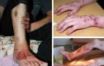
Pathology is accompanied by the following symptoms:
- the patient complains about, which does not disappear, no matter how much he blinks his eyes;
- , even if it didn't happen before;
- dots appear before the eyes;
- loss of peripheral vision;
- visual change in the shape of objects.
The sooner the doctor makes a diagnosis and prescribes the time of the operation, the more likely it is that vision will be restored to 100% after the operation.
Choice of operative intervention
What types of operations exist:

Indications and contraindications for surgery
If the integrity of the shell is damaged in the patient, and the eye gradually loses its functional abilities, then in this case the doctor will prescribe a filling. If the damage is not very significant, or the damage is peripheral, then coagulation is indicated.
Removal of the vitreous body is carried out if it has damage. Such an intervention is carried out if the retinal lesion is severe, or vascular germination is observed in it, as well as in case of bleeding in the vitreous body itself. Each type of surgical intervention has its own characteristics and contraindications.
Vitrectomy (removal of the vitreous body) is not performed when the cornea is clouded, it can be seen visually - a thorn. Also, the operation is contraindicated in case of very strong changes in the retina and cornea, in which case the operation will not have the desired effect. Filling is not done with protrusion of the sclera and with the opacity of the vitreous body.

Laser surgery is contraindicated in cases of severe retinal dissection, vascular pathology in the iris, opacity of the media, and hemorrhages in the fundus. Contraindications to surgery are also intolerance to anesthesia, allergic reactions to the anesthetic, inflammatory processes in the acute stage. In this regard, before planning an operation, it is necessary to undergo all the necessary diagnostic and laboratory tests.
Complications after surgery
Of the possible complications and consequences of surgical intervention, the most common are:
- Inflammation. This phenomenon manifests itself in the form of redness of the eyes, tearing and itching. In order to avoid this, it is recommended to use antiseptic drops, which should be taken a week or two after retinal surgery.
- Vision problems. The first few months after surgery, the eye may not clearly perceive the outlines of objects. In this case, the patient is advised to wear glasses with different diopters, visit an ophthalmologist and periodically check his vision. Usually after a while everything stabilizes.
- Strabismus. This is a common complication after extrascleral filling. The reason is muscle damage or fusion of muscles with the sclera.
- Intraocular pressure. Sometimes an increase in intraocular pressure provokes the occurrence of glaucoma, in which case another operation is performed to remove the installed filling.
- Relapse. Occurs in 20% of cases. Most often, it can be easily corrected during a subsequent operation.
- Violation of visual fields. If the doctor incorrectly chose the power of laser radiation, or the disease began to progress sharply, then the patient's field of view may narrow.

What to do after surgery
The postoperative period should, of course, be discussed with the attending physician, but there are general recommendations that are suitable in any case. It is necessary to observe the regime of the position of the head. After the operation, the doctor introduces some restrictions regarding the position of the head. It is necessary to follow this during sleep. It is not recommended to sleep with your chin down, do not lift weights - the first two weeks you can not lift more than 5 kg.
It is necessary to ensure that when washing, soap and preferably water do not get on the mucous membrane of the eye. When washing your hair, you will have to tilt your head back strongly, you can’t lean forward. If you still didn’t follow, and water or soap got into the eye, then you need to rinse with a solution of levomycetin or furatsilin. It is necessary to visit the doctor on the exact day indicated by him, because it is very important to check the condition of the eye after the operation.

After the operation, the doctor prescribes drops. Rehabilitation works in two directions - the removal of inflammation and the fight against microbes. that can cause infection
In the first few days after surgery, you will have to wear a bandage on the operated eye. This is a necessary measure that will protect the eye from pollution and too bright light. You need to change the bandage a couple of times a day.
A very important point is taking eye drops. With disciplined instillation of eye drops, healing will proceed better, and the patient will return to normal life faster. What drops will be needed, their dosage and duration of use - all this is determined by the doctor in each case individually.
When will normal vision return?
This is also a rather individual question, the terms depend on the patient's body, on the type and complexity of the operation, on age and many other indicators. Someone sees perfectly in three months, while someone needs six months to fully recover. Elderly patients and people with myopia recover longer. During rehabilitation, the patient is advised to wear glasses because objects may double or look fuzzy, but this disappears after a couple of months.
Over time, absolutely all restrictions will be gradually removed, but for some time after the operation it will be impossible:
- drive;
- touch and press on the eyes;
- spend a lot of time at the computer, read a lot, and if necessary, take frequent breaks;
- go out without sunglasses, even if the day is cloudy;
- if a vitrectomy was performed, then you can’t change the temperature dramatically - go to the sauna or bath, swim in the hole, just swim in very hot water, etc.;
- if the replacement of the vitreous body with gas was carried out, then you can not use the subway.

The attending physician may recommend a set of special exercises that will help strengthen the oculomotor muscles. It is necessary to follow all the recommendations of the doctor exactly and on time, not to make independent decisions regarding the postoperative period, on all issues related to the condition of the eye, contact only the attending physician. Your health and vision depend on how disciplined you approach the rehabilitation period.
Video
Retinal detachment is a serious pathology associated with the separation of the retina from the vascular. The disease requires surgical intervention. Therapeutic methods are ineffective. With untimely treatment, blindness may occur. Rehabilitation after surgery is no less important for retinal detachment. It depends on how fully the functions of the eye are restored.
Vision after surgery is restored after a certain period of time. This is due to the complex structure of the retina. For each patient, this period is individual. Immediately after the operation, the patient must stay in bed for several days. After that, he will be allowed to return to his former way of life.
It usually takes one to three months for vision to fully recover after surgery. In some patients, the rehabilitation period may be delayed for 6 months. This is possible in patients aged, as well as in the diagnosis of myopia. Recovery will take much longer if the vitreous is replaced with a low viscosity saline solution.
How well a person will see during the recovery period depends on the following factors:
- timeliness of treatment;
- the state of the retina, the presence or absence of organic changes in it;
- How well does the optic nerve function?
- the degree of transparency of the optical media of the eyeball.
To effectively restore the functions of the eye, it is necessary to follow all the recommendations of the ophthalmologist. The result of the operation is also influenced by the professionalism of the surgeon.
For a speedy recovery after surgery, you need to provide the necessary conditions:
- Less eye strain.
- Reduce physical activity.
- Regular visits to the ophthalmologist.
- Avoid temperature fluctuations.

Air travel is not recommended for certain interventions, such as retinal gas tamponade.
Early rehabilitation period
The recovery period after surgery for retinal detachment is divided into early, which lasts a month, and late. In the first days after the intervention, the patient should devote as much time to his health as possible. The ophthalmologist leaves prescriptions that must be strictly followed.
During the early recovery period, you must follow the recommendations:
- The patient must monitor his own feelings. If signs such as a cloudy image, flashing spots, flies before the eyes, a black veil in the field of vision appear, you should contact your doctor as soon as possible.
- It is worth limiting physical activity - do not lift weights that weigh more than 3 kg, do not exercise too intensely in the gym, exclude strength exercises. In the recovery period, you can practice light exercises, swim, walk in the fresh air.
- The patient needs to constantly control the position of the head - it can not be tilted down. For a while, you will have to give up activities such as gardening, knitting and sewing. Do not tie your own shoelaces and wash your hair, tilting it forward. You can sleep in any position except on your stomach.
- It is necessary to strengthen the immune system and prevent colds. Infection can provoke the development of early postoperative complications. It is best not to visit public places in the first months after the intervention and communicate less with people.
- It is necessary to exclude any thermal effect - do not visit baths, saunas and solariums, do not take a hot bath.
- You need to protect your eyes from sunlight. Since the retina is very sensitive to ultraviolet radiation, the sun can easily damage it. To protect your eyes, you should wear glasses with tinted lenses, and in the summer also a hat.
- The patient must regularly take medications to restore the retina of the eye, prescribed to him by the attending physician. Eye drops are prescribed to prevent infection. You need to bury them by pulling the lower eyelid. Use the drops carefully, without touching the bottle to the eye. If an ointment was prescribed, then apply it to the lower eyelid, and then blink for 10 seconds.
Compliance with these requirements will minimize the risk of re-detachment of the retina after surgery, recovery will be more successful.
In the first days after the operation, discomfort when blinking can provoke stitches on the conjunctiva. After 10-14 days they are usually removed.
late recovery period
A month after the operation, the patient must visit the attending physician without fail, even if he feels fine and there are no suspicious symptoms. The doctor will conduct a thorough examination of the operated eye, examine the fundus. This will reveal any changes in the state of the visual organs.
To avoid recurrent retinal detachment, the patient is advised to continue following the guidelines that have been established for the early recovery period.
In the late rehabilitation period, the following rules apply:
- You need to be careful with the operated eye, avoid situations where it can be damaged, and avoid getting foreign bodies or irritating liquids into the eye.
- Do not stay in the sun for a long period of time.
- Strenuous exercise should still be avoided.
- It is not recommended to drink alcohol, smoke. It is necessary to avoid intoxication of the body, exposure to poisons.
Recovery in the late period may be characterized by the presence of symptoms such as bifurcation or distortion of the outlines of objects. Such phenomena usually decrease after a few weeks after surgery, and sometimes months. In order for rehabilitation after retinal detachment surgery to be faster, the doctor may prescribe the wearing of glasses or contact lenses.
All materials on the site are prepared by specialists in the field of surgery, anatomy and specialized disciplines.
All recommendations are indicative and are not applicable without consulting the attending physician.
Retinal detachment is a common disease. It can practically manifest itself in no way, especially at the beginning of its course, so the patient needs to visit a specialist doctor and conduct an examination of the fundus for diagnosis. However, detachment is dangerous in that, with excessive stress, it can increase in size and cause visual impairment. At later stages, myopia develops, the patient sees poorly peripherally, “flies” fly before the eyes.
Surgery for retinal detachment can be carried out by extrascleral filling. Sometimes it may be necessary to remove all or part of the vitreous (vitrectomy).
Indications
Surgical intervention is performed with retinal detachment. In this case, two layers are separated - neuroepithelium and pigment. Liquid accumulates between them. filling It is designed to restore the integrity of the shell and return the lost functions to the eye.
With minor injuries, peripheral detachment and preservation of vision, coagulation is performed. At the same time, gaps remain, but are “soldered” along the edges. As a result, the separation does not spread and visual impairment does not occur.

Vitrectomy is performed when a change in the vitreous body is detected(a gel-like substance that fills most of the eyeball). This operation can also be indicated for extensive damage to the retina, pathological germination of blood vessels in it, bleeding in the vitreous cavity.
Contraindications
Each of the described types of surgery has its contraindications. Vitrectomy is not performed for:
- Clouding of the cornea of the eye. It is usually visible to the naked eye (in the form of a walleye).
- Gross changes in the retina and cornea. In this case, the operation will not have the desired effect.
 Extrascleral filling is contraindicated in:
- Vitreous opacity.
- Ectasia (protrusion) of the sclera.
Laser coagulation is not carried out with:
- A high degree of retinal detachment.
- Opacity of the media of the eye.
- Pathology of the vessels of the iris.
- Hemorrhages of the fundus.
There are also contraindications in the presence of restrictions on anesthesia, allergies to anesthetic. Operations are not performed in the presence of inflammation in the active stage. That is why it is necessary to pass all the necessary tests before the procedure, do a fluorography, get rid of caries.
Operation progress
Laser coagulation
The operation is performed without anesthesia and lasts about 5-10 minutes. In private clinics, it is not accompanied by hospitalization, the patient can leave the institution on the day of correction. In public hospitals, it is observed within 3-7 days after the procedure.
The operation is performed without anesthesia, only with a small amount of anesthetic in the form of eye drops. Also use drugs that dilate the pupil. After the onset of their action, a special lens is put on the patient's eye, resembling the eyepiece of a microscope. It helps to focus the laser beam and direct it directly to the right place. During the operation, zones of protein destruction and “gluing” of the retina are created, which prevents its separation.

Laser coagulation of the retina
The procedure is performed in a sitting position. The patient feels the action of the laser in the form of bright flashes of light. In rare cases, they can cause dizziness and nausea. For prevention, it is recommended to concentrate on the second eye. Light tingling is possible. Spikes are finally formed in 10-14 days, after this period, and one can clearly judge the success of the operation.
Extrascleral filling
It is advisable for the patient to remain in bed before the operation. At rest, the liquid in the place of separation is absorbed, and the “bubbles” become clearer. This, with extrascleral filling, will help to accurately determine all zones of ruptures.
At the first stage of the operation, the doctor cuts the conjunctiva (the outermost membrane of the eye), produces pressure on the sclera using a special device - a diathermocautery (a device with different tips that allow you to create the necessary electrical discharge on the surface of the tissue). Thus, creating a temporary shaft (a place where the sclera is pressed against the retina), it marks all the places of delamination, after which a filling of the desired size is individually made.
To do this, use a soft elastic material (most often, silicone). The filling is placed on the sclera (the membrane under the retina). As a result, the layers are pressed against each other and the functioning of the visual apparatus is restored. The filling is sewn with non-absorbable threads. The fluid that may be in the gap is gradually absorbed by the pigment epithelium. Sometimes, with its excessive accumulation, it is necessary to make incisions in the sclera to remove it.

In some cases, the retina is pressed additionally, on the other side (as if from inside the eye). To do this, air or another gas mixture is pumped into the vitreous body. The patient may be asked to look in a certain direction with the eye down. This will allow the bubble of gas to stand exactly at the place of the gap. To replenish the volume, it may be necessary to introduce an isotonic solution into the vitreous body. The conjunctiva is sutured.
Despite the great complexity of the operation, its success is quite high. In the textbook "Eye Diseases" (edited by V.G. Kopaeva), released in 2002, it is indicated that “when the operation is performed at the modern technical level, it is possible to achieve retinal adhesion in 92-97% of patients”. To date, the professionalism of surgeons has grown significantly, the equipment has become more advanced and affordable. The main thing is timely diagnosis, which is possible with periodic examinations by an ophthalmologist.
Vitrectomy
The operation is performed in a hospital. It usually complements extrascleral buckling when indicated. Vitrectomy is performed under general or local anesthesia.
Small holes are made in the sclera. Thin scissors and tweezers are introduced into them. The vitreous body is excised, completely or partially removed, and the vacated space is filled with a gas mixture or silicone oil.
Possible complications and consequences
The most common side effects after surgery may include:

- Inflammatory process, manifested in redness of the eye, itching, lacrimation. Antiseptic drops are used as prevention and treatment, which are usually recommended to be taken within 7-10 days.
- Vision change. At first, the operated eye may perceive the contours of objects indistinctly; within a few months, glasses with different diopters will be needed. It is necessary to visit an ophthalmologist periodically and check visual acuity. In a few months, all indicators will stabilize.
- Strabismus. This complication is observed in almost half of the persons who underwent extrascleral filling surgery. Strabismus is caused by muscle damage during surgery, fusion of muscles with the sclera, etc.
- Increased intraocular pressure. In rare cases, it occurs after surgery and can even cause glaucoma. With such a development of events, it is necessary to perform a second surgical intervention and remove the placed filling.
- Re-stratification. The recurrence rate ranges from 9% to 25%. It is usually easily corrected with a second operation.
- Hemorrhage (hemophthalmos). Possible with all types of intervention.
- Narrowing of fields of vision. This occurs as a result of an incorrect choice of radiation power during laser coagulation or due to the progression of the pathological process.
Recovery period
With laser coagulation, practically no restrictions are imposed on the patient. He may be recommended exercises aimed at strengthening the oculomotor muscles. Your doctor may advise you to refrain from strenuous exercise for the first month after the procedure.
With extrascleral filling, the list of rules is much wider:

After vitrectomy, in addition to the above restrictions, it is not recommended:
- Be exposed to sudden changes in temperature, visit a bath, sauna, wash your hair with very hot water.
- Use underground transport (if the vitreous body is replaced by gas).
The speed of rehabilitation depends on the intensity of regeneration processes in the body, the initial area of the lesion, the degree of surgical intervention. On average, it can last from 10 days to several months.
CHI operation, price in private medical centers
Laser coagulation can be done free of charge with a referral from your doctor. After visiting the hospital with the department of eye microsurgery, examination and confirmation of the diagnosis, the patient is assigned a date for the operation. Not earlier than a month before, he must pass all the necessary tests and undergo an examination.

In a private clinic, the process is usually faster. Hospitalization and preparatory period are usually absent. The cost of the procedure is 8,000 - 15,000 rubles for retinal coagulation in one eye.
Extrascleral filling and vitrectomy are free of charge according to the quota. This means that the patient will have to wait in line for the operation, and the very possibility of performing it depends on whether he fits certain parameters (age, general health, aggravation of retinal dissection by other diseases). Prices vary greatly even in Moscow. Extrascleral filling can be performed for 10,000 - 60,000 rubles, vitrectomy - for 50,000 - 100,000 rubles.
Laser photocoagulation of the retina is a common procedure aimed at strengthening the retina. It is performed before laser vision correction and is necessary for patients who suffer from pathologies associated with degeneration or dystrophy of this element of the visual system. There are a number of possible consequences of this procedure.
In this article
One of the most common problems after laser photocoagulation of the eyes is retinal detachment. Practical observations of ophthalmologists show that the problem may not manifest itself in any way, especially in the initial stages after surgery. It is for this reason that the patient must carefully follow all the prescriptions of the ophthalmologist and regularly undergo diagnostics of the visual system and examination of the fundus.
It is important to understand that exfoliation after laser coagulation is dangerous in that with a strong stress on the body, for example, during physical exertion, it can cause a sharp deterioration in vision. So, in the later stages, myopia (nearsightedness) occurs, “flying flies” may appear before the eyes. With timely detection of retinal detachment, doctors perform extrascleral filling or repeated laser coagulation for the patient. Sometimes there is a need for partial or complete removal of the vitreous body, the procedure is called "vitrectomy".
How is the operation going?
Before laser coagulation of the retina, the patient undergoes a complete examination of the visual system, and also passes the necessary tests for the therapist. Preparation for surgery in private clinics may begin with hospitalization on the expected day of the operation. In municipal medical institutions, it may be necessary to observe the patient for a period of 3 to 7 days after the discovery of a retinal detachment.
Before starting the process, the ophthalmologist makes local anesthesia and instills drugs that dilate the pupil. After that, a special type of lens is put on the eyes, which resembles the eyepiece of a microscope. It makes it possible to focus the laser beam and point it to the desired area. During the operation, areas of protein destruction are formed, as well as gluing of the retina, which prevents its further separation. 
Laser coagulation of the eye takes place in a sitting position, at this time the person feels the impact of the device, like bright flashes of light. In exceptional cases, it can cause dizziness and gag reflexes. In order to make it more comfortable for the patient to endure the process, the specialist recommends concentrating on the second eye. The final formation of adhesions occurs after about 10-14 days, only after this period it is possible to judge whether the operation was successful.
Laser coagulation is an absolutely painless process, the patient in rare cases may experience slight tingling.
Possible Complications
Often, after surgery on the retina, there is swelling of the cornea, which can cause a significant change in refractive indices of vision, a person begins to see objects blurry.
However, the edema after coagulation subsides rather quickly, and vision is restored, so this complication is the easiest. There are cases when an ophthalmologist applies coagulants that are too large during a laser operation, while the energy of the device can be transferred to the iris of the visual organ, which provokes an inflammatory process. As a result, the pupil is deformed due to the formation of posterior synechia on the retina, the consequence is corrected by a second operation. The most serious complication after laser coagulation of the retina, according to ophthalmologists, is the closure of the angle of the anterior chamber of the eye, the consequences of this process are jumps in intraocular pressure that occur during detachment of the choroid and swelling of the ciliary body with strong exposure to the laser beam.
There are cases when a specialist performs laser coagulation of the retina with a narrow laser beam that passes through the lens and affects its tissues. The reaction can be individual, sometimes the patient develops a cataract after such an operation with a laser beam. 
Also, microscopic hemorrhages, the appearance of detachment in another place, may appear on the retina itself. Incorrect application of coagulants to the retina often provokes macular edema and impaired perfusion of the nerve of the eye. The consequence is a decrease in vision, a decrease in the ability to see in the dark.
Laser coagulation of the retina is often performed during the formation of the vessels of the optic nerve head. This is fraught with his ischemia and a sharp drop in vision.
The retina is a multilayer formation containing the choroid and pigment epithelium. Therefore, if coagulation is performed by a narrow beam, then the patient may experience ruptures of the Bruch's membrane and hemorrhages in the vitreous body and the retina itself.
Hemorrhages, turbidity, contraction of the boundary membrane and, as a result, detachment of the vitreous body, as the laser beam passes through this formation, are also possible.
After laser exposure to the retina, the patient needs to be regularly observed by a specialist for some time, since anomalies can occur only some time after the procedure. These include progressive atrophy of the retinal pigment layer in the coagulation zone.
Indications for the operation
Laser coagulation is prescribed for people with eye pathologies such as: 
- detachment or rupture of the retina (the eyes become vulnerable to any, even a slight load);
- macular degeneration;
- mechanical damage to the retina, vitreous body, choroid;
- congenital retinopathy (usually in premature babies);
- diabetic retinopathy;
- pathological proliferation of the vessels of the disc of the optic nerve and retina;
- inflammatory processes in the vascular system of the retina with hemorrhages;
- macular lesion;
- retinal anomalies associated with obstruction of the central ophthalmic vein.
Contraindications for coagulation
Doctors will categorically refuse to perform the operation if the patient has: 
- gliosis from the third degree and above. Such a disease provokes the replacement of light-sensitive cells of the retina with connective tissue, there is a strong deterioration in vision;
- severe retinal detachment;
- hemorrhage in the eyeball. This restriction is temporary, if the hemorrhage resolves, the patient is allowed to the procedure. Otherwise, it is necessary to treat the symptom and its underlying cause;
- clouding of the vitreous body, lens or other areas of the visual system due to anomalies, including cataracts. If the deviation is eliminated, then the operation can be carried out.
Restrictions in rehabilitation
In order to avoid possible complications after coagulation as much as possible, it is worth observing a number of rules for one month: