General urine analysis for children is normal. Urine analysis in a child: interpretation, normal. Current standards and deviations
Do you remember when you were still expecting a baby, every time you came to an appointment at the antenatal clinic, you received another coupon for urine testing? And how, during pregnancy, we managed to get sick of these early rises and hikes with endless jars...
During pregnancy, every mother periodically undergoes testing.
Most likely, then you did not think about those lines, numbers and terms that were written in the analysis results. If the gynecologist observing you was satisfied with them, then you were calm.
Now you and your baby have received a referral for an OAM test (this is the abbreviation usually used to mark coupons for a general urine test) from your local pediatrician.

What interesting things can the analysis result show?
You can read how to properly collect “material” for analysis in our. And here we will talk about why this research is carried out, what indicators are analyzed, what ranges are the norm, what you should pay attention to and much more.
Why is this research being conducted?
OAM is one of the simplest and most inexpensive tests for the condition of the urinary system, kidneys and the entire body as a whole. Therefore, this analysis is prescribed for both adults and children, for any suspicion of health problems.

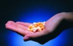
With the help of OAM, you can reveal quite a lot of facts about the health of the baby.
A general analysis of urine (as well as blood), like a litmus test, immediately shows changes and deviations in the functioning of our entire complex system. The color, transparency, and smell of the collected liquid can tell an experienced laboratory technician about what is happening with the baby’s health, whether there is a hidden inflammatory process, endocrine diseases or other problems.
What indicators of the analysis results are indicated in the transcript and what do they mean?
You have in your hands a piece of paper on which everything that you saw in the laboratory at the clinic is written down. You see, some of the words and concepts are clear to you, some of the designations, terms and numbers (even if you managed to read the notorious doctor’s handwriting) may not tell you anything at all. Some words are understandable, but whether this is good or bad in relation to the condition of the cub is unclear. So, let's figure out what they wrote there and what it means?

What can mommy learn from the test certificate?
The indicators analyzed in this test are the same for both adults and children. Some normal options may vary depending on the age of the baby and his diet. There are three main groups of parameters:
- Physical properties of urine(this includes odor, color, transparency and specific gravity of the released liquid);
- Presence and concentration of biological substances(these include glucose, protein, ketone bodies, etc.);
- Sediment microscopy(these are the same red blood cells, epithelium, cylinders and other bodies and cells invisible to the naked eye).
This data is usually enough to avoid unfounded concern for the baby’s health, or, conversely, to prescribe treatment and additional studies in a timely manner.
 In the first years of life, special attention should be paid to the child’s menu. Poor nutrition can lead to the development of dental caries. Some parents do not attach importance to the condition of the baby’s oral cavity, completely in vain! Left untreated, it can lead to irreversible consequences.
Encopresis in young children is a serious psychophysiological problem that can only be solved with the help of a specialist. The reasons for the development of the disease and why this happens are described.
What is the norm, and what indicators can alert you and the doctor?
Let's look at each line received from the laboratory in order:
- Color. A normal indicator is a straw-yellow tint of the liquid. The younger the baby, the lighter the color of his urine. During the newborn period, the liquid secreted by the baby is practically colorless and looks like ordinary water. Gradually, with the transition to a mixed diet, the urine begins to acquire a brighter color and turns yellow.
Too intense coloring should immediately alert an experienced doctor. Thus, dark urine color may indicate liver problems, and reddish, so-called “meat slop color” may indicate kidney disease.
But keep in mind that often, Some coloring products can give your urine an unusual color. Beets color them reddish or brown; the recent introduction of citrus fruits into complementary foods can give them a bright yellow tint. “Abuse” of carrots gives an orange color to both the child’s urine and skin.
- Transparency. For at least 2-2.5 hours, the baby’s urine remains completely transparent. That is why the doctor insisted on freshly collected material for research. After this time, a cloud of turbidity begins to form in the liquid, eventually occupying its entire volume.
The reason for the formation of turbidity can be either the presence of leukocytes (or simply pus) or the loss of salt sediment. And if the latter is the norm, then an increased level of leukocytes is a cause for concern.
- Smell. Normally, a child’s urine has virtually no odor. The permissible maximum is compared with the intensity of the odor of weak meat broth. Further deviation should alert parents. For example, a strong ammonia smell may indicate inflammation of the bladder, the feeling of rotting apples may indicate an increase in the content of ketone bodies in the urine).
- Acidity. The pH of a child’s urine normally fluctuates between readings of 5 and 7.5. That is, the normal acid reaction of a liquid is slightly acidic or neutral. If this indicator increases, leaning towards an alkaline reaction, it is necessary to conduct additional studies to exclude the diagnosis of cystitis or pyelonephritis. A too acidic reaction is possible with diabetes or fasting.
- Density. The specific gravity of urine varies significantly depending on the age of the child. For the newborn period, this figure is 1.001 - 1.003. For children under two years of age, the norm will be numbers from 1.002 – 1.004. By the age of five, this figure grows to values of 1.005 - 1.020. The normal specific gravity of urine for an adult is 1.025.
An unjustified increase in liquid density usually indicates a predominance of meat in the diet. A decrease in density means a “skew” towards plant foods.
- Protein. Ideally, the kidney filter does not allow protein molecules to pass into the secreted fluid; they are too large for this. A small amount of protein in the urine (no more than 5 g/l) is possible after intense physical activity. High levels may indicate inflammatory kidney disease.
- Glucose. Well, in the analysis she should be missing. The presence of glucose in the urine may indicate an excess of carbohydrate foods in the child’s diet. If the levels are elevated, the question of diagnosing diabetes mellitus is raised.
- Ketone bodies. The content of ketone bodies can be observed with fever, vomiting, diarrhea, and anemia. They are also an indicator of excess carbohydrate levels in food.
- Red blood cells. Normally none. Individual 2-3 cells in the field of view are acceptable. An increase in this amount is possible with prolonged physical activity. After rest, these indicators should return to normal.
- Leukocytes in the baby’s urine are the main test for hidden inflammatory diseases of the urinary tract. Normally there are no more than 2-3 of them in the field of view. But if hygiene rules are not followed when collecting urine, this indicator may turn out to be artificially high.
- Epithelium. The presence of epithelial cells in the test contents may also indicate poor hygiene. But it can also be an alarm bell for the presence of infections and kidney diseases.
- Cylinders. There are several types of them - waxy, leukocyte, granular, erythrocyte and hyaline. The latter are found in the urine of children in the first days of life, and subsequently disappear. The subsequent appearance of cylinders in the field of view may indicate many problems in the baby’s health and lifestyle.
For example, any eating disorders - vomiting, diarrhea, constipation, belching - can appear in the form of granular and hyaline casts. Often this is a reaction to a new complementary food, and only requires observation.
- Salt. The release of salts in a child’s urine most often will not be a sign of serious pathologies. Their presence or absence depends on the baby’s lifestyle and nutrition. Thus, urates indicate an abundant meat diet and physical activity, oxalates, on the contrary, indicate an excess of plant foods. Ammonium urate is usually determined during the neonatal period and breastfeeding.

It is important that the collected material is fresh, otherwise the outcome of the analysis may show an inaccurate picture.

Increased acidity of urine does not bode well.

Glucose in the urine - time to change the child's menu.

To ensure that the analysis results are not distorted, the material must be collected under sterile conditions.

In the best outcome, there should be no casts in the child's urine.
There should be no bacteria, mucus or other foreign substances in the baby’s urine.
Or pseudorubella, an unpleasant disease that is accompanied by redness of the skin. The paradox is that roseola does not require treatment. Parents should be patient and wait for the red spots to go away.
The central place in a child's room is occupied by a crib. Choosing a bed for a baby is very difficult; different manufacturers offer many different models. It contains reviews of the most popular cribs and their ratings from parents.
What is torticollis and why does it develop in young children? Follow this link and you will learn everything about this disease.
Urinalysis according to Nechiporenko
If the doctor has doubts about a number of indicators obtained as a result of the OAM, additional tests and studies are prescribed. One of the common follow-up actions is to conduct a urine test according to Nechiporenko. This is a more comprehensive test for assessing the condition of the kidneys and urinary system, compared to a general analysis.
When collecting material for this urine test, It is necessary to thoroughly hygienically prepare the baby for delivery. This will make it possible to eliminate errors and unnecessary worries. For the study, an average portion of the child’s first morning urine is taken.

In this case, water procedures before collecting urine are necessary!
Basically, in this test, a microscopic examination of the sediment is carried out to identify and presence of red blood cells, leukocytes and casts.
Also, in addition, a biochemical, 24-hour urine test may be prescribed to clarify the salt content.
Tatyana, mother of Rita (1.6 years old):
“Exactly, girls, I still remember these endless OAM and OAC during pregnancy! It felt like I had been running to the hospital with jars for the entire 9 months. But it turns out, look how much you can see there!”
Irina, mother of Ilya (4 years old):
“Oh, and I remembered how we caught this OAM with a plate after sleep at Ilyushka’s place, back in his infancy! It’s easier with a girl - hold it over a bowl and write. And with a boyish “faucet” there is both laughter and sin...
It turns out that you can carry out a rapid test for the presence of leukocytes, and therefore inflammation, yourself. If you warm up the collected urine of the baby a little, and the cloud of turbidity dissipates and melts, then this is a salt sediment, no big deal. And if this remains the case, then run to the doctor! It is, rather, pus, leukocytes. Something is inflamed in the cub, we need to look for the cause.”
A general urinalysis includes assessment of the physicochemical characteristics of urine and microscopy of sediment. A general urine test for patients with diseases of the kidneys and urinary system is performed repeatedly over time to assess the condition and monitor therapy.
Indications for the purpose of analysis:
- diseases of the urinary system;
- screening examination during medical examinations;
- assessment of the course of the disease, monitoring the development of complications and the effectiveness of treatment;
Preparing for the study. The day before, it is better not to eat vegetables and fruits, which can change the color of urine, and not to take diuretics. Before collecting urine, it is necessary to perform a hygienic toilet of the genital organs. Morning urine is taken for examination.
1. General properties
1.1. Urine color
Normally, the urochrome pigment gives the urine a yellow color of various shades depending on the degree of saturation of the urine with it. Sometimes only the color of the precipitate may change: for example, with an excess of urates, the precipitate has a brownish color, uric acid - yellow, phosphates - whitish.
Reference values: straw yellow color.
An increase in color intensity is a consequence of loss of body fluids: swelling, vomiting, diarrhea. A change in the color of urine may be the result of the release of coloring compounds formed during organic changes or under the influence of dietary components, medications taken, or contrast agents.
1.2. Urine clarity
Reference values: full.
Turbidity can be the result of the presence of red blood cells, leukocytes, epithelium, bacteria, fat droplets in the urine, precipitation of salts (urates, phosphates, oxalates) and depends on the concentration of salts, pH and storage temperature of urine (low temperature promotes precipitation of salts). If you stand for a long time, your urine may become cloudy due to bacterial growth. Normally, slight turbidity may be due to epithelium and mucus.
1.3. Relative density (specific gravity)
The relative density of urine depends on the amount of excreted organic compounds (urea, uric acid, salts) and electrolytes - Cl, Na and K, as well as on the amount of water excreted. The higher the diuresis, the lower the relative density of urine.
The presence of protein and especially glucose causes an increase in the specific gravity of urine (hypersthenuria). A decrease in the concentration function of the kidneys in renal failure leads to a decrease in specific gravity (hyposthenuria).
Complete loss of concentration function leads to equalization of the osmotic pressure of plasma and urine, this condition is called isosthenuria.
Reference values:
Increase in relative density (> 1030 g/l):
- glucose in the urine in uncontrolled diabetes mellitus;
- protein in the urine (proteinuria) with glomerulonephritis, nephrotic syndrome;
- medications and (or) their metabolites in the urine;
- intravenous infusion of mannitol, dextran or radiocontrast agents;
- low fluid intake;
- large fluid losses (vomiting, diarrhea);
- oliguria.
Decrease in relative density (< 1010 г/л):
- diabetes insipidus (nephrogenic, central or idiopathic);
- acute damage to the renal tubules;
- polyuria (as a result of taking diuretics, drinking too much).
1.4. Urine pH
Fresh urine from healthy people can have a different reaction (pH from 4.5 to 8), usually the urine reaction is slightly acidic (pH between 5 and 6). Fluctuations in the pH of urine are caused by the composition of the diet: a meat diet causes an acidic reaction in the urine; the predominance of plant and dairy foods leads to alkalization of the urine. Changes in urine pH correspond to blood pH; with acidosis, urine is acidic, with alkalosis it is alkaline. Sometimes there is a discrepancy between these indicators.
In case of chronic damage to the kidney tubules (tubulopathies), hyperchloric acidosis is observed in the blood, and the urine reaction is alkaline, which is associated with a violation of the synthesis of acid and ammonia due to damage to the tubules. Bacterial decomposition of urea in the ureters or storage of urine at room temperature leads to alkalinization of urine. The reaction of urine affects the nature of salt formation in urolithiasis: at a pH below 5.5, uric acid stones are more often formed, at a pH from 5.5 to 6.0 - oxalate stones, at a pH above 7.0 - phosphate stones.
Reference values: 5.0
Increase (pH > 7):
- metabolic and respiratory alkalosis;
- chronic renal failure;
- renal tubular acidosis (types I and II);
- hyperkalemia;
- primary and secondary hyperfunction of the parathyroid gland;
- a diet high in fruits and vegetables;
- prolonged vomiting;
- urinary tract infections caused by microorganisms that break down urea;
- administration of certain medications (adrenaline, nicotinamide, bicarbonates);
- neoplasms of the genitourinary system.
Reduction (pH about 4):
- metabolic and respiratory acidosis;
- hypokalemia;
- dehydration;
- starvation;
- diabetes;
- tuberculosis;
- fever;
- severe diarrhea;
- taking medications: ascorbic acid, corticotropin, methionine;
- diet high in meat protein and cranberries.
1.5. Protein in urine (proteinuria)
Protein in the urine is one of the most diagnostically important laboratory signs of kidney pathology. A small amount of protein in the urine (physiological proteinuria) can occur in healthy people, but the excretion of protein in the urine does not normally exceed 0.080 g/day at rest and 0.250 g/day during intense physical activity, after a long walk (marching proteinuria). The normal protein concentration in morning urine is usually considered
Normally, most proteins do not pass through the membrane of the renal glomeruli, which is explained by the large size of protein molecules, as well as their charge and structure. With minimal damage in the glomeruli of the kidneys, there is primarily a loss of low molecular weight proteins (mainly albumin), therefore, with a large loss of protein, hypoalbuminemia often develops. With more pronounced pathological changes, larger protein molecules also enter the urine. The epithelium of the renal tubules physiologically secretes a certain amount of protein (Tamm-Horsfall protein). Some of the urine proteins can come from the genitourinary tract (ureter, bladder, urethra) - the content of these proteins in the urine increases sharply with infections, inflammation or tumors of the genitourinary tract.
Proteinuria (the appearance of increased amounts of protein in the urine) can be prerenal (associated with increased tissue breakdown or the appearance of pathological proteins in the plasma), renal (due to kidney pathology) and postrenal (associated with urinary tract pathology). The appearance of protein in the urine is a common nonspecific symptom of kidney pathology. With renal proteinuria, protein is found in both daytime and nighttime urine. According to the mechanisms of occurrence of renal proteinuria, glomerular and tubular proteinuria are distinguished. Glomerular proteinuria is associated with pathological changes in the barrier function of the glomerular membranes. Massive urinary protein loss (>3 g/L) is always associated with glomerular proteinuria. Tubular proteinuria is caused by impaired protein reabsorption due to pathology of the proximal tubules.
Presence of protein in urine (proteinuria):
- nephrotic syndrome;
- diabetic nephropathy;
- glomerulonephritis;
- nephrosclerosis;
- impaired absorption in the renal tubules (Fanconi syndrome, heavy metal poisoning, sarcoidosis, sickle cell disease);
- multiple myeloma (Bence Jones protein in urine) and other paraproteinemias;
- impaired renal hemodynamics in heart failure, fever;
- malignant tumors of the urinary tract;
- cystitis, urethritis and other urinary tract infections.
1.6.Glucose in urine
Glucose in urine is normally absent or found in minimal quantities, up to 0.8 mmol/l, because In healthy people, all blood glucose, after being filtered through the glomerular membrane, is completely reabsorbed back into the tubules. When the concentration of glucose in the blood is more than 10 mmol/l - the renal threshold is exceeded (the maximum ability of the kidneys to reabsorb glucose) or when the renal threshold is reduced (renal tubular damage), glucose appears in the urine - glucosuria is observed.
Detection of glucose in urine is important for the diagnosis of diabetes mellitus, as well as monitoring (and self-control) of antidiabetic therapy.
Increased levels (glucosuria):
- diabetes;
- acute pancreatitis;
- hyperthyroidism;
- renal diabetes;
- steroid diabetes (taking anabolic steroids for diabetics);
- poisoning with morphine, strychnine, phosphorus;
- dumping syndrome;
- Cushing's syndrome;
- pheochromocytoma;
- major injury;
- burns;
- tubulointerstitial kidney damage;
- eating large amounts of carbohydrates.
1.7. Bilirubin in urine
Bilirubin is the main final metabolite of porphyrins excreted from the body. In the blood, free (unconjugated) bilirubin in plasma is transported by albumin; in this form, it is not filtered in the glomeruli. In the liver, bilirubin combines with glucuronic acid (a conjugated, water-soluble form of bilirubin is formed) and in this form it is released into the bile into the gastrointestinal tract. When the concentration of conjugated bilirubin in the blood increases, it begins to be excreted by the kidneys and found in the urine. The urine of healthy people contains minimal, undetectable amounts of bilirubin. Bilirubinuria is observed mainly with damage to the liver parenchyma or mechanical obstruction of the outflow of bile. With hemolytic jaundice, the urine reaction to bilirubin is negative.
Reference values: negative.
Detection of bilirubin in urine:
- obstructive jaundice;
- viral hepatitis;
- cirrhosis of the liver;
- metastases of neoplasms to the liver.
1.8. Urobilinogen in urine
Urobilinogen and stercobilinogen are formed in the intestine from bilirubin released in bile. Urobilinogen is reabsorbed in the colon and again enters the liver through the portal vein system, and then again excreted along with bile. A small part of this fraction enters the peripheral bloodstream and is excreted in the urine. Normally, in the urine of a healthy person, urobilinogen is determined in trace quantities - its excretion in urine per day does not exceed 10 µmol (6 mg). When urine stands, urobilinogen turns into urobilin.
Reference values: negative.
Increased excretion of urobilinogen in urine:
- increased hemoglobin catabolism: hemolytic anemia, intravascular hemolysis (transfusion of incompatible blood, infections, sepsis), pernicious anemia, polycythemia, resorption of massive hematomas;
- increased formation of urobilinogen in the gastrointestinal tract: enterocolitis, ileitis, intestinal obstruction, increased formation and reabsorption of urobilinogen during infection of the biliary system (cholangitis);
- increased urobilinogen in liver dysfunction: viral hepatitis (excluding severe forms; chronic hepatitis and cirrhosis of the liver;
- toxic damage: - alcohol, organic compounds, toxins during infections, sepsis;
- secondary liver failure: after myocardial infarction, cardiac and circulatory failure, liver tumors;
- increased urobilinogen during liver bypass surgery: liver cirrhosis with portal hypertension, thrombosis, renal vein obstruction.
1.9. Ketone bodies in urine (ketonuria)
Ketone bodies (acetone, acetoacetic and beta-hydroxybutyric acids) are formed as a result of increased catabolism of fatty acids. Determination of ketone bodies is important in recognizing metabolic decompensation in diabetes mellitus. Insulin-dependent juvenile diabetes is often first diagnosed by the appearance of ketone bodies in the urine. With inadequate insulin therapy, ketoacidosis progresses. The resulting hyperglycemia and hyperosmolarity lead to dehydration, electrolyte imbalance, and ketoacidosis. These changes cause dysfunction of the central nervous system and lead to hyperglycemic coma.
Reference values: negative.
Detection of ketone bodies in urine (ketonuria):
- diabetes mellitus (decompensated - diabetic ketoacidosis);
- precomatose state, cerebral (hyperglycemic) coma;
- long-term fasting (complete refusal of food or diet aimed at reducing body weight);
- severe fever;
- alcohol intoxication;
- hyperinsulinism;
- hypercatecholaminemia;
- isopropranolol poisoning;
- glycogenosis types I, II, IV;
- lack of carbohydrates in the diet.
1.10. Nitrites in urine
There are no nitrites in normal urine. In urine, they are formed from nitrates of food origin under the influence of bacteria, if the urine was in the bladder for at least 4 hours. Detection of nitrites in the urine (positive test result) indicates infection of the urinary tract. However, a negative result does not always exclude bacteriuria. Urinary tract infection varies among different populations and is dependent on age and gender.
Reference values: negative.
1.11. Hemoglobin in urine
There is no hemoglobin in normal urine. A positive test result reflects the presence of free hemoglobin or myoglobin in the urine. This is the result of intravascular, intrarenal, urinary hemolysis of red blood cells with the release of hemoglobin, or muscle damage and necrosis, accompanied by an increase in plasma myoglobin levels. It is quite difficult to distinguish hemoglobinuria from myoglobinuria; sometimes myoglobinuria is mistaken for hemoglobinuria.
Reference values: negative
Presence of hemoglobin in urine:
- severe hemolytic anemia;
- severe poisoning, for example, sulfonamides, phenol, aniline, poisonous mushrooms;
- sepsis;
- burns.
Presence of myoglobin in urine:
- muscle damage;
- heavy physical activity, including sports training;
- myocardial infarction;
- progressive myopathies;
- Rhabdomyolysis
2. Microscopy of urine sediment
Microscopy of urine components is carried out in the sediment formed after centrifugation of 10 ml of urine. Sediment consists of solid particles suspended in urine: cells, protein-formed casts (with or without inclusions), crystals, or amorphous deposits of chemicals.
2.1. Red blood cells in urine
Red blood cells (blood cells) enter the urine from the blood. Physiological erythrocyturia is up to 2 red blood cells/μl of urine. It does not affect the color of urine. During the study, it is necessary to exclude contamination of urine with blood as a result of menstruation! Hematuria (the appearance of red blood cells, other formed elements, as well as hemoglobin and other blood components in the urine) can be caused by bleeding anywhere in the urinary system. The main reason for the increase in the content of red blood cells in the urine is renal or urological diseases and hemorrhagic diathesis.
Reference values: none; with microscopy - up to 2 in the field of view.
Red blood cells in urine - exceeding reference values:
- urinary tract stones;
- tumors of the genitourinary system;
- glomerulonephritis;
- pyelonephritis;
- hemorrhagic diathesis (with intolerance to anticoagulant therapy, hemophilia, coagulation disorders, thrombocytopenia, thrombocytopathies);
- urinary tract infections (cystitis, urogenital tuberculosis);
- kidney injury;
- arterial hypertension with involvement of the renal vessels;
- systemic lupus erythematosus (lupus nephritis);
- poisoning with benzene derivatives, aniline, snake venom, poisonous mushrooms;
- inadequate anticoagulant therapy.
2.2. Leukocytes in urine
An increased number of white blood cells in the urine (leukocyturia) is a symptom of inflammation of the kidneys and/or lower urinary tract. In chronic inflammation, leukocyturia is a more reliable test than bacteriuria, which is often not detected. With a very large number of leukocytes, pus in the urine is determined macroscopically - this is the so-called pyuria. The presence of leukocytes in the urine may be due to the presence of secretions from the external genitalia in the urine due to vulvovaginitis, or insufficiently thorough toileting of the external genitalia when collecting urine for analysis.
boys - 0 - 3 in sight
girls< 14 лет - 0 - 5 в поле зрения
An increase in leukocytes in the urine is observed in almost all diseases of the kidneys and genitourinary system:
- acute and chronic pyelonephritis, glomerulonephritis;
- cystitis, urethritis, prostatitis;
- stones in the ureter;
- tubulointerstitial nephritis;
- lupus nephritis;
- kidney transplant rejection.
2.3. Epithelial cells in urine
Epithelial cells are almost always present in urine sediment. Epithelial cells originating from different parts of the genitourinary system vary (usually squamous, transitional and renal epithelium are distinguished).
Squamous epithelial cells, characteristic of the lower parts of the genitourinary system, are found in the urine of healthy people and their presence usually has little diagnostic value. The amount of squamous epithelium in the urine increases with urinary tract infection.
An increased number of transitional epithelial cells can be observed in cystitis, pyelonephritis, and kidney stones.
The presence of renal epithelium in the urine indicates damage to the kidney parenchyma (observed in glomerulonephritis, pyelonephritis, some infectious diseases, intoxication, circulatory disorders). The presence of more than 15 renal epithelial cells in the field of view 3 days after transplantation is an early sign of the threat of allograft rejection.
Reference values: none; under microscopy:
squamous epithelial cells: other epithelial cells - absent
Detection of renal epithelial cells:
- pyelonephritis;
- intoxication, intake of salicylates, cortisol, phenacetin, bismuth preparations, poisoning with salts of heavy metals, ethylene glycol);
- tubular necrosis;
- kidney transplant rejection;
- nephrosclerosis.
2.4. Casts in urine
Cylinders are elements of cylindrical sediment (a kind of cast of renal tubules), consisting of protein or cells, and may also contain various inclusions (hemoglobin, bilirubin, pigments, sulfonamides). Based on their composition and appearance, there are several types of cylinders (hyaline, granular, erythrocyte, waxy, etc.).
Normally, renal epithelial cells secrete the so-called Tamm-Horsfall protein (absent in blood plasma), which is the basis of hyaline casts. Hyaline casts can be found in the urine in all kidney diseases. Sometimes hyaline casts can be found in healthy people. As a pathological symptom, they acquire significance when they are constantly detected and in significant quantities, especially when erythrocytes and renal epithelium are superimposed on them.
Granular casts are formed as a result of the destruction of tubular epithelial cells. Their detection in a patient at rest and without fever indicates renal pathology.
Waxy casts are formed from compacted hyaline and granular casts in wide-lumen tubules. They occur in severe kidney diseases with predominant damage and degeneration of the tubular epithelium, more often in chronic than in acute processes.
Erythrocyte casts are formed when red blood cells are layered on hyaline casts, and leukocyte casts are formed by leukocytes. The presence of red blood cell casts confirms the renal origin of hematuria.
Epithelial casts (rarely) are formed when the tubular epithelium is detached. Occurs with severe degenerative changes in the tubules at the onset of acute diffuse glomerulonephritis, chronic glomerulonephritis. Their presence in a urine test a few days after surgery is a sign of rejection of the transplanted kidney.
Pigment (hemoglobin) cylinders are formed when pigments are included in the composition of the cylinder and are observed with myoglobinuria and hemoglobinuria.
Cylinders are long structures consisting of mucus. Single cylindroids are found in urine under normal conditions. A significant number of them occur during inflammatory processes of the mucous membrane of the urinary tract. They are often observed when the nephritic process subsides.
Reference values: hyaline cylinders – single, the rest – absent
Hyaline casts in urine:
- renal pathology (acute and chronic glomerulonephritis, pyelonephritis, kidney stones, renal tuberculosis, tumors);
- hyperthermic conditions;
- high blood pressure;
- taking diuretics.
Granular casts (nonspecific pathological symptom):
- glomerulonephoritis, pyelonephritis;
- diabetic nephropathy;
- viral infections;
- lead poisoning;
- fever.
Waxy cylinders:
- chronic renal failure;
- kidney amyloidosis;
- nephrotic syndrome.
Red blood cell casts (hematuria of renal origin):
- acute glomerulonephritis;
- kidney infarction;
- renal vein thrombosis;
- malignant hypertension.
Leukocyte casts (leukocyturia of renal origin):
- pyelonephritis;
- Lupus nephritis in systemic lupus erythematosus.
Epithelial casts (most rare):
- acute tubular necrosis;
- viral infection (for example, cytomegalovirus);
- poisoning with salts of heavy metals, ethylene glycol;
- overdose of salicylates;
- amyloidosis;
- kidney transplant rejection reaction.
2.5. Bacteria in urine
Isolation of bacteria in urine has significant diagnostic value. Bacteria remain in the urine for no more than 1-2 days after the start of antibiotic therapy. The first morning urine sample is preferable for testing. The type of bacteria can be determined and the level of bacteriuria can be assessed, as well as the sensitivity of microorganisms to antibiotics can be determined using bacteriological urine culture.
Reference values: negative
Bacteria in urine:
- infections of the urinary system (pyelonephritis, urethritis, cystitis).
Yeasts
The detection of yeast of the genus Candida indicates candidiasis, which most often occurs as a result of irrational antibiotic therapy.
Reference values: negative
2.6. Inorganic urine sediment (crystals), salts in urine
Urine is a solution of various salts, which can precipitate (form crystals) when the urine stands. Low temperature promotes the formation of crystals. The presence of certain salt crystals in the urinary sediment indicates a change in the reaction towards the acidic or alkaline side. Excessive salt content in urine contributes to the formation of stones and the development of urolithiasis. At the same time, the diagnostic value of the presence of salt crystals in urine is usually small. Increased doses of ampicillin and sulfonamides lead to the formation of crystals.
Under physiological conditions, uric acid occurs in high concentrations of urine, after a heavy meat meal, after profuse sweating.
Reference values: none
Uric acid and its salts (urates):
- highly concentrated urine;
- acidic reaction of urine (after physical activity, meat diet, fever, leukemia);
- uric acid diathesis, gout;
- chronic renal failure;
- acute and chronic nephritis;
- dehydration (vomiting, diarrhea);
- in newborns.
Hippuric acid crystals:
- eating fruits containing benzoic acid (blueberries, lingonberries);
- diabetes;
- liver diseases;
- putrefactive processes in the intestines.
Ammonia-magnesium phosphates, amorphous phosphates:
- alkaline urine reaction in healthy people;
- vomiting, gastric lavage;
- cystitis;
- Fanconi syndrome, hyperparathyroidism.
Calcium oxalate (oxaluria occurs with any urine reaction):
- eating foods rich in oxalic acid (spinach, sorrel, tomatoes, asparagus, rhubarb);
- pyelonephritis;
- diabetes;
- ethylene glycol poisoning.
Leucine and tyrosine:
- severe metabolic disorder;
- phosphorus poisoning;
- destructive liver diseases;
- pernicious anemia;
- leukemia
- congenital disorder of cystine metabolism - cystinosis.
Fat and fatty acids:
- degenerative changes in the epithelium of the renal tubules;
- eating large amounts of fish oil.
Cholesterol:
- chyluria;
- fatty degeneration of the kidneys;
- kidney echinococcosis;
- cystitis;
- cholesterol stones.
Indications for
The doctor gives a referral for a general (clinical) urine test in the following cases:
- Routine examination of the baby at the age of one, six and twelve months.
- Urine examination for a child over 1 year of age - 2 times a year.
- Comprehensive examination during medical examinations - admission to kindergarten or school.
- Examination before vaccination.
- Unscheduled - if you suspect kidney or bladder disease.
- In case of a baby’s illness not related to the urinary system, in order to monitor the development of the disease, monitor complications and evaluate the effectiveness of treatment.
- Children who have had a disease caused by streptococcal infection (sore throat, scarlet fever) are prescribed a urine test 1-2 weeks after clinical recovery in order to monitor kidney function.
This type of analysis is most common in pediatric practice. It is quite simple, and its widespread implementation allows for detailed screening and identification of children with various health conditions.

Photo source: http://detskie-analizy.ru
Parameters of general urine analysis in children and adults they are the same, but there are significant differences in the normal variants.
Indicators (parameters) | Note |
|---|---|
General properties | Amount of urine per day, ml, depending on age |
Physical properties |
|
Chemical properties | Determination of the presence and concentration of biological components:
|
Microscopy of urinary sediment | Detection in sediment:
|

Photo source: http://forum.biysk.net
Norms and deviations
General urine analysis by quantitative indicator for newborns must comply with the following data:

For children from 1 month to 14 years The standards for this indicator are as follows:

If the amount of urine excreted differs significantly from the age norm, this may indicate the presence of pathology. But parents should not prepare for the worst in advance. After all, even a completely healthy baby can go to the toilet more than usual. For example, if you drink plenty of water or have melon or watermelon in your diet. And, conversely, reducing the amount you drink can lead to olakisuria - rare urination. It is also observed with excessive sweating, vomiting, diarrhea and is more common in premature babies.
In the first days of life, newborns rarely urinate. But the absence of wet diapers for 12-18 hours is an alarming signal!
The normal daily amount of urine in children is approximately calculated using the formula:
600 + 100 (A - 1) = ml of urine per 1 day, where A is the number of years of the child.
Normal physico-chemical and microscopic indicators A general urine test for children is as follows:

Deviations from the norm in terms of physical and chemical indicators are considered to be:
- Whitish, darkened or uncharacteristic color. This may indicate various disorders in the child’s body. Although the mother should remember that most often this happens when the child eats brightly colored foods - beets, oranges, rhubarb - or certain medications.
- Cloudiness. As a rule, it indicates the presence of bacteria - bacteriuria. There may also be an abundance of salts, mucus, fat or blood cells, and epithelium.
- Specific smell. An ammonia smell most often indicates the presence of an inflammatory process. If your urine smells sharp, reminiscent of rotting apples or acetone, this may be a sign of the presence of ketone bodies.
- Density changes. This indicator is most often physiological and does not indicate pathology. Its increase occurs with excessive sweating, heavy consumption of meat and fatty foods by a child or nursing mother, and a decrease occurs after drinking heavily or with a plant-based diet.
- Uncharacteristic PH reaction. A shift in normal values towards an acidic reaction may indicate kidney failure or high temperature, as well as diabetes. But if the acid reaction is moderate and the child feels well, this is a sign of the predominance of meat products, fats and white bread in his diet. The same reaction can occur during intense physical exertion.
Alkaline urine reaction occurs immediately after eating and with a plant-based diet. It is also considered normal for breastfed babies. Pathological alkalization occurs with diarrhea, vomiting, kidney and bladder infections.
- Presence of protein. May indicate infection, immunological diseases, renal failure. In small quantities it indicates physical activity, and in children under one year of age it indicates orthostatic proteinuria, the normal process of protein appearing in the urine after prolonged standing or the start of independent walking.
- Presence of glucose. Occurs in diabetes mellitus and other endocrine disorders. But it can also appear after eating, with an abundance of carbohydrates in the diet, or with any digestive disorders in infants. Glucose in the urine of newborns is considered normal.
- Acetone (ketone bodies). They are detected with fever, digestive disorders (vomiting, diarrhea, constipation), and anemia. Even errors in diet, nervous or physical fatigue can cause the appearance of acetone in the baby’s urine.
- Bilirubin, urobilinogen, bile acids, indican. These components in a child’s urine are detected when there is difficulty in the outflow of bile and liver damage. This indicator almost always signals pathology.

Photo source: http://fb.ru
Microscopic examination of urinary sediment may indicate pathology:
- Red blood cells. A significant amount may indicate diseases of the kidneys, bladder, hemorrhagic diathesis, and kidney tuberculosis. But hematuria of a non-pathological nature occurs - with heavy physical exertion, playing sports, and in children under one year old - with prolonged standing and walking.
- Epithelium. A large amount of squamous epithelium can tell about the pathology. If there is little of it, it has no diagnostic value. The presence of transitional or renal epithelial cells in the urine is almost always a sign of an infectious disease, intoxication, circulatory disorder or kidney stones.
- Leukocytes. The presence of a large number of leukocytes is a signal of inflammation in the urinary tract. Although sometimes these cells can enter the urine from the external genitalia with vulvitis, vulvovaginitis in girls and with phimosis, balanitis, balanoposthitis in boys.
- Cylinders. Cylindrical sediment elements that normally should not be observed in urine. The exception is hyaline casts, that is, protein coagulated in the lumen of the tubules. But if they are detected in the analysis constantly and in large quantities, this indicates pathology. All other types of cylinders, as a rule, signal kidney disease.
- Salt. Urates (uric acid salts) are detected in large quantities in leukemia, fever, dehydration due to diarrhea or vomiting, and some other pathological conditions. Although this may simply be a consequence of a meat diet.
Amorphous phosphates appear during vomiting or gastric lavage, and calcium phosphate appears during anemia and rheumatism.
Calcium oxalate forms crystals in the urine when there is an abundance of vegetables and fruits containing oxalic acid in the child’s diet.
Cholesterol appears in kidney tuberculosis, cystitis and other diseases.
- Bacteria. They indicate urethritis, cystitis or pyelonephritis, and may also be a consequence of non-compliance with the conditions for urine collection.
- Mushrooms. The presence of fungi (most often of the Candida genus) in a child’s urine indicates candidiasis caused by irrational antibiotic treatment.
- Slime. Appears in urine during inflammatory diseases or due to improper preparation of material for analysis.
Indicators obtained in different laboratories may differ slightly. And, most importantly, only the treating pediatrician can decipher the analysis! He will compare the data from the child’s examination, the interview with his mother and the tests received, and only then make a diagnosis.
Funny video
The 2 year old loves to throw. Look what happened when his parents bought him a basketball hoop!
A child's urine test is the most frequently performed type of examination. Often its purpose is completely unrelated to the need to identify pathologies , deviations, but rather is provided in conjunction with a general blood test, as a study informing about the full development of the child.
Therefore, it is carried out at different age periods, and much more often in infancy than in older age.
Interpretation of a general urine test in childrenallows specialists to determine even the most minor deviations in the development of body systems and internal organs. Timely detection pathologies allows you to prevent their development in time and cure them, ensuring the absence of problems in the future.
General urine test in children
A general urine test is one of the main tests prescribed doctor . It is carried out both during planned preventive examinations and as part of therapeutic measures.
State study urine is the simplest and least painless way to check the general health of the child, which shows How stable are the body's systems functioning?

The role of parents is important here. They must correctly collect the biomaterial and timely deliver it to the laboratory for study. Further transcript biological values liquids child, the consultation is carried out by a pediatrician.
Routine examination in children and urine analysis
Most often, the indication for this type research is a routine examination. U babies it is carried out at the age of one, three months and a year, and then approximately every 2 years. In this way, congenital pathologies in the functioning of body systems, disorders inchild development.
It is very important to conduct an examination in the first months of a baby’s life, since at this age it is easier to diagnose and prevent possible diseases. Analysis of urine The child has identifies a variety of pathologies from cystitis, urethritis to the most serious diseases - cancer.
In addition to routine examinations, the doctor may prescribe tests for long-term bacterial and viral diseases. This decision allows you to understand how effective the treatment is, whether it should be changed, and if so, how.
Rules for collecting urine in infants and older children
To obtain the most correct and reliable research data, it is necessary to follow some rules when collecting material for analysis:
- Before collecting biomaterial (urine), you need to wash genitals baby with warm water without using soap.
- Take biomaterial is necessary in the morning on an empty stomach.
- The containers in which urine is collected must be sterilized. You can also buy a special container at any pharmacy.
- Samples collected Stored in a cool place for no more than three hours.
When collecting urine from infants who do not yet control urination, difficulties may arise. But this problem has already been solved long ago. Pharmacies sell a miniature urinal, which is a sterile bag that is attached externally to the genitals. baby (regardless of gender).
It is enough to attach it with a firmly adhesive line and wait for the natural process of urination. After the bag is filled with urine, the biomaterial is poured into a special container (jar).
In older children, collecting urine is easier. It is necessary that the baby already “goes” to the potty and is able to urinate in it, and then pour the urine into a special container.
Standards in the table
To determine the results of the examination, there are various criteria, each of which has its own specific norm.
| Criterion | Its meaning | Description of the norm |
| Color | A criterion depending on the presence of pigments in the urine. It is significantly influenced by the child’s diet and medications. | Straw yellow (younger) to amber yellow (older) |
| Smell | The indicator, as a rule, depends on components with volatile substances | Specific, slightly reminiscent of the smell of meat broth. Not sharp |
| Transparency | An indicator indicating the presence of sediment in urine | Transparent. Cloudiness can be observed in cases where the biomaterial was not delivered for examination on time |
| Acidity | Parameter indicating the percentage acid-base ratio Constantly changing parameter | Neutral (pH=7) or slightly acidic (pH = 5-7) |
| Density | Depends on the elements and substances present in the collected biomaterial. It is an indicator of the functioning of the filtration function of the renal system, and also determines the degree of dehydration of the body. With excessive consumption of meat products, density increases, and with excessive consumption of fiber, it decreases | Immediately after birth and in the first days – 1.008-1.018; up to six months of age – 1.002-1.003; in children under one year old – 1.006-1.009; from 3 to 5 years – 1.010-1.019; at 7 years of age – 1.008-1.021; over 10 years – 1.011-1.025 |
| Squirrels | Organic substances consisting of amino acids | None. The exception is newborns who are allowed to be present protein (5 g/l) |
| Glucose (sugar) | Organic substance related to simple carbohydrates. It gets into the urine when there is an excessive concentration in the blood. | Absent. In some cases, it is detected after feeding the child |
| Bilirubin | One of the leading components of bile | Not detected |
| Urobilinogen | Component that is formed from bilirubin | Absent |
| Ketone bodies | By-products metabolism of fats and carbohydrates. Toxic | Allowed in tests on a low-carbohydrate diet |
| Indican | Indoxyl derivative | Absent |
| Epithelium | Cells of the upper layer of the epithelium. May enter the sample from the outer surface of the skin during sample collection | Minor amount |
| Cylinders | Micro-impressions of the cavity of the renal tubules. Their presence indicates a disease called cylindruria. Are an indicator of work kidney , or rather, their implementation of the filtration function | Not detected |
| Bacteria | Some microorganisms that enter the biomaterial if collection rules are violated | None |
| Red blood cells | Red "transport" blood elements | Not observed |
| Salt crystals | Oxalates, phosphates, urates. The percentage of their content is influenced by diet and degree of physical activity. | Often found in breastfed children |
| Leukocytes | White bodies | None |
| Slime | A by-product of human mucous membranes. | Absent |
Interpretation of urine test results for children of different ages
The standard values of biomaterial, by which the child’s health is assessed, differ in children, depending on age. The table above presents some criteria indicating normal values for children . Therefore, when deciphering the resultsThis factor must be taken into account.

Research processing ( OAM ) are carried out by qualified doctors, only they will be able to correctly determine why certain indicators are outside the limits normal values. It's not alwaysdeviation from the normmeans the presence of some pathology.
General information about urine test results
Urine (urine) is a biological fluid that contains various organic substances. Its leading component is water (almost 100%), which contains about a thousand different microcomponents. Most of them do not have exact characteristics.
Every day throughgenitourinary system70 g of dry substances are excreted, the largest volume being NaCl (salt) and carbonic acid diamide, otherwise urea.
The daily rate of urine excretion and the percentage of its components depend on the functioning of the renal system, that is, the intensity of filtration in the kidneys, as well as the processes of releasing fluid from the body.

Organoleptic parameters of urine include color characteristics, odor, transparency and quantitative indicator. They are determined without the use of complex equipment. Let's consider the main deviations from the norm and possible reasons for their occurrence.
| Index | Deviations |
| Color criterion |
|
| Smell |
|
| Transparency | Turbidity of the biomaterial occurs with an increased concentration of blood elements (erythrocytes and leukocytes ), urea, in case of accidental contact with the epithelium |
The rate of fluid released when urination varies depending on age. In children, the daily amount of biological fluid in milliliters approximately calculated using the following formula:
600+100x(A–1), where A is the child’s age.

The excreted volume of urine, or diuresis, increases from the consumption of large volumes of liquid, products that stimulate excretion urine , for example, watermelon. A decrease in diuresis occurs when water intake is limited, fluid is removed through sweating, vomiting or diarrhea. In addition, decreased urine output is often present in premature infants. kids.
Pathologies characterized by a decrease in urine volume or its complete or complete absence, such as anuria and oliguria, are diagnosed in children in the first days of life in the absence of urination for 12-18 hours.

A sample of biomaterial is studied here using a microscope. During the study, blood elements, as well as salts and cylinders
| Index | Disease | Peculiarity |
| Red blood cell detection | Disorders of the urinary system, kidneys, cystitis, viral lesions, as well as chemical poisoning of the body | A small number of these blood cells may occur during physical overexertion. |
| Appearance of leukocytes | Kidney diseases – pyelonephritis, cystitis and the like. The highest concentration is in the presence of decay processes. | White blood cells can enter urine from the surface of the external labia in girls |
| Presence of bacteria | Most likely bacterial infectionurinary organs | Penetration of microorganisms into the biomaterial is possible due to violations of the rules for its collection |
| Salts | Urolithiasis disease | Direct dependence on the child's nutrition. Excess salts usually from a large amount of fatty and sweet foods in the diet |
Physicochemical characteristics
In a study conducted in laboratories , evaluate the characteristics of the main physical and chemical indicators biological fluid of the child. These include the density and pH balance of the environment.

The urine density indicator is an indicator of the functioning of the renal system, the ability to filter, and dissolve unnecessary substances. Increased values of this parameter may be due to the presence of the following diseases:
- liver pathologies;
- nephrotic syndrome;
- diabetes mellitus;
- heart rhythm disturbances.
Reduced values are a consequence of improper functioning of the renal system, the presence of various types of infections, as well as dehydration.
A pH value below 5 indicates an increased acid level. It is observed in patients withdiabetes mellitus, with a lack of fluid, as well as with excess meat in the diet.
A pH value above 7 indicates that the acidity in the body is low. Signals serious pathologies in the functioning of the urinary system (renal failure), infections. May occur with a vegetarian diet.
Biochemical research
Results Biochemistry tests include indicators of protein, glucose and bilirubin with urobilinogen. Let's consider causes deviations in these indicators in more detail.
The presence of protein in the urine is unacceptable; even a small amount is a sign of improper functioning of the renal system. However, this phenomenon can occur with a banal increase in temperature in a child or with serious. For the most part, high protein concentrations may indicate the following diseases:
- inflammation of the kidneys (pyelonephritis);
- disturbances in the functioning of the cardiac system;
- allergic manifestations;
- tumor neoplasms.
Elevated glucose levels are also a warning sign. As a rule, this result analyzes indicate the presencediabetes mellitusor galactosemia (congenital pathology ). In addition, it can be observed in premature infants kids due to an incompletely formed renal system, in children with excessive amounts of sweets in the diet.
Exceeding the norm of an enzyme such as urobilinogen (UBG) indicates disorders of the functioning of the gastrointestinal tract.
When conducting biochemistry, by-products of the metabolism of fats and carbohydrates, called ketone bodies, are detected. Pathology , characterized by their increased concentration in urine , called ketonuria, its cause is a deficiency glucose
What measures are taken if there are deviations from the norm in the analysis results?
Under no circumstances should you make hasty decisions or draw hasty conclusions. First of all, you need to consult a specialist in this matter.

As a rule, not all deviations from the norm are a cause for concern. Small discrepancies with the so-called “normal” values are possible and can be caused by completely different, and even quite adequate, reasons. For example, taking medications or vitamins.
Moreover, the doctor draws conclusions not only on the basis of laboratory tests research urine analysis, but also based on the patient’s complaints, data from other examinations, as well as clinical manifestations.
To confirm the diagnosis, the specialist prescribes a repeat analysis, since the possibility of distortion of the results cannot be excluded. Repeated examination will confirm or refute the presence of the suspected illness or disorder.
A general urine test in children is a fairly common test for identifying any diseases, including infectious and colds. Within the framework of OAM, the physical properties of urine, the composition of sediment and the concentration of various substances are studied.
The essence of the study
Clinical urine analysis allows you to study the color, smell, and relative density of biological fluid. When conducting chemical tests, the acidity of urine is determined, the presence of protein structures, sugar, ketone bodies, bile pigments (bilirubin, acids, urobilin, etc.) in the sample. Microscopy of urinary sediment reveals traces of blood, leukocyte cells, cylinders, epithelium, salt formations, mucus, bacteria, etc. The obtained values help identify bacterial and viral infections (sore throat, ARVI, pneumonia), metabolic disorders, and also diagnose diseases of the genitourinary system ( glomerulonephritis, pyelonephritis, cystitis, vulvitis).
When assessing the results of an infant’s analysis, the type of feeding (breastfeeding, artificial, mixed) should be taken into account. After reaching one year of age and weaning the baby, the normal range is similar to that typical for an adult. Urine examination and interpretation of the results is carried out within 24 hours after the biomaterial is submitted. If these deadlines are violated, errors cannot be ruled out; errors are also possible if the sample is taken incorrectly.
In what cases is analysis prescribed?
The analysis is prescribed for almost any clinical examination, both in a newborn and an adolescent. The study is also carried out if:
- there is a suspicion of a disease of the genitourinary system;
- have recently had viral or infectious diseases;
- vaccination against whooping cough and diphtheria is planned at 3 months (especially if there are chronic diseases);
- long-term drug treatment was carried out (to assess how correct and effective the therapy was).
In infants under 1 year of age, clinical analysis is carried out routinely at one month, three months, six months and one year of age.
General analysis indicators
According to the research algorithm, at the initial stage the volume of biological material is studied and an assessment of the physical parameters is given. A light yellow or amber color is considered normal; any change in shade may be a symptom of a serious pathology. However, it is worth considering that this property can be influenced by foods previously consumed (for example, cherry juice, carbonated drinks, red fruits, beets, carrots), as well as taking medications.
 As for the smell, the urine of newborn babies has absolutely no smell. With age, it acquires a characteristic aroma, but not a pronounced one. If the liquid smells too strong, smells like acetone or something chemical, this is a reason to consult a doctor. The foaminess of the biomaterial is also checked in the laboratory. If the foam disappears immediately when shaking, this is normal; if it does not disappear, this indicates the protein content in the sample. At an early age, this phenomenon is normal.
The obtained data on density, urine reaction, as well as other biochemical indicators indicate how correctly the genitourinary organs function. Based on characteristics such as bilirubin, leukocytes, red blood cells, cylindrical particles, amylase, the condition of the kidneys, the presence of injuries and lesions of the excretory tract are assessed. The detection of yeast in the urine indicates the development of candidiasis.
Evaluation of OAM results
It is necessary to decipher test results taking into account clinical symptoms, because in young children the indicators are greatly influenced by nutrition and the process of active formation of all body systems. Therefore, the norms in infants are slightly different, and deviations in indicators do not always appear due to pathologies.
A breakdown of urine analysis in children depending on age is presented in the table:
| Table Norm and interpretation of the results of a general urine test in children | ||||
|---|---|---|---|---|
| Options | Designation on the electronic results form | Normal indicators | ||
| Age | ||||
| Up to 1 year | Over 1 year old | Teenagers over 12 years old | ||
| Color | COLOR | In newborns: colorless, in breastfeeding patients: straw or lemon color | Straw yellow | Straw yellow |
| Density | S.G. | 1.001–1.005 | Up to 2 years 1.001–1.005 2–5 years 1.012–1.020 5–12 years 1.012–1.025 | 5–12 years 1.012–1.025 |
| Acidity | pH | From 4.5 to 7.7 | From 4.5 to 8 | From 5 to 7 |
| Protein | PRO | Absent or up to 0.002 g/l In newborns up to 5 g/l | Absent or up to 0.036 g/l | Absent or up to 0.03 g/l |
| Leukocytes | LEU | up to 3 in p.z. (in sight) | Boys: 5–7 in p.s. Girls: 7–10 in p.z. | up to 6 units in p.z. |
| Glucose | G.L.U. | 0.8 mmol/L or higher | Absent or 0.8 mmol/l | Absent or 0.8 mmol/l |
| Ketone bodies | KET | “–” (absent) | «–» | «–» |
| Red blood cells | BLD | «–» | no more than 2 in p.z. | 1–3 in p.z. |
| Epithelium | Small amount of squamous epithelium | Flat epithelium in a small volume | ||
| Slime | mucus | “–” (negative) | «–» | «–» |
| Bacteria | «–» | «–» | «–» | |
| Urobilinogen, bilirubin | UBG, BIL | «–» | «–» | «–» |
 The results are deciphered by the pediatrician or the specialized specialist who issued the referral for analysis. It is worth noting that in some cases, a change in the color of the biomaterial does not indicate pathology, but may be a consequence of consuming coloring foods or drinks. Red urine is often observed with cystitis, kidney stones, and intoxication. Too light is typical for kidney tuberculosis, as well as cancer. Urine of a brown, earthy hue occurs with stagnation of bile, liver pathology, or purulent inflammatory processes.
 The presence of bilirubin in the sample indicates problems with liver function. If the results showed the content of ketone bodies, this may be a consequence of fasting, a sign of diabetes or a gastrointestinal infection. A small number of red blood cells in adolescence is allowed after prolonged physical exertion. Leukocytes in urine are an indicator of acute genitourinary tract infection. Low mucus is found in case of inflammation of the genital organs or kidneys, glucose - in case of pancreatitis, diabetes mellitus, after overindulging in sweets or flour products.
Preparation and rules for collecting biomaterial
When a doctor prescribes a urine test, parents often have a question: how to properly collect biological material at home? This should be done taking into account the characteristics of gender and age, which will allow you to obtain the most accurate results. For analysis, a morning portion is required, which is collected on an empty stomach; an evening portion is not suitable. Before collecting a sample, it is necessary to perform genital hygiene; it is important to wash the child well to completely remove any remaining soap.
 Collection containers must be sterile and dry. It is best to purchase a special container at the pharmacy. As a rule, this is a container made of transparent glass or plastic with a tightly screwed lid and a label for notes. Another condition: the collected biological material must be stored exclusively in the refrigerator; it must be submitted to the laboratory within 1.5–2 hours after collection.
Do not forget that the results of the study may be affected by taking certain medications, drinking fruits, vegetables and drinks (for example, mineral water). Therefore, in the evening you need to carefully monitor your baby’s diet. If we are talking about a baby, then the mother should give up salted, peppered, smoked foods, as well as beets and carrots.
Urine collection from boys and girls
Biomaterial can be collected in several ways; the sex and age of the child should be taken into account. When carrying out hygienic procedures on a girl before analysis, special attention should be paid to the area between  labia, it can be treated with a cotton swab with a weak solution of potassium permanganate. When collecting a sample from boys, you need to hold the foreskin, release the first drops into the pot, and then place the container under the stream.
It is best to collect urine from a baby girl using a special urine collector. It is a transparent bag with a hole and a hypoallergenic sticky layer around it; there are options with and without a catheter. The technique for collecting material is quite simple and does not require special preparation. After morning hygiene, you need to attach a urine bag to the skin around the genitals. After urination, the bag with urine is removed and the biomaterial is poured into a sterile container.
With the help of such a device, you can collect morning urine from a baby boy. Pharmacies offer universal urine collectors, as well as those designed separately for boys and girls; they differ only in the shape of the hole. To collect urine from older children, you can also use a special urine collector, but if the child is already able to control the urge, you can collect the sample directly into a sterile container. The collected biomaterial should be submitted to the laboratory as soon as possible; the minimum volume of liquid is 20 ml.