Norms for M-echo of the uterus and pathological conditions of the endometrium in menopause. Endometrial hyperplasia in postmenopause Normal thickness of the endometrium of the uterus in menopause
The peak incidence of endometrial cancer occurs at age 60 years. Therefore, endometrial hyperplasia in postmenopause is especially dangerous: this hyperplastic process serves as a background for the development of malignant gynecological pathology.
When does postmenopause occur?
Menopause is the time of the last physiological menstruation.
Postmenopause or menopause is the age period of a woman after the onset of persistent menopause.
In approximately 50% of women, menopause occurs at the age of 45-50 years, in 20% it occurs after 50 years, and in 25% early (before 45 years) menopause occurs.
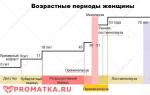
 Periods of female development
 What is endometrial hyperplasia - a brief overview
 Internal genitalia of a woman
 The endometrium is the inner lining of the uterus; more precisely, the mucous layer of the uterine wall adjacent to the myometrium (muscle layer). It is represented by stroma, uterine glands and blood vessels immersed in it.
Endometrial hyperplasia– benign, hormonal-dependent proliferative transformation of the uterine mucosa with disruption of its structure and functions.
The endometrium is a variable tissue highly sensitive to the action of sex hormones. Estrogen stimulation promotes its growth due to the proliferation of the uterine glands. Progesterone, on the contrary, stimulates the maturation and proliferation of the stroma, but inhibits the proliferation of glandular epithelium.
The main volume of estrogen and progesterone in women is produced in the ovaries.
During childbearing age, the key point in the development of typical hyperplasia is hormonal imbalance, or more precisely, estrogeny: hyperstimulation of the endometrium by estrogen with a lack of restraining activity of progesterone.
The causes of endometrial hyperplasia in postmenopause after the extinction of ovarian hormonal activity are not always explained.
Genetic predisposition plays a leading role in the development of cancer of the female genital organs and hyperplastic pathology of the endometrium in postmenopause.
Hyperplastic processes of the endometrium in postmenopause The structure of endometrial hyperplastic processes in postmenopause
 Atypical endometrial hyperplasia is a precancerous process. It can occur independently, as well as against the background of diffuse, focal typical hyperplasia, polyposis and endometrial atrophy.
Causes of diffuse endometrial hyperplasia in postmenopause
The appearance of diffuse hyperplasia of the uterine mucosa in older age first of all forces us to look for the source of pathological secretion of estrogen. Causes of hyperestrogenism in postmenopause:
- Ovarian pathology: hormonally active ovarian tumors, tecomatosis, stromal ovarian hyperplasia.
- Diencephalic pathology: age-related changes in the central nervous system and associated endocrine and metabolic disorders.
- Obesity: extragonadal production of estrogen in adipose tissue.
Causes of focal endometrial hyperplasia in postmenopause
Focal hyperplasia of the uterine mucosa in older age most often occurs in the form of polyposis.
Polyposis is a form of focal hyperplastic process caused by benign transformation of the basal layer of the endometrium.
Typical focal hyperplasia or polyposis of the endometrium in postmenopause develops against the background of chronic inflammation of atrophied areas of the uterine mucosa (chronic atrophic endometritis).
Local factors in the development of local endometrial pathology in postmenopause:
- Changes in the endometrial hormonal receptor apparatus: an increase in the number and sensitivity of estrogen receptors to small doses of the hormone.
- Increased activity of insulin-like growth factors.
- Slowing down planned cell death (apoptosis).
- Violation of local immunity.
Risk factors for endometrial hyperplasia in postmenopause
 Endometrial hyperplasia - risk factors
 Symptoms of endometrial hyperplasia in postmenopause
- Uterine bleeding.
- Bloody discharge from the uterus.
- Sometimes: purulent discharge from the uterus.
- Sometimes: nagging, cramping pain in the lower abdomen.
- Asymptomatic.
Diagnostics
1. Ultrasound transvaginal scanning is the optimal method for the primary diagnosis of endometrial pathology.
Endometrial thickness in postmenopause. Normal M-echo on ultrasoundUltrasound signs of endometrial hyperplasia in postmenopause:
- Magnification of M-echo >5 mm
- Heterogeneity of the endometrial structure.
- Unevenness, unclear boundary between the muscular and mucous layers of the uterine wall.
- Dopplerography: changes in blood flow, high resistance of blood flow in the endometrium.
- Serozometra: fluid in the uterine cavity.
2. Hysteroscopy using a rigid hysteroscope in combination with the endometrium and endocervix (mucous lining of the cervical canal).
3. Histological examination: study of the removed endometrium under a microscope.
4. Detection of ovarian pathology: ultrasound, biopsy, MRI (if necessary).
5. To determine the genetic predisposition to hyperplasia and malignant transformation of the endometrium, a genetic analysis of specific enzymes MMPI, ACE and cytochrome 1A1 (CYP 1A1) is performed.
1. Scraping.Fractional (separate) diagnostic curettage of the uterine mucosa under hysteroscopy control is the first stage of treatment for endometrial hyperplasia and a method recommended for postmenopause to stop uterine bleeding.
The choice of treatment tactics for endometrial hyperplasia in postmenopause depends on the results histological examination endometrial samples.
2. Surgical treatment.At an older age, there is a high risk of degeneration of benign hyperplasia into endometrial cancer. Therefore, in the treatment of endometrial pathology in the postmenopausal period, preference is given to surgical tactics:
- Removal of the uterus and appendages.
- Adnexectomy: removal of the ovaries.
- Endometrial ablation: destruction of the uterine lining.
Ablation (ablation, resection) of the endometrium— a method of gentle surgical treatment of simple diffuse endometrial hyperplasia in postmenopause. The effectiveness of the method is ≈83.4%.
Endometrial ablation is done:
- after a few days, the field is curetted and histological examination of the endometrium;
- in case of relapse of typical endometrial hyperplasia after unsuccessful hormonal therapy.
During ablation, the entire mucous membrane of the uterus is destroyed along with its basal layer to a depth of 3-5 mm. More often the operation is performed electrosurgically.
This operation in some cases serves as an alternative to radical surgical treatment (hysterectomy) for recurrent endometrial hyperplasia.
Indications for removal of the uterus and appendages in postmenopause:- Typical (simple, complex) endometrial hyperplasia in combination with ovarian pathology, fibroids, endometriosis, endocrine and metabolic disorders.
- Relapse of typical simple (complex) endometrial hyperplasia.
- Endometrial hyperplasia with atypia.
- Adenomatous endometrial polyps.
The clear choice of treatment for atypical endometrial hyperplasia and adenomatous polyps in postmenopause is surgical removal of the uterus and appendages.
Conservative hormonal therapy in these postmenopausal cases is carried out only if there are contraindications to surgery.
3. Hormonal treatment.The only indication for conservative treatment of endometrial pathology in postmenopausal women is simple endometrial hyperplasia without atypia.
Hormonal therapy for typical simple endometrial hyperplasia in postmenopause.The effectiveness of treatment is monitored after 6 months:
- aspiration biopsy;
- repeated diagnostic curettage.
Recurrence of typical endometrial hyperplasia in postmenopause is treated surgically.
4. Combined treatment.Indications:
- Typical focal endometrial hyperplasia.
- Simple polyposis.
In postmenopause, against the background of chronic atrophic endometritis, treatment of focal hyperplastic processes of the endometrium with gestagens is ineffective.
Stages of combination treatment
Stage 1Together with hysteroscopy and the diagnostic curettage procedure, the following is carried out:
- Removal of polyps.
- Selective cauterization (destruction) of the basal layer of the endometrium in the area of the removed polyp or focus of polyposis.
- Local anti-inflammatory therapy: washing the uterine cavity with a solution of chlorhexidine 0.02%, etc.
General antibacterial and anti-inflammatory treatment:
- cefazolin + metronidazole;
- levofloxacin,
- ciprofloxacin,
- doxycycline,
- gentamicin,
- Actovegin - to stimulate the healing of injured uterine tissue.
Treatment regimen for endometrial hyperplasia in postmenopause.
 Treatment of endometrial hyperplasia in older age
 Among gynecological oncological diseases, endometrial cancer ranks second after cervical cancer. Therefore, the treatment of choice for endometrial hyperplasia in older patients is surgical removal of the uterus and appendages.
Content
The endometrium is the internal mucous membrane lining the body of the uterus, which is a multicomponent system rich in blood vessels. It is constantly renewed and is very sensitive to changes in the body’s hormonal levels, especially during menopause.
During menopause, the layer usually becomes smaller and thinner. In this case, they speak of normal endometrial involution. However, if the hormonal balance is disturbed during menopause, this will also affect the condition of the endometrium. Various pathological conditions are common, for example, endometrial hyperplasia, when its thickness increases
Climax and its phases
Menopause is a period in the life of the body that is characterized by involution of the reproductive system, which is associated with aging. After menopause, women lose the opportunity to have children and their menstruation stops. This is due to the depletion of the follicular apparatus in the ovaries. As a rule, menopause normally occurs at about 50 years of age. Early menopause is said to occur if it began before the age of 45. Late menopause begins after 55.
If menstruation ends before 40– this is called early ovarian failure syndrome, which is considered a pathological variant of menopause.
During menopause, the ovaries stop synthesizing estrogen hormones, and therefore the functioning of many organs that have receptors for them is disrupted. These include the nervous and urinary systems, mammary glands, skin and even bones. There are periods of menopause.
- Premenopause.
- Menopause.
- Postmenopause.
At the first stage, menstrual cycles become uneven, their frequency and duration change. When comparing them, you can find that adjacent cycles differ by seven or more days. This situation during menopause is observed within 10 cycles of the first extended cycle.
At the last stage, menstruation stops, and the amenorrhea phase begins, which lasts more than three months. It marks the beginning of the postmenopausal period. At this time, the level of follicle-stimulating hormone in the blood increases (more than 25 IU/l). Typically, menopause lasts several years, followed by early postmenopause (5-8 years), and then late postmenopause.
What is endometrium
The endometrium has a complex structure. Normally, it consists of two layers - the main germinal (basal) and functional glandular. It also has a very abundant blood supply, rich in blood vessels.
Normally, epithelial structures are formed from secretory and ciliated cells. The stroma consists of fibroblast-like cells that differentiate during the menstrual cycle and begin to synthesize collagen and extracellular structures, ensuring the integrity of the stroma. It also contains numerous glands (crypts) that open into the lumen of the uterus.
Normally, during the premenstrual phase, the number of glands increases, the endometrium thickens, and its blood supply increases. This is necessary for further implantation of the embryo. During pregnancy, the number of glands and blood vessels increases significantly, which ensures nutrition of the fetus and development of the placenta. If pregnancy does not occur, then the functional part of the membrane is separated and removed during menstruation.
In the early stages of proliferation, it is homogeneous, has low echogenicity, and its thickness, on average, ranges from 3 to 6 mm. On days 8-10 of the cycle, in the late proliferation phase, the functional layer begins to thicken and maintains the homogeneity of its structure. The thickness of the endometrium is normally 5-10 mm. By the end of the second week, the period of late proliferation ends.
The mucous membrane continues to thicken, acquiring increased echogenicity. The thickness during this period is 8-13 mm. After this, the stage of early secretion begins. Tissue growth slows down, it acquires heterogeneous echogenicity - more pronounced in the center and less pronounced in the periphery. The thickness of the endometrium is 10-14 mm. During late secretion, the mucous membrane begins to decrease in size (10-12 mm), it retains high echogenicity.
Age-related changes in endometrial thickness
In children before puberty, the endometrium is in a “dormant state.” Its thickness is insignificant. But even then it is divided into a functionally active and basal layer. After the start of menstruation, the thickness of the endometrium normally increases, it begins to cyclically thicken and be rejected. Its final involution normally occurs during menopause.

With age, the amount of hormones produced by the ovaries decreases, and menopause begins. This is the norm of aging. Since the thickness of the endometrium directly depends on the level of hormonal stimulation, it begins to atrophy and thin out. The size of the uterus itself and its muscular wall also decreases. The endometrium in menopause becomes looser and thinner, the number of glands decreases, and their atrophy occurs. As a result, over time, adhesions and synechiae may appear in the uterus, which are deviations from the norm, which can complicate the course of menopause.
Normal endometrial thickness during menopause
The thickness of the endometrium begins to decrease shortly before menopause. Normally, after the completion of the body's restructuring during menopause, the thickness of the endometrium is 4-5 mm, according to ultrasound.
This is due to a decrease in stimulation of its growth by estrogen. As a rule, the duration of the involution period is from 3 to 5 years. If after this the thickness of the endometrium exceeds the norm of these indicators, they speak of its hyperplasia. This is the most common violation.
Excessive atrophy, on the contrary, develops much less frequently. Also, in the early stages of menopause, excessive menstrual bleeding may occur. This is due to the fact that at this time excessive hypertrophy of the mucous membrane may be observed for a short time. This happens due to hormonal imbalance.
Causes and symptoms of changes in endometrial thickness during menopause
The growth of the endometrium is ensured mainly by estrogens. Some patients experience an increase in estrogen levels during menopause due to certain conditions.
This leads to excessive growth of mucous tissue. It is possible for endometrial cells to penetrate other layers of the uterus, leading to adenomyosis.

Hypertrophy can be provoked by uterine fibroids, ovarian dysfunction, ovarian tumor or polycystic disease, endometriosis and sexually transmitted diseases.
In some patients this condition develops as a result of improper use of hormonal contraceptives.
Factors that negatively affect ovarian function and general hormonal levels are also important:
- smoking;
- woman drinking alcohol;
- diabetes mellitus or impaired glucose tolerance;
- metabolic syndrome, obesity;
- liver pathology;
- hypertonic disease;
- renal dysfunction;
- diseases of the pancreas, adrenal glands;
- autoimmune disorders.
Statistically, endometrial hypertrophy after menopause occurs in 20% of the female population, and the prevalence of the pathology is slowly increasing. This is due to the environmental situation, the predominantly sedentary lifestyle of the urban population, and the widespread prevalence of bad habits.
A woman during menopause most often complains of pain in the lower abdomen, pain during menstruation (if it has not stopped), pain during urination or sexual intercourse. During the period between menstruation, spotting and spotting may occur. Sometimes severe uterine bleeding occurs. Therefore, if such symptoms appear during menopause, you should consult a doctor.
Ultrasound and other diagnostic methods
Examination of patients with endometrial hyperplasia begins with a medical history and gynecological examination in the speculum. After this, a decision is made to prescribe additional examination methods. Ultrasound of the pelvic organs is very informative, as well as safe and convenient. With its help, the doctor has the opportunity to determine the thickness of the layer, its echogenicity and structural features.
In order to find out the type of hyperplasia and growth pattern, it is necessary to take an endometrial biopsy. They also use the method of hysteroscopy - examination of the inner lining of the uterus with a special device, with which you can also take biological samples. A biochemical blood test and determination of hormone levels may also be helpful.
Treatment options for changes in endometrial thickness
When choosing therapeutic tactics, doctors take into account the patient’s age, her medical history, the severity of clinical symptoms, the type of course of the disease and the type of hyperplasia. That is why a thorough diagnosis must be carried out before prescribing therapy. As a rule, treatment has two stages - removal of the endometrium, and further drug treatment, which is aimed at restoring normal hormonal balance and reducing the risk of relapse. In women during menopause, the basal germ layer is most often removed so that the endometrium does not grow.
Various techniques are used. Ablation is used to remove the growth layer. If it is necessary to eliminate hyperplasia of the functional layer, curettage is used.
Surgical intervention to remove hypertrophied mucosa is called curettage. The procedure must be performed in a hospital setting under general anesthesia.
In the postoperative period, antibiotic therapy is prescribed to prevent infectious complications.
The growth and increase in the volume of the endometrium of the female uterus occurs under the influence of changes in hormonal levels, in particular, increased production of estrogen. Therefore, endometrial hyperplasia in menopause is a fairly common phenomenon, since it stems from pathological changes in the female body as a whole.
Enlargement of the endometrium during menopause
The endometrium is the mucous membrane lining the inner surface of the female uterine cavity. The thickness of the endometrium is normally 3-6 mm, but under the influence of various factors it can increase to 15-20 mm. These changes are not always the consequences of the development of pathology - the membrane tends to expand during pregnancy.
In any case, the thickness of the layer is influenced by the hormonal background in the female body, which can change for a number of reasons:
- with the development of symptoms of polycystic ovaries;
- when using medications containing estrogen;
- if you are overweight (obese);
- during menopause.
It is worth noting that there is a whole range of certain factors that significantly increase the risk of hyperplasia. In particular, the development of various pathologies of the uterus, for example, dysplasia and hyperplasia, in most cases is directly related to an unfavorable period of puberty, the presence of bad habits in a woman’s life, previous infectious diseases of the genital and urinary tract, diseases of the endocrine systems, and a general weakening of the immune system.
As for the transformations of the female body during menopause, they only aggravate the possibility of developing various pathological manifestations. The body's resources at this stage are weakened, the ability to resist various diseases is reduced.
At this time, the norm of the endometrium is most often violated. The pathology develops due to existing chronic diseases of the reproductive system, previous surgical interventions, problems with blood circulation and metabolism, and hormonal changes.
Manifestations of endometrial hyperplasia in premenopause are quite common - during the period when a woman’s body is preparing for future dramatic transformations, and the first hormonal disruptions begin.
The development and symptoms of endometrial hyperplasia in premenopause are similar to the usual nature of the formation of pathology, with the only difference being that the uterus at this stage is prone to abnormal bleeding, and the risk of malignant formations is very high.
It is worth noting that some irreversible changes occur in the endometrium during menopause. The layer thickens to an average of 5 mm. If further expansion occurs, this indicates the development of damage to the mucous membrane, and the need for therapeutic measures.

Symptoms of hyperplasia during menopause
Let us consider in more detail the picture of the development of pathology with endometrial hyperplasia during menopause.
Symptoms that suggest the expansion of the endometrium during menopause:
- the appearance of spotting bloody discharge in a small amount;
- prolonged heavy bleeding;
- development of polyps.
It is important to take into account that endometrial hyperplasia in menopause can occur latently, that is, not accompanied by any alarming manifestations. In some cases, hyperplasia is discovered during a gynecological examination completely by accident, when a woman went to the doctor with a completely different problem. If the lesion is not treated, it can lead to serious complications.
Therefore, it is very important to undergo examinations systematically, even if no painful symptoms are felt. The enlarged endometrium in menopause is characterized by increased dynamics of development into a malignant formation. That is, the risk of cancer is very high.
The absence of pain symptoms is characteristic, first of all, of glandular endometrial hyperplasia, accompanied by uterine bleeding of varying intensity. A feature of this type of pathology is the increased development and enlargement of the glands of the mucous tissue, but, in general, adenomatous hyperplasia is not a characteristic manifestation of menopause.
Women over the age of 50 who have high heart pressure, problems with the endocrine system and other accompanying manifestations of menopause are required to carefully monitor their health and undergo regular medical examinations.
Uterine endometrial hyperplasia in menopause is diagnosed using transvaginal ultrasound or using aspiration biopsy. The latter method will not be effective if the lesion is focal.
Ultrasound has a higher accuracy in determining the nature and extent of the lesion. If the detected layer thickness is 6-7 mm, a repeat examination is carried out. In cases where the size of the mucous tissue has expanded to 8 mm, curettage surgery is prescribed. The resulting material is sent for histological analysis. The inner lining of the uterine cavity is examined before and after the intervention using special probes.
Postmenopausal period: the possibility of developing pathology
After the onset of menopause, a postmenopausal period begins in a woman’s life, which can last 3-4 years or longer, accompanied by significant, and sometimes painful, changes in the functioning of the body.
Endometrial hyperplasia in postmenopause is also a fairly common phenomenon. The ovaries finally stop functioning, and the endocrine gland produces a reduced level of hormones, which affects the condition of the mucous membranes.
The consequence of the fact that the ovaries stop functioning often results in the development of a cyst, which can be absolutely painless. In some cases, the cyst is discovered when the pedicle is ruptured or bent.
If the development of a cyst is accompanied by pain symptoms, then it is a very strong and sharp pain. These manifestations still make it possible to detect the lesion earlier and eliminate it. The development of a cyst is accompanied by increased dynamics of expansion of mucous tissue, which, in turn, can provoke a cancerous tumor.
Endometrial pathology in postmenopause is accompanied by bloody vaginal discharge, cramping pain localized in the lower abdomen and lower back, and the development of large single polyps. During postmenopause, the lining of the uterus begins to die.

Treatment of pathology during menopause
For endometrial hyperplasia in menopause, treatment is carried out using three main methods:
- hormonal;
- surgical;
- combined.
In some cases, hormonal therapy demonstrates high effectiveness in combating lesions of the mucous membrane, and allows one to avoid surgical intervention. The use of medroxyprogesterone and megestrol acetate for 3-6 months allows to achieve positive dynamics of the pathology. To determine the effectiveness of treatment, ultrasound is systematically used.
Other drugs of the hormonal spectrum, such as norkolut and duphaston, are prescribed for hyperplasia, but not during menopause, since their use is associated with correction of the menstrual cycle.
As for surgical treatment, it is the most common, although not always effective.

Today, in gynecological surgery, in the fight against pathologies of the mucous membrane, the method of laser cauterization (ablation), curettage (curettage), and complete removal of the uterus (hysterectomy) is used.
Treatment of endometrial hyperplasia in postmenopause is often accompanied by the use of the latter method, which is associated with the need to preserve the health, and often the life of the woman.
Combined treatment is most often used for endometrial hyperplasia in menopause. The use of hormonal therapy at the initial stage of treatment can increase its effectiveness and improve the prognosis of the disease.
When menopause occurs, many changes occur in a woman's body. Some of them are accompanied by severe symptoms that require.
This is explained by the fact that the body produces significantly less estrogen and progesterone, which negatively affects the condition of the female genital organs.
This can lead to partial atrophy of the uterine mucosa, as well as a change in the size of the endometrium.
During menopause, this size must meet certain standards, but if it goes beyond these limits, then this may indicate the presence of certain diseases.
What is the endometrium of the uterus?
The endometrium of the uterus refers to the mucous membrane, i.e. its inner layer. It is extremely important for the normal process of gestation and fetal development. The endometrium is surrounded by numerous blood vessels, and it itself includes many sensitive receptors that ensure proper sensitivity of estrogen from the mucous membrane.
NOTE!
The thickness of the inner layer of the uterus may vary depending on the stage of the menstrual cycle. In particular, the endometrium in the final stage can be 10 times thicker than in the initial stage.
What happens during menopause?
When a woman experiences a climatic period, she experiences a gradual decrease in the frequency of menstrual cycles until they are completely absent. Thus, the uterine mucosa no longer changes according to the stages of menstruation and, accordingly, the thickness of the endometrium also does not change, i.e. it becomes a constant value.
Read about hormonal changes during menopause.
What is the norm?
In order to determine the thickness of the inner layer of the uterus, an ultrasound diagnostic method is used.
An indicator of the norm is the fact that the thickness of the endometrium during menopause decreases compared to normal periods in a woman’s life.
In general, a normal indicator of endometrial thickness is considered to be 5 mm.. If during diagnostic measures it was discovered that this indicator has increased by 1 or 2 mm, then it is necessary to monitor the condition of the patient’s female genital organs for several months.
On the other hand, the characteristics of the body are different for everyone, so it is completely logical if the size of the endometrium will be slightly different in different patients. Accordingly, the criteria for violating the norm may be blurred. In any case, only the attending physician can determine the pathology after receiving the diagnostic results..
Modern medicine identifies several types of this disease.:
- Glandular. This pathology is diagnosed when glandular cells grow, and the connective layer of the endometrium does not change in any way. This form is considered the mildest, because the risk of transition to the malignant stage is practically eliminated.
- Cystic. In this case, damage to the epithelium of the inner layer of the uterus is already observed, which can lead to the development of cancer.
- Glandular-cystic. In addition to the proliferation of glandular cells, cystic formations appear.
- Focal. The proliferation of the endometrium occurs in certain areas where the greatest sensitivity is observed. At this stage, polyps grow, which is already a harbinger of a malignant form.
- Atypical. This type of hyperplasia is considered the most dangerous to a woman’s health. In most cases, to avoid the development of cancerous tumors, the uterus is completely removed.

Diagnostic methods
If a woman during menopause notices certain symptoms, she should immediately consult a gynecologist.
Dangerous symptoms include:
- , which have nothing to do with the menstrual cycle.
- Painful sensations in the genitals.
- Change in the nature of menstruation.
How to distinguish bleeding from menstruation, read.
Quite often it happens that a woman perceives these manifestations as a normal change in hormonal levels during menopause. This is fundamentally incorrect, so if you have at least one symptom, you need to consult a doctor.
After going to the hospital, the gynecologist sends the patient to undergo a certain series of diagnostic measures:
- Ultrasonography. To determine the thickness of the endometrium, the transvaginal ultrasound method is used, i.e. through the vagina. This type of diagnosis is considered the main one in identifying hyperplasia, so it is used everywhere. If the layer has grown to 8 mm, then it is necessary to begin treatment.
- Scraping. This method applies not only to diagnostic, but also to therapeutic measures. The functional layer of the uterine mucosa is scraped for further sending for histological examination. This is done in order to determine the presence of cancer cells.
- Biopsy. This method is performed using a thin tube with a piston, which collects a small amount of endometrial tissue particles. It should be noted that this diagnostic method cannot be used for focal hyperplasia.
- Radioisotope research. In this case, phosphorus is used, which makes it possible to identify foci of the disease.

In addition, the diagnosis includes the usual procedures that any woman has gone through. We are talking about a standard examination of the vagina, taking a smear and a blood test.
Treatment of pathology
It should immediately be noted that the growth of the endometrial layer should be treated only with the help of traditional medicine. In some cases, surgical intervention may be necessary, but there can be no question of any traditional methods.
Conservative treatment is the use of certain. They need to be taken in courses, i.e. do not stop taking it for at least 6 months. Among the drugs used in treatment are Regulon and Logest, which are also oral contraceptives.
If the situation is more severe, the doctor may prescribe surgery.
Surgical treatment can be of several types:
- Endometrial scraping.
- Complete removal of the uterus. This method is considered the most radical measure when there is a risk of developing a malignant tumor.
- Laser ablation, which can be used to treat foci of endometriosis.
It should be noted that curettage of the mucous membrane is the first procedure that is performed if there are indications for surgical intervention. If it does not give a positive effect, then laser treatment is used, during which individual lesions are burned out.
If the doctor understands that these methods have not stopped the progress of the disease, then a decision is made. Unfortunately, in some cases this is the only way out of the situation.

Scraping and consequences
The endometrium consists of glandular and epithelial tissues. During the curettage procedure, only the top layer is removed, while the base layer remains untouched. It is due to this that further restoration of the endometrial layer occurs, so this procedure is not as radical as its name might seem.
There are two types of scraping:
- Normal. This method is carried out almost blindly, so there is a risk of damage to the uterine organ.
- Separate. In this case, the cervix is scraped, and then its cavity. The resulting particles are sent for histological examination.
As a rule, there are practically no complications after this procedure.
But their probability still exists, so we need to talk about them in more detail.:
- Cervical rupture. This may happen due to the procedure being performed incorrectly. If the tears are small, then no action should be taken, but if the damage is extensive, stitches will be needed.
- Spasm. As a result of this complication, blood accumulates inside the uterine organ.
- Inflammation of the uterine organ. This happens if the basic rules of asepsis were violated during the procedure.
- In some cases, damage to the base layer of the endometrium occurs. This complication is difficult to treat, so the woman risks losing her ability to bear children.
After the procedure, discharge of a certain nature may be observed. If they have an unpleasant and pungent odor, this indicates the development of an infection inside the uterus. Yellow discharge should also cause considerable concern, so you should immediately consult your doctor.

Conclusion
After a woman reaches a certain age, various kinds of changes occur in her body. This period is called menopause, so you need to carefully monitor your health.
One of the first points that the doctor pays attention to is the size of the endometrium, which must correspond to a certain norm. If there is a deviation of 3 mm or more, then therapeutic action is necessary.
Useful video
From the video you will learn everything about endometrial hyperplasia:
In contact with
- This is the inner layer of the uterus, very sensitive to hormonal changes in the body. Many women are aware of a disease called endometriosis. It is accompanied by many complications and is difficult to treat.
Overgrowth or pathological thinning of the endometrium is a problem faced by a huge number of women. During menopause, hormonal changes begin, which can affect the endometrium, causing its changes.
Endometrium: functions and normal thickness in menopause
Causes and signs of deviation in endometrial thickness during menopause

During menopause, they only talk about endometrial hyperplasia, since the reduction of this layer during menopause is normal.
However, if the endometrium has a thickness of more than 5 mm during menopause, they speak of a pathological condition that requires.
Almost always, the reason is a hormonal imbalance that controls the growth of the endometrium. This condition can be asymptomatic for a long time or manifest itself in the form of breakthrough pain in the lower abdomen.
Endometrial hyperplasia, like endometriosis, can only be determined using.
There are types of endometrial hyperplasia:
- Glandular. Glandular endometrial hyperplasia is considered a benign disease, accompanied by the proliferation and thickening of glandular tissue due to improper location of the glands. With timely treatment, the prognosis is favorable.
- Cystic. A more serious disease that may be a consequence of the glandular form. At the same time, neoplasms are formed in the endometrium, which can eventually degenerate into a malignant tumor.
- Basal. This is a very rare and serious disease that is difficult to treat. The basal layer of the endometrium grows rarely, as a rule, it is unchanged, and is also difficult to treat with hormonal therapy.
- Polypoid. With this disease, the endometrium thickens not over the entire surface of the uterus, but focally. The lesions are located where the endometrium forms. This condition is often accompanied by bleeding and is treated with curettage, which is also a diagnostic procedure.
- Atypical. The most dangerous hyperplasia, which is rare, but still occurs in women during menopause. At the same time, the endometrium grows very actively, and the cells quickly degenerate. It is difficult to treat such a disease; it is often necessary to resort to surgical removal of the uterus to avoid cancerous tumors.
Diagnostics

If a woman consults a doctor with complaints of bleeding and pain during menopause, she must undergo a series of diagnostic procedures before diagnosis and treatment. In the case of endometrial hyperplasia, it should be comprehensive.
It includes an examination in a gynecological chair, and some invasive procedures that will help clarify the diagnosis and type of hyperplasia:
- . This procedure is considered the main one in the diagnosis of endometrial hyperplasia. To assess the thickness of the endometrium, a transvaginal ultrasound is performed. The procedure is carried out using a special nozzle, which is painlessly inserted into. If the endometrium is more than 5 mm during menopause, the ultrasound procedure is repeated several more times over six months. When the thickness of the endometrium is 8-10 mm, as a rule, treatment and curettage are already prescribed.
- Diagnostic curettage. This procedure is both diagnostic and therapeutic. It is performed under anesthesia. The entire uterine cavity is scraped out, after which after some time the woman stops bleeding. The contents are sent for histology to determine the presence of cancer cells.
- . An endometrial biopsy will be informative only if the endometrium has not grown in patches, but completely over the entire surface of the uterus. This procedure will help determine the exact thickness of the endometrium, pathological processes in it, and cancer. The procedure is carried out using a pipel, which is a flexible thin tube with a piston. Once in the uterus, the pipel absorbs small particles of the endometrium.
- uterus and fallopian tubes. This procedure is very informative in identifying tumors, polyps in the uterus and adhesions in the fallopian tubes. The procedure is invasive because the uterine cavity is filled with a contrast agent before the image is taken. The procedure is unpleasant, but should not cause pain.
Medical and surgical treatment

Serious diseases such as endometritis and are not recommended to be treated exclusively with folk remedies at home. They can only be cured with hormonal therapy and sometimes with surgery.
Before starting treatment, the doctor determines the cause of the disease and the type of hyperplasia. Since the endometrium is very sensitive to changes in hormonal levels, various pathologies should also be treated with the help.
Women's hormones go haywire during menopause. With an increased amount of estrogen and a decreased amount, the endometrium thickens. The risk group includes overweight and overweight women, who often have hormonal problems.Hormonal contraceptives such as Logest, Regulon, etc. are prescribed as hormonal therapy. They are prescribed in courses for up to six months to normalize hormonal levels. It has been proven that taking these drugs does not provoke.
Often, when the endometrium is thickened, drugs such as Duphaston and Utrozhestan are prescribed.
These are hormonal drugs, analogues of progesterone. As mentioned above, the endometrium is sensitive to hormones and grows with a lack of progesterone. They are considered safe and are prescribed to normalize hormonal levels even during pregnancy. The dosage is prescribed by the doctor. The course of treatment for endometriosis lasts a long time from six months to 9 months.
More information about what endometrial hyperplasia is can be found in the video:
Surgery is resorted to only in extreme cases. First, curettage is prescribed. If there is no progress, laparoscopy may be prescribed, during which the foci of endometrial growth are precisely cauterized with a laser.
If treatment does not bring results, there is a risk of cancer, the uterus is removed. There are several types of such operations. Depending on the severity, either only the uterus, or the uterus and cervix, or the cervix and all nearby lymph nodes are removed.
Consequences and prevention
 During reproductive age, endometriosis can lead to. During menopause, this disease is dangerous due to degeneration into a malignant neoplasm. With age, the risk of cancer increases, and endometrial thickening, inflammation and polyps are a precancerous condition. complications
It is worth remembering that for any manifestations of endometriosis or endometritis you should consult a doctor. Uterine bleeding is always an alarming symptom that is not recommended to be ignored.