Anatomical structure of the knee joint in children. Knee joint - anatomy and detailed structure. Pathologies and lack of a cup
The knee joint, along with the hip, is the largest and most powerful articulation of the human skeleton. It combines the bones of the thigh and lower leg, providing a range of motion when walking. The articulation has a complex complex structure, in which each element provides the functioning of the knee in particular and the ability to walk in general.
The device of the human knee joint explains the cause of emerging pathologies, helps to understand the etiology and course of inflammatory and degenerative diseases. Even small deviations from the norm in any element of the articulation can cause pain and limited mobility.
Anatomy
Three bones of the knee joint are involved in the formation of the articulation: the femur, tibia and patella. Inside the joint, on the plateau of the tibia are located, increasing the stability of the structure and providing a rational distribution of the load. During movement, the meniscus springs - they are compressed and unclenched, ensuring a smooth gait and protecting the articulation elements from abrasion. Despite their small size, the importance of the menisci is very high - when they are destroyed, the stability of the knee decreases and arthrosis inevitably occurs.
In addition to bones and menisci, the articulation components are the articular capsule, which forms the torsion of the knee joint and synovial bags, and ligaments. The ligaments that form the knee joint are formed by connective tissue. They fix the bones, strengthen the joint and limit the range of motion. Ligaments provide stability to the joint and prevent displacement of its structures. Injury occurs when the ligaments are stretched or torn.
The knee is innervated by the popliteal nerve. It is located behind the articulation and is part of the sciatic nerve that runs to the foot and lower leg. The sciatic nerve provides sensation and movement to the leg. The popliteal artery and vein, which repeat the course of the nerve branches, are responsible for the blood supply.
The structure of the knee joint
The main joint-forming elements are considered to be the following:
- condyles of the femur
- tibial plateau
- knee cap
- menisci
- joint capsule
- bundles
The knee joint itself is formed by the heads of the femur and tibia. The head of the tibia is almost flat with a slight depression, and it is called a plateau, in which the medial, located along the midline of the body, and the lateral parts are distinguished.
The head of the femur consists of two large, round, spherical projections, each of which is called the condyle of the knee joint. The condyle of the knee joint located on the inside is called medial (internal), and the opposite is called lateral (external). The articular heads do not match in shape, and their congruence (correspondence) is achieved due to two menisci - medial and lateral, respectively.
The articular cavity is a gap, which is limited by the heads of the bones, menisci and the walls of the capsule. Inside the cavity is synovial fluid, which provides optimal gliding during movement, reduces friction of the articular cartilage and nourishes them. The articulating surfaces of the bones are covered with cartilaginous tissue.
Hyaline cartilage of the knee joint is white, shiny, dense, 4-5 mm thick. Its purpose is to reduce friction between the articular surfaces during movement. Healthy cartilage of the knee joint has a perfectly smooth surface. Various diseases (arthritis, arthrosis, gout, etc.) lead to damage to the surface of the hyaline cartilage, which, in turn, causes pain when walking and limited range of motion.
Knee cap
The sesamoid bone, or patella, covers the front of the knee joint and protects it from injury. It is located in the tendons of the quadriceps muscle, has no fixation, has mobility and can move in all directions. The upper part of the patella has a rounded shape and is called the base, the elongated lower part is called the apex. On the inside of the knee is the goose foot - the junction of the tendons of 3 muscles.
joint capsule
The articular bag of the knee joint is a fibrous case that limits the articular cavity from the outside. It is attached to the tibia and femur. The capsule has a low tension, due to which a large amplitude of movements in different planes is provided in the knee. The articular bag nourishes the articulation elements, protects them from external influences and wear. Located on the inside of the knee, the posterior part of the capsule is thicker and resembles a sieve - blood vessels pass through numerous holes, and blood supply to the joint is provided.
The capsule of the knee joint has two shells: inner synovial and outer fibrous. A dense fibrous membrane performs protective functions. It has a simple structure and is firmly fixed. The synovial membrane produces a fluid, which has received the corresponding name. It is covered with small outgrowths - villi, which increase its surface area.
In places of contact with the bones of the joint, the synovial membrane forms a slight protrusion - a torsion of the knee joint. In total, 13 inversions are distinguished, which are classified depending on the location: medial, lateral, anterior, inferior, superior inversion. They increase the articulation cavity, and in pathological processes they serve as places for the accumulation of exudate, pus and blood.
Knee bags
They are an important addition, thanks to which the muscles and tendons can move freely and painlessly. There are six main bags, which look like small slit-like cavities formed by the tissue of the synovial membrane. Internally, they contain synovial fluid and may or may not communicate with the articulation cavity. Bags begin to form after the birth of a person, under the influence of loads in the area of the knee joint. With age, their number and volume increases.
Biomechanics of the knee
The knee joint provides support for the entire skeleton, takes on the weight of the human body and experiences the greatest load when walking and moving. It performs many different movements, and therefore has complex biomechanics. The knee is capable of flexion, extension and circular rotational movements. The complex anatomy of the human knee joint ensures its wide functionality, well-coordinated work of all elements, optimal mobility and shock absorption.
Pathology of the knee joint
Pathological changes in the musculoskeletal system can be caused by congenital pathology, injuries and diseases. The main signs that signal the presence of violations are:
- inflammatory process;
- painful sensations;
- restriction of mobility.
The degree of damage to the articulation elements, coupled with the cause of their occurrence, determines the localization and intensity of the pain syndrome. Pain can be diagnosed periodically, be permanent, appear when trying to bend / straighten the knee, or be the result of physical exertion. One of the consequences of ongoing inflammatory and degenerative processes is the deformity of the knee joint, leading to serious illnesses up to disability.
Anomalies in the development of the knee joint
There are valgus and varus deformities of the knee joints, which can be congenital or acquired. Diagnosis is made by x-ray. Normally, the legs of a standing person are straight and parallel to each other. With valgus deformity of the knee joint, they are bent - an open angle appears on the outside in the knee area between the lower leg and thigh.
The deformity may affect one or both knees. With bilateral curvature of the legs, their shape resembles the letter "X". Varus deformity of the knee joints bends the bones in the opposite direction and the shape of the legs resembles the letter "O". With this pathology, the knee joint develops unevenly: the joint space decreases from the inside and expands from the outside. Then the changes affect the ligaments: the external ones are stretched, and the internal ones atrophy.
Each type of curvature is a complex pathology that requires complex treatment. If left untreated, the risk of excessive knee mobility, habitual dislocations, severe contractures, ankylosis, and spinal pathologies is quite high.
Valgus and varus deformity in adults
It is an acquired pathology and most often appears with deforming arthrosis. In this case, the cartilaginous tissue of the joint undergoes destruction and irreversible changes, leading to loss of knee mobility. Also, the deformation can be the result of injuries and inflammatory and degenerative diseases that caused changes in the structure of bones, muscles and tendons:
- compound fracture with displacement;
- ligament rupture;
- habitual dislocation of the knee;
- immune and endocrine diseases;
- arthritis and arthrosis.
In adults, the treatment of a deformed knee joint is inextricably linked to the underlying cause and is symptomatic. Therapy includes the following items:
- painkillers;
- NSAIDs - non-steroidal anti-inflammatory drugs;
- glucocorticosteroids;
- vasoconstrictor drugs and venotonics;
- chondroprotectors;
- physiotherapy treatment;
- massage.
Drug treatment is aimed at eliminating pain, restoring cartilage, improving metabolism and tissue nutrition, and maintaining joint mobility.
Valgus and varus deformity in children
The acquired varus or valgus deformity of the knee joints that manifests itself by 10-18 months in children is associated with deviations in the formation of the child's musculoskeletal system. As a rule, the deformity is diagnosed in weakened children with muscular hypotension. It appears as a result of a load on the legs against the background of a weak muscular-ligamentous apparatus. The reason for such a deviation may be the prematurity of the child, intrauterine malnutrition, congenital weakness of the connective tissue, general weakness of the body, rickets.
The cause of the secondary pathology that caused abnormalities in the formation of the knee joint is neuromuscular diseases: polyneuropathy, cerebral palsy, myodystrophy, poliomyelitis. Articulation deformity not only causes curvature of the legs, but also has an extremely detrimental effect on the entire body.
Quite often, the feet and hip joints suffer, flat feet and coxarthrosis develop with age.
Treatment of hallux valgus and varus deformity in children includes:
- limitation of loads;
- wearing orthopedic shoes;
- use of orthoses and splints;
- massage;
- physiotherapy, most often - paraffin wraps;
- physical therapy classes.
Conclusion
Having a complex structure, the knee joint bears a heavy load and performs many functions. He is a direct participant in walking and affects the quality of life. Attentive attitude to your body and taking care of the health of all its constituent elements will help you avoid knee pain and maintain an active lifestyle for a long time.
There are many myths associated with the difference between the body structure of a child and an adult. One of them is the opinion that children do not have kneecaps until a certain age. But this information is erroneous, and even an unborn baby already has patellas, but they differ in structure from adults somewhere up to 6 years old, so they are not visible in the picture during an X-ray examination.
The formation of the kneecaps in children occurs by the age of six.
Knee joints of newborns
A newly born baby has cups, but in infancy they are made of thin cartilage, not bone. Therefore, in the first months of a baby's life, it is quite difficult to see them on an x-ray, which gives rise to false information about the structure of the musculoskeletal system in newborns. To avoid damage to the cups, it is not recommended to massage the knees of an infant, because they are fragile and can be damaged.
When do kneecaps appear and what are they in children?
The patella is the largest sesamoid bone of the human body, surrounded by tendons of the quadriceps muscle, located above the cavity of the articular joint of the knee. The patella can be easily felt under the skin, it moves effortlessly in different directions when the leg is relaxed. The main function of the knee cup is to protect against strong lateral displacements of the femur and tibia, which make up the knee joint.
 The development of kneecaps in children can be negatively affected by an unhealthy pregnancy, illness, and injury to the baby.
 Cups are formed during the development of the child in utero approximately in the first trimester at the 4th month of pregnancy. During this period, cartilage is formed, which replaces bone tissue for the time being. At this stage of development in babies, the knee joints are soft and fragile. During pregnancy, there may be problems with the formation of the joint. But such a violation is rare. There are a number of negative factors, both external and internal, that can adversely affect the health of infants.
Common causes of violations:
- abuse or misuse of drugs;
- infectious diseases of the mother during childbearing;
- influence of radiation and unfavorable environment;
- metabolic disturbances.
Exposure to any of these factors during the first 3 months of pregnancy may result in cups not forming at all. If problems with the health of the mother are discovered at such a crucial time, this gives rise to various defects in the knee joints in the child in the future.
The bones of the human skeleton are a reliable support for the whole body and protection for vital internal organs. It is the bones and muscles that enable the human body to move. Muscles have the ability to contract, which, in fact, sets the human body in motion. Thus, the human musculoskeletal system includes:
- bones of the skeleton;
- joints that connect individual bones of the skeleton to each other (the largest are the hip and knee joints);
- muscles.
Human bones are constantly growing and changing. A newborn baby has about 350 bones. In the process of growing a baby, some bones grow together, so in an adult their number is 206. The human skeleton is finally formed by the age of thirty, and in women this process ends earlier than in men.
Anatomy and physiology of the joints of the human skeleton

As mentioned above, the joints of the bones of the skeleton are called joints. Some of them are immobile (cranial bones), others are almost immobile (cartilaginous joints of the spine), but most are mobile and provide various motor functions (flexion, extension, dilution, etc.). Movable joints are called synovial joints. This name is due to the anatomical structure of the joint, which is a kind of complex, including the following composition:
- joint capsule;
- articular surfaces;
- articular cavity;
- articular discs;
- menisci;
- articular lips.
The joint capsule is a complex combination of collagen and elastin fibers and connective tissue. Together, these tissues form a kind of filter, which has a huge number of different functions. The joint capsule is permeated with a complex network of blood vessels and nerve endings that provide nutrition to the joint, its blood supply and signaling function, that is, they send information about its position to the brain.
The articular surfaces are the smooth surfaces of the bones that carry out the connection. The ends of the bones are covered with a thin layer of cartilage and a special lubricant that reduces mechanical friction between the bones.

The movement in the joint directly depends on what its shape is. There is a certain classification, according to which it is customary to distinguish the following types of joints:
- cylindrical (connecting the first two cervical vertebrae);
- flat (connects the tarsal bones of the foot and the carpal bones of the human hand);
- saddle (thumb);
- elliptical (connects the radius to the wrist);
- spherical (shoulder and hip joint);
- articulated (knee joint, elbow joint and finger joints).
The articular cavity is a closed and completely sealed slit-like space that does not communicate with the environment. It is the articular cavity that contains the synovial membrane and synovial fluid. What it is? The synovial membrane is the inner layer of the joint capsule that lines the entire joint cavity, excluding its cartilaginous areas. The main function of the synovial membrane is protective, it is this structure that prevents friction and promotes cushioning. Ensuring the protective function of the synovial membrane is possible due to the fact that it is able to release a special lubricant, which is called the synovial fluid.
Synovial fluid is a special substance that has a complex molecular structure and chemical composition. Without going into details, we note that the synovial fluid is a blood plasma and a protein-polysaccharide component that provides the viscosity and elasticity of this substance. The main function of the synovium is to reduce friction when the joints are loaded and to ensure optimal glide of the articular cartilage. Among other things, synovial fluid provides nutrition to the joint and prevents wear and tear.
Articular discs are biconcave plates that are located between the articular surfaces of some joints and divide it into two cavities. They perform a shock-absorbing function and ensure the elimination of inconsistencies between the articular surfaces. The same function is performed by the meniscus - a kind of cartilage lining. The shape of the menisci depends on the shape of the ends of the bones. Another auxiliary formation of the joint is the articular lip. This formation is an annular fibrous cartilage. There is such a formation only in the hip and shoulder joint.
 The knee joint contains another important structural unit - muscles. Under the influence of nerve impulses, the muscles of the knee joint contract, which ensures the motor function of a person, that is, allows him to walk. The knee joint has flexor and extensor muscles. Flexion occurs due to the muscles located on the back of the thigh and the area of the knee joint. Extension is possible thanks to the quadriceps muscle and the patella, which is an additional point of support.
Human joints are simple (from 2 bones) and complex (more than 2 bones). The largest joints in the human skeleton are the hip and knee joints. The latter has a rather complex anatomical structure, and therefore deserves special attention.
Features of the anatomical structure of the knee
In order to understand the cause of various pathological conditions of the knee, it is worth understanding its anatomical and functional features. The knee joint is the most complex articulation in its structure. It is he who is a vivid example of a complex block-shaped joint. The knee joint is formed at the junction of the distal femur and tibia. Part of the joint is the patella (or patella), which performs a protective function and prevents mechanical damage.
There is some discrepancy between the articular surfaces of the femur and tibia, so the menisci come to the aid of the knee joint, which are trihedral cartilage plates that compensate for the discrepancy between the tibia and femur. The knee joints have two menisci: external (lateral) and internal (medial). It is they who help to evenly distribute pressure when the load on the joint. The outer edge of both menisci almost completely repeats the shape of the condyles of the tibia. The menisci are attached to the joint capsule in a special way, with the inner meniscus attached more tightly and therefore less movable and mobile than the outer meniscus. The medial meniscus tends to move backward when the knee is flexed. The outer meniscus is more mobile, which explains the fact that a lateral meniscus tear is much less common than a similar injury to the medial meniscus.
The structure and shape of the joint is characterized by the presence of several synovial bags (burs), which are located along the tendons and muscles.
The main bursae are located in front of the patella. The largest and most significant synovial bursae are suprapatellar and infrapatellar. Other burses are smaller, but no less significant. Bursae produce synovial fluid, which reduces friction in the joint and prevents wear and tear.
Here are the basic theoretical knowledge that every patient should have.
Functional load on the joint
The lower extremities of a person are the undisputed leaders in terms of the number of injuries and pathological changes, and there is an explanation for this. The hip and knee joints are the largest for a reason. It is these joints that bear the greatest load when walking and moving, and it is the knee that takes on the entire weight of the human body.
 The knee joint is articulated and has complex biomechanics, that is, it provides a fairly large number of various movements (including the knee joint can produce circular rotational movements, which is not characteristic of most joints of the human skeleton).
The main functions of the knee joint are flexion, extension and support. Bones, ligaments and cartilage work as a single coherent mechanism and provide optimal mobility and cushioning of the joint.
Orthopedics as a branch of clinical medicine
Orthopedics studies the etiology and pathogenesis of various disorders and dysfunctions of the musculoskeletal system. Such violations can be the result of congenital pathology or malformations of intrauterine development, injuries and various diseases. In addition, orthopedics studies methods for diagnosing and treating various pathological conditions of the musculoskeletal system.
There are several branches of orthopedics:
- Ambulatory orthopedics. The most significant section, since the majority of orthopedic patients are treated in an outpatient clinic or day hospital.
- Pediatric and adolescent orthopedics. The musculoskeletal system of children and adolescents has certain physiological and anatomical features. The goal of pediatric and adolescent orthopedics is the prevention and timely elimination of congenital pathologies. Among the methods, it is customary to single out conservative therapy and surgical interventions.
- Surgery. This area of orthopedics deals with the surgical correction of various pathologies.
- Endoprosthetics or replacement of damaged joints and their parts with implants.
- Sports orthopedics and traumatology.
Among the diagnostic methods in orthopedics, imaging methods such as radiography, magnetic resonance imaging, ultrasound examination of joints and presenting tissues, computed tomography, as well as podography, stabilometry, densitometry and optical tomography are used.
Laboratory and clinical studies are also widely used, which help to identify the presence of pathogenic microflora, changes in the chemical composition of the synovial fluid and establish the correct differential diagnosis.
Cause of knee pain: the most common pathologies
Pain in the knee is a consequence of its mechanical damage or injury that occurs due to severe overloads. What are and what symptoms should make the patient alert?
The main symptom of the presence of pathological changes in the knee joint is pain and inflammation. The intensity of pain and its localization depends on the etiology of the pathological condition and the degree of damage to the knee joint. The pain may be constant or intermittent or occur during certain loads. Another diagnostic sign of the lesion is a violation of movement in the knee joint (its limitation). When trying to bend or straighten the knee, when walking or leaning on the affected limb, the patient experiences discomfort and pain.
Effusion in the knee joint: etiology, pathogenesis and clinical picture
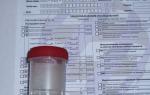
 Among the most common diseases of the knee is an abnormal accumulation of synovial fluid or effusion in the cavity of the knee joint. The main sign of fluid accumulation is swelling, enlargement, limitation of joint mobility, and pain on movement. Such changes are visible to the naked eye and the diagnosis is not in doubt (see photo). If you find such changes, you should immediately seek medical help. Timely differential diagnosis and precise determination of the cause of the accumulation of synovial fluid is the key to successful treatment.
There can be many reasons for the occurrence of such a condition, but most often the effusion of the knee joint is formed as a result of injuries or various general diseases. The human body releases effusion as a response to aggressive external influences. Thus, the cause of the pathological accumulation of fluid can be a fracture, rupture of the tendons or menisci, severe dislocation or hemorrhage. The most dangerous are injuries in which pathogenic microflora enters directly into the joint cavity and purulent inflammation occurs. Synovial fluid is a favorable environment for the active reproduction of various bacteria. This condition is considered threatening and requires immediate medical attention. Also, effusion can be the result of various diseases, often infectious (tuberculosis, chlamydia, syphilis, streptococcus, etc.).
To diagnose the disease and select adequate therapy, it is necessary to find out the cause of its occurrence. The most reliable diagnostic method is a laboratory study of the synovial fluid, which changes its composition and consistency.
Bursitis, or inflammation of the bursae

Bursitis is an inflammation of synovial bags. Quite often, practitioners of sports orthopedics and traumatology encounter such a pathology. Permanent microtraumas and excessive loads are the cause of this pathology in people involved in sports (especially its power types). Moreover, often, ignoring the recommendations of orthopedic doctors to take care of the damaged knee joint, athletes continue intensive training, which only exacerbates the current situation.
Often bursitis is called the knee joint of housewives. From long kneeling while mopping, inflammation occurs in the synovial patella bag. Another fairly common form of this disease is goose foot bursitis or popliteal bursitis. The crow's foot is the junction of certain tendons on the inside of the knee joint. The synovial bag is located under the exit point of these tendons and can become inflamed under a certain load or injury.
With bursitis, the knee joint is painful on palpation, swelling and redness, deterioration of the general condition, local hyperthermia and a general increase in body temperature may occur. There may be slight stiffness or reduced range of motion in the knee joint.
Bursitis develops as a result of trauma and mechanical damage or infection of the bursa. Even a small injury or a shallow cut can cause a disease.
The medical prognosis depends on the degree of neglect of the disease, its ability to spread, and the immune status of the patient.
Meniscal injuries
About half of all knee injuries are meniscal injuries. The anatomical structure of the knee joint, as mentioned above, creates favorable conditions for various traumatic conditions, and traumatization of the medial (internal) meniscus of the knee joint occurs 4-7 times more often. This pathology is called meniscopathy and is a degenerative-destructive pathology.
 The cause of meniscopathy of the knee joint is acute and chronic injuries, which are often an occupational disease of athletes. An acute injury is most often accompanied by a phenomenon such as a blockage of the knee joint or a symptom of blockade. What it is? Immediately after the primary injury, the patient develops severe pain in the joint and a sharp limitation of its mobility. It seems that the patient's lower leg is fixed in the flexion position, there is a feeling of wedging.
Damage to the meniscus can cause the formation of effusion, the occurrence of edema. In a later period, the pain becomes strictly localized directly along the line of the joint space. Differential diagnosis with a bruise or sprain is necessary. If the diagnosis is incorrect, then with repeated injury, the disease passes into the chronic stage, which is characterized by severe pain, a sharp limitation of movement in the joint, and various inflammatory and trophic disorders. In this case, conservative therapy may be ineffective, the patient is shown surgical intervention.
Some pathologies of the knee joint are found only in pediatric practice in adolescent children (from 10 to 15 years). The most striking example is Osgood-Schlatter disease. The most stable diagnostic sign of this pathology is the appearance of a kind of bump, which is located on the knee joint, just below the kneecap. At first, the course of the disease is sluggish, but later the pain constantly increases, the patient's movements become constrained, and the affected knee joint increases in volume.
 The disease occurs as a result of aseptic destruction of the nucleus and tuberosity of the tibia. As a rule, the disease is asymmetrical and affects only one knee joint. The cause of this pathology is a violation due to various causes of blood circulation in the knee joint. The disease has a long course (from several weeks to several months), the knee joint is fully restored only after the completion of the formation of the skeleton (by about 30 years).
Here is a far from complete list of causes that can cause pain in the knee joint. This review does not indicate the methods of treatment of various diseases of the knee joint, since self-treatment is the cause of quite serious complications. Affected knee joints love the cold! If you have any symptoms of damage to the knee joints, then the only thing you can do is apply ice to the injured knee. This helps reduce pain and relieve swelling. You can apply ice every 3-4 hours for 10-15 minutes, and then you should seek medical help as soon as possible. An experienced specialist, having examined the patient's knee joint, can make a preliminary diagnosis and prescribe adequate treatment.
An extensive risk group for diseases of the knee joints are athletes and menopausal women. If you are overweight, have a sedentary lifestyle, or have certain hormonal or metabolic disorders, you may not feel completely safe.
Proper nutrition, a healthy lifestyle and moderate exercise help prevent. You should not endure pain in the knee joint, but you do not need to take painkillers without a doctor's prescription.
^ AGE 3 1/2 -5 YEARS
Age terms of the onset of ossification of the patella and head of the fibula. The centers of ossification of both named anatomical formations appear almost simultaneously in the interval from 3 1/2 to 4 1/2 years. Ossification of the patella occurs from multiple centers of ossification, the head of the fibula - due to a single center. During this age period, there is also another change in the ratio of the rates of ossification of the medial and lateral condyles of the femur. It consists in a faster increase in the vertical size of the bone part of the lateral condyle compared with an increase in this size of the bone part of the medial condyle.

Rice. Fig. 48. X-rays of the knee joint in standard projections of a 4-year-old child (explanation in the text).
^ X-ray in the posterior projection (Fig. 48, a). The shape of the metaphyses of the femur and tibia remains the same. The condyles of the femur are clearly expressed, as well as the intercondylar depression. The height of the lateral condyle is greater than the height of the medial one. This applies only to the bony part of the condyles. Shown in Fig. 48, and pneumoarthrogram of the knee joint indicates the typical anatomical shape of the cartilaginous model of the epiphysis of the femur, characterized by the predominance of the height of the medial condyle. The medial surface of the medial femoral condyle has a wavy outline, which is explained by the activation of the growth zone before the appearance of additional centers of ossification of the marginal sections of the epiphysis. In the central part of the epiphysis of the femur, an area of uneven sclerosis can be traced, which is the result of a projection layering of the ossification points of the patella. Conditional X-ray joint space of irregular shape, the height of its medial section is almost 1.5 times the height of the lateral one. The ratio of the height of the central section of the x-ray joint space to the value of the intermetaphyseal distance is the same as in children of the previous age group (1:7). At the upper surface of the proximal metaphysis of the fibula, the ossification point of its head is visible. The epiphysis of the tibia retains the shape of a cone with a rounded apex, tubercles of the intercondylar eminence are not expressed.
X-ray in the lateral projection (see Fig. 48, b). The image of the knee joint differs from that described in the previous section by the presence of multiple, partially fused, partially isolated centers of ossification of the patella and the presence of the ossification of the head of the fibula.
^ Indicators of the anatomical structure of the knee joint, available for analysis, in principle, the same as in the radiographs of children of the previous age group. The norm of the ratio of the spatial positions of the thigh and lower leg is the valgus deviation of the latter, which is increased compared to the norm in adults. The angle formed at the intersection of the longitudinal axes of the femur and tibia is open to the lateral side, its average values are 165 - 170 °.
An indicator of the correspondence of the bone age to the passport age of the child is the presence of centers of ossification of the central part of the patella and the head of the fibula.
^ The waviness of the contour of the medial surface of the epiphysis of the femur can simulate manifestations of the destructive process. A distinctive feature of the age norm of the named contour is precisely its wavy, and not jagged ("corroded") character, as well as the preservation of the closing plate.
Projective layering on the central sections of the epiphysis of the femur of multiple centers of ossification of the patella can give the impression of pathological changes in the structure of the epiphysis. The strong points of differential diagnosis are, firstly, the absence of a similar area of sclerosis in the structure of the epiphysis on the radiograph in the lateral projection, and secondly, the absence of reactive osteoporosis or osteosclerosis.
^ AGE 6-7
The main manifestations of enchondral bone formation at this age are the appearance of additional centers of ossification of the marginal (lateral and posterior) surfaces of the epiphysis of the femur, complete ossification of the central and dorsal (carrying the articular surface) parts of the patella. Additional centers of ossification of the epiphysis of the femur provide ossification of the lateral and posterior sections of the epiphysis. In the same age period, the ratio of the rates of ossification of the medial and lateral condyles of the femur changes again. It consists in a more rapid increase in the vertical size of the bone part, now not the lateral, but the medial condyle, as a result of which the height of both condyles first becomes the same, and then the height of the medial condyle begins to predominate. Complete ossification of the central part of the patella as a result of an increase in size and fusion between individual centers of ossification ends at about 7 years of age. By the end of this age period, the cartilaginous structure is preserved: a small part of the marginal sections of the distal epiphysis of the femur, subarticular sections of the tibial epiphysis, the apex, lateral edges and anterior surface of the patella, tuberosity of the tibia, about 1/3 of the volume of the head of the fibula and metaepiphyseal growth zones.
^ X-ray anatomical picture. X-ray in posterior view. The transverse size of the metaphysis of the femur almost corresponds to the anatomical one. Its lateral surfaces are slightly concave, epicondyles are not expressed. The edges of the metaphysis are bent upwards, the medial edge is rounded, the lateral edge is pointed (Fig. 49, b). The metaepiphyseal growth zone of the femur may have an uneven height during this age period due to a slightly larger size in the lateral sections. Its medial half is displayed, as a rule, in the form of a single strip of enlightenment, limited by clear trailing plates, the lateral half - in the form of two such strips due to the separate display of the anterior and posterior sections of the growth zone. The area of preparatory calcification is wide. The image of the epiphysis of the femur can have several options depending on the ratio of the heights of the medial and lateral condyles and the size, number and location of additional centers of ossification of the marginal sections of the epiphysis that appear on the radiograph. In children 6 years of age, the predominance of the height of the lateral epicondyle often remains (see Fig. 49, a). The intercondylar depression is poorly expressed. Usually only additional centers of ossification are detected, which form the lateral parts of the condyles. The ossification points located at the lateral contour of the medial condyle are much larger than those located at the lateral contour of the lateral condyle. Both those and others are oval or approximately oval in shape and are surrounded by trailing plates.
Another version of the X-ray image of the epiphysis of the femur, typical for a somewhat later stage of its formation, is shown in Fig. 49b. The height of both condyles of the femur is almost the same, their contours are even, additional centers of ossification of the lateral sections of the condyles are not visible. At the same time, in the structure of the lateral part of the medial condyle, there are several clearly defined areas of increased optical density of small sizes. representing the image of additional centers of ossification of the posterior surface of the condyle. In the lower part of the medial condyle, a relatively large area of increased optical density with a clearly defined end plate, which has a similar anatomical substrate (an additional center of ossification of the posterior part of this condyle), is also visible. In addition to this, the radiograph shows the patella ossification nuclei superimposed on the central sections of the epiphysis of the femur. Rice. 49, c and d illustrates another variant of the age norm of the x-ray image of the epiphysis of the femur, which is more typical for children 6 1/2 - 7 years old. There is a distinct predominance of the height of the medial condyle, the intercondylar depression is clearly expressed. The contours of the lateral surfaces of both condyles are uneven due to the presence of multiple additional centers of ossification. The structure of the lateral sections of the condyles appears to be uneven, as if separate bone fragments of various sizes are visible, but approximately the same round or oval shape, surrounded by clear contours. The anatomical substrate of this heterogeneity of the structure is the projection overlay of additional centers of ossification of the posterior surfaces of the condyles. Against the background of the central part of the epiphysis, partially isolated, partially fused ossification points of the patella can be traced. This variant of the X-ray anatomical picture is quite rare.


Rice. 49. Variants of the image of additional centers of ossification of the femoral condyles on the radiograph in the posterior projection (explanation in the text).
More often in children of 7 years old, an image of the distal epiphysis of the femur is observed, shown in Fig. 49, e and corresponding to the final phase of ossification, namely, the complete fusion of additional ossification centers with the bulk of the condyles. On the radiograph, the height of the medial femoral condyle is somewhat greater than the height of the lateral one, which corresponds to the shape of the cartilaginous model of the epiphysis. The contour of the lateral surfaces of the condyles is moderately wavy; there is no abundance of additional centers of ossification of the lateral parts of the condyles. There is still some heterogeneity in the structure of the lateral sections of the condyles, however, it is rather weakly expressed. The boundaries of individual areas of increased optical density (display of additional centers of ossification of their posterior surfaces not completely merged with the condyles) are almost indistinguishable, only a part of their contours is revealed. Against the background of the central part of the epiphysis of the femur, a uniform clear shadow of the completely ossified central part of the patella is visible.
In addition to the variants of the x-ray image of the epiphysis of the femur in children of the analyzed age period, there is also a variability in the shape and size of the proximal epiphysis of the tibia (semi-oval, as in Fig. 49, a, without signs of the image of tubercles of the intercondylar eminence, trapezoid shape, as in Fig. 48 , b, or a form approaching anatomical with low, but still distinctly differentiating tubercles of the intercondylar eminence, as in Fig. 49, view). The conditional X-ray joint space of the knee joint in most cases has an irregular shape with a predominance of height, depending on the variant of the ratio of the heights of the medial or lateral condyle or its lateral or medial marginal sections. The height of the central part of the x-ray articular them retains the same relation to the height of the intermetaphyseal distance (1: 7). The medial and lateral surfaces of the metaphysis of the tibia have approximately the same concavity, although its medial edge retains a slightly larger transverse size and some sharpness. The nucleus of ossification of the head of the fibula is round in shape, its transverse size is approximately 1/2 of the width of the metaphysis of this bone.
X-ray in lateral projection. The size and shape of the metaphysis of the femur correspond to the anatomical ones. The metaepiphyseal germinal zone of the femur is displayed as a single band of enlightenment with more or less wavy contours. The epiphysis of the femur is displayed on the radiograph in the form of two semi-ovals, of which the larger and with less clear contours corresponds to the medial condyle, the smaller to the lateral (Fig. 50, e). Against the background of the upper part of the epiphysis, the Ludloff spot described above is clearly distinguished. The nature of the contours of the condyles of the femur and the structure of their dorsal sections may have a number of variants associated with the number and localization of additional centers of ossification of their marginal sections. On fig. 50, a and b, a variant of the predominant display of additional centers of ossification of the posterior surface of the condyles is presented. The contours of the condyles are slightly wavy, the structure of the anterior sections is homogeneous. In the structure of the posterior sections of the epiphysis of the femur and near its contour, numerous large additional ossification points are revealed, which are oval in shape and each surrounded by end plates. Despite the presence of a large number of ossification nuclei, the contour of the posterior surface of the condyles can be traced quite clearly. The given version of the X-ray image of the epiphysis of the femur is one of the relatively rare, more often additional centers of ossification of its posterior surface are significantly smaller and fewer, as, for example, in Fig. 50, e. The contours of the condyles are also slightly wavy, the structure of both their anterior and posterior sections is homogeneous. At the posterior and anterior surfaces of the condyles, single small additional centers of ossification of a rounded shape are detected.
A relatively rare case of displaying additional centers of ossification not in the posterior, but in the lateral sections of the condyle on a radiograph made in a lateral projection is shown in Fig. 50, c and d. The contours of both condyles of the femur are clear, in some places slightly wavy. The structure of the marginal sections of the condyles is homogeneous. There are no additional centers of ossification at the contours of the condyles. At the same time, heterogeneity of the bone structure of the epiphyseal area adjacent to the posterior contour of the intercondylar recess is visible, associated with the presence of several rounded areas of increased optical density with relatively clear contours. Such areas of increased optical density are characteristic of the projection layering of additional ossification nuclei of the marginal sections of the condyles. Since they are projected at a considerable distance from the posterior surface of the condyles and, therefore, cannot be regarded as additional posterior centers of ossification, and additional nuclei of ossification of the posterior surface of the intercondylar recess are not described, the anatomical substrate of the described heterogeneity of the structure of the epiphysis of the femur can be only the centers of ossification of its lateral departments.
The proximal epiphysis of the tibia has an approximately oval shape with a slight bulge in the area of the intercondylar eminence. Its structure clearly shows vertically oriented lines of force. The X-ray image of the patella is determined by the completeness of the fusion of multiple ossification points of its central part into a single bone formation. On fig. 50 shows variants of the shape, contours and structure of the patella, observed in the analyzed age period. On fig. 50, and the patella is a single whole, but its dimensions are small, the contours are unevenly wavy. On fig. 50, the dimensions of the patella are close to anatomical (there is no complete correspondence due to the still unossified apex not being displayed on the radiograph). The structure of most of the patella is homogeneous, except for the upper section, where two ossification nuclei that have not yet merged with each other and with the main mass of the patella are visible. On fig. 50, e, the patella is a single bone formation of a rather large size. A feature of its image is the pronounced waviness of the contour of the dorsal surface and the presence in the structure of arcuate, diverging from the dorsal surface, stripes of sclerosis. The anatomical substrate of these strips is the waviness of the lateral surfaces of the patella, which is characteristic of the growth zones in the period preceding the appearance of centers of ossification, in this case, the lateral edges of the patella.
^ X-ray indicators of the anatomical structure of the knee joint available for analysis. X-ray in posterior view. When evaluating the ratios of the spatial positions of the femur and lower leg, the standard values of the angle formed at the intersection of the longitudinal axes of the femur and tibia are used, the same as in adults. When analyzing the image, it is possible to evaluate the following indicators: the shape, size, contours and structure of the ossified parts of the metaphysis of the femur and epimetaphyses of the bones of the leg; the shape of the epiphysis of the femur and the structure of its central part and the contour of the articular surface (analysis of the structure and contours of the lateral sections of the epiphysis is reliable only in the absence of multiple lateral centers of ossification); anatomical relationships in the knee joint in the frontal and horizontal planes, the state of the metaepiphyseal growth zones. Tentatively, the height of the x-ray joint space of the knee joint can also be estimated based on the ratio of the height of its central part to the value of the intermetaphyseal distance (normally 1:7). It is impossible to assess in this age period the true shape, size and contours of the epimetaphyses of the bones that form the knee joint, the shape of the x-ray joint space and the state of the intercondylar eminence.
On the radiograph in the lateral projection, the following can be assessed: the shape, size, contours and structure of the ossified parts of the epimetaphyses of the femur and lower leg bones, the ossified part of the patella (with the proviso that the contour of the posterior surface of the epiphysis of the femur can be assessed only in the absence of multiple additional ossification centers ); the state of physiological enlightenment of the knee joint. It is impossible to assess in this age period the anatomical relationships in the knee joint in the sagittal plane, the true shape, size and contours of the epimetaphyses of the bones that form the knee joint, and the patella, the condition of the tibial tuberosity.
An indicator of the compliance of the local bone age with the passport age of the child is the presence of additional centers of ossification of the distal epiphysis of the femur.


Rice. 50. Variants of the image of additional centers of ossification of the femoral condyles on the lateral radiograph (explanation in the text).
^ Differential diagnosis of X-ray anatomical norm and symptoms of pathological conditions. Certain difficulties in the analysis of images may be associated with additional ossification nuclei of the marginal sections of the distal epiphysis of the femur. Single relatively large ossification nuclei have a number of common features in the X-ray image with the picture of dissecting osteochondritis (Koenig's disease). Differential diagnosis is based on the following differences. With partial or complete layering on the marginal sections of the bone part of the femoral condyles of additional postero-inferior or inferolateral ossification nuclei, it is possible to trace a continuous, smoothly rounded contour of the condyles, the structure of the condyles in the intervals between images of the ossification nuclei is not changed. The ossification nuclei themselves are surrounded on all sides by clear and even end plates. For comparison, in Fig. 51, a and b show an imprint and a skiagram from a radiograph of the knee joint of a child with local aseptic necrosis of the lateral condyle of the femur. At the lateral edge of the lower surface of this condyle, a bone fragment of irregular shape and uneven contours is visible. The end plate is present only on the lower surface of this fragment. The contour of the lateral condyle is concave at the level of the fragment. The vertical and horizontal dimensions of this concavity correspond to the dimensions of the bone fragment. The contour of the niche is sclerotic.
Multiple additional centers of ossification can simulate a picture of dystrophic changes in the bone tissue of the epiphysis, as well as raise suspicions for the presence of tarsomegaly with damage not only to the ankle, but also to the knee joint. The strongholds of the differential diagnosis of multiple additional ossification nuclei with an x-ray picture of tarsomegaly are as follows. Normally, the ossification nuclei of the distal epiphysis of the femur are detected only at the lateral and posterior contours of the condyles or against the background of the structure of their marginal sections. The localization of osteochondral formations near the lower contour of the condyles, and even more so significantly distal to it, is one of the signs of tarsomegaly (see Fig. 51, c and d). Further, the ossification nuclei of the normally formed epiphysis of the femur, projected at its lateral contours, are located in a single chain (one in each of the sections of the condyle contour).


Rice. 51. X-ray picture of aseptic necrosis of the condyle of the femur (a, b) and tarsomegaly (c, d).
The presence of several bone-cartilaginous formations located side by side along the horizontal line testifies in favor of tarsomegaly. Additional ossification nuclei of the epiphysis "fit" into the contour of its cartilaginous model, with tarsomegaly, as seen in Fig. 51, c and d, this regularity is violated (if you draw a general outline of all the bone formations that appear on the radiograph, then the resulting figure will not correspond to the anatomical shape of the distal epiphysis of the femur).
Features of the x-ray image of the patella in the stage of incomplete fusion of individual nuclei of its ossification (similar to that shown in Fig. 50, c) can simulate a fracture. The difference between the age-related X-ray anatomical norm and a fracture lies in the presence of clearly defined end plates in the non-fused ossification nuclei, as well as in the uniformity of the width of the enlightenment strip separating these nuclei from the main mass of the patella.
^ AGE 9-12
Corresponds to the age terms of ossification of the tuberosity of the tibia and the marginal sections of the patella. Ossification of the tuberosity occurs partly due to the spread of the process of ossification from the anterior parts of the metaphysis of the tibia, partly due to independent ossification centers that appear at the age of 9 years. The patella has 4 additional centers of ossification - two lateral, anterior and apical, appearing at the age of 9 years. The fusion of silt with the main part of the patella occurs by 10-12 years. Complete ossification of the epiphyses of the femur, tibia, and fibula is completed somewhat earlier (at about 8 years of age), and by the age of 13, only the metaepiphyseal growth zones and a small part of the tuberosity of the tibia retain the cartilaginous structure.
^ X-ray anatomical picture. X-ray in the posterior projection (Fig. 52, a). The size and shape of the metaphysis and epiphysis of the femur correspond to the anatomical ones. The shape and size of the epiphysis of the tibia also correspond to the anatomical ones, but with the proviso that the tubercles of the intercondylar eminence are relatively low and have rounded tops. In the structure of the epimetaphyses of the bones that form the knee joint, all systems of force lines characteristic of them are revealed. The x-ray joint space of the knee joint has the same shape as in adults, but its height is somewhat greater. Against the background of the epimetaphysis of the femur, a uniform shadow of the patella is revealed, which has an anatomical shape inherent in it. At the lateral contours and at the distal end, on structural radiographs, ossification nuclei of the corresponding marginal sections of the patella can be detected.
X-ray in lateral projection. The size and shape of the epimetaphyses of the femur and tibia and the head of the fibula correspond to the anatomical ones. In children aged 8-9 years, the anterior surface of the metaphysis of the tibia is moderately concave, its contour may be finely wavy (see Fig. 52, b). On radiographs of children aged 9-10 1/2 years, one or several small ossification points of tuberosity of an elongated oval shape are detected near the anterior surface of the tibial metaphysis, surrounded by thin, but still traceable trailing plates (see Fig. 52, c).


Rice. 52. Radiographs of the knee joint in 2 projections. Age period 9-12 years (explanation in the text).
At this age, additional nuclei of ossification of the anterior surface of the patella and its apex, located at the corresponding contours, may be visible. The structure of the anterior sections of the patella can be heterogeneous due to the area of increased optical density of an elongated shape with unevenly wavy contours. Along the edges of this area is a narrow strip of enlightenment. The anatomical substrate of the described heterogeneity of the bone structure of the patella is the projection overlay of the ossification nuclei of its lateral sections (Fig. 53, a and b).
X-ray anatomical analysis is available for the entire complex of radiological indicators of the anatomical structure of the knee joint. The presence of centers of ossification of the tuberosity of the tibia and additional nuclei of ossification of the marginal sections of the patella serve as an indicator of the compliance of the local bone age with the passport age of the child.


Rice. 53. Additional nuclei of ossification of the patella (a, b); calcification of the patella bag (c).
^ Differential diagnosis of X-ray anatomical norm and symptoms of pathological conditions. The nucleus of ossification of the apex of the patella can be mistaken for a bone fragment. An indicator of the age norm of the x-ray image of the patella is the presence of clear end plates in the ossification nucleus and the uniform height of the enlightenment strip separating it from the main part of the patella.
The waviness of the contour of the anterior surface of the metaphysis of the tibia can simulate the manifestation of a destructive process. The presence of a continuous closing plate, as well as the uniformity of the sizes of individual waves and the depression between them, allows to distinguish the age norm of the contour from destruction.
The presence of several ossification points of the tibial tuberosity of different sizes can cause difficulty in terms of differential diagnosis with Osgood-Schlatter disease. The main differential diagnostic sign is the state of physiological enlightenment of the knee joint (rhomboid space). Normally, it has two narrow wedge-shaped protrusions - upper and lower. Pathological processes in the area of the anterior surface of the proximal epimetaphysis of the tibia are always accompanied by shading of the lower projection of the rhomboid space. As an illustration of this situation, an x-ray of the knee joint of a child with calcifying bursitis of the deep bag of the knee joint is given (see Fig. 53, c). At the anterior surface of the epiphysis of the tibia, three structureless intense shadows of an approximately oval shape with clear, even contours are visible. Their location corresponds to the location of the deep bag of the knee joint. The lower ledge of the rhomboid space is shaded. Distinguishes the ossification nuclei of normally forming tuberosity from the areas of its fragmentation in Osgood-Schlatter disease and the presence of trailing plates in them.
^ AGE 12-14
At this age, complete ossification of the tibial tuberosity occurs. Separate points of ossification, gradually merging with each other, fulfill almost the entire cartilaginous model of tuberosity, with the exception of a small area in the lower section. Cartilaginous tissue also remains for some time between the dorsal surface of the bony part of the tuberosity and the anterior surface of the metaphysis of the tibia.
^ X-ray anatomical picture. X-ray in lateral projection. The image of the patella, the metaepiphyses of the femur and tibia, and the head of the fibula corresponds to their image in adults (with the exception of the presence of strips of enlightenment of the metaepiphyseal growth zones and the display of the process of ossification of the tibial tuberosity). The ossified part of the tibial tuberosity has the shape of a relatively wide strip with an expanded and rounded lower end. At the beginning of this age period, it is divided into several parts by transverse stripes of enlightenment (Fig. 54, a), later it represents a single whole (see Fig. 54, b). The lower end of the ossified part of the tuberosity is separated from the lower edge of the recess on the anterior surface of the body of the tibia by a relatively wide gap. A narrower gap separates the proboscis of the tuberosity from the anterior surface of the metaphysis of the tibia. The contour of the latter may be slightly wavy.
X-ray in the posterior projection (see Fig. 54, c). The image of the knee joint as a whole is similar to its image in adults. The exception is two details of the X-ray anatomical picture. The first of these is the presence of the image of metaepiphyseal growth zones mentioned above. The second detail is a relatively common wide transverse band of low optical density against the background of the metaphysis of the tibia with a fairly clear upper contour and an indistinct lower one. This feature of the structure is caused by the projection layering of the non-ossified part of the tuberosity (the gap between the lower end of the bone part of the tuberosity and the lower edge of the depression on the anterior surface of the metaphysis of the tibia - see Fig. 54, a and b).
The set of indicators of the anatomical structure of the knee joint available for analysis is identical to that in adults. An indicator of the compliance of the local bone age with the passport age of the child is complete or almost complete ossification of the tibial tuberosity.
^ Differential diagnosis of X-ray anatomical norm and symptoms of pathological conditions. Transverse bands of enlightenment separating the ossified part of the tibial tuberosity can simulate a fracture or fragmentation of the tuberosity as a manifestation of Osgood-Schlatter disease. The delimitation of the age-related X-ray anatomical norm from both of these pathological conditions is based on the absence of shading of the lower protrusion of the rhomboid space, the presence of trailing plates that limit the mentioned transverse bands of enlightenment, and the even, rather than stepped, contour of the anterior surface of the ossified part of the tuberosity. For comparison, we present an x-ray of the knee joint of a child suffering from Osgood-Schlatter disease (see Fig. 54, d). The structure of the bone part of the tuberosity, as can be seen in the figure, is heterogeneous, its anterior contour is uneven, the continuity of the end plate is broken. At the anterior surface of the tuberosity, an irregularly shaped bone fragment with uneven contours is visible. The total contour of the anterior surface of the tuberosity (taking into account the described bone fragment) is stepped. The diamond-shaped space is shaded.
A transverse band of low optical density in the structure of the tibial metaphysis on a posterior radiograph can simulate pathological changes in the bone structure. To exclude an erroneous conclusion, one should take into account the possibility of such a projection layering of the non-ossified part of the tibial tuberosity.


Rice. 54. Radiographs of the knee joint in 2 projections. Age period 12-14 years (a, b, c); X-ray picture of Osgood-Schlatter disease (d).
^ AGE 15-17
The age period of the final stage of the postnatal formation of the bone components of the knee joint, namely, the synostosis of the metaepiphyseal growth zones and the growth zone of the tibial tuberosity.
Normal X-ray anatomy of the knee joint differs from its X-ray anatomy in adults only in that in the initial stages of the synostosis process, sharply narrowed strips of enlightenment of the growth zones are traced, and after their complete closure, narrow horizontal strips of sclerosis are traced at the site of their former location.
All indicators of the anatomical structure of the knee joint described in the introductory part are available for X-ray anatomical analysis.
^ ANKLE AND FOOT
The ankle joint, as is known, is formed by the articular surfaces of the distal epiphyses of the bones of the lower leg and the block of the talus. The distal epiphysis of the tibia has an approximately square shape with rounded edges, on its medial side there is a protrusion directed downwards - the medial malleolus. On the lateral side of the distal metaepiphysis of this bone there is a notch with a rough surface, to which the fibula adjoins. Articular hyaline cartilage covers the distal concave surface of the epiphysis and the inner surface of the medial malleolus. The distal epiphysis of the fibula is called the lateral malleolus. On its inner side there is an articular surface that does not extend to the top of the ankle. The talus has a body, neck and head. The upper surface of the body of the talus in the frontal plane has the shape of a block with a slightly pronounced depression in the center and two, also indistinctly expressed, shafts - medial and lateral. In the sagittal plane, the upper surface of the body of the talus is convex with a slightly shallower and shorter anterior slope and a steeper and longer posterior one. Articular hyaline cartilage covers the upper surface of the block and the upper part of the lateral surfaces. The upper and medial articular surfaces articulate with the epiphysis and the medial malleolus of the tibia, the lateral articular surface with the lateral malleolus. Thus, the articular space of the ankle joint in the frontal plane has a U-shape, in the sagittal plane it is arcuate.
The skeleton of the foot is divided into three sections - the tarsus, metatarsus and phalanges of the fingers. The tarsus, in turn, is divided into anterior and posterior sections. The posterior tarsus consists of two bones - the talus and the calcaneus, one above the other. The talus, in addition to the parts already noted (body, neck and head), also has two processes - lateral and posterior. In the latter, two tubercles are distinguished - medial and lateral. On the head of the talus there is a navicular articular surface, on the lower surface of the body - the calcaneal articular surfaces, separated by a groove of the sinus of the tarsus. At the calcaneus, a body and a calcaneal tubercle are distinguished. On the medial side of the body is a rectangular bone protrusion - the support of the talus. On the upper surface of the body are the anterior, middle and posterior talar articular surfaces and the groove of the sinus of the tarsus, on the anterior side of the body - the cuboid articular surface. The anterior tarsus consists of 5 bones. The navicular bone has a relatively small thickness, its surface facing the head of the talus is concave, facing the sphenoid bones is convex. There is a fairly large tuberosity on the inferomedial surface of the navicular bone. The articular surfaces do not extend to the tuberosity of the scaphoid. The cuboid bone is shaped like its name. Three of its surfaces are covered with articular hyaline cartilage - the dorsal, with which it articulates with the calcaneus, the ventral, with which the IV and V metatarsal bones articulate, and the medial, with which the club-shaped bone articulates with the lateral sphenoid bone. Ventral to the scaphoid there are three sphenoid bones - medial, intermediate and lateral, articulated on one side with the navicular bone, on the other - with I, II and III mold bones.
X-ray of the ankle joint is performed in two standard (posterior and lateral) projections, foot - in three projections: plantar, lateral and oblique. On radiographs of a fully formed ankle joint, the following radiological indicators of its anatomical structure are analyzed: the shape, size, contours and structure of the distal epiphysis of the tibia, lateral malleolus and talus block; state of the x-ray joint space and anatomical relationships in the joint. The criterion for the correctness of the anatomical relationships in the frontal plane is the uniform height of the x-ray joint space (its horizontal part) and the location at the same level of the lateral edge of the epiphysis of the tibia and the lateral edge of the talus block. In the sagittal plane, the indicators of the correctness of the ratios are considered to be the uniform height of the x-ray joint space and the location at the same level of the centers of the articulating articular surfaces of the epiphysis of the tibia and the trochlea of the talus. On radiographs of the foot after the end of its formation, the following indicators are used to assess the spatial position of the calcaneus and talus bones in the frontal and sagittal planes. In the sagittal plane, the position of the talus is characterized by the magnitude of the talo-tibial angle formed at the intersection of the longitudinal axes of these bones. The normative values of this angle are 90°. The spatial position of the calcaneus (also in the sagittal plane) characterizes the value of the calcaneal-plantar angle formed at the intersection of two lines, one of which is drawn tangent to the lower surface of the calcaneus, the second connects the lower surface of the calcaneal tuber and the lower surface of the head of the first metatarsal bone. The normative values of this angle are 15-20°. In the frontal plane, an indicator of the norm of the spatial positions of these bones is the intersection of their longitudinal axes at an angle of 12-15 ° (heel-talar angle). The size of the longitudinal arch of the foot is characterized by the angle formed at the intersection of lines tangent to the lower surfaces of the calcaneus and I metatarsal bones on a radiograph taken in a lateral projection. An indicator of the norm is the value of this angle in the range from 125 ° to 135 °. In addition, when analyzing images of the foot, the shape, size, contours and structure of the bones of the foot skeleton, as well as the anatomical relationships in the joints of the tarsus, metatarsophalangeal and interphalangeal joints can be assessed. The criterion for the correctness of these ratios is the uniform height of the X-ray joint spaces, and for joints with unequal length of the articular surfaces (talonavicular, metatarsophalangeal and interphalangeal joints) - the location at the same level of their centers, for flat joints - the location at the same level of the edges of the articular surfaces.
A presentation of age-related radioanatomy is given simultaneously for the ankle joint and foot.
^ AGE UNTIL 9 MONTHS
The degree of ossification of the metaepiphyses of the bones of the lower leg and the skeleton of the foot differs little from that which they had by the end of intrauterine development. The cartilaginous structure during this age period is retained by: the epiphyses of the bones of the lower leg and partially their metaphyses, a significant part of the calcaneus, talus and cuboid bones and completely - the navicular, all the sphenoid bones of the tarsus and the epiphyses of the metatarsal bones and phalanges of the fingers.
^ X-ray anatomical picture. Ankle joint. X-ray in posterior view. The lateral surfaces of the metaphysis of the tibia are moderately concave, the distal surface has a slightly pronounced saddle shape. The medial edge of the metaphysis is bent upwards and slightly pointed. The lateral contour of the metaphysis of the fibula is straight, the medial is concave. The edges of the metaphysis are rounded. The epiphyses of the bones of the lower leg are not detected on the radiograph. The upper surface of the block of the talus is straight, the shafts of the block and the groove between them are not expressed. The metaphysis of the tibia and the block of the talus are separated by a wide gap, as are the lateral surfaces of the metaphyses of the bones of the leg.
X-ray in lateral projection. All surfaces of the metaphysis of the tibia (including the distal one) are moderately concave. The epiphyses of the bones of the lower leg are not detected. The upper surface of the trochlea of the talus is arcuate, the posterior edge of the block is rounded, the posterior process of the talus is not pronounced. Against the background of the lower part of the body of the talus, the lateral process is visible, the upper surface of the anterior part of the talus is rectilinear, its differentiation into the neck and head is not expressed.
Foot. X-ray in the plantar projection (Fig. 55). The rounded anterior ends of the calcaneus and talus and the cuboid bone, which has an irregularly oval shape, are visible. The remaining bones of the tarsus are not shown on the radiograph. The metatarsal bones and phalanges of the fingers are represented only by the metadiaphyses.
X-ray in lateral projection. The shape of the ossified part of the calcaneus in general corresponds to the anatomical one. Against the background of the upper part of her body, a rectangular shadow of the support of the talus is visible. The calcaneal tubercle is short, with a slightly convex dorsal contour. The cuboid bone is small in size, with convex dorsal and plantar surfaces and rounded corners. The remaining bones of the tarsus are not detected on the radiograph. The structure of all bones is evenly fine-meshed, without signs of lines of force.
R  is. 55. X-ray of the foot of a child at the age of 1 year.
^
When analyzing images, it is possible to evaluate the following indicators: the shape, contours and structure of the ossified parts of the distal metaphyses of the bones of the lower leg, talus, calcaneus and cuboid bones, the metadiaphyses of the metatarsal bones and phalanges of the fingers; anatomical relationships in the ankle joint in the frontal and sagittal planes. The criterion for the correctness of these ratios in the frontal plane due to the lack of an image of the epiphysis of the tibia and the uneven height of the x-ray joint space is the parallelism of the lines drawn tangentially to the distal surface of the metaphysis of the tibia and to the upper surface of the talus block, as well as the location of the lateral edges of these surfaces. In the sagittal plane, an indicator of the norm of anatomical relationships in the ankle joint is the location on one vertical straight line of the centers of the metaphysis of the tibia and the talus block.
When assessing the spatial position of the talus and calcaneus in the frontal and sagittal planes, the standard values of the tibia-talar and calcaneal-talar angles are used, the same as in adults. The value of the calcaneal-plantar angle due to incomplete ossification of the calcaneal tubercle and non-ossification of the head of the first metatarsal bone differs from the norm in adults and is equal to 10-15°. The criterion for the correctness of the anatomical relationships in the subtalar joint in the sagittal plane is the projection overlay on the body of the calcaneus of the talus head by no more than 1/4 of its vertical size.
The normative value of the angle of the longitudinal arch of the foot for the above reasons is greater than in adults, and equals 130-137 °. It is impossible to assess in this age period the true shape, size and contours of the calcaneus, cuboid and talus bones, the state of the remaining bones of the tarsus, the epiphyses of short tubular bones, the anatomical relationships in the joints of the anterior tarsus and the state of the metaepiphyseal growth zones.
^ AGE FROM 1 YEAR TO 3 YEARS
This period corresponds to the age of the onset of ossification of the epiphyses of the short tubular bones of the foot and the bones of the anterior tarsal. The timing of the appearance of the centers of ossification of the named anatomical formations is not as precise as the beginning of ossification of the bones of the wrist, and can only be named approximately. The nucleus of ossification of the distal epiphysis of the tibia appears first, at the age of approximately 1 year. Then, with an interval of about a year, the center of ossification of the lateral sphenoid bone appears, and after a short period of time, approximately at the age of 2 1/2 years, the medial and intermediate sphenoid bones, the epiphyses of the metatarsal bones and phalanges of the fingers, and the lateral malleolus begin to ossify. The order of appearance of the centers of ossification of the medial and intermediate sphenoid bones does not have a definite pattern. In most cases, the medial sphenoid bone begins to ossify first, however, the simultaneous appearance of centers of ossification of these bones and an earlier onset of ossification of the intermediate bone is also possible. The lateral and intermediate sphenoid bones each have only one nucleus of ossification; ossification of the medial sphenoid bone can occur from one, two or more centers. Ossification of the navicular bone begins at the age of 3-3 1/2 years and occurs more often from one center of ossification, although multiple centers are possible. By the age of 3, the cartilaginous structure is preserved: about 1/3 of the volume of the epiphysis of the tibia, including the medial malleolus; about 1/2 of the volume of the lateral malleolus; marginal sections of the talus, calcaneus (including the apophysis of the calcaneal tuber) and cuboid bones; most of the bones of the anterior tarsus and epiphyses of short tubular bones.
^ X-ray anatomical picture. Ankle joint on the radiograph in the posterior projection (Fig. 56, a). The image of the metaphyses of the bones of the lower leg is similar to that described in the previous section. The epiphysis of the tibia has the shape of a wedge with the base facing the medial side.
Its position relative to the metaphysis is eccentric due to the greater ossification of the medial part of the cartilaginous model. The lateral malleolus is rounded, located closer to the lateral edge of the metaphysis. The upper surface of the talus block is flat with rounded edges. X-ray joint space of the ankle joint is wide, wedge-shaped with the base of the wedge facing laterally. In the structure of the metaepiphyses of the bones of the lower leg and the block of the talus, systems of longitudinal lines of force are clearly traced.


Rice. 56. Radiographs of the ankle and foot. Age period 1-3 years (explanation in the text).
On the radiograph in the lateral projection (see Fig. 56, b), the tibial epiphysis has a rectangular shape with a convex anterior and posterior contours. The lateral malleolus, as in the posterior radiograph, is round in shape. The X-ray joint space of the ankle joint is wide with a lower height in the middle part and the largest at the anterior and posterior edges. The image of the talus and calcaneus is the same as in children of the previous age period, with the exception of a distinct functional formation of the architectonics of the bone structure.
Foot. On the radiograph in the plantar projection, the image of the foot depends on the state of ossification of the bones of the anterior tarsus and epiphyses of short tubular bones. In children aged about 2 years, in addition to the bodies of short tubular bones, cuboid and anterior sections of the calcaneus and talus, ossification nuclei of the lateral sphenoid bone, distal epiphysis of the 1st metatarsal and proximal phalanges of the fingers are also detected (see Fig. 56, c). At a slightly older age (about 2 1/2 years), the ossification nuclei of all three sphenoid bones are usually visible on radiographs. The shape of the lateral sphenoid bone and the cuboid by this time becomes similar to the anatomical one, the intermediate and medial sphenoid bones are round (see Fig. 56, d). In children 3-3 1/2 years old, the X-ray anatomical picture is characterized by significant polymorphism. Its possible options are shown in Fig. 57. On the first radiograph of a 3-year-old child (see Fig. 57, a), the dimensions and shape of the cuboid and lateral sphenoid bones are close to anatomical. The ossification nuclei of the medial and intermediate cuneiform bones and the ossification point of the scaphoid are visible. Ossification of the medial sphenoid bone comes from two nuclei of uneven size. This state of ossification of the bones of the anterior tarsal for the named age is the most natural. On the second radiograph of a child of the same age (see Fig. 57, b), the images of the cuboid and lateral sphenoid bones are approximately the same as in Fig. 57 a. The medial sphenoid bone, which is formed from one ossification nucleus, is also of considerable size.


Rice. 57. Variants of ossification of the anterior tarsal bones in the age period of 1-3 years
(explanation in the text).
At the same time, the center of ossification of the intermediate sphenoid bone and the epiphyses of the metatarsal bones, except for I, has not yet appeared in this child. IV metatarsal and distal epiphysis II and I metatarsal bones. A feature of the X-ray anatomical picture is the disproportionately small size of the intermediate sphenoid bone in comparison with the size of the ossified part of the navicular bone.


Rice. 58. Multiple ossification points of the navicular and medial sphenoid bones of the foot (a); diagram of indicators of anatomical ratios in the joints of the anterior tarsus of an adult (b) and a child 3 1/2 years old (c).
On a radiograph in a lateral projection in children of 3 years old, all the bones of the tarsus can be traced. Rice. 58, a illustrates the X-ray anatomical picture in the case of ossification of the medial sphenoid and navicular bones from several centers of ossification. The navicular bone is represented by one large ossification nucleus and three small ones located at the back surface of the cartilaginous bone model. At the base of the first metatarsal bone, four separate, partially overlapping nuclei of ossification of the medial sphenoid bone are visible, surrounded by clear trailing plates.
^ X-ray indicators of the anatomical structure of the ankle joint and foot, available for analysis. Ankle joint. It is possible to evaluate the following indicators: shape, size, contours and structure of the talus block; anatomical ratios in the joint in the frontal and sagittal planes (the criteria for the correctness of the ratios in the joint are the same as in children of the previous age group). We want to draw attention to the fact that the shape of the x-ray joint space cannot be used as an indicator of the anatomical relationships in the ankle joint in the frontal plane, because due to the age-related peculiarity of the shape of the ossified part of the tibial epiphysis, it also normally has a wedge-shaped shape.
The condition of the metaepiphyseal growth zones of the leg bones is also to be assessed.
Foot. X-ray parameters of its anatomical structure, available for analysis, are different for different states of ossification of the anterior tarsal bones. Before the appearance of the ossification point of the navicular bone, it is possible to assess the spatial position of the calcaneus and talus bones, the size of the longitudinal arch of the foot. The normative values of the angles characterizing these indicators are identical to those given in the description of the normal X-ray anatomy of children of the previous age group. In addition, it is possible to assess the shape, contours and structure of the ossified parts of the calcaneus, talus, cuboid and lateral sphenoid bones, the contours and structure of the ossification nuclei of the remaining sphenoid bones and the epiphyses of the short tubular bones of the foot that are revealed on the radiograph; anatomical relationships in the subtalar, metatarsophalangeal and interphalangeal joints. The criteria for their correctness in the subtalar joint are the same as in children under the age of 1 year; in the last two groups of joints, the correctness of the anatomical relationships is indicated by the location of the centers of the ossified parts of the articulating epiphyses at the same level.
After the appearance of the ossification point of the navicular bone, in addition to the above, it also becomes possible to assess the anatomical relationships in the talonavicular joint in the frontal and sagittal planes and in the Lisfranc joint in the frontal plane. The criterion for their correctness in the first joint is the location on radiographs in both projections of the center of ossification (or ossification nucleus) of the navicular bone at the level of the center of the navicular surface of the talus head. The criterion for the correctness of the anatomical relationships in the navicular-sphenoid joint is the location on the radiograph in the plantar projection of the center of ossification of the navicular bone (or the center of its ossified part) at the level of the gap between the bone parts of the medial and intermediate sphenoid bones (see Fig. 58, c).
It is impossible to assess the true size and contours of the distal epiphyses of the leg bones and short tubular bones of the foot, as well as the bones of the anterior tarsus, during this age period.
An indicator of the compliance of the local bone age with the passport age of the child in children of 2 years is the presence of a nucleus of ossification of the lateral sphenoid bone, in children of 3-3 1/2 years - the presence of a center of ossification of the navicular bone.
^ Differential diagnosis of X-ray anatomical norm and symptoms of pathological conditions. The ossification of the medial sphenoid and scaphoid bones from several centers of ossification deserves special attention, especially when one large ossification nucleus is traced on the radiograph, along the contour of which there are, as it were, several small fragments (see Fig. 58, a). In the presence of an appropriate anamnesis, these features of the image can be mistaken for a bone fracture. An indicator of the X-ray anatomical norm of the image of these bones is the presence of end plates surrounding not only a large ossification nucleus, but also small “fragments”.
^ AGE 5-6
During this age period, almost complete ossification of the bones of the anterior tarsal occurs, the degree of ossification of the epiphyses of the bones of the lower leg and short tubular bones of the foot, as well as the calcaneus and talus, increases significantly. As follows from this characteristic, there are no qualitatively new manifestations of enchondral bone formation in these periods, and the ossification of cartilaginous models of the epiphyses does not end. The basis for the allocation of this age period was the appearance of some features of the X-ray image of the foot on radiographs made in the plantar and, to a lesser extent, lateral projections.
^ X-ray anatomical picture. On the radiograph of the foot in the plantar projection, the dimensions of the head and neck of the talus, the anterior part of the body of the calcaneus, cuboid, lateral and intermediate cuneiform bones, as well as their shape, generally correspond to the anatomical ones. The epiphyses of the metatarsal bones and phalanges of the fingers are almost completely ossified. Almost all systems of lines of force characteristic of them can be traced in the structure of these bones. Special attention should be paid to the possibility of different variants of the norm of the X-ray image of the medial sphenoid and scaphoid bones, shown in Fig. 59 and 60. In fig. 59, and the most typical variant of the X-ray anatomical norm is presented. Both the navicular and medial sphenoid bones have the appearance of a corresponding bone formation. Their shape approaches the anatomical one, the contours are even, the structure is homogeneous with the rudiments of its characteristic lines of force. On fig. 59, b, the medial sphenoid and navicular bones also look like single bone formations. At the same time, their contours are coarsely wavy in places (especially the talar surface of the navicular bone), the shape is irregular - the navicular bone, for example, has a wedge-shaped shape with a lower height of its medial section. As already mentioned, the ossification of these bones often occurs from several centers of ossification. To this it should be added that the ossification of the navicular bone, even in the presence of a single center of ossification, can proceed unevenly. Different phases of the fusion of individual centers of ossification, as well as the difference in the rate of ossification of the medial and lateral parts of the navicular bone, determine both this and subsequent variants of the X-ray anatomical picture.


Rice. 59. Variants of the shape of the ossified part of the scaphoid and medial sphenoid bones of the foot on the radiograph in the plantar projection (explanation in the text).
Rice. 59, c and d are similar to each other in the image of the medial sphenoid bone. In both cases, it consists of two parts of unequal size, each surrounded by end plates and separated by a narrow uniform strip of enlightenment (the stage of incomplete fusion of the two ossification nuclei of this bone).


Rice. 60. Variant of ossification of the navicular bone of the foot (a, b). X-ray picture of osteochondropathy of the navicular bone of the foot in the stage of fragmentation (c).
The image of the navicular bone in these figures is different. On fig. 59, in the navicular bone has the form of a single bone formation, its contours are even, the structure is homogeneous, but its shape is irregular, not corresponding to the anatomical one, due to the significantly smaller longitudinal size of the medial part. On fig. 59, the navicular bone also has the appearance of a single bone formation, also has a homogeneous structure and even contours and a shape that does not correspond to the anatomical one, but this discrepancy is of a different kind. Its presence is determined by the presence of an angular protrusion on the anterior surface of the bone due to the faster rate of ossification of the middle part of the bone and the straightness of the contours. On fig. 60, a and the navicular bone consists of three oval-shaped ossification nuclei that have not yet merged with each other with clearly defined end plates. The location of the middle ossification nucleus can serve as an explanation for the formation of the ossification that is revealed in Fig. 59, a bone protrusion on the anterior surface of the scaphoid. The image of the medial sphenoid bone in this case has no features.
The complex of radiographic parameters of the anatomical structure of the ankle joint and foot available for analysis is the same as in children of the previous age group.
^ Differential diagnosis of X-ray anatomical norm and manifestations of pathological conditions. Almost all of the given variants of the age norm of the X-ray image of the medial sphenoid and navicular bones can cause certain difficulties in the analysis of images. A strip of enlightenment separating two unfused nuclei of ossification of the medial sphenoid bone may suggest (with an appropriate history) that it has a fracture. A distinctive feature of the X-ray anatomical norm is the continuity of the end plates that limit each of the parts of the bone, and the uniform width of the enlightenment strip between them.
The peculiarity of the shape of the navicular bone, and even more so its display in the form of several independent bone parts (non-fused large ossification nuclei) can be mistaken for signs of osteochondropathy. Differential diagnosis of incomplete fusion of the ossification nuclei of a normally forming navicular bone with an x-ray picture of osteochondropathy in the fragmentation stage is based on the following x-ray differences. As already noted, the nuclei of ossification of the navicular bone (see Fig. 60, a and b) are of the correct oval or round shape, each of them is surrounded by a clearly defined, even end plate and has a uniform fine-mesh structure. In contrast, osteochondropathy in the fragmentation stage (see Fig. 60, c) is characterized by the irregularity of the shape of individual fragments, the absence of trailing plates in the presence of uneven contours, and uneven bone structure with a predominance of areas of increased optical density.
^ AGE FROM 9 TO 14 YEARS
The age period of the fourth stage of postnatal formation of this department of the osteoarticular system, which consists V ossification of the apophyses of tubular bones and tarsal bones. Among the apophyses that ossify in these age periods due to independent centers of ossification are: the medial malleolus (its apex), the medial tubercle of the posterior process of the talus, the apophysis of the calcaneal tuberosity, the tuberosity of the navicular and V metatarsal bones. The onset of ossification of these anatomical formations does not have strictly defined age periods, the centers of their ossification can appear in the age range from 8 to 11 years. The first, at the age of 8 - 8 1/2 years, 2-3 points of ossification of the apex of the medial malleolus appear. Somewhat later - at about 9 years old - one nucleus of ossification of the medial tubercle of the posterior process of the talus and one or two nuclei of ossification of the apophysis of the calcaneal tuber are detected. The most recent, at 10-11 years old, the tuberosities of the navicular and V metatarsal bones begin to ossify. The apex of the lateral malleolus has no independent center of ossification. By the age of 14, the cartilaginous structure is retained only by the metaepiphyseal growth zones of the bones of the lower leg and short tubular bones of the foot and the growth zones of the apophyses.
^ X-ray anatomical picture. Ankle joint. X-ray in the posterior projection (Fig. 61, a). The size and shape of the metaphyses of the bones of the leg corresponds to the anatomical ones. The epiphysis of the tibia, with the exception of the medial malleolus, in shape, the nature of the contours and the architectonics of the bone structure corresponds to its image in adults. The medial malleolus in children aged 8-10 years is relatively short, at its lower rectilinear contour, at first, several small centers of ossification are visible, and then a rather large nucleus of ossification of its apex. The dimensions, shape and architectonics of the bone structure of the lateral malleolus correspond to the anatomical ones. The tip of the lateral malleolus, which is an extra-articular formation, does not have a separate center of ossification. In this regard, the presence in this place of a separate bone fragment, even if surrounded by a closing plate, is an indisputable sign of a fracture (see Fig. 61, b). The presence of end plates around the bone fragment and on the distal surface of the lateral malleolus in the presented radiograph is explained by the fact that this is an old ununited fracture. The lateral sections of the metaepiphyseal growth zones of the tibia and fibula in children aged 8-10 years may have a wedge-shaped shape with the bases of the wedges facing outward. The degree of expansion of the marginal sections of the metaepiphyseal growth zones is the same, its edges, due to the mismatch of the images of the anterior and posterior sections, can be two- or even multi-contour.


Rice. 61. Nucleus of ossification of the medial malleolus (a); fracture of the lateral malleolus (b); radiograph of the ankle joint of a 13-year-old child (c).
The proximal surface of the talus has the form of a weakly expressed block. The X-ray joint space of the ankle joint has the same shape as in adults, its height is uniform throughout. By the end of the age period, i.e. in children 13-14 years old, the image of the ankle joint differs from its image in adults only in the presence of metaepiphyseal growth zones of the bones of the lower leg (see Fig. 61, c), which acquire a uniform height throughout its length .
X-ray in lateral projection. The shape and size of the distal metaphyses of the leg bones correspond to the anatomical ones. The anterior and posterior surfaces of the epiphysis of the tibia in children 9-9 "/2 years old are convex, in older children they are rectilinear, with slightly rounded distal edges. The articular surface of the epiphysis is concave, corresponding to the convexity of the talus block. The medial malleolus in children of this age is shortened, against the background of the talus block, the ossification points of the apex of the medial malleolus can be seen.In older age, the image of the medial and lateral malleolus is the same as in adults.The anterior and posterior sections of the metaepiphyseal growth zone of the tibia are sphenoidly expanded (with the bases of the wedges facing outward), the degree of expansion marginal sections of the zone is the same.The right and left edges of the anterior section of the growth zone can be displayed separately.
The image of the talus and calcaneus in children under the age of 9 and after 9 years has a number of differences. In children under the age of 9-10 1/2 years (Fig. 62, a), the talus block has the shape of a hemisphere with the same length of its anterior and posterior slopes. The rear edge of the block is rounded, the posterior process of the talus is not pronounced. The lateral process of the talus has a rounded apex. The upper surface of the anterior part of the talus is straight, the transition of the neck into the head is not differentiated. The shape of the body of the calcaneus basically corresponds to the anatomical one. The calcaneal tubercle is short, the contour of its posterior surface is coarsely wavy, the end plate is sclerotic.
In children over the age of 9-9 1/2 years, the shape of the talus corresponds to the anatomical one. A completely ossified lateral tubercle of the posterior process of the talus is revealed, which is projected on the radiograph, made with the correct placement and direction of the central x-ray beam, below the contour of the posterior calcaneal surface and superimposed on the body of the calcaneus. Somewhat higher and dorsal to it, the nucleus of ossification of the medial tubercle can be traced. The lower edge of the ossification nucleus is located at the same level with the contour of the posterior calcaneal surface of the talus. Between the nucleus of ossification and the posterior surface of the block, a narrow uniform strip of enlightenment is visible, limited by the end plates (see Fig. 62, b). Under other conditions of centralization, the image of the lateral tubercle of the posterior process may be projectively rejected in the proximal direction (see Fig. 62, c). A reference point for differentiation of the lateral and medial tubercles is the contour of the posterior calcaneal surface of the talus - the posterior lateral tubercle is located on its continuation. Under these conditions, the nucleus of ossification of the medial posterior tubercle may be projected below the image of the lateral posterior tubercle. We focus readers' attention on this variant of displaying the nucleus of ossification of the medial tubercle because it is important for the differential diagnosis of the X-ray anatomical norm and apophysiolysis of this tubercle. If we are guided by the usual location of the posterior tubercles (the medial is higher than the lateral) and do not take into account the possibility of their other display, then when analyzing the radiograph shown in Fig. 62, c, one may get the impression of the presence of apophyseolysis (on the basis of displacement of the ossification nucleus in the distal direction).


Fig. 62. Variants of the X-ray image of the nucleus of ossification of the medial tubercle of the posterior process of the talus (explanation in the text).
A relatively rare X-ray image of the ossification nucleus of the medial tubercle of the posterior process of the talus is shown in Fig. 62, d. For the most part, the ossification nucleus is projected against the background of the talus block in the form of a bone fragment of an irregularly oval shape, surrounded by a narrow uniform strip of low optical density. The shape of the calcaneus is generally the same as in children under 9-9 1/2 years of age. At the posterior surface of the calcaneal tuber, one relatively massive nucleus of ossification of its apophysis can be traced, which, as a rule, has a central position (Fig. 63, a).


Rice. 63. Variants of X-ray display of calcaneal tuber apophysis ossification (a, b); core of ossification of the tuberosity of the fifth metatarsal bone (c).
Later, two or three more ossification nuclei of various shapes and thicknesses appear (see Fig. 62, b). With any number and size of the nuclei of ossification of the apophysis of the calcaneal tubercle, they all have clear, even contours and are located at the same distance from the dorsal surface of the calcaneal tubercle. In children 13-14 years old, the apophysis of the calcaneal tuber is detected on the radiograph along its entire length. With a pronounced waviness of the apophyseal growth zone, several contours of the posterior surface of the calcaneal tuber can be detected, partially crossing the image of the apophysis and creating a false impression of fragmentation of the latter (see Fig. 63, b).
Foot. X-ray in the plantar projection. The image of its constituent bones is identical to the image in adults, with the exception of two features, which are the presence of metaepiphyseal growth zones of short tubular bones and the presence of a nucleus of ossification of the tuberosity of the fifth metatarsal bone (see Fig. 63, c).
^ X-ray indicators of the anatomical structure of the ankle joint and foot, available for analysis. In children older than 11 years, in principle, it is possible to evaluate all the indicators listed in the introductory part of this section. In children 8-10 1/2 years old, the true shape, size and contours of the medial and lateral malleolus, the posterior process of the talus, the calcaneal tuber of the calcaneus and the proximal end of the fifth metatarsal cannot be assessed.
^ Differential diagnosis of X-ray anatomical norm and symptoms of pathological conditions. Difficulties in image analysis may arise due to the uneven height of the distal metaepiphyseal growth zones of the leg bones. As is known, the wedge-shaped growth zone as a whole or any of its parts is one of the components of the radiological symptom complex of epiphysiolysis. The delimitation of the age norm and pathology of the form of these growth zones is based on the following radiological differences. Normally, the medial and lateral, as well as the anterior and posterior, marginal sections of both growth zones are expanded to the same extent and are limited by clear trailing plates. The adjacent edges of the metaphysis and epiphysis in children of this age group are located at the same level. In cases of traumatic osteoepiphyseolysis (Fig. 64, a and b - X-ray picture of osteoepiphyseolysis of the distal epiphysis of the tibia, Fig. 64, c - X-ray picture of traumatic epiphyseolysis of the distal epiphysis of the fibula), a pronounced uneven expansion of one of the marginal sections of the metaepiphyseal growth zone (anterior and medial in Fig. 64, a and b, lateral - in Fig. 64, c). The contours of the germ zone at the level of this excess expansion are uneven, serrated, and there are no trailing plates. The location at different levels of the adjacent edges of the metaphysis and epiphysis is also noted.
X-ray imaging of the process of ossification of the calcaneal tuber may present certain difficulties in resolving the issue of the presence or absence of osteochondropathy of the calcaneal tuber apophysis (Shinz's disease). Normally, as noted above, the ossification nuclei of the calcaneal tuber apophysis have a homogeneous structure, smooth contours, and are located at the same distance from the posterior surface of the calcaneal tubercle. Violation of these patterns (all, any two, or only at least one) is a sign of a pathological condition of the apophysis, (see Fig. 62, b, the apophysis of the calcaneal tuber consists of three parts that are different in thickness and length, the upper part of the apophyseal the growth zone is wedge-shaped expanded). The nucleus of ossification of the medial tubercle of the posterior process of the talus and tuberosity of the fifth metatarsal bone can also cause difficulties in image interpretation. The indication for differential diagnosis is, firstly, the exclusion of a fracture of the fully formed named anatomical formations, and secondly, the decision on the presence or absence of traumatic apophyseolysis.


Rice. 64. X-ray picture of osteoepiphyseolysis of the distal epiphysis of the tibia (a, b) and the distal epiphysis of the fibula (c).
The difference between the X-ray picture of normally forming apophyses and avulsion fractures and osteoapophyseolises was presented in Chap. 1. In this section, we provide an illustration of these differences with specific examples. On fig. 65, a shows a radiograph in the lateral projection of the foot of a child with traumatic apophyseolysis of the medial posterior tubercle of the talus. It is clearly seen that the apophyseal germ zone of this tubercle has a wedge-shaped shape, its edges are uneven, the ossification nucleus is displaced upwards - its lower edge is located significantly higher than the location of the posterior edge of the calcaneal surface of the talus.


Rice. 65. X-ray picture of anophysiolysis of the medial tubercle of the posterior process of the talus (a) and tuberosity of the fifth metatarsal (b).
Rice. 63, c and 65, b illustrate the differences in the X-ray picture of a normally forming tuberosity of the fifth metatarsal bone and its traumatic apophyseolysis. On fig. 63, in the apophyseal germ zone has a uniform width, its contours, although sinuous, have clear trailing plates. On fig. 65b, the apophyseal germ zone has a wedge-shaped shape, the integrity of the end plate of the base of the nucleus of ossification of the tuberosity is broken, the nucleus of ossification itself is displaced in the dorsal direction (the dorsal edge of its base is located dorsal to the same-named edge of the germ zone on the surface of the metatarsal bone).
^ AGE 15-17
The age period of the final stage of the postnatal formation of the ankle joint and foot, namely, the synostosis of the metaepiphyseal and apophyseal growth zones. The X-ray image of the ankle joint and foot differs from the image in adults only by the presence of initially narrowed growth zones, and then by the presence of sclerosis stripes at the site of their former location. It is possible to analyze the entire complex of indicators of the anatomical structure of this department of the osteoarticular system.
CONCLUSION
When describing the features of the normal X-ray anatomy of the osteoarticular system in children of different ages, a place was also given to the differential diagnosis of normal X-ray anatomical details with manifestations of pathological conditions. It was given, however, strictly in relation to the specific features of the X-ray image of various joints and sections of the spine and to a certain period of their formation. Due to the great importance for the correct diagnosis of diseases and injuries of bones and joints of a reliable distinction between the norm and pathology, we considered it appropriate to conclude the book with a summary of a number of general provisions of the corresponding differential diagnosis. In accordance with the task of presenting the general provisions in this section, only those features of the X-ray image of a normally forming osteoarticular system that are typical for all or almost all of its departments and, in addition, have a certain similarity with the manifestations of pathological processes or the consequences of traumatic effects are analyzed. . The analyzed features of the X-ray anatomical norm are discussed in the order of the sequence of their manifestation on radiographs.
^ The absence of an image of the epiphyses of tubular bones on the radiograph. In the previous sections of the book, it was noted that in children of the first year of life, the epiphyses of tubular bones are still formed by cartilage tissue that does not have natural x-ray contrast, and therefore the absence of their image on the x-ray is an age-related x-ray anatomical norm. The pattern for the named age period of this feature of the X-ray anatomical picture is not a reason for unconditional refusal to resolve the issue of the presence or absence of dysplastic or destructive processes in a child in a particular joint.
Establishment of the presence of a destructive process, the cause of which is most often hematogenous osteomyelitis of a metaepiphyseal localization typical for this age, is to some extent facilitated by a fairly pronounced characteristic clinical picture. The same applies to a certain extent to destructive tumor processes. Diagnosis of dysplastic changes, such as aplasia, and even more so hypoplasia of the epiphyses, due to the relative scarcity of clinical manifestations, consisting in the presence of often only a slight shortening of the limb and sometimes in limiting the motor function of the joint, is very difficult.
The most complete and reliable information about the state of the cartilaginous models of the epiphyses is provided by artificial contrasting of the joint cavity, however, some indirect and sometimes direct signs of the presence of a pathological process can also be obtained on the basis of the analysis of conventional radiographs, especially since artificial contrasting in children under the age of one year is associated with great difficulties.
A constant component of the radiographic symptom complex of acute and subacute osteomyelitis is, as is known, pronounced, visually determined osteoporosis. Differences in the optical density of the images of paired bones of the extremities are not normally observed. Further, although the inflammatory process is localized mainly in the metaepiphysis, a linear, fringed or multilayer periostitis is usually detected on the radiograph. There are no normal age-related features of the contours of the bones, even remotely resembling periostitis, no.
With hypoplasia, and even more so with aplasia of the epiphysis, the latter has reduced dimensions compared to the age-specific individual norm, including the vertical one. A decrease in this last size can be detected using comparative radiometry of the distance between the surfaces of the ossified part of the bones facing each other, forming the right and left elbow, hip, knee and ankle joints. Normally, this distance in paired joints always has the same value (due to the synchronous increase in the size of their epiphyses). Based on this, a decrease in the named distance in one of the joints can be considered as a sign of a growth retardation of one or both of the epiphyses that form it, or, in other words, as a sign of the presence of hypoplasia or aplasia (depending on the degree of reduction in the distance). Of course, this diagnosis can only be made if the above-mentioned signs of the inflammatory process are absent on the radiograph, often leading to greater or lesser destruction of the epiphyses and, accordingly, to a decrease in their height. It should also be taken into account that a decrease in the distance between the proximal surface of the femoral metaphysis and the bone part of the roof of the acetabulum in one of the hip joints is a sign not only of hypoplasia or aplasia of the femoral head, but also of congenital hip dislocation. Differential diagnosis of these two pathological conditions is based on the presence or absence of other permanent signs of congenital hip dislocation, in particular, the sloping of the roof of the acetabulum (if the visual assessment of the position of the roof is in doubt, then the issue can be resolved on the basis of measuring the angle of its inclination with respect to the line, connects the V-shaped cartilages.Normally, its value does not exceed 25-27°).
It is possible to diagnose a decrease in the height and head of the humerus, although in this case the technique described above cannot be used. A sign of this decrease is the displacement in the cranial direction of the medial angle of the metaphysis of the humerus from its normal position at the level of the lower edge of the glenoid fossa of the scapula. There is no need for differential diagnosis with dislocation in this joint, since due to the presence of bone and soft tissue limiters (the processes of the scapula, the ligament stretched between them and the acromial end of the clavicle), without their destruction, the displacement of the head of the humerus upwards is impossible.
Thus, only underdevelopment of the distal epiphyses of the bones of the forearm and short tubular bones of the hand and foot remains inaccessible for diagnosis.
^ Age features of the shape of the epiphyses of long tubular bones. The peculiarity of the shape of the ossified part of various epiphyses visible on the radiograph, characteristic of different stages of their formation, was described in detail in the main sections of the book, so we only briefly recall its essence. The general regularity of the x-ray image of the epiphyses of long tubular bones before the completion of the process of their ossification is the discrepancy between the anatomical not only their size, but also their shape. This is due to the unequal rates of ossification of different parts of the same epiphysis and is a normal feature of the X-ray anatomy of the osteoarticular system in children aged 1 to 8 years, i.e., in the period from the beginning to complete ossification of cartilage models. The physiological conditionality of the discrepancy between the radiological form and the anatomical one does not exclude another, pathological, genesis. In other words, it does not exclude the possibility that it may be a manifestation of pathological conditions - congenital or acquired, local or systemic disruption of the processes of ossification, deformation or destruction of cartilaginous models of the epiphyses. This circumstance may put the doctor before the need for differential diagnosis of the norm and pathology. The decisive role in solving this issue also belongs to the artificial contrasting of the joint cavity, which makes it possible to reliably assess the shape, size and contours of the cartilaginous model of the epiphyses. However, there are not always sufficient clinical indications or necessary conditions for the use of this method of X-ray examination, so we want to pay attention to those strong points of X-ray diagnostics that can be obtained by analyzing conventional X-rays.
The undeniable importance for the correct differential diagnosis of a good knowledge of the normal X-ray anatomy of the osteoarticular system at various stages of enchondral bone formation hardly needs to be proved. Nevertheless, due to the rather significant age variability of normal changes in the shape of the x-ray image of the epiphyses, this knowledge alone is sometimes not enough, and it becomes necessary to involve additional methods of differential diagnosis of the norm and pathology. The simplest technique is to compare the shape (and size) of the epiphyses that form the paired joints of the limbs. Normally, with the exception of very rare cases, it is the same, therefore, the identification of differences can serve as a fairly reliable indication of the presence of pathology (in a joint with smaller epiphyses or one epiphysis and a discrepancy between their shape and the average age norm). The diagnostic value of this technique is generally limited. Firstly, it is not applicable to identify systemic disorders of osteogenesis due to the lack of standards for comparison, and secondly, its use allows only to state the presence of a deviation from the norm without solving an important question for the clinic, what exactly this deviation is due to - a violation of only the processes of ossification or true deformation , i.e., deformation of the entire cartilaginous model of the epiphysis. Much more informative in this regard, although somewhat more complex, is the comparative radiometry of intermetaphyseal distances in paired joints, i.e., the distances between the surfaces of the metaphyses of the articulating bones facing each other (in the hip joint - between the proximal metaepiphyseal growth zone of the femur and roof of the acetabulum). It is desirable to measure them to identify asymmetric deformations in two places, namely, at both edges of the metaphyses. The same value of the intermetaphyseal distance in a joint with an altered shape of the epiphysis, as in a paired healthy one, is a reliable sign of the absence of true deformity, in other words, a violation of only ossification processes. When the entire cartilaginous model is deformed, the changed shape of the epiphysis is necessarily combined with a decrease in this distance (one or both, depending on the type of deformation).
Comparative radiometry of three values in the knee joint - the height of the bone part of each of the epiphyses forming it and the intermetaphyseal distance - can also help in the diagnosis of mild forms of systemic disorders of epiphyseal ossification, such as pseudoachondroplasia, punctate dysplasia of the epiphyses, etc. True, diagnosis with the help of this technique is possible only in children aged 1 to 3 years, but this is exactly the period when the diagnosis of these diseases is most difficult. We are talking about unsharply pronounced forms for the reason that the diagnosis of severe, manifested by such radiographic signs as the absence of an image of the epiphyses in children 2-3 years old or the presence of their uneven "point" ossification, does not require the involvement of any special techniques. It has been established that the normal height of each of the epiphyses of the knee joint in children under the age of 3 years is 1/3 of the intermetaphyseal distance (see Chapter 3, X-ray anatomy of the knee joint). The lower height of one or both epiphyses is an indication of a delay in ossification, i.e., a violation of the normal course of this process. The identification of signs of such a disorder in both knee joints, even if it has a different severity, is a fairly strong argument in favor of a systemic lesion, since such symmetry is practically not observed with local disorders of ossification.
^ Features of the form of X-ray joint spaces of the joints of the extremities. When applied to the image on radiographs of the joints of children under the age of 8 years, i.e., before the age of completion of ossification of the cartilaginous models of the epiphyses, the term "X-ray joint space" is largely conditional. Its anatomical substrate, in contrast to adults, in addition to the joint space itself and integumentary hyaline cartilages, also consists of parts of the articulating epiphyses that are not ossified and therefore invisible on the radiograph. In connection with the above-mentioned unequal rate of ossification of different parts of the same epiphysis, the x-ray joint spaces are not only higher than on x-rays of adults, but also have an irregular, most often wedge-shaped, shape. This circumstance significantly complicates the solution of the question of the preservation or violation of normal anatomical relationships in the joint, since the generally accepted sign of the correctness of the relationships is the uniform height of the x-ray joint space, and the appearance of wedge shape is considered a sign of subluxation. The age periods of complete ossification of the cartilaginous models of the epiphyses, after which the X-ray joint spaces acquire their characteristic form in adults, are subject to individual fluctuations, which does not allow us to name with absolute accuracy the period when the above-mentioned criteria for assessing the anatomical relationships in the joints become reliable. In connection with these circumstances, the only reliable means of excluding diagnostic errors is the use of not generally accepted criteria for assessing the anatomical relationships in the joints of the extremities in children under 10 years of age, but developed in relation to the features of the X-ray image of the joints of the extremities during the period of incomplete ossification of the epiphyses of tubular bones. A description of the criteria for the norm and pathology of anatomical relationships specific to different joints of the extremities was given in the 2nd and 3rd chapters.
^ Age originality of the contours of the bones. One of the types of this originality, which, from our point of view, deserves the most attention, is the relatively small waviness of the contours of individual sections of the bones, which is revealed on radiographs of children aged 8-12 years. It is explained by an increase in the tuberosity of the surfaces of the growth zones before the onset of ossification of the apophyses. After the appearance of the ossification nuclei, the severity of the waviness of the contour gradually decreases, and then disappears completely. Due to the atypicality of this nature of bone contours in adults and the short duration of its existence, this waviness may cause an erroneous diagnosis of the presence of a destructive process. This peculiarity of contours is most clearly manifested at the end of the acromial process of the scapula, the inferolateral surface of the iliac crest, the symphyseal surface of the pubic bone, and on the cranial surface of the vertebral bodies. The difference between this age-related feature of the contours and the manifestations of destruction is the following. Normally, all "waves" have the same height and length of the bases and smoothly rounded tops. Also, the gaps between them have the same width. For destructive processes, such an orderliness of the contours of the bones is not typical, they are "corroded", with irregularly shaped protrusions and depressions and sharp peaks.
Other features of the image of the osteoarticular system are of a more particular nature, and their differential diagnosis with symptoms of diseases and injuries was given in the main chapters of the book.
^ BASIC LITERATURE LIST
Dyachenko V. L. X-ray steology: norm and variants of the skeletal system in the x-ray image. M., 1954.
Kosinskaya N. S. Development of the skeleton of the foot and ankle joint: X-ray anatomical studies//Vestn. rentgenol. and radiol. - 1958. - No. 1. - S. 27-36.
Lagunova I. G. X-ray anatomy of the skeleton. - M.: Medicine, 1981.
Maykova-Strogonova V. S., Rokhlin D. G. Bones and joints in x-ray image. - T. 1, 2. - M.: Medgiz, 1957.
Fedorov I.I. The processes of ossification of the pelvis in the x-ray image: Abstract of the thesis. dis. cand. honey. Sciences. - 1955.
Fortushnov D.I. Some data on the development of the structure of the spongy substance of the human vertebrae // Proceedings of the department. norms, anatomy of the Saratov State. honey. in-ta. - Issue. 1. - Saratov, 1955. - S. 88-93.
Yukhnova O. M., Durov M. F., Yadryshnikova L.#., Getman L.K. Age features of the spine and spinal cord in children and adolescents//Orthopedist. and traumatol. - 1982. - No. 8. - S. 72-75.
Dawson£., Smith L. Atlanto-axion subluxation in children due to vertebral anomalies//J. bone a joint surg. - 1979. - Vol. 61 A.-P. 4-10.
Fielding J.W. The development of infantile spine//Ztschr. Orthoped. - 1981. - Bd. 119. - S. 555-561.
Gooding H. Growth and development of the vertebral body in the presens and absens of normal stress//Amer. J. Roentgenology. - 1965. - Vol. 93/2. - P. 888-894.
Q Rahilly. The skeletal development of the foot//Clin. Orthop. - 1960. - Vol. 16. - P. 4-14.
Scheller S. Roentgenographic studies on epiphysial growth and ossification in the knee. - Stockholm, 1960.
Foreword
Chapter 1. Anatomical and radiological characteristics of the stages of postnatal enchondral bone formation
Anatomical and X-ray anatomical general characteristics of the stages of postnatal formation of the osteoarticular system
^ Chapter 2. X-ray anatomy of the spinal column
cervical spine
Thoracic and lumbar spine
sacral spine
^ Chapter 3. Normal X-ray anatomy of the shoulder girdle and upper limb
Shoulder girdle and shoulder joint
elbow joint
Wrist joint and hand
^ Chapter 4. Normal radioanatomy of the pelvic girdle and lower limb
Pelvic girdle and hip joint
Knee-joint
Ankle and foot
Conclusion
List of main literature
Vera Ilyinichna Sadofyeva
^ NORMAL X-RAY ANATOMY OF THE BONE AND JOINT SYSTEM OF CHILDREN
Head edited by V. L. Larin
Editor V. I. Kovachev Art editor T. G. Kashitskaya
Hardcover by artist T. G. Kashitskaya
Technical editor E. P. Vybornova
Proofreader A. F. Lukicheva
^ IB No. 5571. MONOGRAPH
Handed over to the set 03/21/89. Signed for publication on 11/22/89. Paper format 60 X 90 1 / 16 Offset paper N 1.
Offset printing. Literary typeface. Conv. oven l. 14.0. Conv. kr.-ott. 14.0. Uch.-ed. 14,45. Circulation 22,000 copies.
Order N° 373. Price 1 rub. 20 k.
Leningrad, Order of the Red Banner of Labor, publishing house "Medicine",
Leningrad branch. 191104, Leningrad, st. Nekrasova, 10.
Printing house. Kotlyakov publishing house "Finance and Statistics"
USSR State Committee for Press.
195273, Leningrad, st. Rustaveli, 13.
The knee joint has a very complex structure, as it consists of many elements. These are ligaments, and nerve fibers, and muscles, and blood vessels, and cartilage. It is the knee joint that is the largest joint in the entire body, and therefore takes on most of the load. In addition, it allows a person to move normally: run, jump, walk.
The structure of the knee joint is special, which makes the joint very vulnerable. Therefore, it is easily exposed to injury and various pathologies. If this joint is damaged, then it will have to be treated for quite a long time, and sometimes this leads to disability. The work of the knee is based on the articulation, so that a person can flex, extend and rotate the leg. All this is possible thanks to the ligament apparatus.
Main parts
First of all, it should be noted that the thickness of the cartilage is from 5 to 6 mm. Cartilage tissue is located over the bones and the back of the cup. Each cartilage has a smooth and shiny surface that helps cushion and reduce friction when moving. The skeletal system is fixed by connective tissues. And in order to strengthen the joint capsule and prevent lateral movements, there are 2 main ligaments: internal and external (medial and lateral). In the central part are the cruciate ligaments responsible for forward / backward movement. Anterior and posterior such ligaments keep the tibia from slipping.
There are 2 tubular bones in the knee joint (tibia and femur), as well as the patella in front. At the bottom of the joint are 2 condyles, which are covered with cartilage. They actively interact with the surface layer of the tibial plateau. The plateau, in turn, is lateral and medial. The condyles contribute to the formation of the patellofemoral recess, due to which the patella moves.
menisci
Structure of the knee joint meniscus:

 Menisci are cartilaginous formations that provide stability to the knee joint and evenly distribute the weight of a person on the surface of the tibial plateau. The menisci are located between the ends of the bones and outwardly resemble cartilage tissue. But their structure is much more elastic. In general, the menisci act as a kind of gasket for the normal functioning of the knee. For example, if this element is damaged, all cartilage begins to deform, therefore, the stability of the joint is also impaired.
Muscular system
The knee joint cannot do without the adjacent muscles that flex, extend, and actuate the knee. The largest muscle in the body is located in front of the thigh and is called the quadriceps. It is attached directly to the kneecap and the superficial layer of the tibia. The knee muscle is a continuation of the quadriceps and is fixed on the joint capsule.
There is also a tailor's muscle, which goes around the cup and is fixed on the tibia. This muscle helps the lower limb to move forward / backward. A biarticular muscle comes to the aid of the tailoring element, starting at the pubic part. It is quite thin, but goes a little further from the transverse axis. In order for the knee to rotate and the lower leg to bend, there are hamstrings and semitendinosus muscles under the knee.
innervation
In fact, the innervation scheme of the knee joint has a huge number of various interlacings:
- The peroneal, tibial, and sciatic nerves contribute to sensation. The tibial nerves innervate the knee from behind with the help of articular branches, the peroneal nerves innervate the outer part of the calyx from the front.
- The nerve plexuses penetrate into the menisci simultaneously with the circulatory system. Their path is located along the periphery of the cartilage body.
- The bundles of nerves in the menisci contribute to the formation of pulpy and non-pulmonic nerve fibers.
- The nervous system of the knee joint, although not very developed, plays a huge role in the functioning of the knee. For example, if one of the nerves in the joint capsule is deformed, then sclerosis occurs.
- The blood vessels in the knee joint are connected through a network of anastomoses. It is the vessels that fully nourish all the elements of the knee joint.
- The venous network, which is located in the periarticular tissue, originates in the articular bag.
- Large vessels are located behind. They provide outflow of blood peripherally and return the flow directly to the heart.


What kind of injuries are
The structure of the human knee joint, as already mentioned, is complex, and therefore it is often injured. The most common injuries are torn ligaments, menisci. A gap can be received by any representative of humanity upon impact, falls or during sports. Often, these injuries are accompanied by bone fractures. Symptoms of all injuries of the knee joint are almost the same. This is a sharp pain, swelling, fluid accumulation and redness. Sometimes at the time of injury, the symptoms do not appear, but only after a few hours. If the knee joint is damaged, then the entire skeletal system of the lower extremities suffers from this. Therefore, it is extremely important to seek help from specialists in a timely manner.

Pathological conditions
The causes of discomfort in the knee joint can be associated with various diseases:
- honoraria;
- menincopathy;
- arthritis;
- bursitis;
- gout.
Gonarthors is a disease in which the cartilage tissue of the knee joint is destroyed. In this case, its deformation occurs, its functions are violated. Pathology develops gradually.
Meniscopathy can develop at any age. Jumps, squats lead to its development. The risk group includes diabetics, patients with arthritis and gout. The main sign of a meniscus injury is a clicking in the knee joint, which causes severe and sharp pain. In the absence of therapy, meniscopathy turns into arthrosis.
Arthritis affects the synovial membranes, capsules and cartilage. If the disease is not treated, the patient will lose his ability to work. Arthritis can manifest itself in various forms, both acute and chronic. In this case, the patient experiences discomfort in the knee. There is swelling and redness. When pus appears, the body temperature rises.
Periatritis affects periarticular tissues, including tendons, capsules, muscles. More often, the disease affects areas that account for the maximum load during movement. The reason for this defeat is a chronic illness, hypothermia, problems with the endocrine system. Periatritis is characterized by pain in the knee joint and swelling.
Tendonitis is manifested as inflammation of the tendon tissue at the site of its attachment to the bone. The reasons for this condition include active sports, including basketball. Pathology can affect the ligaments of the patella. Tendinitis occurs in 2 forms - tendobursitis and tendovaginitis.
Rheumatoid arthritis is a systemic disease characterized by inflammation of the connective tissue. The reasons for its occurrence include genetic predisposition. The active development of the disease occurs at the time of the weakening of the body's defenses. Pathology affects the connective tissue in the joints. In this case, edema appears, an active division of inflamed cells occurs.
Bursitis, gout and other diseases affecting the knee
Bursitis is an inflammatory process that occurs inside the synovial bag. The cause of the disease is the accumulation of exudate, which contains dangerous microbes. Bursitis develops after a knee injury. The disease is accompanied by pain and stiff movements. In this case, the patient loses his appetite, feeling unwell and weak.
Gout is a chronic pathological process that occurs in the area of the knee joint. The disease is characterized by the deposition of sodium monourate, against which an attack of acute pain in the joint is provoked. At the same time, the skin may turn red.
Paget's disease is manifested by a violation of the processes of formation of bone tissue, which provokes deformation of the skeleton. The pathology under consideration can provoke pain in the knee joint. To eliminate it, NSAID therapy is prescribed.
Fibromyalgia is rarely diagnosed. It is expressed as symmetrical pain in the region of the muscles and skeleton, which often appears in the knee. This condition disrupts sleep, causing fatigue and loss of energy. Additionally, convulsions occur.
Osteomyelitis is associated with a purulent-necrotic process of the bone and tissues located around it. The disease develops against the background of a special group of bacteria that produce pus. Pathology can proceed in a hematogenous and traumatic form. Discomfort in the knee is accompanied by general weakness, malaise, high fever.
A Baker's cyst is similar to a herniated knee. Its dimensions vary, but do not exceed a few centimeters. A cyst forms after a severe injury to the knee. Arthritis can lead to it.
Koenig's disease is manifested by the separation of cartilage along the bone and its movement in the knee joint. This phenomenon makes it difficult to move, causing severe pain. At the same time, fluid accumulates in the joint, inflammation and swelling occur.
Osgood-Schlatterl disease is manifested by the formation of a bump in the calyx. Pathology is diagnosed in children and adults. The main symptom is swelling in the knee area. Additionally, there is swelling and sharp pain.
How to treat a knee
The special structure of the knee joint involves complex and lengthy treatment. Before choosing the appropriate technique, it is necessary to fully examine. After receiving the results, the doctor prescribes individual therapy. It depends on the location of the injury, the existing pathology and severity. Age indications and characteristics of the body are also taken into account.
IMPORTANT! Untimely or incorrect treatment leads to serious complications. Pathologies such as arthrosis of the knee joint, arthritis, and so on can develop. In especially neglected cases, atrophy of the lower limb occurs.
Medical therapy
With minor damage to the knee joint, treatment is carried out with the help of injections and tablets. As a rule, the doctor prescribes non-steroidal anti-inflammatory drugs. For example, "Movalis", "Ibuprofen" and the like. Injections are used mainly to eliminate pain and to quickly restore the structure. Be sure the patient must fix the sore leg with a knee brace and apply cooling compresses. You can not lean on the leg, as she needs complete calm. A few days after the bruise, physiotherapy procedures are prescribed. And during the recovery period they are supplemented with special therapeutic exercises.


Operation
If the damage to the knee joint is severe, then surgical intervention is used. Today, several innovative techniques are used that are painless and safe. For example, arthroscopy or meniscectomy. In the first case, 2 small holes are made through which a special optical system with tools is inserted. During the operation, the damaged elements are stitched together from the inside. In the second case, the organ is partially or locally removed.