Acid necrosis of hard tissues of the tooth. Necrosis of tooth tissues (chemical, radiation, computer). Symptoms. Diagnostics. Treatment. Diagnosis and treatment
Dental tissue necrosis - tooth damage, resulting in necrosis of enamel or both enamel and dentin, is a serious disease, often leading to complete loss of teeth.
There are 3 types of necrosis:
1. Acid (chemical) necrosis.
2. Radiation (postradiation) necrosis.
3. computer necrosis.
4. Gingival (cervical) necrosis.
Acid (chemical) necrosis
(ICD-10 code: K03.8. Other specified diseases of hard dental tissues.)
Etiology and pathogenesis
This type of necrosis is the result of exposure to the teeth of chemicals that enter the oral cavity. This pathological process is associated with the production of inorganic and organic acids in production, where safety precautions and preventive measures are not at a high enough level. Vapors of acids, gaseous hydrogen chloride, are in the air of industrial premises, getting into the oral cavity, dissolve in saliva, forming acids. Therefore, the most severe lesions of the teeth are found in the production of nitric, hydrochloric, sulfuric acids and, to a lesser extent, organic ones. These acids, in turn, destroy the organic basis of the hard tissues of the tooth and dissolve minerals. In addition, there is a general intoxication of the whole organism with a violation of tissue trophism. There are lesions of the sympathetic part of the nervous system, endocrine disorders, changes in the cardiovascular system, damage to the respiratory system, suppression of the immune system, a decrease in the pH of the oral fluid to 5, i.e. weakening of its remineralizing function. Currently, due to the mechanization and automation of production, a high level of sanitary technology, chemical necrosis of dental tissues associated with production is observed much less frequently.
Histological examination determines thinning of the enamel, violation of its structure, abundant deposition of replacement dentin with obliteration of the tooth cavity, vacuolar degeneration of the pulp, its mesh atrophy and necrosis.
Clinical picture
Subjective sensations in acid necrosis of the teeth are characterized by the appearance of a feeling of soreness and numbness. The acute course of the process is accompanied by the occurrence of pain when eating, temperature and chemical stimuli. There is a feeling of sticking of the teeth when they are closed. This sensation dulls or disappears with time due to the above-described changes in the pulp and its eventual necrosis. With the chronic development of the process, the exposure of the teeth is slow, and pain does not occur immediately.
The process begins with a change in the color of the enamel, on which chalky spots appear, it loses its luster, becomes chalky, matte, rough and sometimes becomes gray. Gradually, the enamel layer becomes thinner, complete decalcification occurs throughout the thickness of the enamel, mainly on the vestibular surface of the tooth. With this disease of the teeth, the enamel becomes brittle, breaks off in separate pieces with a slight mechanical injury. The cutting edge of the tooth takes on an oval shape, the teeth look "bitten". In the process, the dentin is also resurrected, which is quickly pigmented, its surface becomes smooth and polished. Externally, teeth with enamel necrosis are an alternation of grayish enamel and pigmented dentin. Often around teeth with affected enamel, inflammation occurs in the gums. Teeth with enamel necrosis injure the mucous membrane of the lips. With the rapid development of the process, the pulp of the teeth dies and periodontitis develops. The chronic course of the process is more favorable, since there are no acute inflammatory phenomena from the pulp.
The severity of acid necrosis (Ovrutsky G.D., 1991)
I degree - the disappearance of the gloss of enamel on the upper incisors;
II degree - the disappearance of the luster of enamel, pathological wear I degree (all anterior teeth are affected);
III degree - the disappearance of the gloss of the enamel of the anterior and lateral teeth, discoloration of the enamel of the anterior teeth, pathological wear II - III degree;
IV degree - lack of enamel gloss, the presence of white spots, discoloration of the teeth to dirty gray, enamel chips, pathological wear III degree, exposure of dentin (all teeth are affected);
V degree - the crowns are erased up to the gingival margin, the stump of the tooth is black, the root canals are obliterated; All surfaces of the teeth are affected, but lesions on the lateral surfaces are milder.
Differential Diagnosis
Differential diagnosis should be carried out with superficial, medium and caries in the stain stage, enamel hypoplasia, erosive and destructive forms of fluorosis, hereditary dental lesions (Stenton-Capdepon syndrome, etc.), as well as with enamel erosion.
Prevention
First, it is necessary to warn people about the risk in plants with a high rate of acid necrosis. Workers must comply with safety regulations, use protective equipment, and also be registered with the dispensary. With enamel necrosis due to the appointment of acids, it is necessary to warn patients about the need to take the medicine through a glass tube and thoroughly rinse the mouth after that.
Treatmentacid necrosis can be divided into general and local.
The general treatment, first of all, is the immediate cessation or maximum reduction of the action of the chemical agent. Also ingestion of preparations containing calcium for 3-4 weeks with a break of 2-3 months, as well as multivitamins.
local treatment. First, you need to eliminate hypersensitivity. For this purpose, applications containing calcium and fluorine are used (10% calcium gluconate solution, 0.2-2% sodium fluoride solution). In the presence of softened tissues, they are prepared, and I fill the cavities, it is best to use glass ionomer cement for filling. For preventive purposes, it is recommended to conduct a course of remineralizing therapy 2-3 times a year, as well as treatment of tooth surfaces with fluoride preparations.
Radiation (post-radiation) necrosis
(ICD-10 code: K03.81. Enamel changes due to irradiation.)
Radiation necrosis of hard dental tissues is associated with the action of professional factors, as well as exposure to ionizing radiation in connection with the treatment of malignant neoplasms, diseases of the blood and other organs and systems.
Etiology and pathogenesis
To date, there is no consensus on the mechanism and nature of changes in the tissues of the tooth and oral cavity as a result of radiation. Some researchers tend to classify radiation damage to dental tissues as non-carious lesions. Others believe that after radiation exposure, dental caries actively develops along with non-carious lesions.
The pathogenesis of radiation damage to the teeth has not yet been fully elucidated. Discuss data on vascular, morphological and degenerative disorders in the pulp. Assume the impact on the teeth of xerostomia that develops after radiation exposure. Do not exclude the immunosuppressive effect of ionizing radiation. Some researchers believe that in the irradiated organism there is a specific suppression of metal-containing enzyme systems (primarily iron-containing) involved in the process of tissue respiration in the aerobic phase. Violation of the aerobic phase of tissue respiration entails the accumulation in the tissues of the body, including the dental pulp, of incompletely oxidized metabolic products, as well as a persistent violation of their further oxidation.
Thus, as a result of exposure to ionizing radiation, it is these processes that occur in the dental pulp that lead to disruption of trophism and physiological processes of remineralization of enamel and dentin. This is especially pronounced when combined with a dysfunction of the salivary glands caused by irradiation, followed by an imbalance of remineralizing mechanisms in the enamel-saliva medium.
Clinical picture
Manifestations of post-radiation damage to teeth and tissues of the oral cavity are quite characteristic. First of all, in almost all patients, radiomucositis of the mucous membrane of the lips, cheeks, tongue, loss or perversion of taste sensations, pronounced xerostomia and, accordingly, dryness in the oral cavity are noted. After 3-6 months after exposure to radiation, the enamel of the teeth loses its characteristic luster, becomes dull, grayish-faded. Fragility, erasure of the chewing and vestibular surfaces of the teeth are observed. Against this background, areas of necrosis appear, first local, and then by the type of circular lesions of the teeth. Usually they are dark in color, filled with loose necrotic mass, painless. The absence of a pain symptom is a characteristic feature of radiation damage to the teeth. Gradually, areas of necrosis expand and capture a significant part of the tooth. Removal of necrotic masses from the lesion is usually painless, so this must be done carefully. Without radical therapeutic measures, more than 96% of the teeth are affected in 1-2 years. The intensity of radiation damage to the teeth to a certain extent depends on the area and dose of radiation. These caries-like lesions are painless even on probing;
The cavities formed in the teeth have uneven pitted edges, which are transparent and fragile within the enamel. Cavities are located on surfaces of teeth atypical for caries. The carious cavity is usually filled with a gray mass, its removal is painless or painless. Previously and newly placed fillings fall out.
Treatment
In case of damage to the hard tissues of the tooth crown, the treatment is carried out in several stages. First, necrotic masses are carefully removed from the defects of the teeth manually with an excavator so as not to penetrate into the tooth cavity, and then a calcifying paste is introduced, consisting of equal parts of calcium glycerophosphate powder, zinc oxide and glycerin. The paste is applied in a thin layer on the bottom and walls of the resulting cavity and closed with a temporary filling material. The next stage of delayed dental treatment is carried out after 1–1.5 months. It consists in the removal of non-viable, necrotic tooth tissues to the mineralized area of dentin or enamel, after which a calcifying paste is applied again and the teeth are filled with glass ionomer cements.
With deeper lesions, existing necrotic defects are eliminated with glass ionomer cements and after 3-4 months, if cosmetic restoration of the anterior teeth requires it, part of the glass ionomer is removed, and a composite filling material is applied on top.
Prevention
To reduce the direct effect of radiation on the teeth, an individual lead mouthguard is made, which the patient puts on immediately before each radiotherapy procedure. It is also necessary to reduce the indirect effect of penetrating radiation by preliminary (before irradiation) a monthly course of general and local remineralizing therapy in combination with a complex of antioxidants. If preventive measures were not taken before irradiation, then after radiation therapy it is necessary to carry out the entire course of complex treatment for 5–6 months, combining it with dental interventions. Usually, after 3–4 weeks of complex remineralizing and antioxidant therapy, dentin hyperesthesia appears. This is a good sign, indicating the restoration of the vitality of the dental pulp.
computer necrosis
For the first time computer necrosis of teeth was described by Yu.A. Fedorov and V.A. Drozhzhin in 1997 as necrosis of hard tissues of the teeth, which occurs in people who have been working with computers for more than 3-5 years without observing the work schedule and professional protection.
Etiology and pathogenesis
Modern computers with monitors, like color TVs, are distinguished by soft ionizing radiation, create a special electromagnetic field, have an electrostatic effect and very actively influence the body's resistance.
Necrosis of mineralized tissues, apparently, is associated both with the partial death of odontoblasts or a sharp violation of the function of these cells and other elements of the pulp, and with the direct effect of penetrating radiation and other factors on the protein structures of enamel and dentin. An important negative factor is also a violation of the function of the salivary glands and, accordingly, the processes of physiological enamel remineralization. Antioxidant reserves, buffer systems may not be enough to maintain oxidative homeostasis, especially when there is a shortage of antioxidants in the body.
Clinical picture
Systematic, multiplicity and vastness of damage to the tissues of the tooth are characteristic. Foci of necrosis cover a significant or even most of the crowns of the teeth, especially the surface atypical for caries, their cervical part and roots. These foci are dark brown, almost black, filled with a softened mass of tooth tissues of the same or dirty brown color. They are easily removed with an excavator and are usually painless. Undamaged areas are cloudy white or grayish white, without lively luster. Patients note weak hyperesthesia only at the beginning of the pathological process.
Electroodontometry indicates an extremely weak reaction of the pulp to electrical stimulation (25–30 μA). The absence of a pain symptom, a large employment are the reasons for the belated visit to the doctor in almost all patients. All patients note hyposalivation, sometimes pronounced, turning into xerostomia. Radiologically, fuzzy, more transparent than normal teeth are determined, which indicates hypomineralization.
Differential Diagnosis carried out with radiation and cervical necrosis of hard tissues of the tooth.
Treatment
General treatment includes the appointment of antioxidant drugs (ascorbic acid, beta-carotene), a complex of other vitamins, biologically active substances, calcium glycerophosphate 1.5 g per day (at least 3-4 one-month courses per year), drugs containing macro- and trace elements ("Klamin", "Fitolon").
Local treatment at the first stage is reduced to the removal of necrotic tooth tissues with subsequent remineralization by 2-3-fold applications of phosphate-containing toothpastes; electrophoresis of calcium glycerophosphate; mouth rinses with dental elixirs containing trace elements, calcium, chlorophyll. After 1-2 months, selective treatment of individual teeth is started. At the same time, temporary filling of the cavities is carried out using calcium-containing pads for a period of 1-2 months. Then the treatment ends with the restoration of tooth tissues with glass ionomer cements. The use of composites during the first year of follow-up is contraindicated.
For prevention computer necrosis, it is necessary to observe the mode and rules of working with a computer, to carry out therapeutic and preventive measures.
Gingival (cervical) necrosis
(ICD-10 code: K03.8. Other specified diseases of hard dental tissues.)
Etiology and pathogenesis
It is believed that necrotic changes in the hard tissues of the teeth occur against the background of a violation or restructuring of the functions of the endocrine glands (thyroid, genital), during pregnancy, etc.
Clinical picture
Patients complain of pain that occurs when exposed to thermal, mechanical and chemical stimuli and quickly disappears after their elimination. The disease is characterized by the occurrence of limited foci of enamel necrosis in the area of the necks of the tooth. The manifestation of necrosis begins with a loss of enamel luster and the formation of chalky spots. At first, their surface is smooth, shiny, hard. As the process develops, the size of the chalky area increases, its surface loses its luster, becomes rough and resembles frost in appearance, and then becomes dark brown. In the center of the lesion, softening and the formation of a defect are observed, while the enamel becomes brittle and is chipped off by an excavator. Dentin is also pigmented. The formation of foci of tissue necrosis on the vestibular surface in the area of the necks of the incisors, canines, small molars and much less often large molars is characteristic. Usually many teeth are affected. Often, these areas develop a carious process.
pathological picture. Cervical necrosis is characterized by the appearance of typical zones of superficial demineralization. When studying thin sections of teeth with a white spot with polarizing microscopy, pronounced subsurface changes are found with a preserved outer layer of enamel, Retzius lines are clearly visible, a central dark zone is determined with lighter areas along the periphery, i.e. characteristic signs of carious lesions. Based on this, we can assume that enamel necrosis is nothing more than a rapidly progressive carious process.
Differential Diagnosis
Cervical enamel necrosis must be differentiated from the pronounced stages of the wedge-shaped defect and erosion. These diseases are similar only in the localization of the elements of the lesion on the neck of the tooth or near it, however, the appearance of the lesions in all three types of pathology has significant and characteristic features. Also, differential diagnosis of cervical necrosis is carried out with superficial and medium caries.
Treatment
General treatment consists in the treatment of general somatic diseases, and patients are also prescribed calcium preparations, i.e. general remedial therapy.
Local treatment includes local remtherapy. Chalk-like spots and small areas of enamel decay are treated with 75% fluoride paste. Cervical areas of necrosis of a large size or in case of complications of a carious process are prepared with the removal of softened tissues and sealed with glass ionomer cement. Teeth lacking enamel are covered with artificial crowns.
Preventionis to prevent general somatic diseases and their timely treatment. For the same purpose, remtherapy is carried out twice a year.
Conclusion
Necrosis of tooth tissues can be caused by local causes (in people engaged in the production of nitric, hydrochloric, sulfuric and, to a lesser extent, organic acids), but more often the cause is diseases of the central nervous system, disruption of the endocrine system (marble disease, diseases of the gonads, pituitary gland) , chronic intoxication of the body (for example, endemic and industrial fluorosis). There are cases of tooth enamel necrosis in case of liver disease, late chlorosis. Cervical necrosis of hard tissues of the tooth is observed during pregnancy or in patients with thyrotoxicosis. With anacid gastritis, as a result of improper intake of hydrochloric acid, mainly the frontal teeth are affected. And depending on the etiology of necrosis, specialized treatment is carried out. A favorable prognosis can be achieved only with timely diagnosis and treatment. But the results will be even better if preventive measures are taken. 1. Diagnosis, treatment and prevention of dental diseases: Proc. allowance / V.I. Yakovleva, T.P. Davidovich, E.K. Trofimova, G.P. Prosveryak. – Mn.: Vysh. school, 1992. - 527 p.: ill.
2. Therapeutic dentistry: Textbook / E.V. Borovsky, Yu.D. Barysheva, Yu.M. Maksimovsky and others; Ed. Prof. E.V. Borovsky. - M.: Medicine, 1988. - 560 p.: ill.: l. ill. - (Study literature. For students. Med. In-comrade. Stomat. Fak.).
Chemical(acid)tooth necrosis- This is a non-carious lesion that develops under the direct influence of various acids or acidic products on tooth enamel and dentin. It is more common in people working with chemical acids. Moreover, the formation of chemical necrosis under the influence of inorganic acids is more likely than from organic acids. In addition, this type of necrosis occurs in pregnant women and in patients who suffer from frequent vomiting, hyperacid gastritis, achilia.
The pathogenesis of acid necrosis
With acid necrosis of the teeth, the formation of decalcified zones of enamel is observed, under the chronic influence of acids. Further, these areas soften and gradually lose the enamel layer.
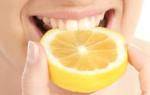
Occupational acid necrosis is commonly seen in factories where there is a high concentration of acid fumes and hydrogen chloride gas in the air. These substances enter the oral cavity and dissolve in saliva, as a result of which saliva oxidizes and demineralizes the hard tissues of the tooth. It should be noted that excessive consumption of citrus fruits and acidic drinks affects the enamel in the same way as acid fumes in factories.
Acid Necrosis Clinic
Clinical picture acid necrosis of teeth develops slowly. With chemical necrosis, canines and incisors are more often affected: in the region of the cutting edges, the enamel disappears, resulting in sharp edges. In the initial stage, patients develop a feeling of soreness on the teeth, and there may also be a feeling of sticking of the teeth, which is associated with the “washing out” of minerals from the tooth enamel, resulting in a feeling of softness of the teeth when closing. In the later stages of the disease, pain from temperature and chemical irritants appears. However, it should be borne in mind that pain may not be observed, in view of the fact that tertiary dentin is being produced. With the progression of necrosis, the enamel loses its luster, becomes dull and rough. And after the complete loss of enamel, the dentin is pigmented and becomes dark. In advanced cases, complete dissolution and erasure of the crown of the tooth can be observed.
Differential diagnosis of acid necrosis
Acid necrosis of the teeth must be differentiated from enamel erosion. Erosion is characterized by a hard, shiny surface, and with necrosis, softening of the enamel occurs.
Prevention of acid necrosis
Prevention acid necrosis is to improve working conditions, the use of individual means, conducting regular alkaline rinses.
Acid Necrosis Treatment
In the initial stages of acid necrosis, it is necessary to minimize the effect of acids on tooth enamel and conduct remineralizing therapy every 3-6 months. The patient is prescribed calcium glycerophosphate (1.5 g for 30 days), multivitamins and phosphate-containing toothpastes. The course of treatment should be repeated every 3 months. Also, if necessary, restorative treatment is carried out using glass ionomer cements, and if the bite is reduced, prosthetics are performed.
Tooth necrosis is a severe disease that often leads to complete loss of teeth. This lesion can be caused by both exogenous and endogenous causative factors. The latter include a violation of the activity of the endocrine glands, diseases of the central nervous system, chronic intoxication of the body or hereditary disorders of the development of teeth. One of the varieties of such non-carious pathology of hard tissues of the tooth is cervical necrosis.
This pathology of the teeth most often occurs in patients with hyperthyroidism and in women during pregnancy, and sometimes after it. This disease is especially intense when pregnancy is combined with hyperthyroidism. Severe symptoms of thyrotoxicosis are violations of protein and mineral metabolism.
Characteristic is the formation of foci of tissue necrosis on the vestibular surface in the area of the necks of incisors, canines, premolars, and much less often molars. Initially, small chalky stripes with a smooth, shiny surface appear on the vestibular surface of the necks of the teeth. Gradually, the area of such altered areas of enamel increases, their surface loses its luster and becomes rough, and the enamel itself becomes matte. Over time, in the area of the affected area, the enamel completely disappears and the dentin is exposed. The boundaries of the defect are not stable; there is an upward trend. In some patients, in the absence of proper oral care, a carious cavity forms in the defect area. The necrotic process can spread to the entire vestibular surface of the crowns. The enamel of the entire tooth becomes so loose that it is easily scraped off by an excavator.
The occurrence of cervical necrosis, especially at the stage of loss of the enamel, is usually accompanied by increased sensitivity of the teeth to all types of stimuli (temperature, chemical, mechanical).
A patient with cervical enamel necrosis should be carefully examined by an endocrinologist.
With severe hyperesthesia of the necks of the teeth, agents are used to help eliminate it or at least weaken the intensity. In cases where the dentin is affected, i.e. a carious cavity has formed in the area of the necrotic focus, they resort to filling the teeth. However, it must be borne in mind that in the future the enamel around the filling may undergo necrosis, therefore, before filling, it is desirable to conduct a course of remineralizing therapy to strengthen the tooth tissues.
Acid (chemical) necrosis of teeth is the result of local influences. This lesion is usually observed in inorganic acids (hydrochloric, nitric, sulfuric) and somewhat less often in organic acids that work for a long time in production. In the workshops of such industries, in the absence of proper ventilation, acid vapors and gaseous hydrogen chloride accumulate in the air, which, getting into the oral cavity, dissolve in saliva, which acquires an acidic reaction and leads to decalcification of the hard tissues of the tooth.
Already in the initial stages of acid necrosis, patients develop a feeling of numbness and soreness in the teeth. Pain may occur when exposed to temperature and chemical stimuli. Sometimes there is a feeling of sticking of the teeth when they are closed. Over time, these sensations become dull or disappear due to the deposition of replacement dentin, degenerative changes in the pulp or its necrosis.
During an objective examination, the enamel of the teeth of the frontal group becomes dull and rough or acquires a dirty gray tint. Erasure of tooth tissues is pronounced.
The enamel in the area of the cutting edges of the crowns disappears, while sharp, easily broken off areas of the crown of the tooth are formed, then the process of destruction and abrasion extends to the enamel and dentin not only of the vestibular, but also of the lingual surface of the incisors and canines. Gradually, the crowns of the anterior teeth are destroyed to the gingival margin, and the group of premolars and molars is subjected to strong abrasion.
Prevention of acid necrosis of teeth is carried out primarily by designing supply and exhaust ventilation in order to seal production processes. Alkaline water columns are installed in workshops for frequent rinsing of the mouth.
In the treatment of chemical necrosis of the tooth, the effect of the acid agent is eliminated and then complex remineralizing therapy is carried out, followed by restorative treatment using glass ionomer cements.
Necrosis of tooth tissues more common in recent years than 10-15 years ago. Its prevalence reached 5.1% of all non-carious disorders of hard dental tissues and 9.7% of non-carious lesions of the 2nd group. Moreover, its varieties have increased, new forms of necrosis have appeared, still unknown to us. Along with this, questions regarding this nosological form remained unresolved.
Usually necrosis of the tissues of the tooth is a complex and severe disease, often resulting in a significant deterioration or loss of chewing function. It is believed that tooth necrosis is a consequence of negative endogenous (dysfunction of the endocrine glands, diseases of the central nervous system, chronic intoxication, etc.) and exogenous (chemical, radiation, toxic, etc.) factors.
Consider some of the most common forms of necrosis of hard tissues of teeth.
Chemical necrosis of teeth(or, as it is called, acid necrosis of the teeth) develops as a result of the action of various acids or acidic products on the enamel and dentin itself. This is carried out either as a real negative production factor (high concentration of acids and other substances in the workplace), or as a household option for frequent or constant exposure to acid-containing foods, drinks, and drugs.
acid necrosis(it is better to call this phenomenon the dissolution of hard dental tissues under the influence of acids) is observed not only in individuals exposed to acids at work, but also in patients who for a long time suffered from frequent, recurring vomiting or belching of acidic stomach contents in case of peptic ulcer, hyperacid gastritis, as well as in people who took hydrochloric acid for achilia.
Decalcified hard substances of tooth crowns, chronically exposed to acids, soften, gradually lose the enamel layer, and dentin is exposed, which is a softer tissue. Naturally, under the influence of chewing pressure and the formation of a food bolus, in the presence of antagonists, the process begins abrasion of teeth. Occupational chemical (acid) necrosis occurs in many industrial enterprises where workers are exposed to vapors of inorganic and organic acids, as well as some other chemicals.
Studies by various authors have shown that the main symptom of this pathology is the progressive loss of enamel substance. At the same time, the leading factor is the leaching of mineral components (macro- and microelements) after denaturation of the protein substance of the enamel. .
It should be noted that the household unfavorable factor associated with the constant intake of hydrochloric acid in diseases of the gastrointestinal tract, the consumption of a significant amount of citrus fruits, acidic juices, dosage forms affects the enamel and dentin in the same way as a professional one.
The clinical manifestation of acid (chemical) necrosis develops gradually. In the very initial stage of acid necrosis, patients have a feeling of sore teeth. One of its leading signs may be a complaint of a "feeling of a feeling of sticking." This is quite understandable, minerals are washed out of the enamel, and the protein component, when closed, creates a feeling of softness of the teeth.
In the future, pain sensations from exposure to temperature and chemical stimuli appear. They can periodically decrease, as replacement dentin is produced. With the progression of the process, the enamel loses its luster, becomes dull and even rough. With the loss of enamel, areas of dentin become pigmented and dark. If the harmful action of acids and acid-containing substances continues, then almost complete dissolution and abrasion of the tooth crowns is inevitable.
Prevention of industrial acid necrosis comes down to improving working conditions in the workplace, sealing derivative processes, as well as the use of personal protective equipment, the organization of alkaline rinses in workshops and the use of phosphate-containing toothpastes for cleaning and applications immediately after work. These measures can significantly change the situation for the better and achieve a reduction in occupational dental diseases.
To prevent domestic acid necrosis of the teeth it is recommended to use glass or plastic tubes for taking acidic drugs, rinse the mouth with alkaline solutions, and also apply phosphate-containing pastes daily to the teeth.
Treatment of chemical necrosis of teeth depends on the degree of its manifestation and severity. Thus, the volume of therapeutic measures in the initial forms of chemical necrosis, when there is still no significant destruction of tooth enamel, is limited to the termination or maximum decrease in the effect of a chemical agent on the teeth and the implementation of complex remineralizing therapy for 3-6 months. The patient is prescribed: calcium glycerophosphate at a dose of 1.5 g per day for 30 consecutive days; klamin (1-2 tablets) or fitolone (30 caps) - 2-3 times a day 15 minutes before meals for 60 days in a row; multivitamins kvadevit or complivit - 2-3 tab. per day for 30 consecutive days. Teeth brushing and applications (15 minutes each) using phosphate-containing toothpastes (Pearl, Cheburashka, Bambi, etc.) daily for 5-6 months. contract. The course of general treatment is repeated every 3 months. In the presence of defects in the tissues of the tooth (reduction in size, change in shape, chipping of crowns, etc.) after complex remineralizing therapy, restorative treatment is carried out after 3-6 months. with the use of SIC, and with a significant decrease in bite - by rational prosthetics.
This group of patients requires dispensary observation and repeated courses of complex remineralizing therapy.
Radiation (post-radiation) necrosis of hard tissues of teeth occurs after exposure to ionizing radiation in connection with the treatment of malignant tumors, diseases of the blood, skeleton and other organs and systems, as well as with the action of professional factors.
Damage to the hard tissues of the teeth, some authors associate both with the direct effect of radiant energy on them, and with violations of mineral and protein metabolism in the body, with a change in the composition of saliva and the functional state of physiological systems.
However, there is no consensus on the mechanism and nature of changes in the tissues of the tooth and oral cavity to date. Some researchers tend to attribute radiation damage to dental tissues to non-carious lesions, others believe that ionizing radiation causes acute dental caries, and, finally, some authors believe that after radiation exposure, dental caries actively develops along with non-carious lesions.
The pathogenesis of radiation damage to the teeth has not yet been fully elucidated. Thus, data on vascular, morphological and degenerative disorders in the pulp that precede the damage to the hard tissues of the teeth are discussed. It is assumed that radiation necrosis of the lower jaw occurs when the xerostomia that develops after radiation exposure affects the teeth; the immunosuppressive effect of ionizing radiation is not excluded.
According to the concept, in the irradiated organism there is a specific suppression of metal-containing enzyme systems (primarily iron-containing) involved in the process of tissue respiration in the aerobic phase. Violation of the aerobic phase of tissue respiration entails the accumulation in the tissues of the body, including the dental pulp, of incompletely oxidized metabolic products, as well as a persistent violation of their further oxidation to CO 2 and H 2 0. This concept of 30 years ago organically fits into modern concepts about the important pathogenetic role of activation of lipid peroxidation and the accumulation of free radicals in body tissues under the adverse effects of endo- and exogenous factors, including penetrating radiation.
As a result of the action of ionizing radiation, it is these processes that occur in the dental pulp that lead to a violation of trophism and physiological processes of remineralization of enamel and dentin. This is especially pronounced when combined with impaired function of the salivary glands caused by irradiation, followed by an imbalance of remineralizing mechanisms in the enamel/saliva medium.
Clinical signs of post-radiation damage to the teeth and tissues of the oral cavity are quite characteristic. First of all, almost all patients have radiomucositis of the mucous membrane of the lips, cheeks, tongue, loss or perversion of taste sensations, severe dryness in the oral cavity.
L. A. Ivanova (1989) found that in such patients the level of non-specific resistance and immune protection of oral tissues is significantly reduced.
Usually after 3-6 months. after radiation exposure tooth enamel loses its characteristic luster, becomes dull, grayish-faded color. Fragility, wear of the chewing and vestibular surfaces of the teeth are noted. Against this background, areas of necrosis appear, first local, and then by the type of circular lesions of the teeth. These lesions are usually dark in color, filled with loose necrotic mass, painless. The absence of a pain symptom is a characteristic feature of radiation damage to the teeth, indicating the suppression of the function of odontoblasts. Gradually, areas of necrosis expand and capture a significant part of the tooth. Removal of necrotic masses from the lesion is usually painless, so this must be done carefully.
If radical interventions are not used, then in 1-2 years more than 96% of the teeth will be affected.
The intensity of radiation damage to the teeth to a certain extent depends on the area and dose of radiation. Thus, under the action of penetrating radiation on the area of the head, neck and shoulders, extensive necrotic lesions of the teeth develop. When irradiating the chest, pelvic organs, limbs, the pathological process in the teeth develops in most cases according to the type acute caries with some clinical features.
Firstly, these lesions of the teeth, resembling acute caries, are painless, even when probing, the indicators of electroodontometry are reduced to 15-25 μA.
Secondly, the cavities formed in the teeth have uneven, corroded edges, which are transparent and fragile within the enamel, the carious cavity is usually filled with a softened dirty gray mass, its removal is painless or painless at all. The loss of previously existing and newly placed fillings is typical, which indicates the inappropriateness of immediate filling of these defects.
With chronic, fractional, ionizing radiation, usually associated with professional features of work, tooth damage is chronic.
The appeared defects on the teeth are flat, located on atypical, usually immune to caries surfaces of the teeth, covered with a gray coating, under which one can feel quite hard enamel and dentin. These lesions are mostly painless and patients present because of a cosmetic defect.
Questioning the patient allows you to differentiate these lesions and outline treatment and prevention measures.
Prevention of radiation damage to the teeth consists, firstly, in reducing the direct effect of radiation on the teeth by making an individual lead mouth guard, which the patient puts on immediately before each radiotherapy procedure. Secondly, by reducing the indirect effect of penetrating radiation by preliminary (before irradiation) appointment of a monthly course of general and local remineralizing therapy in combination with antioxidants.
As a treatment, these patients are prescribed: calcium glycerophosphate - 1.5 g per day for 1 month; klamin (table 2-3) or fitolone (30 caps) - 2-3 times a day during the entire period of treatment and exposure (it should be remembered that these drugs are radioprotectors); multivitamins - 3-4 tablets per day during the entire period of treatment and exposure; antioxidants (aevit or separately vitamins A, E) - 4-6 capsules per day along with ascorbic acid 0.5 g per day during the entire period of treatment and exposure.
Local prophylactic effect consists in daily, preferably double, applications of phosphate-containing pastes such as "Pearl", "Cheburashka", "Bambi" and others on the teeth during the entire period of treatment and irradiation. After irradiation, it is necessary to repeat the course of taking calcium glycerophosphate. Under these conditions, even after radiation therapy, usually the teeth are preserved without any special changes.
However, these therapeutic measures must be continued for several more months. Unfortunately, doctors of related specialties do not always follow these rules of dental prophylaxis in this severe group of patients, therefore, suffering from the underlying disease is joined by torment due to subsequent treatment and extraction of teeth, which poisons these people, perhaps, the last years and months of life.
Not so long ago, such a dental problem as necrosis of hard tissues of the teeth was not yet widespread. Over the past ten to twenty years, the number of people who are susceptible to this severe and difficult to treat disease has increased significantly. It began to be diagnosed much more often in the younger generation.
The concept of necrosis of hard tissues of the teeth and the causes of its occurrence
Under the necrosis of the hard tissues of the teeth, a serious pathology is meant, during the development of which the cells of enamel and dentin slowly die off. As a result, this affects the chewing function of a person, which in the future leads to problems in the work of the entire digestive system. It is also possible violations of diction.
At the initial stage, the disease affects the area characteristic of a particular type of tooth necrosis. Subsequently, necrosis begins to affect the entire surface of the enamel. Without timely treatment, the dental crown is completely destroyed, and after the loss of a tooth, necrosis in some cases can go to the gum.
There are a number of reasons that can provoke the development of necrosis of hard tissues of the tooth. They are usually divided into external and internal factors. Internal include:
- malfunctions of the central nervous system;
- pregnancy, especially several cases in a row;
- dysfunction of the thyroid gland, for example, hypothyroidism;
- imbalanced hormone production, which is often observed in adolescents;
- constant intoxication of the human body;
- predisposition.
External factors negatively affect the dental tissue itself and come down to:

Classification and symptoms of necrosis
There are a number of varieties of necrosis of hard tissues of the teeth, which is due to the factors that provoked it and the location of the lesion (see also: non-carious lesions of the teeth: classification, treatment of pathologies of hard tissues). There are three main types of the disease:
- acid;
- computer;
- radiation.
Each of these types of pathology has its own etiology and distinctive symptoms. The latter is especially important in the differential diagnosis of the disease.
Acid

The second name for acid necrosis of teeth is chemical. This type of pathology develops as a result of prolonged exposure to acids or strong chemicals. For this reason, it is more often encountered by people who work at chemical enterprises and are exposed to vapors circulating in the air and saturated with acids, hydrogen chloride.
Also, acid necrosis of the teeth affects those categories of people who often vomit:
- pregnant women;
- patients with achilia;
- suffering from gastritis.
The disease affects mainly the canines and front incisors, begins with the appearance of decalcified, gradually collapsing areas. The consequence of this is the exposure of dentin and the formation of a sharp edge of the cutting part.
Computer
The computer type of tooth necrosis is a fairly young type of pathology that has been diagnosed quite recently. People who spend 8 hours a day in front of a computer for 3-5 years are subject to this disease. Experts explain the reason for the development of the computer type by ionizing radiation from monitors.

The disease is painless and almost asymptomatic, except for the changed appearance of the enamel. Due to the fact that in addition to the dental crown, root and jawbone, the pulp is initially affected, the teeth, even healthy ones, become dull and grayish. In the lesions, softening of the dentin tissue is observed.
Radiation
Radiation therapy, aimed at combating oncological diseases, which also affects other organs of the human body, leads to the radiation type of necrotic thinning of the hard tissues of the tooth. Workers with radiation devices are also at risk.
The degree of damage and the rate of development of the disease is determined by the dose of radiation received by the person. The higher the dose received, the faster the hard tissues are destroyed.
Pathology is accompanied by:
- dimeneralization of the tooth;
- deterioration in well-being;
- deviation of trophic processes of soft tissues;
- numbness or burning sensation in the enamel area and on the mucous membrane;
- anemia
- high dryness of the oral mucosa;
- hemorrhagic syndrome;
- periodontal inflammation;
- puffiness.
Diagnostic methods
The main methods for diagnosing tooth necrosis are a visual examination by a dentist and the use of radiography.
An important step in establishing the diagnosis is the differentiation of enamel necrosis from other diseases with similar symptoms. Below is a table of dental diseases and their differences from necrosis:
| Disease | How to distinguish from necrosis |
| Marble disease and Stanton-Capdepon syndrome | Speed of distribution. Necrotic death is faster. |
| Fluorosis and enamel hypoplasia (we recommend reading: dental fluorosis: symptoms and methods of treatment) | The period of laying the disease and its manifestations. Fluorosis and hypoplasia are laid during the period of gestation and appear with teething. Symmetry is also characteristic of their manifestations, while the properties of enamel are preserved. |
| Caries | Localization of the lesion. Caries affects only one area, and with necrosis, the entire surface undergoes changes. |
It is also necessary to determine what kind of tooth necrosis the patient has. To do this, the doctor draws attention to the specific symptoms that are characteristic of each particular variety. For example, computer necrosis immediately penetrates into the pulp of the tooth, and with acid necrosis, sharp edges are formed and the disease itself develops slowly.
Treatment
Treatment of any type of necrotic tissue death is to restore their integrity and increase density according to the schemes that the doctor must prescribe.
Initially, it is necessary to stop the destructive processes in the body and eliminate external factors that lead to the disease. Next, the doctor carries out the processing of dental crowns, due to which the dentin tissues are saturated with minerals and nutrients, trophism is restored (we recommend reading: the structure of dental tissue). In severe cases, it may be necessary to install prostheses or restore the crown using polymer materials.
Preventive measures
Prevention is reduced to the elimination or maximum limitation of the impact of those factors that lead to the disease. Each specific type of necrotic tissue death entails its own preventive measures. For example, during radiation therapy, patients put special mouthguards made of lead on their teeth, thereby reducing the effect of radiation rays.
Harmful enterprises should install powerful exhaust ventilation and alkaline water columns for workers to rinse their mouths. Compliance with sanitary standards when working at a computer will prevent the development of computer necrosis. Plus, you need to take care of the oral cavity and monitor the diet.