What should be the thickness of the endometrium of the uterus? Endometrium by cycle days: what is Endometrium 12 mm of menstruation determined for
The endometrium is the inner mucous membrane of the body of the uterus, which has two layers: functional and basal. The basal layer has a constant thickness and structure. The stem cells that make up its composition are responsible for the restoration (regeneration) of the layers of the endometrium. The functional layer has different dynamics, sensitively reacts to the concentration of female hormones. Thanks to the changes taking place in the functional layer, menstruation comes every month. It is she who is an indicator of women's health. If any pathology of the endometrium occurs, failures in the menstrual cycle often occur.
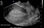
Thickness of the endometrium
To put it figuratively, the endometrium can be compared to a cradle, which at a certain period is ready to accept a fertilized egg. If this does not happen, then the functional layer is rejected, which is reborn after menstruation.
The endometrium, the norm of the thickness of which is different, has different indicators for the days of the cycle:
- 5-7 day. In the early proliferation phase, the thickness of the endometrium does not exceed 5 mm.
- 8-10 day. The endometrium thickens up to 8 mm.
- 11-14 day. In the phase of late proliferation, the thickness reaches 11 mm.
After this, the secretion phase begins. During this period, if there is no pathology of the endometrium, the layer becomes looser and thickens.
- 15-18 days. The thickness reaches 11-12 mm.
- 19-23 days. The maximum thickness of the endometrium. The average is 14 mm, but can reach a maximum of 18 mm. The layer becomes looser, "fluffy".
- 24-27 days. The thickness begins to decrease slightly, it becomes from 10 to 17 mm.
Such are the phases of the endometrium. During menstruation, the thickness of the endometrium decreases, reaching only 0.3-0.9 mm.

If a woman has menopause, what should be the endometrium? The standard layer thickness is 5 mm. The slightest deviations of 1.5 or 2 mm should cause alertness. In this case, it is better to see a gynecologist.
What to do if the endometrium is thin?
Very often thin endometrium is the cause of female infertility. It is quite possible to cure this, you just need to persistently go towards your goal. Treatment can be carried out in several alternative ways: hormonal drugs, herbal decoctions, pseudohormones.
Herbal treatment
Some women do not want to resort to medical treatment of thin endometrium and use folk remedies in this case.
Thin endometrium is well restored with the help of sage. Drink it in the first phase of the cycle. 1 teaspoon should be brewed in 200 g of water, taken throughout the day.
The upland uterus is transformed as a pseudohormone in a woman's body. In addition, it has an anti-inflammatory effect.
Drops "Tazalok" from a series of homeopathy help in the normalization of the menstrual cycle, are a regulator of the synthesis of endogenous gonadotropic hormones.
Build-up of a thin endometrium with the help of drugs
How to build up a thin endometrium, the norm of the thickness of which changes in different phases of the cycle? In the first phase of the cycle, doctors prescribe the drug "Proginova", "Femoston", etc. For the second phase of the cycle, "Dufaston" is suitable. This drug contributes to the formation of the structure of the endometrium, it acts like a synthetic progesterone.
Before using all these synthetic drugs, you should definitely consult a gynecologist and assess the risk yourself, since they all have some contraindications.

There are cases when a thin endometrium is detected after taking oral contraceptives. Refusal of them and the use of Regulon tablets for two months often gives a positive result and helps to restore the thin endometrium.
Anatomical reference
A healthy endometrium is the key to a successful onset and development of pregnancy. Currently, many women experience some kind of endometrial disease and, as a result, suffer from infertility. What does the term "endometrial pathology" mean, what consequences does this phenomenon lead to, how to overcome this problem? About everything in order.
The main function of the endometrium in the female body is the successful, safe implantation of the embryo. For pregnancy to occur, it must attach to the wall of the endometrium. That is why, with various pathologies of the endometrium, infertility can occur, the successful attachment of the embryo becomes simply impossible. But the pathologies are different, there are several diseases of the endometrium. Which one, in each case, should be determined by a specialist.
Deviations from the norm
From the nature of the occurrence of the disease, gynecologists-endocrinologists distinguish two benign disorders. The pathology of the endometrium of the uterus is inflammatory in nature, this includes endometritis. Non-inflammatory - these are hyperplastic processes. These include endometrial polyps, hyperplasia, and endometriosis.
It happens that several pathologies are combined in the female body. What is the reason for this? First of all, a violation of the endocrine system or a genetic predisposition. In many cases, after successful treatment, pregnancy becomes possible.
endometritis
Inflammatory disease of the mucous membrane (endometrium) of the uterus. What is the cause of the disease? Penetration into the uterine mucosa of various pathogens. There are several underlying factors contributing to the disease:
- Any infectious processes that exist in the body.
- Perfect intercourse without contraception.
- Erosion of the uterus.
- Examination of the uterus, tubes by hysterosalpinography.
- Chronic gynecological diseases.
- Non-sterile instrument during a gynecological examination.
- C-section.
- Curettage of the endometrium.
Typical symptoms of endometritis:

If endometritis is detected during pregnancy, it requires immediate treatment. The disease can affect the fetal membranes of the embryo and lead to its death.
Hypoplasia - thinning
If on certain days of the cycle the thickness of the endometrium is underestimated, gynecologists diagnose hypoplasia. The cause of the disease is hormonal disorders, poor blood supply, inflammatory processes. Such a pathology of the endometrium can occur as a result of frequent abortions, infectious diseases, prolonged use of the intrauterine device. The main task in the treatment of hypoplasia is the thickening of the endometrium.
Hyperplasia - thickening
The cause of the disease is most often hormonal disruptions in the body or hereditary factors. With hyperplasia, the layers of the endometrium change their structure.
There are several types of hyperplasia:
- Glandular hyperplasia.
- Fibrous hyperplasia atypical (precancerous condition).
- Glandular cystic hyperplasia.
The glandular endometrium is often found in diseases of the adrenal glands, ovaries, thyroid gland. Most often, hyperplasia affects women with diabetes mellitus, polyps in the uterus, fibroids, arterial hypertension.
Why is hyperplasia dangerous? Uncontrolled cell growth, which can lead to terrible consequences - endometrial cancer. Hyperplasia is treated with both medical methods and surgical intervention.
Endometrial polyps
Benign growth of endometrial cells. Polyps are able to deploy not only in the uterus itself, but also on its neck. The reasons for their formation are hormonal disorders, the consequences of surgical interventions, abortions, infections of the urogenital area. Polyps most often form in the endometrium. There are several types of polyps:
- Glandular. They are formed in the tissues of the glands, usually diagnosed at a young age.
- Fibrous. Formed in connective tissue. More common in older women.
- Glandular fibrous. Consists of both connective and glandular tissue.
The only way to get rid of polyps is through surgery. This should be done as soon as possible, since the cells are able to degenerate into malignant ones. Modern equipment allows you to perform operations quickly, efficiently, painlessly.

endometriosis
A female disease in which nodes form outside the uterus, similar in structure to the layer of the endometrium. Nodules may appear on nearby organs. It happens that when the uterine tissues are rejected, they are not completely removed with menstruation, penetrate the tubes and begin to grow there. endometriosis develops.
The main causes of the origin of the disease:
- Excess weight.
- Frequent stress.
- Bad habits.
- Disruptions in the menstrual cycle.
- Inflammation in the genitals.
- Operations on the uterus.
- Heredity.
- Hormonal disruptions.
- Thyroid problems.
Symptoms of endometriosis include:
- Infertility.
- Painful urination and bowel movements.
- "Smearing" selection in the middle of the cycle.
- Pain before menstruation.
- Pain during intercourse.
Removal of the endometrium - ablation
Currently, an increasing percentage of women suffer from various pathologies of the endometrium. Suffer from prolonged, profuse, painful menstruation, hyperplastic processes, polyposis. Unfortunately, it is not always possible to achieve effective treatment with hormone therapy or curettage of the uterine body. The alternative in this case is ablation, or removal of the endometrium. This is a minimally invasive procedure that destroys or completely removes the lining of the uterus (endometrium).
Indications for the operation:
- Massive, repetitive, prolonged bleeding. In this case, there is no effectiveness of the treatment. The presence of malignant processes in the genital area in women older than 35 years.
- Relapses of hyperplastic processes during premenopause or postmenopause.
- The impossibility of hormonal treatment of proliferative processes in the postmenopausal period.
What factors should be considered when performing ablation?
- The impossibility of complete removal of the uterus or the refusal of this type of surgical intervention.
- Unwillingness to preserve childbearing function.
- The size of the uterus.

Biopsy of the endometrium
For diagnostic purposes, small amounts of tissue are taken from the body in special ways. To make a correct diagnosis based on the results of a biopsy, the doctor must comply with a number of necessary conditions during the procedure. Based on the results of the scraping examination, the pathologist evaluates the functional and morphological state of the endometrium. The results of the study directly depend on how the endometrial biopsy was performed, what material was received. If strongly crushed pieces of tissue are obtained for research, then it is difficult for a specialist, sometimes it is impossible to restore the structure. It is very important when performing curettage to try to get non-crushed, larger strips of the endometrium.
How is an endometrial biopsy performed?
- As a complete diagnostic curettage of the body of the uterus with the expansion of the cervical canal. The procedure begins with the cervical canal, then the uterine cavity is scraped. With bleeding, curettage should be carried out with a small curette, special attention should be paid to the tubal corners of the uterus, where polyposis growths often form. If crumb-like tissue appears during the first scraping with a curette from the cervical canal, then the procedure is stopped due to suspicion of carcinoma.
- Stroke scrapings (chain technique). The goal is to find out the causes of infertility, control the results of hormone therapy. This technique should not be used for bleeding.
- aspiration biopsy. Suction of pieces of mucous tissue of the endometrium. The method is most often used for mass examinations, the goal is to identify cancer cells.

If any endometrial pathology is detected in a woman's body, treatment should be started immediately. The treatment process started in time gives the most promising prognosis. Even such a sentence as infertility may not be terrible if you turn to a gynecologist in a timely manner, undergo a full examination, and a course of treatment. Watch your health!
In a woman's body, cyclical changes occur monthly, associated with the transformation of the inner layer of the uterus. What should be the structure and thickness of the endometrium?
The inner surface of the uterus, which is called the endometrium, consists of the basal and functional layers. The upper functional layer is rejected monthly in the form of bloody menstrual discharge. The basal layer is a layer of cells with a high regenerative ability, it is thanks to them that the functional layer of the endometrium is updated.
What is the main function of the inner layer of the uterus?
The main function of the endometrium is to create the most favorable conditions for the implantation of the fetal egg into the uterine wall and the further development of pregnancy. Here, its transverse dimension is of fundamental importance. What determines the thickness of the endometrium? First of all, sex hormones affect his condition: estrogens, progesterone, to a lesser extent testosterone.
Depending on the phase of the menstrual cycle (proliferative or secretory stages), the structure of the inner layer of the uterus may change. Below is a linear relationship between its thickness and cycle days:
- In the proliferation phase (day 5-7 of the cycle), the parameters range from 2 mm (sometimes only 1 mm) to 6 mm, on average - 5 mm (sometimes in the ultrasound report you can find data in centimeters, for example, if 0.38 is indicated, it means 3.8mm)
- The phase of medium proliferation (8-10 days of the cycle) - an average of 8.5 mm (thickness ranges from 4 mm to 9 mm) according to ultrasound, this is the so-called three-layer endometrium
- The phase of late proliferation (11-14 days of the cycle) - the thickness of the inner layer of the uterus is up to 11 mm (fluctuations from 8 mm to 14 mm)
- In the second phase of early secretion (15-18 days of the cycle) -10-16 mm, on average 13 mm
- Phase of medium secretion (19-23 days) - the endometrium reaches its maximum thickness, on average 14 mm (fluctuations from 12 mm to 16 mm)
- The phase of late secretion (24-27 days of the cycle) - the endometrium becomes a little thinner, 12 mm (fluctuations from 10 mm to 17 mm).
When are deviations in the thickness of the endometrium observed?
Cases when a discrepancy between the thickness of the endometrium and normal parameters is detected arise due to functional and pathological factors. The functional reason is the onset of pregnancy. During this period, there is a physiological thickening of the inner layer of the uterus. It should be noted that a change in the thickness of the endometrium occurs after fertilization on the seventh day of pregnancy, at which point the fetal egg is not yet in the uterus itself.
On the 30th day of pregnancy, the thickness of the endometrium, which is optimal for the development of pregnancy, is 20 mm. Not only thickness is also important, but also its structure, this is important for fertilization.
There are cases when there is a lack of menstruation with an endometrial thickness of 10 mm. If there is a delay, a re-examination is necessary, usually after a month.
Such a discrepancy in thickness indicates a hormonal "failure" in the body. The pathological causes of changes in the thickness of the endometrium include its hypoplasia and hyperplasia.
Additional information about endometrial diseases is posted in the video:
What are the causes of endometrial hypoplasia?
The main reasons for the decrease in the thickness of the endometrium are:
- Congenital diseases (infantilism, gonadotropic nanism), when there is insufficient proliferation of the endometrium
- Damage to the uterus and its inner layer after an abortion
- Postponed inflammatory diseases of the uterus.
With such a pathology, a small thickness of the endometrium is noted, it does not reach 7 mm. In such cases, the main complaint of patients is the lack of pregnancy. This means that the fertilized egg is not able to attach to the wall of the uterus, and pregnancy does not occur.
What are the causes of endometrial hyperplasia?
In some cases, ultrasound can determine too much, exceeding the norm, the thickness of the endometrium. This indicates the presence of endometrial hyperplasia.
The main reason for this pathology, in which there is a discrepancy between the thickness of the inner uterine mucosa and normal indicators, is today considered to be excessive formation of the hormone estrogen in the body of a woman. Additional factors provoking the disease are genetic predisposition, inflammatory gynecological diseases, diseases of the endocrine glands.
What are the symptoms of the disease?

Endometrial hyperplasia is characterized by menstrual irregularities with a tendency to increase bleeding: menstrual blood loss becomes more abundant and longer than usual, the discharge has an admixture of lumps or particles of the epithelium. In addition, there may be spotting from the genital tract, not associated with the menstrual cycle. There may be a delay in menstruation or their early onset.
External factors can provoke the occurrence of bleeding, for example, a hot bath affects the amount of blood loss. Repeated cases of bleeding, as a rule, lead to the development of iron deficiency anemia. Particular vigilance should arise with uterine bleeding in women who are in the menopausal period, their presence may indicate the development of a malignant neoplasm of the uterus - adenocarcinoma.
Infertility is also one of the symptoms of endometrial hyperplasia. It is associated with excessive formation of estrogen in the body and the formation of an anovulatory (without maturation of the egg) cycle.
Methods for diagnosing endometrial hyperplasia
The main diagnostic method is ultrasound. Ultrasound of the pelvic organs allows you to find out the parameters of the thickness of the inner layer of the uterus at different phases of the cycle.
It is better to study the echo in the second phase of the menstrual cycle, it is better to determine the thickness on these days, otherwise the results will be inaccurate. Some patients say the width of the endometrium, meaning the thickness, this is an incorrect term.
It is also possible to visualize the presence of concomitant cysts and polyps of the uterus, to identify areas of uneven thickness of the endometrium. The unevenness of the basal layer indicates the presence of an inflammatory process in the uterus, endometritis.
Sometimes the question arises why it is impossible to measure the endometrium in the presence of volumetric formations in the uterus (myoma, tumor). This is due to large errors in the parameters, in such cases, computed tomography of the pelvic organs is performed, it allows you to most accurately measure the thickness of the inner layer of the uterus. Without its results, curettage of the uterine cavity is not recommended.
Normal parameters during diagnostics range from 9 mm to 11 mm. If the indicator does not correspond to the normal thickness and increases to 15 mm, one can think about the presence of hyperplasia. If the ultrasound determines the endometrium with a thickness of 21 mm (or more, for example, 24 mm or 26 mm), and its structure is uneven, one can assume the presence of a malignant neoplasm - adenocarcinoma.
In any case, an echo study can only suspect the presence of a pathology, the final stage of diagnosis is hysteroscopy, followed by diagnostic curettage and histological examination of the material. The procedure is not dangerous, it is performed under short-acting intravenous anesthesia. In the future, after cleaning, the morphological structure of the endometrium will be determined, it allows you to find out about the presence of atypical cells in it.
Medical and surgical treatment
Surgical treatment is in many cases the preferred method because it is more effective. The methods of treatment include diagnostic curettage (cleaning) of the uterine cavity, followed by a histological examination of the material obtained. Cleaning is especially indicated at a thickness of 21 mm. If hyperplasia is combined with the presence of polyps, they are removed simultaneously during the operation under the control of a hysteroscope. After scraping (“cleansing”), slight spotting may occur for several days, this does not pose a health hazard.
About the treatment of endometrial disease is described in the video:
One of the most radical methods of treatment is ablation (destruction) of the endometrium. It is carried out for women, usually in the menopausal period (50-52 years), who have repeated recurrences of bleeding after previous surgical treatment.
Drug treatment includes a number of hormonal drugs that affect the thickness and condition of the endometrium. Most often, combined oral contraceptives (COCs) are used to treat this pathology, for example, Zhanin, Yarina. They are also used as a means to eliminate bleeding in cases where therapeutic curettage is not recommended. For example, in young nulliparous women.
With the ineffectiveness of drug therapy, cleaning is done (curettage of the uterine cavity). Against the background of coc, the thickness of the endometrium and its structure are normalized. When taking oral contraceptives, various side effects can be observed: liver dysfunction, vein thrombosis, discoloration of the skin.
The second group of drugs for the treatment of hyperplastic processes in the endometrium is progesterone derivatives, gestagens. These include Utrozhestan and Dufaston. The thickness of the inner layer of the uterus changes after taking utrogestan in the direction of its reduction. This group also includes the Mirena intrauterine device, which contains a progestogen and is capable of exerting a local effect on the endometrium.
The third group of drugs is the so-called gonadotorpin-releasing hormone agonists (Zoladex, Buserelin). They are administered as injections once a month, while also normalizing the thickness of the endometrium. The disadvantages include the presence of side effects in the form of a sensation of "hot flashes", while the mood often changes. The correct dosage of the drug allows you to avoid these unpleasant consequences.
Preparation of the endometrium before in vitro fertilization
In the last few decades, the most effective method of infertility treatment has been the method of in vitro fertilization. With IVF, success largely depends on the state of the endometrium at the time of the procedure. In the presence of pathological changes (hyperplasia, hypoplasia), appropriate treatment is carried out using estrogens and progesterone, depending on the phase of the cycle. If there is no pathology, in any case, medical preparation of the endometrium is necessary before embryo transfer (during cryotransfer), the required thickness should be 6-8 mm.
Each organ of the female reproductive system has its own functions and purpose. The uterus has a special role, it is responsible for the secure attachment and full development of the embryo.
The endometrial layer lines the uterine cavity from the inside, creates optimal conditions for the fetal egg and supports the course of pregnancy. The normal thickness of the endometrium depends on the day of the cycle. The size of the mucosa may be lower and higher than normal. Both conditions are abnormal and require correction.
Women learn about the importance of the size of the endometrium during the menstrual cycle after problems with conception begin or gynecological diseases are detected. This can be avoided. Modern diagnostic methods allow you to accurately and quickly assess the condition of the uterus and existing abnormalities. The endometrium can be brought back to normal. To do this, it is necessary to regularly undergo ultrasound, and in case of identified pathologies, be treated under the supervision of a doctor.
Ultrasound is the fastest, safest and most informative method for determining the thickness of the uterine mucosa. During a routine examination by a gynecologist, it is impossible to obtain accurate indicators. Only ultrasound allows you to analyze the echographic signs of the inner layer of the reproductive organ. Doctors observe how the endometrium grows and changes, and also detect pathological changes, including tumor growths.
In the absence of contraindications, specialists resort to the transvaginal method, when the organ is examined through the vagina. The most important condition is the study on the day appointed by the doctor. This is due to the fact that the norm of the endometrium in each of the days of the menstrual cycle is different. Normal indicators of mucosal thickness during ovulation differ from the thickness parameters before menstruation. The difference is insignificant, but even the slightest deviation affects reproductive capabilities and health in general.
Signs of thinning
A healthy endometrium, the thickness and structure of which correspond to the day of the cycle, ensures reliable implantation of the embryo, but not all women understand the significance of the measured indicators and pay attention to signs of a decrease in the layer thickness. Specific manifestations have not been identified, but some symptoms should alert and become a reason to see a doctor.
One of the main signs of thinning of the mucosa is the failure of the menstrual cycle, when there are no periods at the right time, and delays are observed regularly.
In addition to cyclic deviations, a decrease in thickness may be accompanied by the following manifestations:
- pain in the lower abdomen;
- the presence of blood clots in the discharge;
- bleeding outside of menstruation.
The mucous layer of the uterus promotes the attachment of the embryo and is the structure that supplies the embryo with nutrients. When the endometrium does not correspond to the phase of the cycle and its thickness is insufficient, pregnancy is impossible. There is simply no chance for an egg to implant safely in the uterus. The fetal egg is rejected, and doctors in such cases diagnose a miscarriage in the early stages. For those who want to get pregnant, such expert opinions are another missed opportunity to have a baby. The situation could have turned out differently if the measures for correcting the thin endometrium had been taken on time.
Norms of the endometrium by phases
The endometrium is updated monthly and has a two-layer structure. The basal (deep) layer does not change and contributes to the regeneration of the functional layer, the thickness of which is not constant.

The size of the mucosa in the first days of the cycle averages 3-4 mm. The endometrial layer reaches its maximum thickness after the egg has formed and left the follicle. During the period of ovulation, the indicators may vary, on average they are 12-19 mm. With successful fertilization, these parameters are optimal for successful attachment and further implantation of the embryo.
In cases where pregnancy does not occur, the overgrown endometrial layer is rejected and comes out during menstruation.
The indicators that are studied to assess the size and structure of the mucosa are considered average, but when comparing the result with the norm of the thickness of the endometrium of the uterus, they allow drawing conclusions about the state of the inner lining and the prospects for conception.
If the hormonal background is in order, the process of mucosal growth successively goes through three periods: menstruation (bleeding), proliferation, secretion. Each phase has its own terms, features and functions.
Bleeding phase
In the phase of menstruation with a failed conception, the functional layer is torn off and comes out with the blood. The onset of bleeding is considered the first day of a new cycle. Menstruation lasts 3-7 days. Rejection begins in the first 2 days, the size of the endometrium during this period ranges from 6 mm to 9.
On the 3-5th day of the menstrual cycle, gradual tissue regeneration begins. The thickness grows and reaches 3 mm by the end of the bleeding phase. The compliance of the mucous layer with these parameters is considered the norm.
proliferative phase
Lasts 2 weeks. During this time, the follicles responsible for the production of estrogen have time to mature. This hormone stimulates the active growth of the uterine membrane. As a result, the functional layer thickens and by the end of the period its size reaches 11–13 mm. In parallel with the increase in size, the sound permeability of the mucosa changes. By the end of proliferation, this indicator is 9–11 mm.
Proliferation begins on the fifth day of the cycle. The phase includes early, middle and late stages. All 3 periods should always take place in a clear sequence. The absence or failures in the course of any of the stages indicate the development of pathological processes in the body.
The thickness of the uterine endometrium of 7 mm is considered the threshold for possible fertilization. If the size is smaller, conception does not occur.
In the proliferation phase, the thickness is almost twice as large, but this is not the most successful period for fertilization. The woman's body is vulnerable, reacts to any negative phenomena and stimuli. Diseases, stress, overwork can stop the natural maturation of the follicle and provoke untimely rejection of the inner layer of the uterus.

The most favorable time for fertilization is the third (secretory) phase, which begins after the proliferation of the endometrium.
Secretory
Secretion is a period of intensive growth of the mucous membrane. The phase lasts from the 15th to the 30th day and is accompanied by the active production of progesterone, which stimulates the growth of endometrial tissues. The mucous layer increases, swells, becomes dense, spongy and vascular. The shell size can reach 21–26 mm. This is a normal thickness, sufficient for secure attachment and nourishment of the embryo.
The secretory phase includes three stages:
- Early is the 15-18th day. The normal thickness parameter for this period is 12 mm.
- In the phase of medium secretion (from the 19th to the 23rd day), the maximum size of the endometrial layer is observed, after which the thickening stops. The norm for this period is 15–21 mm.
- The late period of the secretory phase occurs on the 24th day from the onset of menstruation and lasts 3–4 days. The size of the endometrium begins to decrease and reach 10–17 mm.
If fertilization does not occur, the menstrual phase begins again, and the uterine mucosa is shed during menstruation. This sequence is considered a physiological norm. In all women of reproductive age, these periods are repeated regularly.
Thickness by day of cycle
The hormonal background is responsible for the thickness of the functional layer of the endometrium. If an imbalance is not observed, on different days of the cycle, the size of the mucosa will correspond to the norm.
With menstruation, the endometrium changes markedly:
- in the first days it is seen as an inhomogeneous structure 5–9 mm thick. There is no clear layered composition of the inner lining. This is due to the fact that during this period the cells are located non-standard;
- 3-4th day of menstruation - cells acquire a clear structure, echogenicity increases, and the thickness of the endometrial layer decreases to 3-5 mm;
- 5-7th - normal endometrial thickness ranges from 6 to 9 mm. With the onset of the proliferative stage of the cycle, sound conductivity increases, echogenicity decreases, and the size of the endometrium grows;
- 8–10th - gradual thickening of the mucosa continues. A clear hyperechoic structure appears in the center of the endometrial layer. Indicators of the norm of thickness vary within 8-10 mm;
- 11-14th - the echographic picture almost does not change. This is a late stage of proliferation with a characteristic increase in echogenicity and thickening of the endometrium of the uterus up to 9–13 mm;
- 15-18th - the functional membrane of the uterus thickens up to 10-15 mm. Significant changes in the echogenicity and structure of the endometrium are not observed;
- 19–23rd - the normal parameter varies from 10 to 18 mm. This is the highest figure for the entire period. After this, the thickening of the endometrium stops;
- on the 24th–28th day of the monthly cycle, a decrease in the size of the endometrium is observed. Its thickness is reduced to 12 mm, during ultrasound, heterogeneity of the structure and increased echogenicity are noticeable.

Delay rate
The main ones are hormonal disorders. It is impossible to exclude the influence of other factors, such as stressful situations, gynecological diseases, problems of the endocrine system, unbalanced nutrition.
The cyclic period with late menstruation is lengthened. The production of hormones is disrupted. As a result, the size of the endometrium after ovulation does not change and corresponds to the natural level of the secretory phase (12–14 mm).
Possible pathologies
If we analyze the results of ultrasound studies, the digital values of the thickness of the endometrium by day of the cycle show an increase trend. Growth is gradual - and this is normal. But, unfortunately, not all women have such an ideal picture. The size of the uterine mucosa often differs from the normative indicators. This happens under the influence of a variety of reasons and factors, including:
- hormonal disorders;
- injuries of the mucous and uterine cavity;
- impaired circulation;
- inflammatory and infectious diseases of the uterus.
Pathologies of the endometrium are detected by ultrasound and during additional laboratory examinations. After the cause of the deviation is determined and confirmed, the doctor prescribes treatment taking into account the stage and type of the disease, as well as age, physiological characteristics and the state of the body.
The discrepancy between the thickness of the endometrium is usually divided into 2 types: hypoplasia and hyperplasia.
Hyperplasia
Hyperplasia is the pathological growth of the endometrium. Anomaly in the thickness of the mucous layer of the uterus is reflected in the density. It increases, and the structure becomes heterogeneous. Such changes complicate the implantation of the fetus and the flow of other processes that contribute to the normal development of the embryo.
The disease is dangerous because the rapidly growing endometrium before menstruation does not come out during menstruation. This can lead to perforation (breakthrough), severe bleeding, and hospitalization.
Hyperplasia can be glandular and atypical. The latter form is more dangerous and is considered as a precancerous condition.
The main reason for the inconsistency of the endometrium with the norms is hormonal disruptions. Thickening is provoked by the active production of estrogen and a deficiency of progesterone. Other causes include tumors and polycystic ovaries, diseases of the endocrine system, metabolic disorders, prolonged hormone therapy, weakened immunity, abortions and uterine injuries.

hypoplasia
Abnormally thin endometrium in medicine is defined by the term "". This disease refers to congenital pathologies arising from insufficient synthesis of hormones.
Hypoplastic endometrium has no symptoms. The disease does not manifest itself until the woman has a desire to become pregnant. Difficulties can arise with this, and only an experienced doctor is able to determine what provoked the development of endometrial pathology. Among the signs of the disease are:
- long absence of pregnancy;
- frequent miscarriages;
- late menstruation (after 16 years);
- pathological discharge from the vagina;
- irregular periods.
For life, hypoplasia does not pose a danger, but with a thin endometrium, there is practically no chance to bear a child. The thinned shell prevents the onset of pregnancy and the full attachment of the embryo.

Thickness Mismatch
Indicators of the normal thickness of the endometrium are individual, depending on the state of the reproductive system, age and other characteristics of the body. Parameters that go beyond the established limits are considered to be a violation. Similar phenomena are noted during miscarriage and the development of gynecological diseases.
The only pleasant reason for the discrepancy in the thickness of the endometrium may be the conception that has taken place. Growth stimulates the active production of progesterone (pregnancy hormone). The mucous membrane becomes overgrown with vessels, the secretion becomes more abundant, and the endometrial layer increases to 20 mm or more. In other cases, any deviations from the norm are usually attributed to pathological conditions.
Complications and consequences
If no abnormalities are detected on ultrasound and the thickness of the endometrium is normal, a woman has a chance to become pregnant and give birth to a healthy baby. Unfortunately, not everyone is attentive to their health. Rare visits to the doctor, ignoring alarming symptoms and self-medication often lead to the development of gynecological pathologies that are dangerous for the reproductive system. The most severe consequence is infertility. The inability to conceive develops due to untimely diagnosis and treatment of progressive diseases.
With hyperplasia in women, the cycle is disrupted, the intensity and duration of monthly discharge increases. Frequent bleeding that occurs between periods leads to anemia. In addition, the abnormal growth of the inner lining of the uterus causes endometriosis, cysts, polyps and other neoplasms.
No less dangerous complications of hypoplasia. As a rule, they do not appear in the first days and months after the diagnosis of the disease. The vulnerability of the thin endometrium contributes to the unhindered penetration of pathogenic microorganisms into the uterine cavity. This causes infectious and inflammatory processes, provokes an ectopic pregnancy and frequent miscarriages.
Treatment of disorders
The thickness of the endometrium is successfully corrected. If during an ultrasound examination, deviations are detected by the days of the cycle up or down, the doctor determines the type, stage and echo signs of the pathology.
Treatment of hyperplasia can be medical and surgical. The specialist prescribes the dosage and suitable drugs after determining the type and extent of the disease. The most effective is hormone therapy with the use of progesterone preparations. With a decrease in estrogen levels, the endometrium reaches normal levels.

Surgical intervention is resorted to in cases where conservative methods are not effective. Doctors may remove the endometrium. In difficult cases of atypical hyperplasia, hysterectomy is performed.
The use of hormonal agents gives good results in the treatment of hypoplasia. The thin layer of the endometrium is corrected by agents that contain excessive doses of the hormone estrogen. If the disease has arisen due to inflammatory processes of the reproductive organs, therapeutic measures are aimed at stopping and eliminating the source of inflammation. Severe forms of hypoplasia require surgical intervention.
The reproductive health of a woman depends on many factors. The indicator of endometrial thickness is one of the most important and significant parameters, since it is with it that the possibility of becoming pregnant, bearing and giving birth to a child is associated. Regular ultrasounds will help track the normal and abnormal conditions of the endometrium, as well as identify other gynecological abnormalities.
The thickness of the endometrium is a relative value, but nevertheless, it is an indicator of ongoing processes and hormonal balance in the female body. Knowing the thickness of the inner lining of the uterus, it is possible to determine the phase of the menstrual cycle, age, and also draw preliminary conclusions about the general state of a woman's health.
But, as a rule, gynecologists go from the opposite, or rather, compare the actual value with the established norms. Each age group is characterized by its own indicators, for example, the thickness of the endometrium, which is considered the norm during menopause, is not suitable for conceiving a child and indicates obvious violations.
We will talk in more detail about the norms of the endometrium characteristic of a certain age period in this article.
The norm of the endometrium for conception
The endometrium of a woman of reproductive age regularly undergoes cyclic changes. The thickness of the functional layer of the inner membrane changes mainly, which actively thickens, up to the onset of ovulation and several days after it, and then gradually atrophies and is rejected during menstruation.
This complex process is completely regulated by hormones, so it immediately reacts to the slightest hormonal disruption.
The thickness of the endometrium is of fundamental importance for women planning a pregnancy. Since normally, the thickness of the endometrium reaches its maximum value at ovulation, thereby creating favorable conditions for the implantation of a fertilized egg. In addition, in order for the embryo to attach and begin to develop, the mucosa must be mature, and its structure must be appropriate.
So, depending on the phase of the menstrual cycle, the thickness of the endometrium varies:

If pregnancy has occurred, and the fetal egg is securely in the uterine mucosa, then the latter continues to actively develop. Normally, the endometrium thickens during pregnancy, enriched with blood vessels. For a period of 4-5 weeks, its value will reach 20 mm, and even later it will be converted into which will serve as protection and supply the fetus with nutrients and oxygen.
The norm of the endometrium in menopause
First of all, menopause is characterized by a decrease in estrogen production, which cannot but affect the organs of the reproductive system. In particular, the uterus, ovaries, vagina and mammary glands react to changes.
During menopause, the inner layer of the uterus becomes thin and loose, and eventually completely atrophies. Normally, the thickness indicators during this period are 3-5 mm. If the actual values are increased, then we are talking about pathological hypertrophy. The symptoms of this condition can be different in intensity of bleeding, starting with a brown daub, ending with profuse blood loss. In the first case, the condition is corrected by hormonal therapy, in the latter - by surgical intervention.
Article plan
The reproductive system of a woman is a complex mechanism in which each organ must clearly perform its function. The value of the uterus should not be underestimated, the unborn child develops in it. In order for a woman to perform a reproductive function, her health was in order, the inner mucous layer of the uterus (endometrium) is updated monthly. Only a healthy layer is able to create conditions for the development of the baby. In this article, we will figure out what the thickness of the endometrium should be on the days of the cycle, it is important for a woman to know this, since only if the inner mucous layer is healthy, a fertilized egg can be fixed in it.
Structure of the endometrium
The endometrium contributes to the implementation of the mechanism of the menstrual cycle. Another of his tasks is to provide the most suitable conditions for fixing a fertilized egg in the uterus, its full development, and obtaining everything necessary from the mother's body.
Consists of 2 layers:
- basal - layer directly adjacent to the uterine walls;
- functional - the surface layer that is torn off during menstruation. The basal layer ensures its complete recovery before the start of the next cycle.
The hormonal background of the female body is responsible for the thickness and structure of the inner mucous layer. It increases monthly - this happens during the 2nd phase of the monthly cycle. It also enhances the process of its blood supply. This indicates that the body is ready to receive the fetal egg. Rejection of the functional layer occurs when a woman does not become pregnant - menstruation begins.
Stages of endometrial development
The process of cyclic changes in the uterus in a woman's body occurs monthly. The size of the endometrium differs depending on the onset stage of the monthly cycle. There is a division of the menstrual cycle into phases:
- stage of bleeding - desquamation;
- phase of change in the basal area - proliferation;
- growth of the functional surface - secretion.
At the 1st stage, the process of rejection begins, the upper (functional) layer is removed. First there is a detachment, then comes the process of restoration. The new layer begins to actively develop from the cells of the basal layer.
At the second stage, the functional layer grows, tissues grow. There are only 3 stages that he goes through every month - early, middle, late.
In the third stage, blood vessels and glands develop. The mucous membrane thickens, this is facilitated by its swelling. The process is also divided into 3 stages - early, middle, late. In gynecology, there are average indicators of the norm for the size of the mucous layer of the uterus.
How and why to measure the thickness of the mucous layer
It is impossible to find out the thickness of the endometrium at a routine examination by a gynecologist. The specialist prescribes an ultrasound scan, designed to be carried out on certain days of the menstrual cycle. In the process, the doctor sees the state of the uterus, can detect neoplasms located in the organ, factors that affect the thickness and density of the endometrium. Examines the structure of the mucosa.
Women who are faced with the problem of conception, infertility, must know these indicators. Ultrasound is prescribed on the days of ovulation. Thickness indicators change daily during the monthly cycle. Experts have approximately average values that can show the state of a woman's reproductive function, what problems exist.
A special table has been developed that has such indications, according to which a specialist can see how the patient's indicators differ from the approximate indicators of the norm. It should be noted that a deviation, a pathology, is considered a large difference between the available and average indicators, from which specialists are repelled.
Table of norms
| Phase of endometrial development | Cycle day (stage of development) | Thickness index (mm) |
| Bleeding | Desquamation - 1-2 days of the cycle | 5-9 |
| Regeneration - 3-4 days | 2-5 | |
| Proliferation | Early stage - 5-7 days of the cycle | 3-7 |
| Middle stage - 8-10 days | 7-10 | |
| Late stage - 11-14 days | 10-14 | |
| Secretion | Early stage - 15-18 | 10-16 |
| Middle stage - 19-23 | 10-18 | |
| Late stage - 24-27 | 10-17 |
The probability of a future pregnancy depends on the size of the endometrium. Next, we will figure out how many millimeters should be its thickness, creating favorable conditions for fertilization.
Norms for the phases of the menstrual cycle
The endometrium grows in accordance with the phases of the cycle. Ultrasound allows you to track the indicators, it is prescribed at different periods of the menstrual cycle, because the thickness of the mucous layer differs in the phases of the cycle. The results of the study allow the specialist to assess the internal state of the body. Depending on the size of the endometrial layer, the specialist makes a diagnosis. There is an average, which is considered the norm, but in each individual case, it may differ.
Bleeding phase
The beginning of the cyclic period in a woman coincides with the first day of menstruation. Bleeding occurs as a result of the release of the functional layer. It can last 4-7 days. The stage is divided into 2 periods:
- rejection;
- regeneration.
Rejection is 1-2 days of menstruation, the endometrium reaches 5-9 mm. On the 3-5th day, the regenerative process starts. The inner layer begins to grow, showing a minimum thickness of 3 mm.
proliferative phase
Starts on the 5th day of the cycle. Its duration is up to 14-16 days. The endometrial layer increases. There are 3 periods in the second phase of the cycle:
- early - from 5 to 7 days of the cycle. On the 5th day, the layer thickness is 5-7 mm, on the 6th day - 6 mm, on the 7th day - 7 mm;
- medium - during this period, the endometrium begins to actively grow, thicken. On day 8, its size is 8 mm. The end of the stage falls on the 10th day of the cycle, the size is 10-12 mm;
- final - this stage completes the period of proliferation, it lasts from 10 to 14 days of the cycle. The thickness of the functional layer increases, the height of the inner lining of the uterus reaches 10-12 mm. The process of maturation of follicles in the egg begins. The diameter of the follicles on the 10th day is 10 mm, on the 14-16th day - about 21 mm.
Secretory
This period is important for the female body. It lasts from the 15th to the 30th day. It is divided into early, middle, late stages. At this time, the structure of the inner mucous layer of the uterus changes significantly.
- Early rebuilding lasts from 15 to 18 days. Gradually, slowly, the process of growth of the mucous layer occurs. Values may vary, on average 12-16 mm.
- The average period lasts from the 19th to the 24th day of the cycle. Standard thickness up to 18 mm. The inner layer thickens. Normally, a woman should not have an excess of this indicator. On average, it can be 14-16 mm.
- The late stage begins on the 24th day of the cycle, ends on the first day of the new phase. A gradual decrease in the shell is observed, the norm of thickness during this period is on average up to 12 mm, it is possible that the dimensions will be lower. During this period, the mucous layer is at its densest.
Delay rate
With a delay in menstruation, its cyclic period is lengthened. Often this provokes a hormonal failure. It is impossible to exclude such factors as stressful situations, malnutrition, problems of the endocrine system, gynecological diseases.
In the process of delay, the necessary hormones are not produced in the body, the size of the uterine epithelium remains at the level of the secretion phase. The average value is 12-14 mm. There is no decrease in this indicator, there is no process of rejection, menstruation.
Thickness before menstruation
The endometrium is in the secretion phase before menstruation. Its approximate size is 1.2 cm. Estrogen and progesterone act on the functional layer, causing rejection. In the process of rejection, the endometrial membrane becomes thinner by about 3-5 mm, one of its levels is lost.
During pregnancy
If the egg is not fertilized, the functional layer exfoliates during menstruation. If a woman becomes pregnant, then the normal thickness of the endometrial layer in the first days remains at the same level. After a few weeks, the figure rises to 20 mm. A month later, an ultrasound scan may show a small fertilized egg.
If a woman experiences a delay, and pregnancy tests show a negative result, you can find out about it by the level of mucosal enlargement, 2-3 weeks after the embryo is fixed on the walls of the uterus.
What to do if the thickness does not match
The discrepancy between the thickness of the endometrium is detected by the doctor during an ultrasound scan. It is often observed during pregnancy, the mucous membrane is overgrown with vessels. By the 2nd week of pregnancy, the layer grows to 2 or more centimeters. Any changes in thickness may be pathological. There are two types of violations:
- - for therapy use drugs with a large amount of estrogen. Small amounts of aspirin are also prescribed. Leeches, acupuncture, and physiotherapy have proven themselves in the treatment of pathology. Experts note the stimulation of endometrial growth when using sage;
- hyperplasia - hormonal drugs are used as drug therapy. Surgical intervention (scraping) of an excessively large mucous layer is not excluded. In the most severe cases, a woman is offered removal of the uterus. Combination therapy (curettage and hormonal drugs) show good results.
The mucous layer undergoes the greatest changes during menstruation; female sex hormones contribute to this. If there is no imbalance in the hormonal background, menstruation proceeds without deviations.
In menopause
Menopause becomes the cause that provokes changes in the state of the mucosa, a decrease in the layer of the endometrium (sometimes atrophy), and the cessation of menstruation. The norm of the layer in menopause is 5 mm. If the indicator is exceeded, there is a risk of developing pathologies.
Endometrium while taking COCs
The use of COCs has become commonplace in the life of a modern woman. However, few people know what happens to the body when taking contraceptives and how they prevent conception. To understand this, you need to understand how oral contraceptives work:
- During ovulation, the mature egg moves into the fallopian tube, where it is fertilized with seminal fluid. Contraceptives inhibit the maturation process, so the egg is in a dormant state and ovulation does not occur.
- Taking birth control makes the mucus in the cervix too thick, preventing sperm from entering the uterus. Therefore, even in cases where a woman forgets to take a pill, the risk of becoming pregnant is extremely low, even if ovulation has occurred.
- Poor peristalsis of the fallopian tubes, which is caused by contraceptives, reduces the likelihood of seminal fluid reaching the egg.
- COCs have a direct effect on the endometrium. In a normal state, the fetal egg enters the uterus, is attached to the endometrium. After menstruation, in the first half of the menstrual cycle, the endometrium is restored. During the second half of the cycle, it actively grows, ensuring the successful fixation of the egg on the walls of the uterus. However, under the influence of contraceptives, the restoration of the mucous layer is inhibited - fertilization becomes impossible, even if it has occurred, the fetal egg has no way to gain a foothold.
At what thickness do scraping
The endometrium consists of 2 layers - functional, basal. It is the functional layer and the vessels under it that a woman sees when menstruation occurs. If fertilization does not occur, this layer exfoliates and comes out during menstruation, blood appears as a result of rupture of blood vessels. With hyperplasia, there is an increase in the layer, its cells.
When the endometrial layer reaches 26 mm, its structure changes, active cell division occurs, it is necessary to do scraping, which helps to eliminate severe bleeding that accompanies menstruation. This prevents the formation of malignant cells, and hormone therapy reduces the risk of recurrence.
Pathologies
Among the most common pathologies of the endometrium, experts note two - hypoplasia and hyperplasia. Both pathologies have different characteristics and methods of treatment.
Hyperplasia
- this is a pathology, during which there is a thickening of the upper (functional) layer of the uterine mucosa up to (26 mm), compaction, and a change in structure. Hyperplasia prevents, does not allow a fertilized egg to gain a foothold in the uterus, the fetus does not have the opportunity for development.
Pathology often provokes a failure in menstruation, its duration and intensity of discharge are disturbed. Often, it provokes the development of anemia, a woman observes spotting between menstruation of varying intensity. An enlarged endometrial layer often becomes the root cause of the appearance of polyps and other neoplasms.
hypoplasia
The thinned endometrial membrane does not allow a woman to realize the reproductive function - to become a mother. Hypoplasia prevents the egg from attaching to the uterine wall. The egg does not receive the necessary nutrition that the blood vessel system provides, which is why the fetus dies some time after formation. Thin mucous often causes the development of inflammatory, infectious processes in the uterus, as it becomes less protected from the penetration of various microorganisms. Hypoplasia often causes poor development of the external genital organs, ectopic pregnancy.
The process of changing the endometrium is one of the most important in a woman's body. It passes all periods correctly if the hormonal balance is normal. When the first deviations appear, if you feel worse, you should consult a doctor. Preserving your health is an important task that every woman should pay enough attention to.