Transverse vein of the face. Topographic anatomy of vessels and nerves of the face. When is an ultrasound examination necessary?
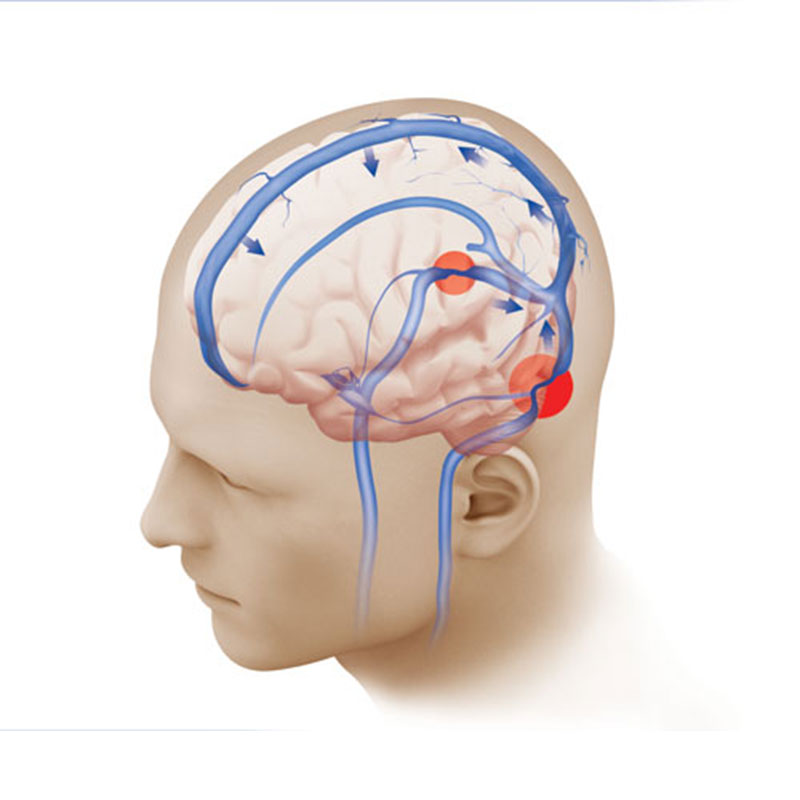
As you know, for normal functioning the brain needs a certain amount of oxygen, glucose and other substances. This is precisely what explains the presence of a developed network of arteries that carry blood to the tissues. Timely drainage of fluid is very important, so it is worth examining the main veins of the head and neck.
Many people are interested in additional information. What are the features of the anatomy of the head and neck? What vessels supply blood from different parts of the brain? In what cases do doctors recommend ultrasound of veins? What complications result from disruption of normal blood flow in the veins? The answers to these questions will be useful to many readers.
Anatomy of the head and neck: brief information
To begin with, it is worth considering general information. Before studying the veins of the head and neck, you can familiarize yourself with the anatomical features.
As you know, the head is located at the top of the spinal column. articulates with the atlas (first cervical vertebra) in the area of the foramen magnum. The spinal cord passes through this hole - the structure of the skeleton ensures the integrity of the central nervous system.
The skeleton of the head and neck consists of the skull, auditory ossicles, and hyoid bone. The skull itself is conventionally divided into parts:
- brain part (consists of the frontal, occipital ethmoid, sphenoid, as well as paired temporal and ;
- facial part (consists of the vomer, lower jaw, as well as paired zygomatic, palatine, maxillary, lacrimal, nasal bones).
The skeleton is covered with muscles that allow flexion, rotation and extension of the neck. Of course, when considering anatomical features, one cannot fail to mention nerves, brain, glands, blood vessels and other structures. By the way, we will look more closely at the veins of the head and neck.
Internal jugular vein
This is a fairly large vessel that collects blood from almost all areas of the neck and head. It begins at the level of the jugular foramen and is a direct continuation of the sigmoid sinus.
Slightly below the source of the vessel there is a small formation with expanded walls - this is the superior bulb of the jugular vein. This vessel runs along the internal carotid artery, and then passes behind (this vessel lies in the same fascial sheath as the carotid artery, the vagus nerve). Slightly above the place where the jugular vein merges with the subclavian vein, there is another extension with two valves - this is the inferior bulb.
Blood from the entire system flows into the sigmoid sinus, in which, in fact, this vessel begins. In turn, the cerebral veins, as well as the labyrinthine vessels and ophthalmic veins carry blood to them.
These are wide vessels with thin walls. They do not have valves. The vessels begin in the area of the spongy substance of the cranial vault and collect blood from the inner surface of the bones. Inside the cranial cavity, these veins communicate with the sinuses of the dura mater and meningeal vessels. Outside the skull, these vessels connect with the veins of the outer integument.
The frontal veins are the largest diploic vessels - they flow into the sagittal sinus. This group also includes the anterior temporal vein, which carries blood to the sphenoparietal sinus. There are also posterior temporal and occipital diploic veins, which drain into emissary vessels.
Features of blood flow through emissary vessels
Emissary veins provide connection between the sinuses and vessels located in the tissues outside the skull. By the way, these vessels pass through small bony valves and exit to the outside of the skull, where they communicate with other vessels.
- The parietal emissary vein, which connects the superior sagittal sinus with the external vessels. In their skulls it comes out through the parietal opening.
- The mastoid emissary vein exits through the opening of the mastoid process. It connects the sigmoid sinus with the occipital vein.
- The condylar vein leaves the skull through the condylar canal (it is part of the occipital bone).
Brief description of the superior and inferior ophthalmic veins
The superior ophthalmic vein is the larger one. It includes vessels into which blood flows from the tissues of the forehead, nose, upper eyelid, membranes and muscles of the eyeball. Approximately at the level of the medial corner of the eye, this vessel communicates with the facial vein through an anastomosis.
Blood from the vessels of the lower eyelid and adjacent eye muscles drains into the inferior vein. This vessel runs along the lower wall of the orbit, almost under the optic nerve, and then flows into the superior ophthalmic vein, which carries blood to the cavernous sinus.
Extracranial tributaries

The internal jugular vein is quite large and collects blood from many vessels.
- Pharyngeal veins, which collect blood from the pharyngeal plexus. This vascular structure collects blood from the tissues of the pharynx, auditory tube, occipital part of the dura mater, and soft palate. By the way, the pharyngeal vessels are small and do not have valves.
- The lingual vein, which is formed by the sublingual, deep and paired dorsal veins of the tongue. These structures collect blood from the tissues of the tongue.
- The thyroid vein (superior), which collects blood from the sternocleidomastoid and superior laryngeal veins.
- The facial vein communicates with the internal jugular at the level of the hyoid bone. This vessel collects blood from almost all tissues of the face. Small vessels drain into it, including the mental, supraorbital, angular, external palatine and deep facial veins. Blood also flows here from paired vessels, including the upper and lower labial, external nasal, as well as the veins of the parotid gland, upper and lower eyelid.
- The mandibular vein is considered a fairly large vessel. It begins in the area of the auricle, passes through the parotid gland, and then flows into the internal jugular vein. This vessel collects blood from the pterygoid plexus, the middle ear vein, as well as the middle, superficial and deep temporal vessels, the vein of the temporomandibular joint, and the anterior ear veins.
Features of blood flow through the external jugular vein

This vessel is formed by the confluence of two tributaries, namely:
- anterior tributary (it forms an anastomosis with the mandibular vein);
- posterior (this tributary collects blood from the occipital and posterior auricular veins).
The external jugular vein forms approximately at the anterior edge of the sternocleidomastoid muscle. From here it follows along the anterior surface of the muscle, pierces the plate of the cervical fascia and flows into the confluence of the internal jugular and subclavian veins. This vessel has two paired valves. By the way, it also collects blood from the suprascapular and transverse veins of the neck.
Anterior jugular vein
When considering the superficial veins of the head and neck, one cannot fail to mention the anterior jugular vein. It is formed from small vessels that collect blood from the tissues of the chin area, follows down the front of the neck, and then penetrates into the space above the sternum.
At this point, the left and right veins are connected through a transverse anastomosis, resulting in the formation of the jugular venous arch. On both sides, the arch flows into the external jugular veins (left and right, respectively).
Subclavian vessel

It is an unpaired vessel that starts from the axillary vein. This vessel passes along the surface of the anterior scalene muscle. It starts at approximately the level of the first rib and ends behind the sternoclavicular joint. It is here that it drains into the internal jugular vein. At the beginning and end of the subclavian vessel there are valves that regulate blood flow.
By the way, this vein does not have constant tributaries. Most often, blood enters it from the dorsal scapular and thoracic venous vessels.
As you can see, the tissues of the neck and head have a very developed venous network, which ensures the timely outflow of venous blood. However, if the functioning of certain organs is disrupted, natural blood flow may be disrupted.
When is an ultrasound examination necessary?

You already know how the veins of the head and neck work. Of course, a violation of the outflow of blood is fraught with stagnation and dangerous complications, which primarily affect the functioning of the central nervous system. If you suspect various circulatory disorders, doctors recommend undergoing examinations. And ultrasound of veins today is one of the simplest, most accessible and informative tests.
In what cases are patients sent for such a procedure? The indications are as follows:
- periodic dizziness;
- frequent fainting;
- headache;
- increased cholesterol levels along with hypertension;
- constant weakness, fatigue;
- diabetes;
- suspicions of the presence of tumors, atherosclerotic plaques, blood clots and other formations that impair vascular patency;
- the procedure is carried out before surgery, as well as during one or another therapy, in order to monitor the effect of treatment.
Of course, in order to make an accurate diagnosis, additional tests and laboratory tests are carried out. It is worth noting that most often stagnation and disturbances in blood outflow are associated with thrombosis and atherosclerosis.
Description of the ultrasound procedure

Duplex scanning technique is used to diagnose various vascular diseases. This ultrasound procedure allows you to check the speed and nature of blood flow in the veins, as well as visualize them and determine the causes of disorders. For example, this procedure makes it possible to diagnose thrombosis, narrowing of a vessel, thinning of its wall, dilation of veins, etc.
The procedure is absolutely painless and lasts about half an hour. During this time, the doctor moves a special sensor over the neck, back of the head, temples and closed eyes, which directs ultrasonic waves, and then captures and records their reflection from moving red blood cells.
The veins of the head and neck perform very important functions, so their condition is worth monitoring. If you have any alarming symptoms, you should consult a specialist and undergo an examination. Diseases diagnosed in the early stages of development are much easier to treat.
(v. facialis communis) see List of anat. terms.
- - Neat brickwork, designed to be perceived in its natural form - without plaster, coating, whitewashing, painting or cladding...
Architectural Dictionary
- - standard brickwork, leaving the main spoons visible...
Architectural Dictionary
-
Large medical dictionary
- - B. in the facial area, regardless of its origin...
Large medical dictionary
- - see List of anat. terms...
Large medical dictionary
- - a paired venous duct in the embryo, formed by the fusion of the anterior and posterior cardinal V., flowing first into the venous sinus of the heart, and later into the atria...
Large medical dictionary
- - see List of anat. terms...
Large medical dictionary
- - see List of anat. terms...
Large medical dictionary
- - see List of anat. terms...
Large medical dictionary
- - see List of anat. terms...
Large medical dictionary
- - see List of anat. terms...
Large medical dictionary
- - see List of anat. terms...
Large medical dictionary
- - see List of anat. terms...
Large medical dictionary
- - see List of anat. terms...
Large medical dictionary
- - see List of anat. terms...
Large medical dictionary
- - adj. ...
Synonym dictionary
"common facial vein" in books
author Betina VladimirThe front and back of penicillinase
From the book Journey to the Land of Microbes author Betina VladimirThe front and back sides of penicillinase We have already met penicillinase in the 19th chapter. This enzyme is a shield of bacteria, reflecting destructive arrows in the form of penicillin molecules. How much trouble she caused to the doctors, who with disappointment had to
PART THREE Vienna, Kevecses, Tunbridge Wells, Vienna (1938–1947)
From the book The Hare with Amber Eyes: Hidden Heritage author Waal Edmund dePART THREE Vienna, Kevecses, Tunbridge Wells, Vienna
a) Front side
From the book The Only Begotten Word. Experience of understanding the ancient Russian faith and history based on language author Moleva Svetlana Vasilievna27. Self – the front side of Consciousness
From the book The Universal Key to Self-Awareness. Adhyatmajnanacha Yogeshwar author Siddharameshwar Maharaj27. The Self is the front side of Consciousness The Self is the front side of consciousness. This is life itself. This is who we are, ourselves. Who is God? The meaning of “I Am That” is this: When I awaken from sleep, what awakens, the body or the Self? God performs all actions like eating and drinking.
Face mesh
From the book Beekeeping for Beginners author Tikhomirov Vadim VitalievichFace mesh There should be several of them in stock. The beekeeper himself and all members of his family must be provided with them. Sometimes it happens that bees, either because of bad weather or because of a bad bribe, do not allow anyone to pass through, and a face mesh is the best way to remain without traces of a sting on
Fas (front side)
From the book Great Soviet Encyclopedia (FA) by the author TSBFas (front side) Fas (from the French face - face), 1) the front side of something. 2) In fortification, the side of a long-term or wood-earth fire structure facing the enemy. F. are also called straight sections of wire fences and anti-tank
Maxillofacial trauma
From the author's bookMaxillofacial trauma Maxillofacial injuries are divided into closed (bruises, hemorrhages, ruptures of muscles, tendons and nerves, closed fractures of the bones of the facial part of the skull, dislocations of the lower jaw) and open (wounds, open fractures). Bruises of the soft tissues of the face
“VIENNA, VIENNA – ONLY YOU ONE...”
From the book Vienna. Guide author Striegler Evelyn“VIENNA, VIENNA – ONLY YOU ONE...” “... you will be the city of my dreams!” Vienna is amazingly diverse. The former residence of the emperor is also a modern arts center. A city that invites you to shop during the day, and in the evenings the eternal sounds of
Facial neuralgia
From the book Homeopathic Handbook authorFacial neuralgia Pain of a convulsive nature - Colocint. Left-sided facial neuralgia: pain increases or decreases with the rise and fall of the sun; lacrimation from the eye on one side - Spigelia. Severe facial neuralgia, usually right-sided -
Facial neuralgia
From the book Life without a cold author Nikitin Sergey AlexandrovichFacial neuralgia Facial neuralgia is most often caused in sensitive people due to colds of the facial skin and rheumatic inflammation of the soft parts of the face, which spreads to the nerve. Sometimes it is associated with dental diseases and aches. Only felt in
Chapter 1 FACIAL DIAGNOSTICS
From the book Mirror of the Soul and Health. Facial diagnostics and reflexology by Chen LiChapter 1 FACIAL DIAGNOSTICS
Front part of the head
From the book Rottweilers author Sukhinina Natalya MikhailovnaFacial part of the head The front part of the head includes the eyes, ears, and neck. All of them must strictly comply with the specified standards, especially if the dog is planned to be prepared for participation in various exhibitions. It is also important that the dog does not have any flaws if
2. REVERSE AND FRONT SIDE
From the book Book 10. Fruits of Wisdom (old edition) author Laitman Michael2. BACK AND FRONT SIDES A person is designed in such a way that he always looks forward, at his future growth, advancement, his path seems to him to be an ascent “from the bottom up”, he perceives each of his future states as greater, better than the present, today. If he feels
Practice "Front side"
From the book Walking Through the Fields, or Moving Your Legs Alternately author Krass Natalya AlexandrovnaPractice “Face” 1. This practice is a facial massage to remove blocks. Performed sitting on a chair or on the floor “Turkish style”.2. Place the pads of the fingers (except the thumbs) of both hands parallel to each other in the middle of the forehead. Slow and smooth
In this article we will look at the topography of blood vessels and nerves in relation to the facial muscles, but we will go from deep layers to superficial ones.
Rice. 1-41. The external carotid artery passes anterior to the auricle and continues into the superficial temporal artery, which divides into parietal and anterior branches. Also, the maxillary and facial branches depart from the external carotid artery, most of which are not visible when viewed from the front. departs from the external carotid and, bending over the edge of the lower jaw, goes to the corner of the mouth, where it gives off branches to the upper and lower lips, and itself goes up and inward to the inner corner of the palpebral fissure. The portion of the facial artery that runs lateral to the external nose is called the angular artery. At the inner canthus, the angular artery anastomoses with the dorsal nasal artery, which arises from the supratrochlear artery, which, in turn, is a branch of the ophthalmic artery (from the internal carotid artery system). The main trunk of the supratrochlear artery ascends to the middle of the forehead. The area of the superciliary arches is supplied with blood by the supraorbital artery, which emerges from the supraorbital foramen. The infraorbital region is supplied with blood by the infraorbital artery, emerging from the foramen of the same name. The mental artery, which arises from the inferior alveolar artery and emerges from the mental foramen, supplies the soft tissues of the chin and lower lip.

Rice. 1-42. The veins of the forehead form a dense, variable network and usually merge anteriorly into the supratrochlear vein, also called the frontal vein. This vein runs in the midface medially from the orbit to the edge of the mandible and ultimately connects with the internal jugular vein. The name of this vein varies depending on the anatomical region. On the forehead it is called the frontal vein. In the region of the glabella, it connects with the supraorbital vein, and inward from the orbit - with the superior orbital vein, thus providing outflow from the veins of the orbit and cavernous sinus. Near the bony part of the external nose, it connects with the veins of the upper and lower eyelids (venous arch of the upper and lower eyelids) and is called the angular vein. On its way along the external nose, it collects blood from the small veins of the nose and cheeks, and also anastomoses with the infraorbital vein, which emerges from the infraorbital foramen. In addition, blood from the zygomatic region enters this vein through the deep vein of the face. On the cheek, the main vein connects with the superior and inferior labial veins and is called the facial vein. Connecting with the veins of the chin, the facial vein bends over the edge of the lower jaw and flows into the internal jugular vein on the neck. The veins of the parietal region unite into the superficial temporal vein, which, in turn, flows into the external jugular vein.

Rice. 1-43. The face is innervated by fibers of the trigeminal (mainly sensory fibers; motor fibers innervate the masticatory muscles) and facial nerves (motor fibers). In addition, the large auricular nerve, which belongs to the spinal nerves, takes part in the sensitive innervation of the face.
The trigeminal nerve (5th pair of cranial nerves, CN V) has three branches: the ophthalmic (CN V1), maxillary (CN V2) and mandibular (CN V3) nerves.
The optic nerve is divided into the frontal, lacrimal and nasociliary nerves. The frontal nerve runs in the orbit above the eyeball and divides into the supratrochlear and supraorbital nerves. The supraorbital nerve has two branches, the larger of which, the lateral one, exits the orbit onto the face through the supraorbital foramen or supraorbital notch and innervates the skin of the forehead to the vertex, as well as the conjunctiva of the upper eyelid and the mucous membrane of the frontal sinus. The medial branch of the supraorbital nerve emerges from the orbit medially through the frontal notch and branches in the skin of the forehead.
Another branch of the frontal nerve, the supratrochlear nerve, emerges from the inner canthus and innervates the skin of the nose and conjunctiva.
The outer canthus is innervated by the lacrimal nerve. It separates from the optic nerve in the orbital cavity and before exiting it gives off branches to the lacrimal gland. The nasociliary nerve, a branch of the ophthalmic nerve, gives off the anterior ethmoidal nerve, the terminal branch of which, the external nasal nerve, in turn passes through the cells of the ethmoidal labyrinth.
Through the infraorbital foramen, the infraorbital nerve, a large branch of the maxillary nerve (CN U2), enters the face. Its other branch, the zygomatic nerve, passes laterally in the orbit and enters the zygomatic region through separate canals in the zygomatic bone. The zygomaticotemporal branch of the zygomatic nerve innervates the skin of the temple and forehead. The zygomaticofacial branch of the zygomatic nerve exits through the zygomaticofacial foramen (sometimes there may be several holes) and branches in the skin of the cheekbone and lateral canthus.
The auriculotemporal nerve, a branch of the mandibular nerve, runs under the foramen ovale. Having passed along the inner surface of the branch of the lower jaw, it bends around it from behind, innervates the skin in the area of the condylar process and the external auditory canal, pierces the parotid salivary gland and ends in the skin of the temple. The teeth of the upper jaw are innervated by the maxillary nerve. The teeth of the mandible are innervated by the inferior alveolar nerve, which arises from the mandibular nerve (CN, V3) and enters the mandibular canal through the mandibular foramen. The branch of the mandibular nerve emerging from the mental foramen is called the mental nerve; it provides sensitive innervation to the skin of the chin and lower lip.
Facial muscles are innervated by the facial nerve(CN V2). It emerges from the stylomastoid foramen and gives off numerous branches to the facial muscles. The branches of the facial nerve include the temporal branches, going to the temporal region and innervating the muscles of the forehead, temple and eyelids; zygomatic branches innervating the zygomatic muscles and muscles of the lower eyelid; buccal branches to the muscles of the cheeks, the muscles surrounding the oral cavity, and the muscle fibers around the nostrils; the marginal mandibular branch, innervating the muscles of the chin, and the cervical branch to the platysma.

Rice. 1-44. General view of the arteries, veins and nerves of the face.

Rice. 1-45. Deep arteries, veins (right) and nerves of the face (left).
Rice. 1-45. The vessels and nerves of the face passing through the bone canals and openings are located close to each other. On the right side of the face, deep arteries and veins and their exit points onto the face are shown. The branches of the ophthalmic artery from the internal carotid artery system pass through the orbital septum in one or several places - the supratrochlear artery and the medial arteries of the eyelids (pass through the upper edge of the septum). Veins of the face also pass through the orbital septum, forming the superior orbital vein.
The supraorbital artery and vein pass through the supraorbital foramen. Sometimes this opening may not be closed and is called the supraorbital notch, by analogy with the medially located supratrochlear notch, through which the supratrochlear artery and vein pass. Even more medial are the branches of the dorsal nasal artery and the superior branches of the ophthalmic artery, connecting with the arterial arch of the upper eyelid. Venous drainage occurs in the superior ophthalmic vein.
The lateral and medial arteries of the eyelids extend from the ophthalmic artery to the lower eyelid, forming the arterial arch of the lower eyelid and giving off branches to the dorsum of the nose. All arterial branches are accompanied by veins of the same name. The infraorbital artery and vein pass through the infraorbital foramen. They branch in the tissues of the lower eyelid, cheek and upper lip and have many anastomoses with the angular artery and vein.
The zygomaticofacial vessels exit into the face through the zygomaticofacial foramen.
Through the mental foramen, which opens the canal of the lower jaw, the mental branches of the mandibular artery and nerve pass. Through the same opening, the mental branch of the inferior alveolar vein enters the mandibular canal. In the figure, the facial artery and vein at the edge of the lower jaw are crossed. The transverse facial artery is shown at the lower edge of the zygomatic arch. The superficial temporal artery and vein are divided at the entrance to the temporal fossa.
The left side of the face also shows the exit points of the nerves. The supraorbital nerve passes through the supraorbital foramen, arising from the ophthalmic nerve (the first branch of the trigeminal nerve CN V1), which provides sensory innervation to the supraorbital region. Inside the orbit, the supratrochlear nerve arises from the optic nerve, which, passing through the opening in the orbital septum (septum), is divided into medial, lateral and palpebral branches. Through the infraorbital canal, which opens with the infraorbital foramen, passes the infraorbital nerve, a branch of the maxillary nerve (second branch of the trigeminal nerve, CN V2). It provides sensory innervation to the lower lip, cheek and part of the nose and upper lip.
Thus, the lower eyelid is innervated by two nerves: the palpebral branch of the infratrochlear nerve (from the ophthalmic nerve) and the inferior palpebral branches of the infraorbital nerve (from the maxillary nerve).
The zygomaticofacial nerve enters the face from the foramen of the same name and provides sensitive innervation to the zygomatic region. The mental nerve exits the mandibular canal through the mental foramen and carries sensory fibers to the mental region and lower lip. To avoid loss or impairment of sensitivity in the lower lip due to damage to this nerve when performing complicated wisdom tooth extraction and osteotomy of the mandibular branch, it is necessary to have a good knowledge of its topography in the mandibular canal.
Rice. 1-46. Individual branches of the supratrochlear and supraorbital arteries and veins run very close to the bone and are covered with fibers of the corrugator muscle. Other branches run in a cranial direction above the muscle. The lateral and medial branches of the supraorbital and supratrochlear nerves pass under, above, and through the fibers of the corrugator brow muscle. The motor innervation of this muscle is provided by the anterior temporal branches of the facial nerve (CN VII).
The temporalis muscle is supplied by the deep temporal arteries and veins. The sensory innervation of this area is carried out by the deep temporal nerve (from CN V3). The muscle receives motor innervation from the temporal branches of the facial nerve.
The superficial temporal artery and vein, together with the temporal branches (from the facial nerve) go above the zygomatic arch and are crossed in this figure.
The vessels and nerves emerging from the infraorbital foramen (artery, vein and infraorbital nerve) supply the area around it and also branch into the tissues of the lower eyelid (branches of the lower eyelid), the muscles of the nose and the upper lip.
The facial artery and vein bend over the edge of the lower jaw anterior to. More medially, they cross the buccal muscle and branch in an arcuate manner in an oblique direction, located superficially to the branches of the infraorbital artery and vein. At the intersection of the branch of the lower jaw, the pulsation of the artery is palpated.
The buccal muscle is innervated by the buccal branches of the facial nerve.
The neurovascular bundle of the mandibular canal exits onto the face through the mental foramen. The mental artery, the mental branch of the inferior alveolar vein and the nerve of the same name branch in the soft tissues of the lower lip and chin. The motor innervation of the adjacent muscles is carried out by the marginal branches of the mandible, arising from the facial nerve (CN V2).

Rice. 1-47. Topography of arteries and veins (right half) and facial nerves (left half) in relation to facial muscles.
Rice. 1-47. The branches of the supratrochlear and supraorbital arteries and veins pass through the frontal belly of the occipitofrontal muscle. The lateral and medial branches of the supratrochlear and supraorbital nerves pass through and above the muscle. The motor innervation of this muscle is carried out by the anterior temporal branches of the facial nerve.
The dorsum of the nose is innervated by the external nasal branches arising from the anterior ethmoidal nerve. This nerve passes between the nasal bone and the lateral nasal cartilage and runs along the surface of the cartilage. The branches of the infraorbital nerve (external nasal branches) branch in the wings of the nose. The motor innervation of the muscles is carried out by the zygomatic branches of the facial nerve (CN V2).

Rice. 1-48. Topography of arteries and veins (right half) and facial nerves (left half) in relation to facial muscles.
Rice. 1-48. Additional venous drainage from the forehead occurs through accessory branches of the supratrochlear nerve.
The orbicularis oculi muscle, covering the orbital septum (septa), is supplied by thin branches of the medial and lateral eyelid arteries, and venous outflow is carried out through the venous arches of the upper and lower eyelids. The lateral artery of the eyelids arises from the lacrimal artery, and the medial artery arises from the ophthalmic artery. Both of these arteries belong to the internal carotid artery system. Venous blood from the upper and lower eyelids flows into the veins of the same name, which flow medially into the angular vein, and laterally into the superior ophthalmic vein (upper eyelid) and inferior ophthalmic vein (lower eyelid).
The lateral and medial branches of the supratrochlear nerve pass through the proud muscle and the depressor brow muscle, which are located in the glabella and supraorbital region. The muscle receives motor innervation from the temporal branches of the facial nerve (CN, V2).
The nasal muscles are supplied by branches of the angular artery. Somewhat more cranially from the angular artery its terminal branch, the dorsal nasal artery, departs. Venous blood flows through the external nasal veins, which drain into the angular vein. Also, part of the venous blood flows into the infraorbital vein. Sensitive innervation is carried out by the branches of the external nasal nerve, extending from the ethmoidal nerve (a branch of the frontal nerve), motor innervation of the adjacent muscles is carried out by the zygomatic branches of the facial nerve.
The levator anguli oris muscle, covering the superior and lateral parts of the orbicularis oris muscle, is supplied by the facial artery and vein, and innervated by the superior labial branches, which arise from the infraorbital nerve, which runs along the surface of this muscle.
The mental foramen is closed by the muscle that depresses the lower lip.

Rice. 1-49. Topography of arteries and veins (right half) and facial nerves (left half) in relation to facial muscles.
Rice. 1-49. Venous drainage from the superficial epifascial layers of the forehead and parietal region occurs through the parietal branches of the superficial temporal vein. Here it also anastomoses with the supratrochlear vein. The main artery of this area is the superficial temporal. At the inner corner of the palpebral fissure, the angular vein connects with the supratrochlear vein. Thus, the superficial veins of the face connect with the superior ophthalmic vein, which opens into the cavernous sinus. It is also possible to connect with the subtrochlear vein, which is also called the nasofrontal vein. The external nasal vein collects blood from the dorsum of the nose and opens into the angular vein.
The angular vein accompanies the angular artery, which lies more medially. When it reaches the levator labii superioris muscle, the vein passes above it and the artery below it.
Blood from the upper lip flows into the superior labial vein, which, in turn, connects to the facial vein. The infraorbital vein enters the infraorbital foramen, closed by the levator labii superioris muscle. Its branches connect with the branches of the angular vein and thus connect the superficial veins of the face with the pterygoid venous plexus. Blood from the lower lip flows into the facial vein through the inferior labial vein. The arterial blood supply to the upper lip is provided by the superior labial artery, and the lower lip by the inferior labial artery. Both of these vessels arise from the facial artery. The inferolateral part of the chin is closed by the depressor anguli oris muscle, which receives motor innervation from the marginal mandibular branch of the facial nerve. Sensory innervation of this area is carried out by branches of the mental nerve, which arise from the inferior alveolar nerve.

Rice. 1-50. Topography of arteries and veins (right half) and facial nerves (left half) in relation to facial muscles.
Rice. 1-50. In the forehead area, the supratrochlear vein also forms anastomoses with the anterior branches of the superior temporal vein.
The angular artery and vein run in a long groove between the levator labii superioris and alae nasi muscle and the orbicularis oculi muscle and are partially covered by the medial edge of the latter. The facial vein passes under the levator labii superioris muscle, and the artery runs above it. Both of these vessels pass under the zygomaticus minor, with the exception of individual arterial branches that may run along the surface of the muscle and then pass under the zygomaticus major. The topography of neurovascular formations in this area is very variable.
Next, the artery and vein are located in the space between the masticatory muscle and the depressor angle oris muscle, and cross the lower edge of the lower jaw.

Rice. 1-51. Topography of arteries and veins (right half) and facial nerves (left half) in relation to facial muscles.
Rice. 1-51. Most of the masseter muscle is covered by the parotid salivary gland. The gland itself is partially covered by the laughter muscle and platysma. All the arteries, veins and nerves of the area pass through these muscles.

Rice. 1-52. Topography of arteries and veins (right half) and nerves of the face (left half) in the subcutaneous fat layer.
Rice. 1-52. The muscles and superficial fascia of the face are covered with a subcutaneous fat layer of varying thickness, through which blood vessels are visible in some places. Small arteries, veins and nerve endings go through the layer of fat to the skin.

Rice. 1-76. Arteries of the face, side view.
Rice. 1-76. The external carotid artery runs anterior to the auricle and gives off the superficial temporal artery, which branches into parietal and anterior branches. Also from the external carotid artery branches extend to the face and upper jaw: the posterior auricular artery departs under the auricle, even lower is the occipital artery, at the level of the lobe is the maxillary artery, which goes medially under the branch of the mandible, at the level between the lobe and the external auditory canal – transverse artery of the neck, which runs along the branch of the mandible. The facial artery bends over the lower edge of the lower jaw and goes to the corner of the mouth.
The main artery of the face is the maxillary artery, which gives off many large branches, which will be described below.
The inferior and superior labial arteries extend from the facial artery to the corner of the mouth. The terminal branch of the facial artery leading to the external nose is called the angular artery. Here, at the medial canthus, it anastomoses with the dorsal nasal artery, which arises from the ophthalmic artery (from the internal carotid artery system). In the upper part of the face, the supratrochlear artery runs to the middle of the frontal region. The supraorbital and infraorbital regions are supplied with blood, respectively, by the supraorbital and infraorbital arteries, emerging through the foramina of the same name. The mental artery, a branch of the inferior alveolar artery, enters the face through the opening of the same name and supplies the soft tissues of the chin and lower lip.
Facial vessels The main source of blood supply to the face is the external carotid artery. From the neck area, the facial artery comes to the face, which is projected onto the skin from the middle of the body of the lower jaw to the inner corner of the eye. Gives large branches: the arteries of the upper and lower lips and the final branch - the angular artery, anastomoses with the orbital artery through the nasal arteries.
The second large artery - the maxillary artery (a. xillaris) - departs from the external carotid artery in the thickness of the parotid gland at the level of the neck of the articular process of the lower jaw, goes into the deep region of the face, lies on the outer surface of the external pterygoid muscle and lies first in the temporo- pterygoid tissue space, then - in the interpterygoid space.
A. maxillaris is the largest branch of the external carotid artery; it gives off 19-20 branches and supplies blood to the entire deep area of the face with the masticatory muscles and the dentofacial apparatus. The artery is inaccessible for ligation, therefore, if necessary, they resort to ligation of the external carotid artery in the neck in the carotid triangle. In the deep area of the face near the artery, it is customary to distinguish three sections:
1) Mandibular (pars mandibularis) - behind the neck of the articular process. The largest branch is the inferior alveolar artery (a. alveolaris inferior);
2) Pterygoid (pars pterygoidea) - between the temporal muscle and the external pterygoid. Branches:
a) middle meningeal artery (a. meningea media);
b) deep temporal artery;
c) masticatory artery;
d) superior alveolar artery;
e) buccal arteries;
e) pterygoid arteries.
3) Pterygopalatine (pars pterygopalatine) - in the pterygopalatine fossa. Branches: infraorbital, pharyngeal, palatine, etc.
The venous system of the face is divided into two layers. The first layer of veins is formed by the facial vein system, v. facialis, the origins of which are the angular vein, supraorbital, external nasal, tube veins, nose, as well as the retromandibular vein, v. retromandibularis, located in the thickness of the parotid gland. In the region of the root of the nose, the facial vein has wide anastomoses with the superior orbital veins and through them with the vein-sinuses of the dura mater. Infection can enter the sinus veins due to carbuncles and boils of the upper lip, nose, with the development of thrombophlebitis (sinus thrombosis) and inflammation of the membranes of the brain.
The deep venous network of the face is represented by the pterygoid venous plexus (plexus pterygoideus). It drains into the postmaxillary vein. Thus, both systems are interconnected. It should be noted that the pterygoid plexus, located in the intermaxillary space, is connected with the vein-sinuses of the dura mater. The postmaxillary and facial veins merge posteriorly from the angle of the mandible into the common vein of the face, which flows into the internal jugular vein.
Nerves of the face. The facial innervation is carried out by the facial, trigeminal, glossopharyngeal nerves, and cervical plexus.
The facial nerve (7th pair of cranial nerves) primarily supplies motor innervation to the facial muscles. The nerve leaves the pyramid of the temporal bone through the stylomastoid foramen and forms the posterior auricular nerve 1 cm below.
The main trunk of the facial nerve enters the thickness of the gland and here it is divided into superior and inferior branches, from which five groups of branches arise. The branches extend radially from a point 1 cm downward from the auditory canal. Inflammatory processes in the gland can cause paralysis and paresis of the facial nerve. Incisions on the face are made only taking into account the course of the branches of the facial nerve. The nerve lies relatively shallow, and there is a high risk of damage to its branches, which also leads to paralysis of the facial nerve or its individual branches.
The trigeminal nerve (5th pair of cranial nerves) is mixed (sensory-motor) in structure and function. After leaving the brain stem, the nerve forms the semilunar gasserian ganglion. The node is located on the anterior surface at the apex of the pyramid of the temporal bone and lies in the cavity formed by the dura mater. Three main branches of the trigeminal nerve depart from the anterior edge of the ganglion: I) orbital; 2) maxillary; 3) mandibular.
According to its topographic and anatomical structure, the trigeminal nerve is one of the most complex. Its branches pass through hard-to-reach anatomical areas and enter into complex relationships with blood vessels. At the same time, since the nerve carries sensitive pain innervation for the dentofacial apparatus, anesthesia of the branches of the nerve is necessary during operations on the face. Therefore, we will consider the places where the large branches of the nerve exit to the face.
It should immediately be noted that the skin of the face receives pain innervation from the trigeminal nerve.
The first branch innervates the skin of the frontal and orbital areas.
The second branch of the trigeminal nerve provides pain innervation to the infraorbital region, nose, upper lip, teeth, and upper jaw. It leaves the skull through a round hole in the pterygopalatine fossa and gives off the main branches. The infraorbital nerve exits through the inferior orbital fissure, enters the orbit, lies in the infraorbital groove and exits through the infraorbital foramen. It is located 0.5 cm below the middle of the edge of the orbit, forms a “crow’s foot”, from which the labial and nasal branches extend to the lower eyelid. Along the way, the nerve gives off the superior posterior, middle and anterior alveolar nerves; they enter the upper jaw in the area of the tubercle. These nerves connect in the canaliculi of the alveolar process of the upper jaw and form the superior dental plexus.
In addition, in the pterygopalatine fossa, the pterygopalatine branches and branches of the maxillary nerve (n. petrosus major and n. facialis) form the autonomic pterygopalatine ganglion, from which the palatine nerves depart: large (exits through the greater palatine foramen), middle and posterior (enters through the lesser palatine hole), innervating the gum, soft and hard palate.
The posterior nasal nerves, a large branch of which, the nasopalatine nerve, exits through the incisive foramen and innervates the anterior part of the palate.
The mandibular nerve exits through the foramen ovale. The mixed nerve carries motor innervation for the masticatory muscles: temporal, masseter, and pterygoid muscles. Its largest branches are: buccal, auriculotemporal, inferior alveolar and lingual nerves. The inferior alveolar nerve runs down the inner surface of the external pterygoid muscle, then between the pterygoid muscles it enters the mandibular foramen and exits into the mandibular canal along with the artery. Provides pain innervation to the teeth of the lower jaw, its final branch is the n. mentales (mental). This nerve exits through the mental foramen. The lingual nerve goes to the tongue from below.
The mental nerve innervates the skin of the lower lip, the gums in the area of the canines and premolars, and the skin of the chin. The mental foramen is located midway between the lower edge of the jaw and the alveolar process.
Projection anatomy of vessels and nerves of the facial part of the head:
1. The facial artery (a. facialis) is projected from the intersection of the anterior edge of the masticatory muscle with the lower edge of the lower jaw in an ascending direction to the inner corner of the eye.
2. The mandibular foramen (foramen mandibulare) is projected from the side of the oral cavity onto the buccal mucosa in the middle of the distance between the anterior and posterior edges of the mandibular branch, 2.5-3 cm upward from its lower edge.
3. The infraorbital foramen (foramen infraorbitalis) is projected 0.5-0.8 cm downward from the middle of the lower orbital margin.
4. The mental foramen (foramen mentalis) is projected in the middle of the height of the body of the lower jaw between the first and second small molars.
5. The trunk of the facial nerve (truncus n.facialls) corresponds to a horizontal line drawn through the base of the earlobe.
Incisions for purulent parotitis
Indications. Cellulitis and abscess of the parotid gland.
Technique. The patient is placed on his back, his head is turned to the side. Three radial incisions 5-6 cm long are made. The incisions begin at the tragus of the ear: the upper one - along the lower edge of the zygomatic arch, the middle one - in the direction of the corner of the mouth, reaching the anterior edge of the masticatory muscle (m. masseter), the lower one - in the direction from the middle of the distance between the angle of the lower jaw and the chin, also reaching to the anterior edge of m. masseter
The direction of the cuts coincides with the course of the branches of the facial nerve (Fig. 83).
The skin with subcutaneous fatty tissue is dissected. Use hooks to widen the wound. The parotid-masticatory fascia is dissected using a scalpel along a grooved probe. Then the capsule and the superficial layer of the substance of the parotid salivary gland are dissected. The main danger with incisions is damage to the branches of the facial nerve that penetrate radially through the thickness of the parotid salivary gland.
Nerve branches cannot be crossed. It should be borne in mind that the stenon duct is projected along the line connecting the lower edge of the external auditory canal with the corner of the mouth or the wing of the nose; within these limits, the incision should be made with extreme caution to avoid injury to the excretory duct of the parotid salivary gland. Gauze strips (tampons) are inserted into the incisions.
When the abscesses are localized in the deep parts of the gland (mandibular fossa), an incision is made according to Voino-Yasenetsky. A 3 cm long incision is made with the head thrown back, from the earlobe downwards between the posterior edge of the ascending ramus of the mandible and the anterior edge of the sternocleidomastial muscle. The incision should be 1-1.5 cm behind the edge of the mandible so as not to damage the inferior branch of the facial nerve, which remains in front of it.
The edges of the wound are stretched with sharp hooks and a blunt instrument (forceps), passing to a depth of 2.5 cm towards the styloid process and the posterior wall of the pharynx, penetrating through the tissue of the parotid gland (see Fig. 83).
Test tasks (choose the correct answer)
1. The transverse sinus corresponds to the anatomical formation of the bones of the skull:
1) external occipital protuberance;
2) mastoid process;
3) upper nuchal line;
4) lower nuchal line.
2. The arteries of the soft tissues of the head have the following directions:
1) axial;
Topographic anatomy of the neck. Neck fascia and fibrous spaces. Neurovascular bundles of the neck. Neck organs
Boundaries and external landmarks. The upper border of the neck area is drawn along the edge of the base of the lower jaw, through the apexes of the mastoid processes and posteriorly along the upper nuchal line. The lower border is drawn along the jugular notch of the sternum, along the upper edges of the clavicles, through the humeral processes of the scapula (acromion) to the spinous process of the 7th cervical vertebra.
To facilitate orientation in the complex topography of the neck area, and above all in the numerous vessels and nerves, various external landmarks are used, which can be divided into five groups: bone, cartilage, muscle, vascular and skin folds. Landmarks allow you to divide the neck into sections and areas, and also help plan surgical approaches to the neck.
The midline divides the neck into right and left halves. The frontal plane, drawn through the transverse processes of the cervical vertebrae, divides the neck into anterior, visceral and posterior muscular (neck) sections. The transverse plane, drawn through the hyoid bone, divides the anterior part of the neck into the suprahyoid and infrahyoid regions.
The muscles of the anterior neck form a special coordinate system in the form of triangles (Fig. 84).
The boundaries of the triangles are drawn along the contours of large muscles. The sternocleidomastoid (sternocleidomastoid) muscle divides each half of the anterior neck into an inner and outer (lateral) triangle. Within the internal triangle, the submandibular triangle is distinguished, bounded by the bellies of the digastric muscle. The unpaired mental triangle is distinguished between the anterior bellies of the digastric muscle. In addition, the internal triangle contains the carotid and scapulotracheal triangles. In the outer triangle, the scapular-trapezoid and scapular-clavicular triangles are distinguished. Triangles help navigate the complex anatomy of the neck. Each triangle is distinguished by its unique layer-by-layer anatomy and arrangement of neurovascular elements.
Layers. In the layer-by-layer anatomy of the neck area, the issue of fascia and cellular spaces as anatomical elements that determine the course of purulent-inflammatory processes should be highlighted.
The fascia of the neck is an anatomical element that makes the neck a single whole. The most widespread and practically acceptable is the classification of neck fascia according to V.N. Shevkunenko (Fig. 85), according to which five fascia are distinguished on the neck (Table 12). Between the sheets of fascia there are fatty tissue and lymphoid tissue, so the fascia determines the location of phlegmon in the neck (mainly adenophlegmon) and the direction of purulent leaks.
Neurovascular bundles of the neck. There are two large neurovascular bundles in the neck: main and subclavian.
The main neurovascular bundle of the neck consists of the common carotid artery, internal jugular vein, and vagus nerve. It is located in the neck in the area of the sternocleidomastoid (sternocleidomastoid) muscle and the carotid triangle. Thus, the main socistonervous bundle along the carotid artery has two sections: 1st section in the area of the sternocleidomastoid muscle, 2nd section in the carotid triangle. In the area of the sternocleidomastoid muscle, the neurovascular bundle lies quite deep, covered by the muscle, the 2nd and 3rd fascia. The sheath of the bundle is formed by the parietal leaf of the 4th fascia and, in accordance with Pirogov’s laws, has a prismatic shape, with spurs the sheath is fixed to the transverse processes of the cervical vertebrae.
The relative position of the elements of the neurovascular bundle here is as follows: a vein lies in front and outward of the artery, the vagus nerve is located between the vein and artery and posteriorly.
Above, the main neurovascular bundle is located in the carotid triangle (Fig. 86), which is bounded above by the posterior leg of the digastric muscle, in front by the upper belly of the omohyoid muscle, and behind by the anterior edge of the sternocleidomastoid muscle. The neurovascular bundle is not covered by muscle and third fascia. With the head thrown back, the pulsation of the carotid artery is clearly visible on the neck, and upon palpation the pulse can be seen here.
determine even with a significant decrease in blood pressure. The relative position of the elements of the neurovascular bundle remains the same, the venous elements lie more superficially, and the common facial vein flows into the internal jugular vein here. The common carotid artery in the carotid triangle at the level of the upper edge of the thyroid cartilage (according to Pirogov) is divided into internal and external branches. It is practically important to know their differences. An anatomically reliable sign of the external carotid artery is the presence of lateral branches in the carotid triangle, of which the superior thyroid, lingual and facial arteries are constant. Ligation of the external carotid artery to stop bleeding throughout injuries to the maxillofacial area is performed immediately after the separation of the superior thyroid artery. The internal carotid artery in the neck does not give branches. The internal carotid artery is usually divided into three sections:
1) from the bifurcation of the common carotid artery to the hypoglossal nerve;
2) from the hypoglossal nerve to the entry into the carotid artery canal and 3) intracranial. To perform surgical interventions, the internal carotid artery is accessible only in the first section.
An anatomical feature of the carotid triangle is the presence of large nerve trunks. As part of the main neurovascular bundle, the vagus nerve (10th pair of cranial nerves) runs here. Forming an arch, the external carotid artery is crossed by the hypoglossal nerve (12th pair of cranial nerves), here it gives off a descending branch lying on the anterior surface
the common carotid artery, which then anastomoses with the cervical plexus (cervical loop). In the bifurcation of the common carotid artery lies the carotid glomerulus, the so-called intercarotid paraganglia, the receptor body (glomus caroticus). Behind the internal carotid artery lies the superior node of the sympathetic trunk. The location in a narrow space of large vessels, cranial nerves, receptor formations, and the sympathetic trunk makes it possible to distinguish the carotid triangle as a reflexogenic zone of the neck.
Sympathetic trunk. The cervical sympathetic trunk has 3-4 nodes. The upper node is located at the level of the 2nd and 3rd cervical vertebrae, lies on the 5th fascia and the longus colli muscle. The middle node is unstable, it is located at the intersection of the common carotid and inferior thyroid arteries, at the level of the 6th cervical vertebra, and lies in the thickness of the 5th fascia. The intermediate node lies on the surface of the vertebral artery before entering the transverse processes, at the level of the upper edge of the 7th cervical vertebra. The inferior, or stellate, node is located behind the subclavian artery, at the level of the lower edge of the 7th cervical vertebra.
The close proximity of the main neurovascular bundle to the sympathetic trunk and the presence of anastomoses with the vagus nerve explains the effect of vagosympathetic blockade according to Vishnevsky. In some cases, vagosympathetic blockade can cause acute reflex cardiac arrest, which is associated with the departure from the superior sympathetic ganglion of the upper cervical cardiac nerve, and from the vagus nerve - the depressor nerve to the heart, the so-called Zion nerve.
The subclavian neurovascular bundle is formed by the subclavian artery, subclavian vein and brachial plexus. Along the course of the subclavian artery and according to its relationship with the anterior scalene muscle, three sections are distinguished. The subclavian neurovascular bundle is located in the internal and external triangles of the neck. In the internal triangle of the neck, elements of the subclavian neurovascular bundle occupy the deep intermuscular spaces of the neck.
Deep intermuscular spaces of the neck. On the neck in the inner triangle in the deep layers of the sternocleidomastial region, the following deep intermuscular spaces are distinguished: I) prescalene fissure; 2) scalene-vertebral triangle; 3) interscalene gap.
The first intermuscular space - the prescalene fissure (spatium antescalenum) is limited from the front and outside by the sternocleidomastoid muscle, from the back - by the anterior scalene muscle, from the inside - by the sternohyoid and sternothyroid muscles. In the spatium antescalenum there is the lower section of the main neurovascular bundle (a. carotis communis, v. jugularis interna, n. vagus), the phrenic nerve and the venous angle of Pirogov - the confluence of the internal jugular vein and the subclavian vein. On the surface of the body, the venous angle is projected onto the sternoclavicular joint. All large veins of the lower half of the neck (external jugular, vertebral, etc.) flow into the venous angle. The thoracic lymphatic duct flows into the left venous angle. The right lymphatic duct flows into the right venous angle. The thoracic lymphatic duct (HLD) is an unpaired formation. It is formed in the retroperitoneal space at the level of the 2nd lumbar vertebra. Two variants of the final section of the GLP at the point of its confluence with the venous angle are described: scattered and main.
The terminal section of the subclavian vein is located in the prescalene fissure. The vein crosses the clavicle at the border of the inner and middle third of the clavicle and lies on the first rib. The subclavian vein starts from the lower border of the first rib and is a continuation of the axillary vein. The topography of the right and left subclavian veins is almost the same. The subclavian vein can be divided into two sections: behind the clavicle and at the exit from under the clavicle in the trigonum clavipectorale. The subclavian vein passes between the anterosuperior surface of the first rib and the posterior surface of the clavicle. The length of the subclavian vein is 3-4 cm, diameter 1-1.5 cm or more. The subclavian vein lies anterior to the anterior scalene muscle. The vein is distinguished by its constant location, its walls are fixed in the space between the first rib and the collarbone, the periosteum of these formations and the spurs of the fifth fascia. In this regard, the subclavian vein does not spasm, its walls never collapse. This makes it possible to perform puncture and catheterization of the subclavian vein during severe hypovolemia (shock, massive blood loss). The high volumetric velocity of blood flow in the subclavian vein prevents the formation of blood clots and fibrin deposition on the catheter. At the lower edge of the middle third of the clavicle there are subclavian ar
teria and vein are separated by the anterior scalene muscle. The artery is removed from the vein, which avoids mistakenly hitting the artery instead of the vein. At the same time, the artery separates the vein from the trunks of the brachial plexus. Above the clavicle, the vein is located closer to the dome of the pleura; below the clavicle, it is separated from the pleura by the first rib.
Immediately behind the sternoclavicular joint, the subclavian vein connects with the internal jugular vein, the brachiocephalic veins are formed on the right and left, which enter the mediastinum and, having united, form the superior vena cava. Thus, along the entire front, the subclavian vein is covered by the clavicle. The subclavian vein reaches its highest point at the level of the middle of the clavicle, where it rises to its upper edge. In front, the subclavian vein is crossed by the phrenic nerve; in addition, on the left, above the apex of the lung, the thoracic lymphatic duct passes into the venous angle formed by the confluence of the internal jugular and subclavian veins.
Features of the subclavian vein in young children. In newborns and small children, due to the high position of the chest (the jugular notch of the sternum is projected onto the 1st thoracic vertebra), the neck is relatively short. Its shape is cylindrical. The subclavian vein is thin-walled, tightly adjacent to the 1st rib and clavicle directly posterior to the costosubclavian ligament. The final segment of the subclavian vein at the venous angle lies directly on the dome of the pleura, covering it in front. In newborns, the diameter of the vein ranges from 3 to 5 mm, in children under 5 years old - from 3 to 7 mm, over 5 years old - from 6 to 11 mm. The subclavian vein is covered in front by the clavicle and only in young children can it protrude slightly above the clavicle. The subclavian vein is accompanied throughout its entire length by loose fiber, which is especially well developed in children. In children of the first five years of life, the subclavian vein is projected onto the middle of the clavicle; at older ages, the point of projection of the vein shifts medially and is located on the border of the middle and inner third of the clavicle.
The second intermuscular space - the scalenovertebral triangle (trigonum scalenovertebrale) - is located posterior to the prescalene fissure. The outer edge of the triangle is formed by the anterior scalene muscle, the inner edge by the longus capitis muscle, the base by the dome of the pleura, and the apex by the transverse process of the 6th cervical vertebra. The 1st section of the subclavian artery lies in the triangle. The importance of this department is very great, since three important branches pass here: the vertebral, thyrocervical trunk, and internal thoracic artery. The anatomical features of the position of the vertebral artery allow relatively free manipulation only in a small area from its mouth to its entry into the bone canal of the cervical vertebrae, i.e. in the scalene-vertebral triangle - its first section. The second section is located in the bone canal, the third - at the exit from the atlas with the formation of a siphon, and the fourth - intracranial. The scalene-vertebral triangle is the second reflexogenic zone of the neck, since behind the subclavian artery lies the lower node of the sympathetic trunk, in front of the vagus nerve, outside on the anterior scalene muscle is the phrenic nerve (Fig. 87).
middle scalene muscles. Here lie the second section of the subclavian artery with the outgoing costocervical trunk and the bundles of the brachial plexus.
The third section of the subclavian artery is located in the outer triangle of the neck, here the transverse artery of the neck departs from the artery, all elements of the subclavian neurovascular bundle join together to pass into the axillary fossa of the upper limb. Inwardly from the artery lies a vein, posteriorly, above and outwardly, 1 cm from the artery, there are bundles of the brachial plexus. The lateral part of the subclavian vein is located anterior and inferior to the subclavian artery. Both of these vessels cross the upper surface of the 1st rib. Behind the subclavian artery is the dome of the pleura, rising above the sternal end of the clavicle.
https://qualitymedicine.ru https://fullmedicine.ru https://honeymedicine.ru https://firehealth.ru https://elmedicino.ru https://plusmedicine.ru https://youmedicine.ru https://primemedicine.ru https://enjoyhealth.ru https://caremedicine.ruCommon carotid artery,a.carotiscommunis,passes in the carotid triangle and at the level of the upper edge of the thyroid cartilage or the body of the hyoid bone is divided into a.carotis externa and a.carotis interna (bifurcation). To temporarily stop bleeding, a.carotis communis is pressed against the tuberculum caroticum of the VI cervical vertebra at the level of the lower edge of the cricoid cartilage.
External carotid artery, a.carotis externa, supplies blood to the outer parts of the head and neck. From the external carotid artery, slightly above its beginning, the superior artery of the thyroid gland, a.thvroidea superior, departs and goes down and forward to the thyroid gland. Along the way, it gives off a.laryngea superior, which supplies the mucous membrane of the larynx. The lingual artery, a.lingualis, arises at the level of the greater horns of the hyoid bone and goes upward through Rogov's triangle of Pi (formed by the posterior edge of the m..mylohyoideus, the posterior belly of the m.digastricus and the trunk of the n.hypoglossus) to the tongue. The facial artery, a.facialis, departs slightly above the lingual artery at the level of the angle of the lower jaw, passes inside from the posterior abdomen of the m.digastricus and goes to the m.masseter, where at its anterior edge it bends over the lower edge of the jaw onto the face. Then this artery goes to the medial corner of the eye, where its final branch, a.angularis, anastomoses with a.dorsalis nasi (branch of a.ophthalmica from the internal carotid artery system). Supplies blood to the pharynx and soft palate, palatine tonsils, submandibular gland, muscles of the floor of the mouth, sublingual glands, upper and lower lips. The ascending pharyngeal artery, a.pharynqea assendens, starts from the inner surface of the external carotid artery at its very beginning and supplies the lateral wall of the pharynx, the soft palate and partially the palatine tonsil, and its branches penetrate into the cranial cavity to the meninges. A.stemocleidomastoidea is directed and supplies blood to the muscle of the same name. The occipital artery, a.occipitalis, begins on the posterior surface of the external carotid artery and under the posterior abdomen of the m.digastricus goes to the back of the head, supplies blood to the skin and muscle of this area, the auricle, and the dura mater. The posterior auricular artery, a.auricularis posterior, passes over the posterior abdomen of the m.digastricus and goes to the skin behind the auricle, supplying blood to the skin and muscles of this area, the facial nerve and the middle ear. Superficial temporal artery, a.temporalis superficialis, one of the two terminal branches of the external carotid artery. It passes in front of the external auditory canal into the temporal region, located under the skin on the fascia of the temporal muscle. Its terminal branches are ramus frontalis and ramus parietalis. They supply blood to the m.temporalis and the soft integument of the cranial vault. Along the way, this artery gives off branches to the parotid salivary gland, to the lateral surface of the auricle, to the external auditory canal, soft tissues in the area of the outer corner of the eye, m.orbicularis oculi and to the zygomatic bone. The maxillary artery, a.maxillaris, is another terminal branch of the external carotid artery. Gives off the following branches: middle meningeal artery, a.meninqea media, (to the dura mater of the brain); inferior alveolar artery, a.alveolaris inferior (before entering the canal of the lower jaw, it gives off the ramus mylohyoideus to the muscle of the same name, in the mandibular canal it gives off branches to the teeth, interalveolar septa and mucous membrane, and leaving the canal, a.mentalis branches in the soft tissues of the lower lip and chin); infraorbital artery, a.infraorbitalis. enters through the fissura orbitalis interior into the orbit and through the canalis infraorbitalis exits onto the anterior surface of the maxillary bone (supplies blood to the upper teeth, the mucous membrane of the alveolar process and the maxillary sinus); pterygopalatine artery, a.sphenopalatina. penetrating through the opening of the same name into the nasal cavity, it branches in the nasal mucosa. A.maxilaris also gives off branches to the palate, pharynx, and auditory tube; some of the vessels descend down into the canalis palatinus majores et minores and branches in the hard and soft palate.

Internal carotid artery, a.carotis interna, departs from the common carotid artery and rises upward, entering the canalis caroticus of the temporal bone. It does not give off branches in the neck area. In the skull it gives off the following branches:
Carotid-tympanic branches, rr.caroticotvmpanici. penetrate into the tympanic cavity;
Ophthalmic artery, a.ophthalmica. penetrates through the canalis opticus into the cavity of the orbit and supplies blood to the dura mater of the brain, the lacrimal gland (a.lacrimalis), the eyeball and its muscles, to the eyelids (aa.palpebrales laterales et mediales), to the mucous membrane of the nasal cavity (aa.ethmoidales anterior et posterior), to the skin of the eyebrow (a.supraorbitalis), to the skin of the nose (a.dorsalis nasi);
The anterior cerebral artery, a.cerebri anterior, supplies blood to the cerebral cortex;
The middle cerebral artery, a.cerebri media, supplies the brain with blood;
Artery of the choroid plexus, a.chorioidea:
Posterior communicating artery, a.communicans posterior.

Venous blood from the organs of the oral cavity and tissues of the maxillofacial area flows through the jugular vein system. Internal jugular vein, v.jugularis interna receives blood from the head and neck. The tributaries of the internal jugular vein are divided into intracranial and extracranial. The first include the sinuses of the dura mater of the brain and the veins of the brain, cranial bones, orbit, and dura mater flowing into them. To the second: facial vein, v.facialis (corresponds to the course of the corresponding artery, synonym - v.facialis anterior), retromandibular vein. v.retromandibularis (collects blood from the temporal and parotid regions); pharyngeal veins, w.pharvnqeae; lingual vein, v.lingualis; superior thyroid veins, vv.thvroideae superiores (corresponds to the course of the corresponding arteries); middle thyroid vein, v.thvroideae media.
Common facial vein(v.facialis communis) - a vein that is the immediate common trunk of v.facialis anterior et v.retromandibularis (facialis posterior), flowing into v.jugularis interna.
External jugular vein, v.jugularis externa,begins behind the auricle at the level of the angle of the lower jaw in the region of the retromaxillary fossa (descends covering the m.platysma), crosses the m.stemocleidomastoidea and along the posterior edge of this muscle at the level of the hyoid bone connects to a common trunk withanterior jugular vein, v.jugularis anterior,which collect blood from small veins below the chin and go down the front surface of the neck, flowing into the v.subclavia.
Pterygoid venous plexus, plexus venosus pterygoideuslocated in the infratemporal fossa. Collects blood from the membranes of the brain, from the upper pharyngeal plexus, from the inner, middle and outer ear, from the parotid gland, masticatory muscles, partially from the orbital vein, from the mucous membrane of the nasal and oral cavities, as well as from the teeth. Joins v.retromandibularis. v.facialis communis. and then in v.iuqularis interna.
Sources:
1. “Guide to maxillofacial surgery and surgical dentistry” - A.A. Timofeev, Kyiv 2002
2. “Atlas of Human Anatomy”, volume III. The doctrine of vessels. R.D. Sinelnikov. Moscow, 1996
Edited by: