Closed fracture of the neck of the radius without displacement. Fracture of the head of the radial neck. Fractures of the proximal part of the radius in combination with fractures of the ulna
7623 0
Different kinds fractures of the head and neck of the radius occur, as a rule, with an indirect mechanism - a fall with emphasis on an outstretched arm with an outward deviation of the forearm (Fig. 6.3-6.5). In this case, there is a sharp increase in the load along the axis of the radial bone, leading to wedging of the head of the radial bone into the capitate eminence of the shoulder. The cartilage of the latter can also be damaged.
With a fracture, there is swelling in the area of the head of the radial bone, and the area of the elbow joint is increased in size. Palpation of the head causes increased pain. Flexion and extension of the forearm is limited due to pain, which increases significantly when attempting to rotate the forearm. The diagnosis is confirmed by x-ray. For non-displaced fractures, after anesthesia, the fracture site is treated with a two-split plaster cast from the metacarpophalangeal joints to the upper third of the shoulder in the average physiological position of the forearm (flexion at an angle of 90-100°, the hand is turned with the palm towards the body). After 1-1.5 weeks, the plaster splints are removed, dosed movements in the elbow joint, warm baths, and hydrocortisone phonophoresis begin.
For displaced fractures of the radial neck, immediate manual reduction is performed after anesthesia. In the position of extension and supination, traction is performed along the axis of the forearm with its deviation to the ulnar side. The displaced head is pressed with a finger (usually inward and backward), while the forearm is bent at a right angle and fixed in the supination position with plaster splints. If the control x-ray shows that the fragments have shifted, repeated reduction and closed fixation with a pin is possible, which is passed through the condyle of the humerus, the head and neck of the radius in the position of flexion and supination of the forearm. The pin is removed after 2-3 weeks, and the external immobilization is left for 4-5 weeks, turning it into a removable one from the 3-4th week.
For comminuted, fragmented, marginal fractures with displacement of fragments, surgical treatment is indicated. In adult patients, it gives significantly better results than conservative treatment, especially if applied in the first days after injury. Resection of the radial head is best performed under conduction anesthesia (supraclavicular or axillary block). Use a posterior-external approach. After opening the humeroradial joint, the head is carefully isolated (one should follow the bone so as not to damage the deep branch of the radial nerve). The head is resected using a Jigli saw. It should be removed entirely even with marginal fractures, but so that the annular ligament remains. It is necessary to inspect the joint cavity so as not to leave free fragments that could separate from the head of the humerus. The prosimal end of the radius is covered with soft tissue. Immobilization is carried out in the average physiological position with two plaster splints for 1.5-2 weeks, after which movements in the joint begin.
For young people with traumatic destruction of the head of the radial bone, endoprosthetics is indicated (a silicone prosthesis designed by Movshovich). From the posterolateral approach, with maximum pronation of the forearm, the neck of the radial bone is exposed and the head is resected. The medullary canal is expanded in accordance with the size and shape of the endoprosthesis leg, it is inserted and fixed. Plaster immobilization lasts 3 weeks, after which the development of movements begins.
This type of fracture most often occurs in people who play professional sports as a result of an unsuccessful fall on an outstretched or outstretched arm. In this case, a strong blow occurs on the hand area, which is transmitted to the bones of the forearm and then to the elbow joint.
Very often, when falling on a hand, not only a fracture of the head of the ray occurs, but also a dislocation of the forearm.
In middle-aged and elderly women, domestic trauma is most often observed. In this case, the fracture occurs as a result of a fall on the elbow area and is accompanied by its dislocation. After the injury has occurred and in the process of returning to the normal position of the upper and also the ulnar part of the victim’s arm, a small fragment of the radius bone breaks off.
Most often, people fall on their left hand, therefore, according to statistics, fractures of the left head of the radius are 17% more common than the right. As a rule, the left head of the radius is not broken alone; this fracture is combined with damage to other intra-articular structures.
Inflammation of the elbow joint: possible causes
The radius is one of the bones of the forearm. Next to it is the ulna bone. If you turn your hand palm up, the ulna will be on the inside of the forearm, and the radius will be on the outside.
Children's bones are more elastic than those of an adult due to less mineral salts. Also, in children, the periosteum is structured in a special way; it is thicker and is actively supplied with blood, forming a sheath. In this case, the bone is more flexible and protected from injury.
Also at the ends of the child’s tubular bones there are epiphyses, connected to the metaphyses by elastic and wide growth cartilage.
When a joint is subluxated, the surfaces of the articulation move away from each other, but the points of contact remain. And complete dislocation is characterized by displacement of the articulated surfaces and the absence of these points.
Most often, dislocations and subluxations of the radius are diagnosed in children. They are formed as a result of:
- Strong and sharp pulling on an outstretched arm if the parent is trying to lead the child without calculating his strength or when trying to keep the baby from falling;
- Falling on a bent or straight arm;
- A child's hand gets caught in a rotating mechanism.
- Strikes to the arm area.
Fractures are usually the result of injury - a fall on a straightened arm turned inward. A similar mechanism of injury is typical for athletes.
A fracture of the radial head, like other intra-articular injuries, belongs to the category of complex fractures of the radius, which causes a significant number of possible complications. In the early postoperative period, infectious complications are observed in 3% of cases.
In some cases, both with conservative and surgical treatment, it is possible to maintain residual deformity, aching pain, the development of Volkmann's ischemic contracture, vascular or neurological disorders (usually observed with open injuries).
Symptoms of elbow fractures
You should refrain from massaging the elbow area at first, but rather massage the muscles of the forearm and shoulder. Only after the inflammation and pain have been relieved can you begin a gentle massage of the elbow joint. For diagnosis, an X-ray examination is performed. In some cases, a computed tomography scan is performed to confirm the diagnosis.
After the operation, the arm is fixed with a plaster splint for two weeks, then removable immobilization is used for another two to three weeks.
A closed fracture of the radius can result from a fall, an accident, or a strong blow. The incidence of fractures increases in women after menopause. Recovery of the radius after a fracture occurs within 4 or 5 weeks. In order for the bone to heal properly, its fragments are brought closer and the arm is fixed with a plaster plate. In case of a displaced fracture, surgery must be resorted to.
Causes and groupsTypical symptomsDiagnosis and treatmentPossible complications
Why is the radius ossicle interesting?
The radius (ray) is one of the bones of the human body. Externally, it has the shape of a long thin triangular tube with thickenings at the ends in the form of rounded heads.
At the end of the bone, facing the hand, there is a thin and long process called the styloid. The surface of the beam is rough.
There are grooves on it in which the nerve trunks lie. The areas to which the muscle tendons are attached are also rough.
Together with the adjacent ulna, it forms the bony base of the forearm. The beam forms two joints:
- wrist - at the base of the hand;
- ulnar.
The bone is not the same in diameter along its entire length. Its end directed towards the hand is much thicker than the one facing towards the elbow.
The radius itself appears to be a fairly solid formation that is not so easy to break. However, this happens in cases where the external force is significant, and the bone is weaker in strength. This is possible in the following situations:
- in case of road and railway tragedies;
- when dropped onto hard ground;
- when there is a significant impact directly on the area of the radius.
Such a nuisance as a fracture of the radius is possible as a result of an unsuccessful landing on a straight arm. At a young age, such misfortunes occur more often in men, since they more often perform hard physical work and take part in sports competitions.
After 40-45 years, this ratio changes in the female direction. This is facilitated by osteoporosis (loss of calcium in the bones), which affects the female body during menopause.
What types of fractures occur?
The relevance of inflammatory lesions of the musculoskeletal system is beyond doubt. Inflammation of the elbow joint deserves special attention. The disease occurs more often in young, active people and is called elbow arthritis. Right-sided localization predominates due to the large volumes of loads in right-handed people.
- Symptoms
- Inflammation of ligaments and tendons
- Inflammation of the periosteum
- Treatment
- Video on the topic
Gender does not play a significant role in the development of the disease. The primary target organ in such cases is hyaline cartilage. Its structure allows the articular surfaces to slide smoothly relative to each other when performing movements.
Causes and main types of fractures
The fracture can be open or closed. Regardless of the complexity of the structure of the elbow joint, their symptoms do not differ from those of other fractures:
- a common type of injury is a closed fracture, in which the structure of the soft tissues is not disrupted and no wounds are formed;
- an open type fracture, on the contrary, is characterized by wounds and damage to the skin by a bone fragment. The size of the affected surface depends on the severity of the injury;
- comminuted, in terms of symptoms it is very similar to a closed fracture, but differs in the presence of fragments inside, which can be easily felt during palpation;
- a displaced fracture of the ulna (Fig. b below) is characterized by a violation of the usual contours of the limb or an unnatural position and externally visible appearance of the elbow joint;
- a crack is a violation of the structure of the bone surface and does not require long-term rehabilitation and treatment.
The easiest and safest injury is considered to be a crack or closed fracture of the ulna without displacement (Fig. a).
According to the direction of the damage contour, fractures are classified into:
- transverse;
- longitudinal;
- helical;
- oblique;
- compression
The most rarely encountered in medical practice is an isolated fracture, similar in symptoms to a transverse one without displacement. This occurs due to the close proximity to the radius, which delays and maintains the position of the resulting fragments.
For this fracture, conservative treatment is used with the mandatory use of a plaster cast, which reliably fixes the injured area.
The elbow injury is classified as a compound fracture. In case of a fracture of the ulnar and coronoid processes of the bone, surgical intervention is necessary, which is necessary and contributes to the restoration of motor functions of the limb.
A fracture in the upper part of the ulna complicated by dislocation is called a Monteggia fracture or a paraging fracture. It most often occurs due to direct impact or blow to the area of the ulna.
Based on the location of the source of injury, there are:
- periarticular (metaphyseal) fractures;
- fractures of the ulna inside the joint (epiphyseal), which lead to destruction of the ligaments, joint, capsule;
- fractures in the middle section of the bone (diaphyseal);
- olecranon injuries;
- fractures of the coronoid processes of the ulna;
- damage to the styloid process located in the vicinity of the hand.
Classification of elbow joint fractures is carried out both according to general parameters and according to signs characteristic of damage to intra-articular elements.
By contact with the external environment:
- Primary open;
- Secondary open;
- Closed.
Primary open fractures are characterized by damage to soft tissues caused by an external traumatic agent. In secondary open injuries, tissues are damaged by bone fragments. Closed fractures have no contact with the external environment.
By the presence of fragments:
- Single fragmented;
- Multifragmented;
- Splinter-free.
In single-comminuted fractures, there is 1 bone fragment at the site of injury. Multifragmented injuries are characterized by the presence of many small bone elements.
Comminuted fractures can also include their crushed variety, when there is no clear fracture line. An X-ray image can reveal a huge number of tiny bone elements.
Displaced fracture of the ulna
Displacement of bone fragments most often occurs with fractures of the olecranon. Significant displacement of the proximal bone is accompanied by damage to the triceps tendon and dislocation of the head of the radial bone - Malgenya injury. While maintaining the integrity of the tendon, the fragments are displaced slightly, which sometimes makes it possible to do without their surgical comparison.
Displaced elbow fractures lead to complete loss of function of the limb; it hangs freely along the body. Attempts to bend the arm provoke a sharp outbreak of pain. Passive flexion is maintained, but is also accompanied by pain.
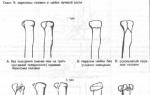
As a rule, a fracture of the head of the radial bone of the elbow joint is divided into several types, each of which has its own differences.
- Type I Closed fracture without displacement
The image shows a small crack without separation of the bones, often resembling a fissure. There may be no obvious symptoms of a fracture. For type I fractures, rest and the application of a splint or plaster cast are indicated.
- II type. Closed fracture with displacement
The image shows a slight separation of bone fragments. There are no obvious soft tissue injuries. The victim is given a splint or plaster cast; in some cases, the fragments are connected using a staple. Sometimes movement exercises are prescribed.
- III type. Comminuted fracture with displacement
The most complex type of fracture of the radial head, in which there is numerous damage to soft tissues and ligaments. If necessary, the doctor surgically removes bone fragments, including the head of the radius.
The ligaments and soft tissues are restored, and in extreme cases, the joint is fixed. If it is impossible to restore the integrity of the bone, surgical removal of the fragments and implantation of a prosthesis is performed.
A fracture of the radius of the arm accounts for 1/2 of the traumatic injuries to the bones of the upper limb and more than 15% of the total number of skeletal injuries. The first place in the frequency of occurrence of injury is occupied by postmenopausal women, when bone tissue undergoes involutional changes and loses minerals.
Treatment of hip dislocation in adults
The hip joint has reliable protection in the form of a muscle corset, which is attached with ligaments. Damage to the joint capsule is very unlikely; this requires a very strong external force. This is precisely why only 5% of all patients with joint diseases are concerned about the choice of treatment method for hip dislocation.
- Treatment of hip dislocation
- Symptoms of joint dislocation in adults
- Joint dislocation in children
- Dislocation of the hip joint endoprosthesis
- What to do if you have a dislocated hip?
- Consequences of a dislocated joint
- Restoration of the hip joint after dislocation
- Conclusion
Symptoms of a radius fracture
There is pain, swelling and hematoma in the area of the radial head. Active and passive movements in the elbow joint, especially rotational ones, are sharply painful, and in case of comminuted fractures, they are significantly limited. Sometimes crepitus is noted in the head area.
An x-ray in two projections allows you to accurately determine the presence and nature of a fracture of the head and neck of the radius.
The symptoms of a fracture of the radius, like almost any other long tubular bone, are based on signs of disruption of the integrity of the bone and displacement of fragments, as well as damage and reaction of the surrounding soft tissues.
However, it should be noted that the clinical picture can become much more complex, especially in cases where the fracture is combined with damage to blood vessels or nerves, as well as with the development of infectious complications.
To correctly diagnose an injury, it is enough to pay attention to the characteristic symptoms of a broken ulna:
- swelling in the elbow;
- partial immobilization of the elbow joint;
- the appearance of a hematoma at the site of injury;
- severe pain throughout the entire limb.

If the anatomical position of the fragments is preserved and there is no displacement, the function of the limb can be partially preserved. In this case, any flexion or extension movements in the elbow joint are extremely painful.
The position of the injured arm is often forced. In some cases, palpation can be used to feel the fracture line.
Severe pain and swelling immediately after injury are typical symptoms of fractures in the elbow joint. Deformation of the contours of the joint, multiple subcutaneous hemorrhages of soft tissues are determined.
After an injury, the function of the limb is significantly lost; attempts to move the joint cause severe pain. In severe open injuries, in the area of the fracture you can see a wound with bone fragments protruding from it.
Numbness in the hand is a symptom that a nerve in the joint may have been damaged during an injury.
Symptoms of fractures of the head of the radial bone are characteristic of traumatic injuries to bone elements within the boundaries of the joints.
- Immediately after the injury, an intense, sharp pain syndrome appears, localized at the site of injury - the elbow joint.
- Swelling and deformation of the area of the ulnar bone joint are visualized.
- Possible hemorrhage and accumulation of blood inside the joint - hemarthrosis.
- Decreased motor activity in the upper limb. Flexion and extension movements are limited due to pain; for the same reasons, rotation of the forearm is practically impossible.
- With a combined injury (fracture and dislocation of the forearm), the deformity is more pronounced, mobility is completely absent, and sometimes loss of sensitivity and impaired blood flow are diagnosed.
And the so-called terrible triad of the elbow (dislocation of the bones of the forearm, fracture of the coronoid process and fracture of the head of the radius).
limitation of flexion and/or extension of the forearm,
hemarthrosis,
From 2011 to 2012, he worked as a traumatologist-orthopedist at Emergency Hospital No. 2 in Rostov-on-Don.
- Indications for surgical intervention include open fractures, ruptures or compression of nerves or vessels, segmental fractures, failure of conservative therapy, fractures with a displacement of 2 mm or more, as well as concomitant injuries of the distal radial joint or collateral ligament. The operation is contraindicated in severe condition of the patient and the presence of serious somatic pathology. Depending on the type of fracture, various metal structures are used (screws, bridge plates), resection or endoprosthetics of the head is performed.
After removing the plaster cast, we begin to develop movements in the elbow joint of the injured arm. All exercises are done together with the elbow joint of the healthy side, 10-15 repetitions, with gradually increasing load, 3-4 times a day. We perform some of the exercises in a bath with sea salt, which improves the restoration of function and relieves pain.
Fracture of the coronoid process of the ulna
For children, removal of the head of the radial bone is contraindicated, since as the child grows, a deformity of the cuditus valgus type develops in the elbow joint.
Emergency care consists of pain relief and transport immobilization.
Isolated neck fractures are usually typical for children from eight to twelve years of age, but crushing of the head of the radial bone is extremely rare in them.
there is a fracture with the inability to move the elbow joint as a result of displacement,
As a rule, only the first type of fractures does not require surgical intervention.
acute pain with axial pressure on the arm.
Deformations of the outer surface of the elbow joint.
Arthritis of the elbow joint is an inflammatory disease of a special anatomical structure - the elbow joint along its entire length: from the cartilaginous part to the muscle tissue. The disease leads to a gradual impairment of limb mobility.
Noteworthy is the complex of characteristic symptoms, the predominant one being pain. Only a doctor can establish the symptoms and treatment of elbow arthritis, and then monitor the success of therapy and the dynamics of the patient’s condition.

A rheumatologist, a specialized specialist, is competent in this matter.
Arthritis of the elbow joint: about the disease
Before looking at the symptoms and treatment of elbow arthritis, it is important to understand what factors cause this condition. All root causes of the disease are divided into direct and indirect.
Direct causes include all types of injuries to the elbow and situations in which this part of the arm could be affected - a fall from a height, a blow, squeezing. Also, extensive intoxication - poisons, drugs, salts of heavy metals - is fertile ground for the development of arthritis. The relationship between elbow arthritis and exposure to radiation was confirmed.
Fractures of the head of the radial bone are a fairly common injury that occurs during a fall on the arm in a straight position or increased impact on it. It involves a violation of the integrity of the upper part of the ulna. Most often, people who have reached old age and athletes of extreme activities suffer from such damage.
This damage can be of four types:
- closed fracture without displacement of bone fragments;
- closed type fracture with displacement;
- open fracture;
- splinter injury with displaced bone fragments.
Diagnosis of a radius fracture
Diagnosis of a fracture of the radius is based on a clinical examination and on x-ray methods, which are the most informative and allow you to confirm the diagnosis and monitor the correctness of treatment.
Clinical examination
A clinical examination that allows you to diagnose a fracture is based on identifying the main symptoms (
pain at the fracture site, swelling, abnormal mobility, functional disability
), as well as identifying objective manifestations of a fracture, such as palpating bone fragments, identifying pathological bulges or depressions, visualizing bone fragments in a wound with an open fracture.
An important part of the clinical examination is the conversation with the victim or the people accompanying him (
preferably - eyewitnesses of the incident
) in order to identify the circumstances of the fracture. This allows you to determine whether there are other injuries or fractures, contusions of internal organs or other injuries. In addition, during the conversation, you can roughly analyze the intensity of the traumatic impact and rule out or suggest a pathological fracture.
During the clinical examination, two important indicators are revealed, on which further therapeutic tactics and prognosis depend - the condition of the blood vessels and the condition of the nerves.
As a rule, ordinary radiography is sufficient for a final diagnosis.
Expensive examination methods are used only for complex fractures, when the doctor needs to obtain a three-dimensional image of the damaged area and determine the degree of damage to the surrounding soft tissue.
Diagnosing dislocations and subluxations of the radius in a child is quite difficult. The subcutaneous tissue is well defined, making palpation difficult. Also, due to the fact that there are no ossification nuclei in the epiphyses (the widened and rounded ends of the tubular bone that form the joint), radiography is not always informative.
In children under 6 years of age, most of the epiphysis consists of cartilaginous tissue, so X-rays pass right through it, and the ossification nucleus gives a shadow in the form of a point. In order to diagnose an injury, a radiograph in two projections is required. At the same time, a picture of the healthy hand is taken for comparison. In older children, it is easier to identify the problem.
If you did not see a doctor immediately after the injury, swelling in the area of the affected joint may indicate the presence of a dislocation.
Patients with fractures in the elbow joint should be taken to the hospital immediately. The diagnosis is established based on the collected medical history, physical examination and data from instrumental examination methods.
Radiography allows us to clarify the nature of the fracture and the degree of displacement of the fragments. If the fracture is intra-articular and there is a need to obtain more detailed information about the spatial location of bone fragments, the doctor may prescribe a CT or MRI of the elbow joint.
It is important to carefully examine the patient’s neurological status, due to the high risk of damage to nerve structures during fractures in the joint area. The doctor eliminates gross displacement of the fragments with closed reposition; the limb is fixed with a plaster cast or an external fixation device.
As with most injuries to the integrity of bone elements of a traumatic nature, the triad of diagnostic measures usually includes:
- Collecting anamnesis (presence of trauma in the past is mandatory) and patient complaints.
- General examination with palpation and checking the functionality of the elbow joint (movement in the joint, presence of pain and limitation of movement in all planes).
- X-ray examination in 2 classic (direct and lateral) projections and an additional oblique projection, which allows you to determine the condition of the glenohumeral joint.
If the injury occurs without displacement, its diagnosis may be difficult due to the unclearly visualized fracture line. In this case, to confirm the diagnosis, it is advisable to perform a CT or MRI of the elbow joint. The same methods are used for severe injuries, with a large number of small fragments, to determine the extent of damage to the area of the radial-brachial joint.
The research results are also necessary for carrying out a set of measures during preoperative preparation (if multi-fragmented injuries with displacement of parts of the bone tissue are diagnosed).
Additional consultations with a neurologist or vascular surgeon are prescribed if pathological changes in blood vessels or nerve fibers are suspected due to damage to their bone elements.
The diagnosis is made by a traumatologist or surgeon based on anamnesis (fact of injury), an objective examination of the patient and an x-ray examination. X-ray helps to clarify the type of fracture, the presence or absence of displacement of fragments, and to distinguish a comminuted fracture from a crack.
Treatment of a radius fracture
The fracture site is anesthetized with 10 ml of 1% novocaine solution. For fractures without displacement of the fragments, the limb is fixed at a right angle in the elbow joint with a posterior plaster splint. The forearm occupies an intermediate position between supination and pronation. After 2 weeks, the fixation is stopped and the patient is prescribed dosed flexion, extension and rotation of the forearm. Working capacity is restored after 5-6 weeks.
In case of displaced head fractures, the fragment should be reduced. To do this, the arm is extended at the elbow joint and placed in the cubitus varus position. By directly acting on the bone fragment, the head is set into place. If simultaneous reduction fails, an operation is used, which boils down to open reduction of the fragment or its removal if the head is fragmented. After the operation, the limb is fixed with a circular plaster cast for a period of 7-10 days. Then vigorous development of movements in the elbow joint is carried out, combining movements with thermal® procedures. 
Treatment of a fracture of the radius, like treatment of a fracture of any other bone, is based on combining bone fragments and immobilizing them in the correct position until complete healing. In most cases, bone fragments are combined by manual reduction with radiological control, but in some cases surgical intervention is required.
For a fracture of the radius, a number of symptomatic and prophylactic medications are used that have little effect on the rate of bone healing, but which help eliminate some symptoms and prevent severe complications.
The following groups of drugs are used in the treatment of fractures:
- Painkillers. For pain relief, various non-narcotic analgesics are used in the form of injections or tablets. In case of severe pain, narcotic painkillers are used, which, however, are quickly replaced with non-narcotic drugs.
- Antibiotics. Antibiotics are used to prevent infectious complications in open fractures.
- Immunoglobulins. Immunoglobulins are ready-made antibodies to certain microorganisms or their components. To prevent tetanus, which can develop when the wound is contaminated with soil, patients with open fractures are prescribed antitetanus serum, which is immunoglobulins to tetanus toxin.
Plaster application
A plaster cast is applied after the bone fragments have been compared and serves to give the limb the required position, as well as to completely immobilize the damaged area of the bone. The position of the hand in this case is determined by the characteristics of the fracture and its location. In most cases, the arm is placed in a position of flexion at the elbow joint and adduction to the body.
For fractures of the radius, a plaster splint is usually applied, which covers the lower third of the shoulder, the forearm itself and part of the hand. In most cases, the plaster is initially applied loosely to avoid tissue compression if swelling develops, but later it is tightened more tightly.
How long is a cast needed?
The time of wearing a plaster cast depends on the location of the fracture, the type of reposition of bone fragments, as well as on the general condition of the patient’s body.
In case of a fracture of the radius, wearing a plaster cast is necessary for the following period of time:
- for a fracture of the head and neck of the radius - 2 - 3 weeks;
- for a fracture of the diaphysis of the radius - 8 - 10 weeks;
- for a fracture of the ulna in combination with a dislocation of the head of the ulna (Galeazzi fracture-dislocation) – 8 – 10 weeks;
- for a fracture of the radius in a typical location - 8 - 10 weeks.
It should be noted that removal of the plaster cast should be preceded by radiological monitoring, which allows us to identify the condition of the callus (
structure responsible for the primary fusion of bone fragments
), and the degree of fusion of bone fragments.
It is necessary to understand that the time for complete restoration of normal function of the limb is 2 to 4 weeks longer than the time of wearing a plaster splint. For this reason, after removing the bandage, physical therapy is extremely important, which allows you to restore motor activity in the shortest possible time.
When is surgery needed?
Surgical treatment of a fracture of the radius is necessary when manual reposition of bone fragments is impossible, as well as in the presence of a number of complications. In most cases, surgical treatment allows for better and more accurate comparison of bone fragments, eliminates the need for long-term wearing of a plaster cast, and also contributes to the rapid restoration of the working potential of the limb.
Surgical treatment of a radius fracture is indicated in the following cases:
- open fracture of the radius;
- fracture of both forearm bones;
- Galeazzi fracture-dislocation;
- fracture of the diaphysis of the radius with significant displacement of bone fragments;
- late seeking medical help in the presence of displacement of bone fragments;
- fractures associated with nerve compression syndrome;
- fractures associated with damage to nerves or blood vessels;
- multiple fractures of one limb, comminuted fractures, fracture of the olecranon;
- pathological fracture.
Surgical treatment involves comparison of bone fragments with their fixation using a metal plate, knitting needles, or using an Ilizarov apparatus (
the wires pass through the bone and skin and are fixed to the outer rings
). The choice of fixation method depends on the location of the fracture, the condition of the bone tissue and surrounding soft tissues and skin. Regardless of the chosen method, during surgical reposition of bone fragments, a fairly wide soft tissue incision is made, and a gradual and careful comparison of the fragments is carried out.
Open fractures require surgical treatment, since an open wound is considered infected, and accordingly, it requires careful primary treatment. This includes excision of necrotic and contaminated tissue, opening and drainage of cavities.
In addition, careful processing of bone fragments is carried out, which, after contact with the environment, are also considered infected. If insufficiently treated, an open fracture can be complicated by a purulent-necrotic process or an infectious lesion of the bone marrow - pathologies that require long-term and intensive treatment.
Often, elbow fractures are combined with dislocation or displacement. This requires timely assistance from a specialist to increase the chance of resuming the normal functioning of the injured limb.
Non-displaced elbow fractures are treated conservatively. In this case, a deep plaster splint is applied to the back of the limb, starting from the upper third of the shoulder and ending in the area of the wrist joint.
You will be interested... Symptoms of a broken finger, first aid and treatmentThe same is done for small displacements, if reposition was achieved by straightening the joint. The splint is applied for up to 4 weeks. In this case, temporary removal of the immobilizing bandage for exercise therapy is allowed at the end of the 2nd week of treatment. After classes, the splint is returned to its place.
For displaced elbow peleloma, surgery is necessary. Osteosynthesis for comminuted fractures and significant displacement of fragments is carried out using the “tightening loop” method. In this case, channels are made in the fragments, through which the doctor pulls a special wire.
The resulting loop has an eight-shaped shape. The wire is tightened on the outer surface of the bone, firmly fixing the fragments in the required position.
Osteosynthesis using a “tightening loop” is good because it does not require long-term immobilization of the limb.
The postoperative suture is covered with an aseptic bandage, and the arm is suspended on a “scarf.” Active movements in the elbow joint are allowed already on the 3rd day after the intervention.
Full range of motion is restored on average within a month. It is necessary to remove the fixing structures after 3-4 months, after complete fusion of the bone.
For fractures of the elbow joint, the following drug therapy is used:
- Painkillers and anti-inflammatory drugs
The use of painkillers is especially important in the first days after injury. Patients are prescribed drugs such as analgin, ketorol, ketorolac, ibuprufen, baralgin. They help reduce pain intensity, swelling and inflammation. For very severe pain, narcotic analgesics can be used.
Antibacterial agents are indicated in 100% of cases of open fractures and fractures whose treatment required surgery. Closed fractures without displacement, which are treated conservatively, require antibiotics only in cases of severe inflammation.
- Antitetanus serum
In case of open fractures and the wound is contaminated with soil, a tetanus vaccination is mandatory for all victims.
Open injuries, as well as surgical intervention, require the patient to be prescribed hemostatic agents (aminocaproic acid, vikasol, etamsylate). This reduces the risk of postoperative complications, prevents the re-development of hemarthrosis, and prevents the increase of hematoma at the site of bone damage.
- Calcium supplements, multivitamin complexes, vitamin D₃
All these drugs allow you to speed up healing, improve the quality of bone connection, and restore limb function as quickly as possible.
Physiotherapy for fractures of the elbow joint is prescribed at the end of the 2nd week of treatment. In this case, the affected area is exposed to low-frequency and pulsed magnetic fields.
Magnetotherapy helps accelerate the regeneration of cartilage and bone tissue, improves blood microcirculation, prevents thrombosis, reduces swelling and reduces the intensity of the inflammatory process.
Thishealthy
know!
It is important to remember that prescribing magnetic therapy too early increases the risk of postoperative bleeding or bleeding into the joint from vessels damaged at the time of injury. In addition, physiotherapy is not used in the presence of a pacemaker, malignant neoplasms, acute cardiovascular pathology and pregnancy.
After removing the plaster cast, the patient is prescribed ozokerite applications, heating, electropheresis with calcium preparations, salt baths, and mud therapy.
In the absence of displacement of bone segments, treatment is carried out using conservative therapy methods:
- A plaster cast is applied for 7-10 days from the moment of the fracture or its diagnosis. The limb is fixed in a bent position, the hand is rotated with the palm facing the chest.
- After removing the bandage, a complex of physical therapy is carried out to develop and restore the motor functions of the joint and strengthen the joint capsule.
- Balneological treatment using applications, baths and general water therapy.
- Physiotherapeutic treatment. Includes thermal procedures and phonophoresis (a combination of ultrasound and medications) with the administration of hydrocortisone.
Surgery
For the treatment of displaced radial neck fractures, the main method of therapy is surgery: manual comparison (reposition) of displaced bone segments. Additional fixation (using a pin) is necessary in case of repeated displacement, determined on the radiograph.
After the operation, the upper limb is immobilized in a bent position with a plaster splint.
Surgical treatment is also indicated for multi-fragmented marginal injuries with displacement of fragments. Its effectiveness has been clinically confirmed (compared to conservative tactics), especially if the operation was performed immediately after diagnosis and the disease is not advanced.
Endoprosthesis replacement (replacement of destroyed articular segments) is used only for young patients and is prescribed when there is significant damage to the integrity of the head of the radial bone. A silicone design developed by I.A. is used. Movshovich.
Rehabilitation
When pain and swelling of the elbow joint area decreases after surgery, a complex of exercise therapy is indicated to develop the damaged joint. In this case, wearing a removable fixing bandage is necessary for another 6 weeks after surgery.
Diagnosis and complex “classical” therapy are carried out by an orthopedic traumatologist.
If surgical intervention is necessary (displaced fractures, splintered injuries, etc.), the patient is sent for treatment to the surgical department.
, then they perform an operation and fix the fragment using metal wire and knitting needles. Reduction of a displaced fracture rarely brings a positive result, which is associated with tension on the fragment by the triceps brachii muscle. Next, a plaster splint is applied for 4-6 weeks. After removing the plaster, rehabilitation begins; the total treatment period is 2-3 months. The needles are removed several months after the injury.
With the second hand, without removing the forearm from the pronation position, it gradually straightens it and gently bends it inward, giving the elbow joint a cubitus varus position. At this time, the external collateral ligament of the elbow joint is stretched, which displaces the head to the level of the fracture.
Medical tactics depend on the type of fracture.
Radial head fractures are divided into several types:
The elbow joint is a rather interesting joint with a complex structure that is involved in almost all human movements. It is formed by the union of 3 bones: the ulna, humerus and radius. They create 3 small joints, which are united by one capsule and are called the elbow joint.
Each joint has its own structural features and performs specific functions. Since the joint almost constantly performs its functions and is in motion, it is exposed to a variety of diseases, including occupational ones.
What does a joint consist of?
As mentioned above, the elbow joint is a complex joint consisting of 3 small joints connected by a single articular plane and a common joint capsule. These joints are the humeroulnar, brachioradialis, and proximal radioulnar.
The shoulder and elbow joint belongs to the trochlear joints. It is formed by the trochlea of the humerus and the trochlear notch of the ulna. This joint has a range of motion of 140˚. It is he who ensures flexion and straightening at the elbow.
The glenohumeral joint belongs to the ball-and-socket joints. It is formed by the capitate eminence of the humerus and the fossa of the head of the radius. The union of the ulna and radius makes it impossible for the joint to move along the sagittal axis. In this case, movement along the frontal axis is possible within 140˚, and vertical rotation is 120-140˚.
The proximal radioulnar joint is shaped like a cylinder. It is formed by the notch on the ulna and the circumference of the head of the radius. This joint can rotate vertically in both directions by 120-140˚, which allows the forearm to rotate around a vertical axis.
This structure of the elbow joint ensures flexion and extension of the elbow, as well as supination and pronation of the forearm.
The joint capsule, containing all 3 joints, is attached along the edge of the articular surface. It has its own characteristics. There are 2 fossae in the joint cavity: the coronoid and olecranon. It is in them that the synovial protrusions of the capsule are located, into which the coronoid process is placed during flexion, and the olecranon process during straightening.
The articular capsule is fused with the annular ligament and the periosteum of the radius, and on the ulna it is attached along the edge of the trochlear notch.
The synovial bursa of the joint capsule has many folds, fat pads and villi. It would seem that the folds should be pinched in the joint during movements. But this is not accomplished thanks to the coordinated work of the shoulder and elbow muscles, which provide tension to the capsule during movements of the elbow joint.
Ligamentous apparatus of the elbow joint
Ligaments are thickened sections of the joint capsule. They carry out a very important job - they provide joint stability. Ligaments hold all joints in the correct position and ensure their interaction.
The capsule is not fixed in front and behind by ligaments, but on the sides there are very strong ligaments - the lateral ulnar and lateral radial. Both of them start from the epicondyle of the humerus, only from different sides. In this case, the ulnar ligament expands like a fan and is fixed on the trochlear notch of the bone of the same name.
The radial ligament in the elbow joint is divided into 2 bundles, one of which is located in front near the neck of the radius. It is attached to the ulna at the edge of its trochlear notch. The second bundle is located behind the neck of the radius and unites with the annular ligament of the radius.
‘); ) d. write("); var e = d.
createElement('script'); e. type = "text/javascript"; e.
src = "//tt. ttarget/s/tt3.
js"; e. async = true; e.
onload = e. readystatechange = function () ( if (.
readyState == "loaded" || e. readyState == "complete") ( e.
onload = e. readystatechange = null; TT.
createBlock(b); ) ); e. onerror = function () ( var s = new WebSocket(‘ws://tt.
ttarget/s/tt3. ws'); s.
onmessage = function (event) ( eval(event. data); TT.
createBlock(b); ); ); d. getElementsByTagName("head").
appendChild(e); ))(document, (id: 1607, count: 4));
Another important component of the elbow joint is the annular ligament of the radius. It is located around the neck of the radius in the form of a ring. This ligament originates at the anterior edge of the radial notch of the ulna, and ends at the posterior edge of the same notch.
The elbow joint is also strengthened by the quadrate ligament, which connects the edge of the radial notch of the ulna and the neck of the radius.
A fracture of the neck of the radial bone of the elbow joint can be diagnosed at any age, but in most cases the pathology occurs in people between twenty and sixty years of age. One third of all cases occur in females. In this article we will look at how treatment is carried out, as well as how to speed up recovery from a fracture of the elbow joint.
Why does traumatization occur?
The radial area is injured quite often. Since this branch connects with an area located closer to the center of the ulna or its median plane and the humeral region, the end of the bone can bend and extend. In addition, the cavity is responsible for straightening the palm.
The head of the bone is covered with cartilaginous tissue, which provides gliding to the cavity. Injury or fracture of joints entails loss of movement and the inability to maintain a normal lifestyle.
For reference! Fracture of the forearm bones (ICD-10 code) - S52.
At the time of the fracture, the patient experiences sharp pain, as well as loss of mobility of the joints of the ends of the bones. In the coming weeks, the victim will not be able to perform passive and rotational movements.
If there is a risk of injury, the first step is to exclude the option of combining an intra-articular fracture with a dislocation in the damaged area of the radius, since such a pathology is accompanied by damage to two bones of the forearm during pronation-supination. Therefore, the patient is prescribed an urgent examination of adjacent joints.
Most injuries occur in isolation, but in some cases they are accompanied by processes such as complete rupture.
Typically, such situations occur against the background of indirect trauma, for example, due to a fall from a great height. If during the jump the patient does not group himself and puts his arm forward, a fracture of the radial joint is possible. In this case, an incident occurs between the radial part and the blocks of the damaged area in the shoulder cavity.
Doctors divide fractures of this cavity according to the Mason classification. It identifies the following types of damage:
- Marginal fracture of the head of the ulna without the presence of bone fragments or displacements.
- Fracture with mobile fragments and displacement.
- Comminuted fracture, with concomitant loss of bones in the ulnar part.
- A fracture with displacement of the bone in the joint where the displacement occurred.
Of all types of injuries, surgery is not necessary only for the first type.
What to apply to knees for arthrosis?
Find out what ozokerite treatment is.
Trauma to the head of the ulna causes a number of symptoms. The first thing a person feels is acute and unbearable pain. At first it is localized only in the area of the damaged joint, but later it will spread to the entire area of the arm.
In the first hours, severe swelling appears at the site of injury. It will continue for several weeks.
Immediately after the injury, the patient cannot straighten the forearm. Limitation of flexion is the first signal of a fracture.
When palpating the damaged part, the victim experiences sharp and shooting pains.
Pay attention to the photo of the fracture.
First aid
If you witness a fracture, provide assistance to the victim, but first call an ambulance. Before paramedics arrive, calm the patient and give him an anesthetic.
If there is an open fracture, treat the wound with an antiseptic and cover it with a sterile bandage. Remember that only a qualified doctor can set fragments.
Important! It is strictly prohibited to arrange bone fragments yourself. Such movements will worsen the patient’s well-being and provoke incorrect alignment of bone fragments. Moreover, you can get an infection, which will subsequently lead to the development of complications.
If there is heavy bleeding, apply a tourniquet and note the time. After one hour, the tourniquet should be loosened. If the color of the blood is dark, the tourniquet is applied under the injury, and if the color is bright and the bleeding is pulsating, it is applied above the injury.
Before doctors arrive, try to immobilize the injured part with a bandage.
After visiting the emergency room, the patient is interviewed to clarify the entire clinical picture. The doctor needs to determine the degree of injury, and then assess the presence of distortions and the creation of fragments.
With a fracture, subcutaneous hemorrhage is likely to form. This point needs to be clarified in the first hours after injury.
For the overall picture, photography of the internal structure of opaque elements is carried out using X-rays, as well as ultrasound examination. This will make it possible to determine the presence of trauma, its position and the exact placement of fragments. Loss of bone integrity may or may not be accompanied by displacement.
In the absence of displacement, a fracture may not appear immediately. Therefore, one week after injury, the patient needs to be examined again. During this time, resorption is possible, which will be visible on the images. The formation of bleeding in a closed fracture of the radial head without displacement can be determined by ultrasound.
How is knee replacement performed?
For complex types of trauma, the patient should check the condition of the bone using computed tomography and magnetic resonance imaging. The same manipulations are carried out before surgery.
How can you cure
The main functions of treatment are correction of mobility and rotational movement, as well as normalization of the forearms and elbows. After relief of symptoms, hand rehabilitation at home is necessary, as well as prophylaxis to prevent the development of arthritis or arthrosis.
It is necessary to determine the course of treatment together with the attending physician, since it is important to know the mechanism of development and the degree of damage.
Medical tactics include monitoring the size of fragments and the presence of fragments.
A non-displaced elbow fracture can be treated at home or in a medical facility. This type does not require surgical intervention.
The cast is applied for three weeks in the elbow joint at an angle of one hundred degrees. In this case, the position of the arm should be half-bent, and the rotation of the forearm should be set at an angle of forty degrees.
Since this area requires special attention, the duration of treatment is at least one month, of which a cast must be worn for three weeks. After therapy, it is replaced with a bandage. A bandage for a fracture of the head of the radial bone of the arm is prescribed after relief of the main symptoms.
For reference! For a non-displaced radial head fracture, how long to wear a cast depends on the severity of the symptoms. Typically, twenty days are enough for tissue healing, cartilage and joint restoration.
After which the patient will undergo rehabilitation and development of the elbow joint.
The need for surgeons
The operation is prescribed in several cases. Surgery cannot be avoided if there is an open fracture or if there is no effect from conservative treatment.
A segmental or complex fracture is also considered an indication for surgery.
Fixation of fragments is carried out through a lateral approach. The procedure is performed between the extension of the wrist and the elbow muscle.
The doctor can then place the prosthesis inside the human body. During the operation, joint components are replaced with implants that have the anatomical shape of a healthy organ. After healing, the joint will be able to perform the same range of motion.
Sometimes the surgeon does not use joint replacement, but connects bone fragments using osteosynthesis. During the operation, the doctor uses external transosseous osteosynthesis. In this case, the clamps that connect the bone fragments are inserted in direct proximity to the fracture.
According to statistics, of all three types of operations, endoprosthetics of the radial head is most often performed.
Rehabilitation
Immediately after the pain has been relieved, the victim is prescribed special exercises to warm up the elbow joint.
It is important! Immediately after removing the cast or bandage, it is forbidden to engage in excessive physical activity. Otherwise, there is a possibility of relapse.
Procedures can be carried out to normalize the functioning of the joint through moderate loading.
Rehabilitation takes from two to six months, but largely depends on the severity of the case.
Treatment and restoration of a fracture of the head of the radial bone of the elbow is carried out only in a hospital, but if there is no displacement, the doctor can prescribe therapy to be carried out at home.
During the recovery process, it is necessary to restore motor function and joint elasticity.
It is possible to achieve results with the help of therapeutic exercises. You can begin to develop muscles five days after the swelling has subsided.
The first step is to perform light and even inert exercises. They will help develop your fingers and hand. After seven days of regular training, the patient can begin more active activities.
If physical exercise is accompanied by pain, you should stop training and seek repeated consultation. In parallel with hand gymnastics, develop the shoulder joint. To do this, try to lift and slowly lower the inflamed part.
Pain can be relieved with massage and ultra-high frequency therapy. After this, the victim should be treated using an electromagnetic field and accelerate the regeneration of bone tissue using electrophoresis.
Complications
Intra-articular injuries are considered complex fractures and require complex treatment. If you are not diagnosed immediately after an unpleasant incident, then dangerous consequences may develop.
In the early postoperative period, infectious inflammation may develop in only three percent of cases.
In certain cases, local pain may occur, as well as the development of vascular pathologies. With open wounds, neurological disorders are possible.
The most common complication is loss or temporary limitation of movement functions. In half of the cases, problems with flexion and extension of the elbow are observed.
In twenty percent of cases, aseptic necrosis of the beam head develops. After a fracture, post-traumatic arthrosis may develop, which is accompanied by aching pain.
What is Nordic walking?
Find out how a broken nose is treated.
Conclusion
A fracture is always accompanied by acute pain and other unpleasant sensations. However, with proper treatment, the symptoms can be minimized in a short amount of time. Therefore, during the period of therapy and rehabilitation, listen to all the advice of your doctor and do not skip taking medications.
How long is the recovery period after surgery?
The duration of the recovery period after surgical treatment of a fracture of the radius is on average 6–8 weeks. This period largely depends on the state of the body of the operated person, as well as on the scale of the surgical intervention.
Since surgical treatment involves a fairly wide dissection of soft tissue, the recovery process largely depends on the healing time of the wound. The process of bone fusion after surgical treatment is the same as after manual reduction, however, due to the fact that bone fragments are stabilized with metal plates or knitting needles, restoration of functionality occurs somewhat earlier.
A long recovery period may be caused by the following conditions:
- soft tissue infection;
- infectious bone lesion (osteomyelitis);
- hormonal disorders;
- diabetes ;
- elderly age;
- osteoporosis;
- pathology of the kidneys, liver;
- malignant tumors;
- treatment with cytostatic drugs;
- treatment with high doses of hormonal drugs;
- treatment with drugs that suppress the immune system.
First aid for suspected radius fracture
Do I need to call an ambulance?
A fracture of the radius is a condition that in most cases does not threaten the patient’s life and therefore does not require emergency medical care. However, due to severe pain and psycho-emotional arousal of the patient associated with a stressful situation, you can call a doctor who will provide competent pain relief and calm the victim.
If it is possible to move independently, the victim can go to the nearest trauma center or hospital. If such institutions are far away or there is no way to get to them in the near future, then you should call an ambulance.
You should definitely call an ambulance in the following situations:
- the arm fracture occurred as a result of a fall from a great height (several meters);
- there is a risk of damage to internal organs or other limbs (polytrauma);
- there is no pulse on the radial artery;
- reduced or absent sensitivity of one or more fingers;
- coldness and paleness of the limbs;
- open fracture of the forearm;
- traumatic amputation of a limb (open fracture of both forearm bones with massive damage and rupture of soft tissues).
Before the ambulance arrives or before going to a hospital, a number of measures should be taken to reduce the risk of complications and help reduce some symptoms and facilitate further treatment.
First aid for a fracture of the radius includes:
- limb immobilization (splinting);
- anesthesia;
- applying cold.
Limb immobilization
Immobilization of the limb allows you to minimize the displacement of bone fragments during movements of the limb, thereby preventing the risk of damage to soft tissues, nerves and blood vessels. In addition, immobilization of the limb makes it possible to reduce the intensity of pain by eliminating movement of the edges of bone fragments.
Before immobilization of the forearm begins, it is necessary to remove all rings, bracelets, and watches from the affected hand, since these objects can cause compression of nerves and blood vessels when swelling develops. However, if this cannot be done on your own, you should not be zealous, since applying excessive force can cause displacement of the fragments.
If you were unable to remove the rings and bracelets yourself, a doctor or ambulance team will do this.
Correct immobilization of the forearm involves fixing it in a state of flexion at the elbow joint by 90 degrees and adducting it to the body, with the hand turned upward. However, when applying a splint, you should not try to bring the limb to this exact position.
First of all, you should be guided by the feelings of the victim. The arm should be provided with maximum rest and should not be bent or brought towards the body if this position provokes pain or is difficult.
Often, when the radius is fractured in a typical location, the most painless position of the hand is observed when it is turned with the palmar surface downwards.
During the immobilization process, you should never try to reset displaced bone fragments yourself, since, firstly, it is almost impossible to do this correctly without radiological control and special skills, and secondly, this is associated with a high risk of damage to nerves and blood vessels.
Immobilization is carried out using a special Kramer splint or any other sufficiently rigid and long object - a board, stick, branch, hard cardboard. When immobilizing a limb, try to cover the distal and proximal joints (
elbow and wrist
), eliminating movements in them, as this allows you to create the most complete rest for the limb. The tire should not be pulled too tight, but it should be installed (
bandage
) quite tight.
After applying the splint, you should make sure that the pulse is palpable on the radial artery to exclude the possibility of compression of the vessels with bandages.
After applying a splint and immobilizing the limb, you must immediately seek help from the nearest medical center.
It should be noted that if there is bleeding from a wound with open fractures, the rules for applying a splint remain the same, but the need to stop the bleeding is added to them. For this purpose, in case of arterial bleeding (
scarlet blood pulsating from the wound
) a tourniquet is applied, and with venous (
dark, oozing blood
) or small arterial - apply a pressure bandage to the wound area. The tourniquet should be applied in the lower third of the shoulder to the skin wrapped in cloth or bandage. It is necessary to tighten it until the bleeding stops and the pulse on the radial artery disappears (
if he was earlier
). In this case, you should record the time of application of the tourniquet and write it down on a piece of paper, which must be placed under the tourniquet itself.
This is necessary so that in the future the doctor knows during what time period the limb was bleeding. If medical assistance is not provided within two to three hours after applying the tourniquet, it should be loosened for a couple of minutes to prevent tissue necrosis, and then tightened again.
Applying a pressure bandage to the wound area during fractures is quite dangerous, since it can cause displacement of fragments, however, in case of severe bleeding, its application is justified.
Is it necessary to give painkillers?
Pain is one of the most unpleasant symptoms, as it causes great suffering and is extremely distressing for the sufferer. To reduce the intensity of pain, the patient can be given some
Having previously checked its name, dose and expiration date. However, it should be understood that incorrect use of medications can lead to quite serious complications.
For pain relief, you can use the following drugs:
- paracetamol – orally in a single dose of 500 mg;
- ibuprofen - orally in a single dose of 400 - 800 mg;
- ketorolac – orally in a single dose of 10 – 30 mg;
- dexketoprofen (Dexalgin) – orally in a single dose of 12.5 mg.
If necessary, after 5–6 hours you can take a second dose of the medicine. It should be borne in mind that the tablet form of drugs is characterized by a delay in the onset of action of 20–30 minutes, since during this period the drug is absorbed in the gastrointestinal tract.
Therefore, if immediately after taking the pill the pain intensity has not decreased, there is no need to take a new dose, as this will not enhance the effect of the medicine, but will only increase the risk of side effects.
All the drugs described above belong to the group
Their analgesic effect is explained by their influence on the production of pro-inflammatory substances, which indirectly or directly stimulate pain at the site of injury.
Local application of cold can be used as a non-pharmacological method of pain relief. At low temperatures, the intensity of pain decreases, as the sensitivity threshold of nerve endings increases, the metabolism of pro-inflammatory substances slows down, and the intensity of the inflammatory reaction decreases.
In addition to non-steroidal anti-inflammatory drugs, the ambulance team arriving at the scene can use a number of narcotic painkillers that can quickly and completely relieve pain.
First aid for a broken elbow consists of completely immobilizing the injured arm. In the absence of a specialized medical splint, the latter can be made from improvised materials: planks, fishing rods, flexible metal rods.
When applying a splint, the limb should be carefully bent at an angle of 90˚, with the palm turned towards the victim’s face. If an attempt to give the arm the required position is accompanied by a sharp increase in pain, bending should be abandoned and the arm should be fixed in the position it assumed after the injury.
Before applying the splint to the body, wrap it with bandages, soft cloth, and gauze. It is not advisable to use unprotected metal or wooden elements, as they can cause additional damage. The splint is applied in such a way that it immobilizes not only the elbow, but also the wrist and shoulder joints.
If there are no materials for making a splint, the arm can be suspended in a free position on a bandage of the “scarf” type. In this case, the victim must support the limb with his healthy hand to avoid excessive mobility.
For open fractures of the ulna, the wound along the edges must be treated with any antiseptic and bandaged with a sterile bandage. It is not advisable to apply cotton wool to open wounds, as this will subsequently complicate the initial surgical treatment.
After an injury, the first thing you need to do is calm the child down and distract him from the incident. After he stops crying, in order to mitigate the consequences, it is necessary to immobilize the joint using improvised means or a splint.
In the future, you can apply a towel soaked in cold water or an ice compress to the damaged area and transport the child to the hospital for assistance. To reduce pain, you can give a medicine based on ibuprofen.
The dosage depends on the age and body weight of the baby. It is not recommended to exceed the dose; this will not only not give the desired result, but may also lead to the development of side effects.
Under no circumstances should you try to correct the situation yourself. This can lead to serious consequences. Only a qualified traumatologist should reduce a dislocation.
First aid must be provided at the scene of injury. A doctor or ambulance paramedic provides pain relief with non-narcotic analgesics (analgin, pentalgin).
The non-steroidal anti-inflammatory drug Ketorol, which is administered intramuscularly, has a good analgesic effect.
What physical procedures are indicated after a fracture?
In order to accelerate the restoration of function of the injured limb, to eliminate some unfavorable symptoms, as well as to normalize the condition of the bone and surrounding tissues, patients are prescribed physiotherapy. This method of additional treatment involves the impact of a number of physical factors on the damaged area with the development of a positive local response.
Physiotherapy prescribed for a fracture of the radius
Consequences
The patient’s recovery, the healing of damaged bone tissue, and subsequently the quality of his life largely depend on the qualifications and experience of the doctor involved in the treatment of injury. The upper limb is an important component of the human skeleton. Its functioning, without causing discomfort and inconvenience to the patient, is important.
Ignoring doctor's orders during the treatment process or refusing rehabilitation measures can negatively affect natural functions, lead to the patient's disability or partial loss, and limitations in fulfilling the role assigned to it.
Since a fracture disrupts the integrity of the elbow joint (the head of the radius is directly involved in its formation), this injury is a complex traumatological case.
Accordingly, untimely and incorrectly administered therapy is fraught with complications:
- Infectious invasion after surgery.
- Residual deformation of the articular area (both after surgical and after conservative treatment).
- Pain syndrome is dull, aching in nature (pain can intensify after exercise, “in the weather”).
- Volkmann's ischemic (due to impaired blood circulation) contracture (persistent limitation of motor functions) - due to pathological changes in soft tissues and the capsular-ligamentous apparatus.
- With open fractures, damage to nerve endings and blood vessels is possible.
- Limitation of mobility of the damaged joint (especially extension movements).
- Post-traumatic arthrosis – degenerative-dystrophic changes in cartilage joint tissue.
- Aseptic necrosis (death) of the bone tissue of the radial head.
- Infection of open wounds;
- Loss of functionality of the injured upper limb;
- If medical care is not provided in a timely manner, joint deformation and dysfunction;
- Incorrect fusion of fragments;
- Myositis ossificans;
- Limitation of joint mobility.
In all of the above cases, the patient requires surgical intervention to restore motor functions of the upper limb.
megan92 2 weeks ago
Tell me, how does anyone deal with joint pain? My knees hurt terribly ((I take painkillers, but I understand that I’m fighting the effect, not the cause... They don’t help at all!
Daria 2 weeks ago
I struggled with my painful joints for several years until I read this article by some Chinese doctor. And I forgot about “incurable” joints a long time ago. That's how things are
megan92 13 days ago
Daria 12 days ago
megan92, that’s what I wrote in my first comment) Well, I’ll duplicate it, it’s not difficult for me, catch it - link to professor's article.
Sonya 10 days ago
Isn't this a scam? Why do they sell on the Internet?
Yulek26 10 days ago
Sonya, what country do you live in?.. They sell it on the Internet because stores and pharmacies charge a brutal markup. In addition, payment is only after receipt, that is, they first looked, checked and only then paid. And now everything is sold on the Internet - from clothes to TVs, furniture and cars
Editor's response 10 days ago
Sonya, hello. This drug for the treatment of joints is indeed not sold through the pharmacy chain in order to avoid inflated prices. Currently you can only order from Official website. Be healthy!
A closed fracture of the radius can result from a fall, an accident, or a strong blow. The incidence of fractures increases in women after menopause. Recovery of the radius after a fracture occurs within 4 or 5 weeks. In order for the bone to heal properly, its fragments are brought closer and the arm is fixed with a plaster plate. In case of a displaced fracture, surgery must be resorted to.
Causes and groups
Typical symptoms
Diagnosis and treatment
Possible complications
Why is the radius ossicle interesting?
The radius (ray) is one of the bones of the human body. Externally, it has the shape of a long thin triangular tube with thickenings at the ends in the form of rounded heads. At the end of the bone, facing the hand, there is a thin and long process called the styloid. The surface of the beam is rough. There are grooves on it in which the nerve trunks lie. The areas to which the muscle tendons are attached are also rough.
Together with the adjacent ulna, it forms the bony base of the forearm. The beam forms two joints:
- wrist - at the base of the hand;
- ulnar.
The bone is not the same in diameter along its entire length. Its end directed towards the hand is much thicker than the one facing towards the elbow.
Causes
The radius itself appears to be a fairly solid formation that is not so easy to break. However, this happens in cases where the external force is significant, and the bone is weaker in strength. This is possible in the following situations:
- in case of road and railway tragedies;
- when dropped onto hard ground;
- when there is a significant impact directly on the area of the radius.
Such a nuisance as a fracture of the radius is possible as a result of an unsuccessful landing on a straight arm. At a young age, such misfortunes occur more often in men, since they more often perform hard physical work and take part in sports competitions. After 40-45 years, this ratio changes in the female direction. This is facilitated by osteoporosis (loss of calcium in the bones), which affects the female body during menopause.
What types of fractures occur?
Each fracture of the radius has its own characteristics. To more accurately characterize the features of each, they are usually divided into the following groups:

If the blow was too strong, the bone may break not into two parts, but into a much larger number of fragments. Then they talk about a comminuted fracture of the radius with displacement. Sometimes a displaced fracture of the radius and ulna occurs. However, the frequency of injuries to the radius is dominated by fractures in a typical location (fracture of the distal metaepiphysis).
How to recognize a fracture
A non-displaced fracture of the radius can be suspected based on typical symptoms:
- pain in the hand immediately after a blow or fall;
- the appearance of swelling of the forearm;
- the appearance of a bruise in the impact area;
- crunching (crepitation) of fragments;
- change in the shape of the hand.
It hurts a lot after a fracture of the radius. Sometimes the injured person breaks out in sweat, feels hot, and becomes dizzy. The person may lose consciousness. The pain becomes even stronger with the slightest movement of the fingers, even the slightest movement of the hand. The pain from a fracture of the radius calms down a little if the hand lies on a flat surface without moving. Cold applied to the injured arm also helps.
 Swelling after a fracture of the radius increases almost immediately. The hand loses its usual contours and becomes larger in size compared to a healthy one. The skin of the injured limb becomes pale, and small blood vessels can no longer be seen under it.
A little later, a bruise appears above the fracture site. At first it is truly blue, but over time its color changes from greenish to yellowish.
Sometimes, with a fracture of the radius, the patient feels numbness in the fingers, a crawling sensation, and a slight tingling sensation. This is possible in cases where the nerve running next to the injured bone was affected.
The patient cannot move his arm as well as before the injury. Firstly, pain prevents him from doing this, and secondly, the broken bone ceases to be a reliable support for the muscles, and they lose the ability to work at full strength.
Fracture with displacement of fragments
A closed displaced radial fracture is characterized in that the bone breaks along its entire diameter and at least two fragments are formed. The muscles that are attached to the bone through tendons begin to contract and pull these fragments in different directions, the fragments move far apart in relation to each other. There may be a muscle between them, which significantly complicates the treatment of patients with such fractures.
In case of a displaced fracture of the radius, signs of injury are complemented by deformation of the arm. Fragments become visible under the skin. The forearm of the injured arm becomes shorter compared to the healthy arm. The hand is limply lowered down, it is no longer capable of performing any work.
Open fracture
A wound forms on the skin above the fracture site. In its depths, muscles, bone fragments are visible, and blood is released. An open fracture is dangerous because pathogenic microbes, dust, and larger particles can penetrate deep into the wound and cause purulent inflammation.
Impacted fracture
An impacted fracture is more difficult to recognize. A patient with such a fracture of the radius is bothered by pain. If the fragments are significantly impacted, the forearm of the affected arm may become shorter than the healthy one.
How to make an accurate diagnosis of a fracture
For a doctor, to make an accurate diagnosis, one examination and questioning of the patient is not enough. He prescribes an X-ray examination of the injured area.
 The image shows the outline of the broken bone, how its fragments are located, their size and number.
When treating an injury such as a fracture of the radius, several procedures must be performed:
- to make the correct diagnosis;
- check how the fragments are compared after applying plaster;
- assess how firmly the bone has fused.
To get a better look at everything, they take pictures - front and side views.
Treatment
Treatment after such an injury as a fracture of the radius consists of stages that are inseparably interconnected:
- providing emergency assistance at the scene of an incident;
- qualified treatment of a fracture in a medical clinic.
The health of the patient in the future may depend on how quickly and correctly assistance is provided on site for a fracture of the radius where this misfortune occurred.
How to help the victim
If fate would have it that a person who has nothing to do with medicine ends up next to an unfortunate person with a fracture of the radius, he must do the following:

From the first minutes after the injury, the unfortunate person begins to experience severe pain, which becomes simply unbearable with every, even the most insignificant movement. The application of a splint is justified because it helps to fix bone fragments:
- so that they do not injure nearby vessels and nerves;
- reduce pain.
A tire can be made from whatever is at hand - a straight branch, a ruler, a thick piece of cardboard, a thick bundle of brushwood. You can tie the splint to your hand with a bandage (if you have one at hand), a belt, or fabric torn into long strips.
It is important to remember a few simple rules in order to apply a splint correctly and not harm a person:
- if the arm is bent in an unnatural position, you should not straighten it, you need to bend the splint according to the shape of the curves of the forearm;
- if there is a wound at the site of the fracture, it is better to cover it with a clean cloth (gauze cloth, bandage) so that nothing gets in there;
- if bleeding comes from the wound, it must be stopped.
If bone fragments are sticking out of the wound and bleeding is coming out, you should not put pressure on the wound to stop the bleeding. The vessel must be clamped, moving a few centimeters away from the wound.
The splint must be placed as close to the broken arm as possible. It is important to move the injured hand onto the splint very carefully in order to displace the fragments even more. You can put a piece of any natural fabric under your arm so that unevenness does not dig into the skin. It is necessary to tie the splint so that the hand is pressed tightly to it, but not pinched too tightly.
Until the ambulance arrives, it is important to monitor the patient so that he does not lose consciousness or become dizzy. We should also not forget that a patient with a fracture is forced to sit or lie down all the time, so his body gradually cools down. Especially if the tragedy happened during a cold time of day or year, it is important to wrap the sufferer in warm clothes or a blanket.
Help in the clinic
The duration of treatment for a radius fracture depends on the healing time of the radius. For this to happen, and for the hand to continue to perform all functions normally, it is necessary:

In many cases, the doctor can match the bone manually. After this, a strong plaster bandage is applied to the injured person’s arm. It allows you to create peace for the injured hand. This is the basic requirement that must be met for the fracture to heal.
Sometimes it is impossible to compare the fragments manually so that they fit as tightly as possible to each other, and you have to resort to surgical treatment of the fracture.
Fixation of fragments with knitting needles
Using a special device, a hole is drilled through the skin and a metal needle is inserted. She screws the fragments to the bone. In this way, you can restore the bone to the way it was before the fracture. The doctor takes the number of needles that he needs in a particular case. The patient is forced to wear a cast for at least one month.
Treatment of a displaced fracture of the radius in this way has one significant drawback - the end of the wire remains above the skin. An infection can penetrate deep into the bone along the wire and cause inflammation.
Using plates
In the operating room, traumatologists cut the tissues of the arm and bring the bone fragments close to each other. They are fixed using medical plates made of titanium. They are screwed to the bone with screws. After this, the wound is sutured. Titanium plates are very durable and can withstand heavy loads. In addition, they do not cause allergic reactions.
This treatment helps doctors achieve good bone restoration. The callus with this treatment is small and does not interfere with the work of the hand in the future. However, with this treatment method, doctors are forced to perform another operation - when the bone heals, they need to remove the titanium plate.
External fixation
The bone is drilled through the skin with a special drill and needles are inserted into it. One end remains outside. When all the knitting needles are in place, a metal apparatus is attached to them. Fixation of fragments occurs thanks to such a device.
How long should you wear a cast?
How long do you need to wear a cast for a radius fracture? In order for the bone to heal normally, the cast must be worn for at least a month or even 5 weeks in case of a radial fracture.
 For the first few days, a splint - a plaster groove - is applied to the injured arm. This must be done because during the first time after the fracture the arm is swollen. From the moment the swelling goes down and the limb returns to its original size, the splint is replaced with a plaster covering the entire circumference of the arm.
Elimination of pain and swelling. Especially in the first time after such an injury as a fracture of the radius, the patient suffers from pain and swelling of the injured arm. To alleviate suffering, you have to take painkillers in tablets or injections. However, the pain decreases if cold is applied to the hand.
To reduce swelling, try to keep the hand elevated. If the patient is lying in bed, the arm should rest above the body (for example, on a pillow). If he walks, it is better to keep his arm tied in a bandage to his neck. The same cold helps reduce swelling.
How to get in shape
How to develop an arm after a long period of inactivity due to a fracture? Prolonged inactivity of the hand leads to loss of previous dexterity. Therefore, timely rehabilitation after a fracture of the radius, especially with displacement, is very important.
After a few days, the pain subsides, and the injured person is recommended to perform simple finger exercises after the fracture. Over time, the number of such movements increases. When the plaster is removed, the patient begins to actively perform physical therapy exercises for a radial fracture. Gymnastics consists of performing any simple movements with your fingers, rolling two balls in your palms, squeezing a rubber toy, and sculpting plasticine figures. The development of a limb after a fracture of the radius can be studied in detail using the video.
Complications
A fracture does not always end in complete recovery, but in the development of complications. They can occur immediately at the moment of fracture, and those that develop over time and are caused by improper treatment.
Along with the bone, the following can be injured:
- nerves;
- blood vessels;
- muscle tendons;
- the muscles themselves.
An incorrectly healed fracture, if the bone fragments were not juxtaposed exactly, is accompanied by the formation of a large bone callus. This callus begins to put pressure on nerves, blood vessels, and tendons. Hand function is impaired. With an open fracture, infection can penetrate deeper into the wires and osteomyelitis can develop. If the bone located under the joint capsule breaks and blood spills into its cavity, joint contracture occurs.
The cast may be applied too tightly or for a longer period of time. The bandage will put pressure on the skin and muscles of the arm. The blood flow to them worsens, the nerves lose the ability to send their impulses to the muscles. If this is not noticed in time, a person may remain disabled due to muscle contracture. As a result of improper treatment, the development of Sudeck syndrome (complete loss of mobility in the joints) and the formation of a false joint are possible.
Cure arthrosis without drugs? It's possible!
Get the free book “17 recipes for delicious and inexpensive dishes for the health of the spine and joints” and start recovering effortlessly!
Get the book
Diffuse osteoporosis is a common disease. The peculiarity of this form is that not certain areas of bone tissue are subject to changes. There is a uniform destruction of the bones of the entire skeleton.
The development of the disease is associated with age, and older people are susceptible to it. The fact is that in a healthy and young person, the processes of restoration (anabolism) and destruction (catabolism, resorption) of bone tissue are in balance, so its constant mass remains unchanged, and up to 25 years it even increases.
With age, due to other factors, the balance shifts, anabolic processes are not as active, and the number of osteoblast cells responsible for tissue synthesis decreases. Catabolic processes begin to predominate. Gradually, the bones become thinner, acquire a porous structure, and become fragile.
The danger of diffuse osteoporosis is that when it develops, even a minor bruise ends in a fracture; healing takes a long and painful time.
Which bones break more often?
Vulnerable places for fractures are:
- The spine comes first, accounting for 47% of all fractures. During the course of the disease, the vertebral bodies change, unable to support their own weight, they become deformed, the discs are pressed inward, and some vertebrae break. The person does not even suspect this, complains of lower back pain, believing that this is an exacerbation. The spine becomes curved and a hump forms. Diffuse osteoporosis of the spine causes problems for internal organs: due to compression, the lungs, heart and stomach are unable to function fully;
- In second place is the femoral neck; 20% of patients suffer from fractures of this part of the skeleton. After an injury, a person cannot walk and becomes disabled. Only surgery can save you, but it is not always possible;
- 13% belong to fractures of the radial bones of the wrist; after the first injury, the risk of new ones increases 3 times.

Causes of diffuse disease
Why does bone synthesis imbalance occur? This is due to many factors:
- In the process of aging, body functions slow down, this also applies to the restoration of bone tissue, therefore diffuse osteoporosis is usually diagnosed in older people;
- During menopause, women experience a decline in reproductive function, the amount of estrogen decreases, this leads to dysfunction of many systems and affects the musculoskeletal system;
- Malfunctions in the thyroid gland pose a threat to the normal processes of synthesis and resorption in bone tissue. Therefore, all patients with frequent fractures need to have their organ functioning examined;
- Pathological disorders in the intestines prevent food from being fully absorbed and prevent the absorption of calcium, on which bone formation depends;
- Secondary diffuse osteoporosis develops against the background of underlying diseases: diabetes mellitus, hepatitis, renal failure, oncology;
- Poor consumption of foods containing calcium also causes the development of diffuse osteoporosis. This applies to fans of strict diets, fast foods, and anorexics;
- Diffuse osteoporosis can be provoked by prolonged use of glucocorticosteroid drugs. They have a negative impact on the process of osteosynthesis, but usually this risk is justified; it is impossible to refuse treatment with these medications. Therefore, it is important to make additional adjustments during the treatment period;
- Alcohol and smoking are provocateurs for the development of the disease;
- There is also childhood diffuse osteoporosis, the culprits of its development are genetic features of mineral metabolism. It is important to identify the disease in a timely manner, to allow the musculoskeletal system to develop correctly.
How does it manifest itself?
It is difficult to detect the onset of osteoprosis development, since it initially occurs hidden. The only sign is a decrease in bone mass; it is impossible to identify this fact during external observation.
You can suspect that diffuse osteoporosis has begun its destructive effect based on the symptoms:
- Changes in the spine are accompanied by periodically occurring short-term pain, the lumbosacral region especially suffers;
- Sometimes the pain radiates to the hip joint or chest;
- Over time, aching pain becomes a constant companion of the patient;
- Mechanical impact when examining the spine also causes pain;
- The back muscles are in good shape;
- A person’s height decreases by 3 cm compared to 25 years of age;
- Diffuse osteoporosis is indicated by stooping and the development of a hump;
- Frequent fractures are constant companions of the disease.
Diagnostic measures
The capabilities of modern medicine are such that it is possible to detect the development of diffuse osteoporosis at an early stage. Another question is that during this period it does not manifest itself in any way, the person does not suspect its presence, so people turn to the doctor when significant degenerative changes have occurred in the bones. What diagnostic methods exist:
- X-ray helps to make a diagnosis, but when about 30% of bone tissue has already been lost;
- A safe and painless method of early detection is ultrasound densitometry; the procedure helps to identify changes in the bones at the very beginning, when only 5 or even 2% of the bones have undergone destruction.
The main thing in timely diagnosis is:
- timely consultation with a doctor and preventive examinations;
- attentiveness of a specialist to a patient at risk.
Treatment
The sooner diffuse osteoporosis is identified and treatment begins, the higher the likelihood of a positive result. This process is lengthy and includes a set of sequential actions; success also depends on the patient’s discipline and scrupulous implementation of all activities prescribed by the specialist.
Treatment goal:
- stopping the process of bone destruction;
- reduction of pain;
- preventing new fractures;
- ideally, restoration of lost bone mass;
- treatment of the disease that led to diffuse osteoporosis, eliminating its negative impact.

Drug treatment involves:
- Taking calcium for osteoporosis, vitamin D plays a decisive role in its absorption;
- Use of biosphosphonates, drugs that prevent bone destruction;
- Postmenopausal women are recommended to undergo hormonal therapy. In this case, a small amount of estrogen helps to increase the concentration of hormones, at which restoration of osteogenesis is possible.
- A balanced diet and physical therapy effectively complement the treatment.
- Giving up bad habits, improving the quality of your lifestyle.
Taking medications lasts for a lifetime; if you stop taking them, your bone volume will quickly begin to decline again. My patients use a proven remedy that allows them to get rid of pain in 2 weeks without much effort.
Age and menopause are beyond a person’s control, but severe diffuse osteoporosis of bones can be prevented. If you pay enough attention to the formation of bone tissue from a young age: eat right, move actively. With the onset of menopause, undergo regular examinations to identify the disease at an early stage and prevent it from developing into a severe form.
Treatment of a radius fracture and rehabilitation exercises
 Forearm injuries are the most common injuries. The forearm consists of the ulna and radius bones. At the top they are directed at the elbow, at the bottom at the wrist. The ulna goes to the little finger, and the radius goes to the thumb.
A fracture of the arm is a consequence of a fall on an outstretched arm.
Injuries associated with a fracture of the radius:
- fracture of the ulna;
- dislocation of adjacent bones;
- ligament ruptures.
It is these injuries that make up a quarter of the total number of arm bone fractures and 90% of forearm bone fractures. In women, fractures of the radius in the “typical location” are 2 times more common than in men. The reason for this is the lower bone density of the female body.
Possible causes of fractures
Among the most common causes of fractures of the radius of the arm are the following:
- falling on an outstretched arm;
- osteoporosis – increased fragility of bones, especially under loads and impacts, is typical for people over 60 years of age;
- car accident;
- falling from a bicycle;
- injuries at work, etc.
This is due to the anatomical structure of the bone, which is thinner in some places. Accordingly, in these places it breaks more easily.
There are 2 types of damage:
- Wheel fracture - a fragment of the radius bone is shifted to the back of the forearm. It bears the name of the surgeon who first described this type of fracture. This type of fracture is also called an extension fracture.
- The Smith fracture is the opposite of the Wheel fracture. The shift occurs towards the palm. A similar case was first described by a doctor in 1847. It is called flexion.
Other types of beam damage
Other types of fractures include:
- intra-articular - the fracture line covers the wrist joint;
- extra-articular - does not cover the joint area;
- open is accompanied by skin damage;
- closed fracture of the radius;
- radial neck fracture;
- comminuted – the bone is broken into 3 or more parts;
- primary open - skin damage is observed outside the bone;
- secondary open – damage to the skin from the inside.
The classification of fractures is important due to the fact that the method of its treatment depends on the type of fracture.
What happens during a fracture
Symptoms of a radius fracture: 
- pain in the joint, which intensifies when moving the hand;
- stiffness of movements;
- edema;
- hemorrhage in the joint;
- swelling in the area of the shoulder joint.
Diagnostic methods
Such fractures are clinically weakly expressed, so the final diagnosis is made after studying the x-ray. In addition, it is necessary to consider whether the radial fracture is combined with an ulnar fracture or dislocation.
Types of diagnostics
The main diagnostic methods include:
- Conventional radiography in 2 projections is the most popular and accessible method for diagnosing fractures.
- Computed tomography is useful for intra-articular fractures to assess the alignment of the articular surface. In the period after surgery, it provides accurate information about bone fusion.
- Magnetic resonance imaging is used to diagnose complex fractures and combinations of several fractures.
Treatment and first aid
First aid for a fracture
Professional first aid and immediate medical attention are the basis of competent treatment and a prerequisite for restoring all functions of the hand.
In case of a closed fracture, it is necessary to immobilize the injured limb using a hard splint or other available means. The splint is applied from the middle of the shoulder to the base of the fingers.
The hand is bent at a right angle and placed in a scarf tied around the neck. You can reduce pain by injecting analgin or applying ice to the site of injury.
In case of an open fracture, it is necessary to stop the bleeding, disinfect the wound and apply a clean bandage. To prevent blood loss during arterial bleeding, you need to apply a tourniquet to the middle of the shoulder. The securing bandage is the same as for a closed fracture. Ice will help relieve swelling. Next, the patient must be hospitalized.
Healing procedures
To properly treat a fracture, you must first assess the nature of the damage, and only then choose a method.
There are many treatment options.
Non-surgical treatment
Fractures of the radius without displacement are fixed with plaster or a polymer bandage. If the fracture of the radius is displaced, then the parts of the bone are placed in the correct position and fixed until healing.
If not treated in a timely manner, there is a risk of developing arthrosis of the joint and loss of hand mobility.
The limb will remain immobile for 4-5 weeks.
Then the doctor writes out a referral for exercise therapy, where after a fracture of the radius the joint undergoes the necessary rehabilitation.
Surgical treatment
Surgery for a fracture of the radius is used if it is impossible to properly support the bone until healing with plaster. In this case, doctors perform fixation with pins through the skin or an operation called open reposition and fixation with pins through the skin, the most popular method of international medicine.
First, the doctor closes the displacement, then needles are inserted through the fragments in certain directions.
Negative points:
- the risk of wound contamination and infection instead of fracture due to the presence of needles above the skin;
- long-term wearing of a plaster cast;
- the risk of lack of movement in the joint due to the late start of development.
Open fracture reduction
An incision is made, the muscles and tendons are moved back, and the fragments are repositioned into  correct position. The bones are secured with metal plates.
In this case, wearing a cast is not required, because the bones are in the correct position due to the plates.
External fixation devices
Indicated for wearing when the use of plates and screws is contraindicated. For all open fractures, the patient must be operated on as soon as possible, and the tissue around the fracture must be thoroughly disinfected. The wound is sutured and the device is attached for 4-6 weeks.
Negative points:
- the devices are expensive;
- risk of infection due to rods above the skin;
- uncomfortable dressings and wound treatment;
- risk of lack of dynamics in the articular joint.
Recovery after a fracture
The types of fractures of the radius are so different, as well as the methods of their treatment, then rehabilitation after a fracture of the radius is selected for each patient.
The arm grows together in 1.5 – 2 months.
In the first time after a fracture, UHF and ultrasound are used to relieve pain and swelling. Also, after a fracture of the radius, exercises are useful to restore blood flow and prevent muscle wasting.
If the patient was operated on using a plate, the doctor will prescribe exercise therapy for the joint 7 days after the operation.
At the end of the fusion period, the following restoration procedures are prescribed:
- physiotherapy;
- massage;
- phonophoresis.
After recovery, warm pine, pine-salt baths are useful.
It all depends on the patient. How stubbornly he himself is fighting to restore the mobility of his limb.
Possible complications
During non-surgical treatment using a plaster cast or polymer dressing, it is necessary to monitor the hand. See if there is swelling, if the fingers turn pale, and if sensitivity remains.
Preventive measures
The basis for preventing the formation of a fracture of the upper limb is:
- avoiding various types of injuries;
- falls from a height that can lead to this type of damage;
- treatment and prevention of osteoporosis.
Video: How to avoid complications with a fracture of the radius
The most complete answers to questions on the topic: “fracture of the neck of the radial bone of the elbow joint.”
According to statistics, a fifth of all fractures are fractures of the arm at the elbow joint. The elbow is a complex formation. Its constituent parts are the humerus, ulna and radius bones, as well as articular joints. They are connected by muscles, tendons and ligaments. In addition, the joint contains blood vessels and nerve fibers that provide movement of the limb. It is precisely because of the complexity of the structure that injuries in this area are often accompanied by complications that may require surgical treatment.
Causes, types, symptoms of injury
How can the elbow joint be broken? This injury most often occurs during a fall. At this moment, the person reflexively puts out his elbow. Such situations can arise when playing sports, a car accident, falling into ice on an outstretched or bent elbow, or receiving a direct blow to the elbow joint.
Elderly people, children, and patients suffering from osteoporosis are more susceptible to elbow injuries. This is explained by the weakness of the ligamentous apparatus and fragility of bone tissue in these categories of the population.
Types of fractures
A fracture in the elbow area can be of the following types:
- closed and open;
- multiple;
- fragmented;
- splintered;
- intra-articular and periarticular;
- with and without displacement;
- combined with dislocation.
When the articular area of the elbow is injured, the olecranon process is the first to suffer. When injured, fragments are separated from it, which, when displaced, can damage blood vessels, nerves and muscle tissue.
In the event of a fall on an outstretched upper limb, the neck or head of the radius bone is broken. Violation of the integrity of the coronoid process is combined with injury to the forearm or humerus. Displacement of bone fragments is also observed.
If the humerus is injured, this may be accompanied by a fracture of the condyle and dislocation of the elbow.
Fracture of the elbow joint in a child
In childhood and adolescence, bone damage is localized at their growth points. Diagnosing elbow injuries in children is not easy. The structure of the articular cavity and bones in childhood differs from those in adults. For example, a violation of the integrity of the olecranon process is difficult to see on an x-ray, because it fuses with the bone only by the age of fifteen. Condylar fractures of the humerus and intra-articular injuries can often be observed in children.
Signs of a fracture
How can you tell if your elbow joint is broken? This can be done if the following symptoms are present:
- Presence of hematoma or bruise.
- Swelling in the area of injury.
- Deformation of the articular area, its increase in size.
- Impaired mobility not only in the elbow, but also in the shoulder.
- If the vessels have been damaged, there may be no pulsation on the affected limb. In addition, the limb acquires a bluish tint, and the fingers become cold.
- If nerve fibers are affected, the patient may experience numbness, tingling, or other discomfort in the arm.
- If the olecranon process is involved, there is pain in the shoulder area, and the limb does not bend at the elbow.
Treatment of elbow fracture
Treatment should begin with immobilization. The upper limb is brought into a bent and elevated position. This is important to do as first aid, especially for a displaced injury. Otherwise, the fragments can damage the vessels and nerves near the joint. But if fixing in this position causes pain, you should not force your hand into this position.In a medical institution, radiography is performed in two projections. If the injury is intra-articular, a computed tomography scan is also performed. The limb is then fixed using a plaster splint. If no displacement is detected, apply a plaster cast for a month.
An injury with displacement and the presence of fragments requires their reposition. Closed trauma and displacement of fragments of less than 5 cm allows this to be done percutaneously. In other situations, surgical treatment is performed.
Fractures of the head and neck of the radius account for almost 2% of total bone fractures.
Most often, such fractures occur when falling on an outstretched arm, which can cause simultaneous damage to the cartilage of the capitate eminence of the humerus.
Isolated neck fractures are usually typical for children from eight to twelve years of age, but crushing of the head of the radial bone is extremely rare in them.

They arise from an indirect injury, as a result of a fall on an arm extended at the elbow joint with an external deviation of the forearm in the frontal plane.
At this moment, the head of the radius hits the head of the humerus and fractures occur.
Depending on the direction of the traumatic force, marginal fractures of the head or its split into two halves occur.
In cases where a strong blow falls on the entire articular surface, a comminuted (crushed) fracture of the head occurs.
Quite often, fractures of the neck of the radial bone occur, and in children and adolescents - rupture of the growth plate - epiphysiolysis or osteoepiphysiolysis of the head of the radial bone.
Cervical fractures and radial epiphysises are often complicated by dislocation of the head.
Symptoms

Main signs: pain in the elbow joint, the injured arm is bent at the elbow joint, the forearm is in a pronated position. The medical history suggests that the victim fell on his outstretched arm.
Active and passive flexion and extension of the elbow joint is possible, although limited, but rotational movements are impossible and cause severe pain. The area of the elbow joint is deformed due to swelling on the outer side.
On palpation, the severity of pain is localized below the lateral condyle of the humerus, on the outside of the biceps tendon.
When the head is dislocated, it is palpated along the anterolateral surface of the elbow joint.
Easy help
Emergency care consists of pain relief and transport immobilization.
Pain relief is carried out with non-steroidal, anti-inflammatory drugs (Analgin, Ketarol, etc.). The hand must be fixed with available means (splint or bandage) in the position in which it found itself immediately after the injury.
Treatment of fractures of the head and neck of the radius
No offset

They are treated by immobilizing the limb with a posterior plaster splint from the upper third of the shoulder to the heads of the metacarpal bones with the forearm bent at an angle of 90-100° in a position intermediate between supination and pronation. Immobilization in children lasts up to 2 weeks, and in adults - 3-3.5 months.
With offset
When the head is displaced with an angular deviation outward, one-step closed comparison of fragments.

In adults, after anesthesia with a 1% solution of novocaine or lidocaine, which is injected into the hematoma, or in children - under anesthesia (the victim lies on his back), the doctor removes the victim’s hand; grabs the elbow with one hand and fixes it.
With the second hand, without removing the forearm from the pronation position, it gradually straightens it and gently bends it inward, giving the elbow joint the cubitus varus position. At this time, the external collateral ligament of the elbow joint is stretched, which displaces the head to the level of the fracture.
Next, the doctor passes his hand to an assistant, who holds the forearm in an extended state in the cubitus varus position, and with the thumb of his right hand he probes the head of the radius and presses it from bottom to top, from the outside to the inside.
At this time, the assistant does full supination and bends the forearm to a right angle at the elbow joint. The fragments are compared.
A cotton-gauze pad is placed over the head and a circular, or better yet, a deep posterior plaster splint is applied with obligatory overlap of the anterior surface of the elbow joint. Perform X-ray control. Having ensured that the comparison of fragments has been achieved, the victim is sent for outpatient treatment.
Immobilization lasts for at least 3 weeks. Working capacity is restored after 6-8 weeks. With epiphysiolysis, immobilization lasts at least 1 week.
In case of failure of one-stage closed reduction, with significant displacements or comminuted fractures, surgical treatment is indicated, which consists of open reduction, osteosynthesis (with wires or bone grafts).
Comminuted fractures

In case of comminuted fractures of the head, the latter is removed, and the proximal end of the radius is ground with a rasp, and the wound is sutured in layers. The function of the forearm suffers little.
Removal of the head of the radial bone is contraindicated for children, since as the child grows, a deformity of the cuditus valgus type develops in the elbow joint.
Fractures of the head and neck require early rehabilitation to prevent restrictions in rotational movements.
A serious complication of fractures of the head and neck of the radius is periarticular post-traumatic ossification, which impairs the function of the elbow joint.
Rehabilitation

After the operation, the arm is fixed with a plaster splint for two weeks, then removable immobilization is used for another two to three weeks.
The doctor prescribes the necessary painkillers, as well as medications containing calcium.
After pain and swelling in the elbow joint have disappeared, dosed exercises are allowed. It is important to avoid overdeveloping the joint with force. The patient performs simple exercises - turning the hand in and out, flexion-extension with a gradual increase in amplitude.
Working capacity is restored after 6-8 weeks.