Sperm structure, physiology and biochemistry of sperm. What is a spermatozoon and what are the features of its structure? Types of spermatozoa
During every minute, a man's body produces 50,000 sperm. During every hour, his testicles produce 3,000,000 sperm. During each day - 72,000,000 sperm. This amazing process, coupled with incredible performance, begins at puberty and continues until death. Compare it with the maturation of eggs within 28 days, that is, once a month, in a woman’s body (and even then before menopause).
However, due to the size of the sperm, the amount of sperm released is not at all that large. If you were to collect together all the sperm that contributed to the conception of every human being who has ever lived or is alive, there would only be enough of them to fill a thimble. The sperm produced by a man during the day, collected together, would be no different from a grain of sand. Naturally, they are not visible to the naked eye, and their structure can only be studied using an electron microscope.
Male sperm is a complex substance consisting of more than 30 different components, including citric acid, fructose, highly concentrated potassium and such an important element as zinc. The composition of sperm also includes sulfur, copper, magnesium, calcium, vitamin C and B12, i.e., all the most important chemical elements for human health. In addition, the seminal vesicles contain 15 different prostate secretions that stimulate muscle contractions and dilation of blood vessels. Despite the presence of citric acid, semen has a slight alkaline property.
There are two types of sperm: some contain the sex chromosome X, others Y. Fusion with an egg of a Y-sperm leads to the birth of a boy, but an X-sperm? girls.
A study by Israeli scientists confirmed that the sex of an unborn child can most likely be determined at conception. It is believed that Y-sperm are more mobile, but have a shorter life expectancy. Therefore, if conception occurs during the period of ovulation, i.e., when a mature egg leaves the ovary, they manage to achieve their goal faster than X-sperm. Then a boy will be conceived. Conversely, if conception occurs a day before ovulation, then there is a greater chance of fertilization of the egg with an X-sperm, which has a longer life expectancy. And a girl will be conceived.
Newborn sperm
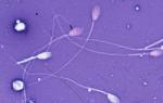
"Newborn sperm" are microscopic germ cells. They are grouped in the testicles in ranks, like soldiers on parade. As they develop, they form an oval-shaped head, a thin neck and a tail (flagellum), long compared to their microscopic size. The sperm contains a set of 23 chromosomes, which are located in the head and contain genes that transmit family resemblance traits to future generations. Spermatozoa move using a flagellum. The blows, reminiscent of a whip, propel them forward on the long journey to the awaiting egg.
Of the multi-million army of sperm released during one ejaculation (ejaculation), only one can penetrate the egg. A fertilized egg develops a special kind of protection that prevents other sperm from penetrating into it. For the normal process of fertilization, it is important not only the formation of a sufficient number of full-fledged sperm, but also a certain composition of the liquid part of the sperm: the optimal concentration of fructose, zinc and calcium ions, biologically active peptides and a low level of acidity. The state of these indicators is influenced by the level of hormones and radiation, the action of certain chemicals and even the psycho-emotional state.
The sperm's tail moves like a snake, bending in several places at once. The lower part of the tail must wag from side to side 800 times for the sperm to move forward 1 cm.
Testicular activity
Testicles can be compared to a conveyor belt, as they work without interruption. The activity of each sperm-producing seminiferous tubule does not stop for a minute. The huge conveyor line moves tirelessly forward without smoke breaks, lunch breaks or overnight downtime. When the finished product comes off the line, some of the cells left behind are halfway through, while others are just beginning to live. At each stage of development, a specific rhythm and speed of movement is observed, which can neither be slowed down nor accelerated. The formation of a germ cell takes a long time, about 72 days. At the end of the production process, not all of them are in perfect shape. Some have no flagellum, others have an underdeveloped head, and others are deformed. This was to be expected with such mass production. Several million poorly formed, unformed sperm do not reduce a man's ability to fertilize. Inside the testicles, male cells can only make small movements.
Activities of the epididymis
The epididymis are long, narrow tubules that lie curled up above both “twins.” When sperm production ends, they move from the testicles to the epididymis. They are not yet sufficiently developed, unable to move properly and fertilize an egg. Sperm motility is an important factor in fertilization ability. To win the race, the male cage must move forward and only forward, without changing direction. Spermatozoa acquire motility only in the initial part of the epididymis. The walls of the epididymal duct secrete fluid, under the influence of which the sperm begin to move. But they still have a poor sense of direction, which forces them to swim in a circle, that is, to remain in place. This means that they would lose the race to the egg in disgrace. The maturation of sperm in the tubules of the epididymis lasts twelve days before they sufficiently learn to swim. At this time, the most sensitive muscles located in the walls of the tubules push them forward. The enormous distance they have to overcome is about 6 m. The liquid nutrient medium serves as food for them, helps them mature and gain the necessary mobility. To sum it up, we can say that the epididymis is a real school of courage.
Short shelf life of sperm
Sperm need to spend 72 days in the testes and 12 days in the epididymis to reach maturity, a total of almost 3 months. Only after this are they ready to set off on the long journey to the seminal vesicles and further to the prostate gland. Mature germ cells accumulate in the epididymis, but not for long. They have a limited shelf life. They remain “fresh” and active for less than a month. After this, they age sharply and soon die. Dead sperm decompose, and the nutrients they contain, including proteins, are absorbed by the testicles. If a man ejaculates only once a month, it seems to him that he is no longer able to impregnate a woman. He thinks his sperm are too old, or dying, or have already died. But in fact, the production of male reproductive cells is a continuous process. Millions of new sperm enter and travel through the epididymis in an endless stream. Although the ejaculated sperm may contain old sperm, along with them there are also completely new ones, ready to begin the race to the egg and realize their chances.
The male reproductive cell develops about 75 days from the moment of its occurrence. Therefore, the consequences of harmful effects may take several months to appear. Some relative guarantee of the correct development of germ cells is provided by strict adherence to nutritional standards. Scientific research suggests that excess weight in men leads to changes in testosterone and estrogen levels? the main hormones responsible for the formation of sperm. In addition, with excess weight, the temperature of the testicles increases, which for successful sperm formation must be lower than body temperature. For the same reason, frequent hot baths are undesirable.
Sweet seed
Sperm (seminal fluid), produced by the male gonads, consists of sperm, seminal vesicle fluid and prostate secretion. Sperm make up on average only 3% of the ejaculate. The remaining 97% is the secretion of the prostate gland and the fluid of the seminal vesicles. In the first portion of ejaculate, the sperm content is higher than in subsequent ones, and especially in the last. The ejaculate contains approximately 300 to 500 million sperm. Sperm is a complex liquid, saturated with various compounds and sugar, and not all components are known. Fructose (a sugar found in semen) may serve as an energy source for sperm, but this remains to be proven. Semen is alkaline, while vaginal secretions are acidic. It is generally accepted that the alkaline substance coats the sperm and protects them while they are inside the vagina. Prostate secretion contains strong antibacterial compounds. The sperm is released in a liquid state, then it quickly turns into a jelly-like state, and after 20 minutes the sperm liquefies again. It is possible that this helps germ cells survive in the vagina. The average volume of ejaculate, provided that orgasm occurs with 3-day intervals, is from 3 to 5 cm; the quantitative expression of the ejaculate may vary depending on age, health status, amount of fluid drunk, and so on. In a partner, sperm can cause an allergic reaction. The allergy manifests itself in the form of a rash or prolonged itching of the reproductive organs. This happens extremely rarely; most often, such symptoms indicate the presence of an infection.
In addition to its direct function of fertilizing the egg, sperm has a positive effect on the woman’s body, except, of course, in those cases when it becomes a carrier of diseases (AIDS, hepatitis, sexually transmitted diseases). Based on this, hormonal contraceptives, on the one hand, are preferable to condoms, on the other? the latter remain the most effective means of preventing infectious diseases transmitted through sexual intercourse.
In a partner, sperm can cause an allergic reaction. The allergy manifests itself in the form of a rash or prolonged itching of the reproductive organs. This happens extremely rarely; most often, such symptoms indicate the presence of an infection.
It is no secret that some French manufacturers use sperm to make cosmetics. This cosmetics is very effective and is not cheap. The thing is that in nature there is no more valuable and unique product than sperm. The cosmetic value of sperm is determined by the presence of extremely useful substances in its composition.
It turns out that the world-famous Viagra and some other popular drugs for impotence do not increase sperm activity, as one might expect, but inhibit it, which negatively affects the ability to fertilize.
Small leak
Before ejaculation, a small drop of liquid wets the end of the penis. It comes from the Cooper's gland and produces a strong alkaline reaction that neutralizes all traces of acid after urination. It cleans and rinses the urethra, preparing it for the passage of sperm. This fluid contains several thousand sperm. There is a theory that this is a "team of superstars" ready to win the race. To avoid conception, even a small part of this fluid should not enter the vagina, otherwise sperm may find their way to the egg. Removing the penis from the vagina just before ejection of semen is called coitus interruptus. This method is often used by young couples trying to avoid pregnancy. However, they are at great risk of becoming mom and dad in nine months. The culprit is often a small drop from the Cooper's gland. Coitus interruptus requires skills and the ability to control one's reactions and manage orgasm, which are most often absent in youth. This can cause a lot of stress for partners. However, many experienced, mature couples choose this particular method of protection, the most ancient and widespread. But it does not protect against infection with sexually transmitted diseases and AIDS, while a condom provides at least partial protection.
The strongest survives
It is generally accepted that only 200 sperm survive the journey to the egg. Some are unable to overcome the very first obstacle - the cervix, while others die while moving through the uterus. Still others may get confused and not get into the right oviduct. A sperm can live in a woman’s birth canal for 2 to 7 days. This is how long it may take for an egg to be fertilized. When it comes to sperm, it is true that quality is more important than quantity. The key issue is the problem of mobility: the cell must swim in only one direction, that is, forward. The average sperm speed is 3 mm per minute. The faster ones have a better chance of reaching the goal before they die. So, speed and mobility are the main conditions for winning races. Those lucky enough to survive accumulate in the widest part of the oviduct. This is where they eagerly await the arrival of the egg. If she is already in place, they gather around her, selflessly trying to break through her protective shell. Wriggling, the sperm sharply hits the outer wall of the cell, releasing chemical compounds that dissolve its protective layer. Eventually small holes appear in the wall and a few lucky sperm make their way inside the egg. From those who succeed, only microscopic heads remain. Now they face the last obstacle, the last bastion that needs to be taken. This thin outer shell protecting the nucleus of the egg is the most difficult obstacle. And only one sperm can overcome it. Perhaps they really will be the best of the best. Its head moves to the middle, and its nucleus connects with the nucleus of the egg. Conception occurs - total implosion, perfect fusion, complete union of two nuclei. According to generally accepted ideas, this reunion is a manifestation of a powerful, all-encompassing micro-force. It is this that determines all the parameters of our personality. Chromosomes join in pairs, once and for all determining a set of hereditary traits. The new life is a perfectly proportional, democratic mixture of the genes of both parents.
Infertility problem
Infertility is the body's inability to produce offspring. According to one study, 15% of American and 12% of English couples face infertility problems, and in 35% of cases this is due to male infertility. In 10-15% of cases, the reason lies in the infertility of both partners. Experts say that there is cause for concern only if conception does not occur within a year of intense sexual activity. Some of them believe that this period should be extended to a full 18 months. Currently, male infertility is becoming more common, and the cause of this phenomenon is unknown. In 1950, the average number of sperm per semen was 40 million higher than in 1988. One of the most important reasons may be overheating of the testicles (staying in hot water is a primitive method of contraception). Tight clothing can act in a similar way, increasing the temperature in the groin and perineum area. Studies to establish the relationship between the type of underwear and fertility have shown that men who wore boxer shorts had a higher sperm count than those who wore tight underwear. Unfavorable environmental factors (radiation, air pollution with lead compounds and other toxic substances, etc.) also have a harmful effect on the quality of sperm. Currently, the prevailing opinion is that they cause much more damage to health than was commonly believed. The testicles are more exposed to harmful environmental influences than the internal organs. Therefore, you should remember that the testicles are an extremely sensitive organ, and avoid anything that involves unnecessary risk.
Insufficient intake of vitamin C (less than 60 mg per day) has a negative effect on sperm health and is believed to affect the occurrence of various disorders in the offspring. Well-known risk factors are tobacco, alcohol and drugs. Anabolic drugs that bodybuilders are addicted to are also very dangerous. Not all men remember the health of their offspring when choosing a profession. And statistics show: among painters, floor polishers and other people who work with paints and varnishes, the quantity and quality of sperm changes, and anomalies are more common in their children. And, for example, the wives of dentists have an increased risk of miscarriage due to the fact that their husbands inhale fumes of narcotic substances that are administered to patients. Studies of the sperm and offspring of computer scientists have so far yielded conflicting results. And yet, experts advise both men and women engaged in such work to interrupt or limit it at least a month before possible conception.
Sperm are most mobile in autumn and winter; at the same time, sperm contains the maximum concentration of germ cells. Scientists recommend the months from October to February as the most suitable for conception. In addition, during these months the likelihood of conceiving a boy is greatest because in the summer, due to the heat, the Y chromosomes, carriers of the male genetic code, are much inferior in viability to the female X chromosomes.
A change in the maturation process of sperm, a decrease in their number, motility, and the presence of chromosomal abnormalities in them can cause male infertility, which, although somewhat less common than female infertility, requires no less thorough research and treatment.
Sperm volume
The amount of sperm sufficient for conception is from 2 to 5 cm. If the ejection volume is less, the sperm becomes thick and viscous, and the sperm are poorly protected from the effects of acidic vaginal secretions. If the volume is larger, then the sperm is too diluted, and there is a high probability of germ cells scattering into the vagina. Don't lose hope! If the results of the analysis are not in your favor, do not despair. In vitro, sperm die much faster than in the body. In vitro they live only from 2 to 6 hours. The stress associated with taking the test and the fear of being diagnosed with infertility can negatively affect the results. People tend to make mistakes, and this can easily happen within the walls of a laboratory. The results may be affected by poor quality packaging, errors in calculations, or improper storage. Perform several (2 to 3) tests over 6-7 weeks, changing laboratory technicians. Only after this, if all the results are clearly negative, decide what to do next. Rare congenital anomalies include dysfunction of the testicular tubules that produce sperm. The germ cells begin to turn into sperm, but most of them do not mature. Currently, highly qualified specialists can separate mature sperm and use them to fertilize an egg outside the woman’s body. Male infertility remains a poorly understood problem. Therefore, try to avoid treatment in clinics that have not received official recognition. Instead of surgery to remove the nodes of the vas deferens or testicular biopsy, you can resort to artificial insemination of the partner with your own or donor sperm. However, these operations are expensive both materially and psychologically and do not always give a positive result. Regardless of your decision, try to feel like a man. Drive away gloomy thoughts, they only increase the state of tension and weaken self-confidence. Don't lose hope and keep trying. You should know that there have been cases where men with hopelessly low sperm counts surprised specialists, their partners, and themselves with unexpected paternity.
Myths about sperm
“You may run out of sperm” This naive and ridiculous idea of the processes occurring in the body is widespread among boys who frequently masturbate. But a surprisingly large number of mature men believe this. Moreover, although the vast majority of men know that the body produces sperm throughout life, this opinion cannot be dispelled. Abstinence does not affect sperm quality in any way. Recently, studies were carried out on sperm 12 and then 120 hours after the last sexual intercourse. Analyzes showed that abstinence had no effect on the shape, motility, or number of sperm. However, long-term abstinence causes a decrease in the number of high-quality sperm.
"Ejaculation depletes the body"
This misconception is closely related to the previous one. For a long time, coaches and sports team leaders demanded that their players abstain from sex at best 4-5 days before the start of important sports competitions. Recently, scientists at Colorado State University studied the physical fitness of athletes who: a) abstained from sex for 5 days, b) had sex within the last 24 hours. They were tested: endurance, readiness for effort, mobility, reaction speed, balance, muscle strength and other indicators important for athletes. The researchers noted "no significant or measurable differences" in both groups of athletes.
"In old age, sperm are no longer produced"
At age 70, sperm production declines. But studies show the presence of sperm in the ejaculate of 48% of men aged 80 to 90 years. Currently, most scientists agree that older men have less viable sperm than younger men. There is a slight increase in the number of deformed sperm, which can cause developmental defects in the conceived child. The degree of risk in such cases cannot be determined, since a man at this age no longer strives to become a father.
Health
When it comes to sperm, people always seem to have questions. Some people want to kill sperm, some want to get them or sell them, some are worried about the work of their “little helpers”. After all, a world without sperm would be a very lonely place. Here are some surprising facts you may not yet know about sperm.
1. Abnormal sperm are normal
The sperm production mechanism in humans is quite lazy. How else can we explain the fact that 90 percent of the sperm in a man’s seminal fluid are deformed? Two heads, two tails, huge heads, a pin-shaped head, a spiral tail - truly this list of sperm deformations can be continued for a long time.
In truth, this is the price we paid for monogamy. In those species where the female receives sperm from more than one male, the sperm have a more uniform appearance. In humans, as a rule, the sperm of two men does not end up in the same woman at the same time.
2. Half a teaspoon
This is the volume that usually comes out when a man ejaculates. It's not much, but one way or another, the sperm manage to do their job.
3. Sperm have tough helmets.
Of course, this is not exactly a helmet, but an oval structure called an acrosome. It contains strong chemicals that are produced when a sperm attaches to an egg. The substance dissolves the outer shell of the egg, drilling a hole through which the sperm can enter the egg.
4. Sperm and sperm
 Some people use the terms sperm and sperm interchangeably. But sperm are just a component of semen or seminal fluid. The seminal fluid also contains substances from the prostate gland, as well as seminal vesicles. Sperm, which are produced in the testicles, require a lot of fuel to move their tail. Luckily, they get this fuel from the sugar fructose, which is supplied by their seminal vesicles. Fluid from the prostate gland, or prostate, contains substances that help the seminal fluid liquefy as it enters a woman. Without this, sperm would not be able to move.
5. One testicle is enough
If a man loses one testicle for medical reasons, the other is usually able to produce enough sperm to conceive a child. Perhaps the most famous example of this was the famous American cyclist Lance Armstrong, who lost one testicle to cancer and became the father of five children.
6. 200 million competitors
It only takes one sperm to fertilize a woman's egg, but there is fierce competition for the honor of doing so. In fact, the average semen contains about 200 million sperm.
7. The factory never closes
 Women are born with a limited number of eggs. But things are completely different for men. Men produce sperm all day, every day, throughout their lives.
As a man ages, sperm become slower and DNA more fragmented, but the factory never closes.
8. Sperm are tiny
Want to see sperm? It is better to get a microscope, since these living creatures are very small to be seen with the naked eye. How small? The length of the sperm is approximately 0.05 mm from head to tail.
Of course, what sperm lack in length, it makes up for in quantity. If it were possible to line up all the sperm released during ejaculation, they would stretch for 9.5 km.
9. Sperm need protection
Sperm look like any other cell in our body, but by the time they leave the testicles, they have half as much DNA as other cells in our body. All this looks suspicious for the immune system. To prevent immune cells from attacking sperm, the testes provide them with special cells that surround them, creating a fence.
10. Dead sperm can create living babies.
 To fertilize an egg the traditional way, sperm need to be able to swim. However, the situation is different in the case of in vitro fertilization. In reality, experts use tiny robotic glass rods to implant a single sperm into an egg. Sometimes they even hit the sperm until it stops moving. After all, the main thing you need is the DNA inside the sperm.
11. Which way to go?
Spermatozoa are capable of pushing themselves, but many have difficulty moving in one direction. In fact, only half of the sperm manage to do this. Others swim in circles, others sway with the movements of the seminal fluid.
But since most of them start, many still get to the egg. This is despite the fact that the tubes connecting the uterus to the ovaries contain small hair cells that create barriers to sperm. If you've ever seen salmon swim against the current, you'll understand what we're talking about.
12. Sperm lives for several days
How long can sperm live inside a woman's body? About two to three days.
13. Y has no equal
 Once the sperm connects with the egg, the chromosomes exchange pieces of DNA, meaning there is a mixture of DNA from the mother and the father. But there is an exception: the Y chromosome has no analogues in the DNA of the egg, and therefore it is passed on practically unchanged from father to son. Because the Y chromosome looks the same as the chromosome of the father, his father's father, and so on through generations.
14. Keep cool
No matter how hot sex is, a man's testicles need to be kept cool, that is, cooler than body temperature, which is important for the production of healthy sperm.
A man's body maintains the ideal temperature of the scrotum with the help of veins, which drive heat away from the muscles of the scrotum that raise and lower the testicles to either bring them closer to or away from body heat.
If a man crosses his legs, the temperature of the scrotum increases. The same thing happens when he wears swim trunks.
15. Two months to create a sperm
How long does it take to produce sperm? According to recent studies, it takes about two months.
Sperm production is continuous, just like a conveyor belt. But just like with a conveyor belt, it takes time to go from start to finish.
A sperm is a male reproductive cell (gamete). It has the ability to move, which to a certain extent ensures the possibility of meeting different-sex gametes. The dimensions of the sperm are microscopic: the length of this cell in humans is 50-70 microns (the largest is in the newt - up to 500 microns). All sperm carry a negative electrical charge, which prevents them from sticking together in the sperm. The number of sperm produced in a male individual is always colossal. For example, the ejaculate of a healthy man contains about 200 million sperm (a stallion produces about 10 billion sperm).
Structure of sperm
In terms of morphology, sperm differ sharply from all other cells, but they contain all the main organelles. Each sperm has a head, a neck, an intermediate section and a tail in the form of a flagellum.. Almost the entire head is filled with a nucleus, which carries hereditary material in the form of chromatin. At the anterior end of the head (at its apex) there is an acrosome, which is a modified Golgi complex. Here, the formation of hyaluronidase occurs, an enzyme that is capable of breaking down the mucopolysaccharides of the egg membrane, which makes it possible for the sperm to penetrate into the egg. In the neck of the sperm there is a mitochondrion, which has a spiral structure. It is necessary to generate energy, which is spent on active movements of the sperm towards the egg. The sperm receives most of its energy in the form of fructose, which the ejaculate is very rich in. The centriole is located at the border of the head and neck. On a cross section of the flagellum, 9 pairs of microtubules are visible, 2 more pairs are in the center. The flagellum is an organelle of active movement. In seminal fluid, the male gamete develops a speed of 5 cm/h (which, relative to its size, is approximately 1.5 times faster than the speed of an Olympic swimmer).
Electron microscopy of the sperm revealed that the cytoplasm of the head has not a colloidal, but a liquid crystalline state. This ensures the sperm's resistance to unfavorable environmental conditions (for example, the acidic environment of the female genital tract). It has been established that sperm are more resistant to the effects of ionizing radiation than immature eggs.
The sperm of some animal species have an acrosomal apparatus, which shoots out a long, thin filament to capture the egg.
It has been established that the sperm membrane has specific receptors that recognize chemicals secreted by the egg. Therefore, human sperm are capable of directed movement towards the egg (this is called positive chemotaxis).
During fertilization, only the head of the sperm, which carries the hereditary apparatus, penetrates the egg, and the remaining parts remain outside.
An egg or oocyte is a specially differentiated cell, adapted for fertilization and further development. Unlike sperm, eggs are not capable of active movement and have a uniform shape: in most animals they are round, can be oval or elongated. The nucleus, as a rule, follows the shape of the egg. It is characterized by a large amount of cytoplasm, which, in addition to the usual organelles, contains a large amount of yolk - a reserve nutritional material for the development of the embryo. Eggs with a large amount of yolk are usually large (fish, reptiles, birds), eggs with a small amount of yolk (lancelet) or without any yolk (mammals) are not large, but are always larger than sperm. The structure of eggs is determined by the content and location of the yolk. Based on these characteristics, the following types of eggs can be distinguished. Alecithal eggs contain no yolk at all. Such eggs are characteristic of placental mammals. Homolecithal eggs contain a small amount of yolk, more or less evenly distributed throughout the cytoplasm (lancelet). The next type is telolecithal. They are characterized by the content of a medium or large amount of yolk, located polar. This type is divided into two subtypes: “medium” telolecithal and “extreme” telolecithal. “Medium” telolecithal eggs contain an average amount of yolk, located in the vegetative part (amphibians). The “extremely” telolecithal type contains a large amount of yolk, also concentrated in the vegetative part (bony fish, reptiles, birds). The centrolecithal type of egg is also characterized by the presence of a large amount of yolk, which is located in the center of the egg (insects).
The presence of a large amount of yolk determines the polarity of the eggs (with the exception of centrolecithal cells). The polarity of eggs is well expressed in amphibians, reptiles, and birds. The upper part of the egg, poor in yolk, is called the animal pole, and the lower part, containing a large amount of yolk, is called the vegetative pole. The mental line connecting the animal and vegetative poles and passing through the center of the egg is called the axis of the egg.
A characteristic feature of the structure of eggs is the presence of membranes. The shells retain the shape and structure of the egg, protect its contents from drying out, and protect from mechanical and chemical influences of the external environment.
Oocyte membranes are divided into three groups: primary, secondary and tertiary.
The primary shell of the egg is formed by the egg itself and represents its superficial compacted layer, it is called the vitelline membrane and is formed before fertilization in the process of oogenesis.
Secondary membranes are produced by the cells that nourish the egg. An example is follicular cells. Often these membranes can be dense and then they have micropiles - openings for sperm penetration.
Tertiary membranes serve to protect the egg; they are formed during the passage of the egg through the oviduct. An example of tertiary membranes is the albumen, subshell and shell in birds.
Eggs are very sensitive to temperature fluctuations, ultraviolet rays, X-rays and radium.
With a relatively small increase in temperature, which animals tolerate painlessly, the eggs die. Increasing the dosage of X-rays, radium, ultraviolet rays is fatal to eggs. It has been established that if the development and fertilization of germ cells is still young, then it is more sensitive to radiation.
Plant tissues
The cells of higher plants are also differentiated and organized into tissues. Botanists distinguish four main types of tissue: meristematic, protective, basal and conductive.
Meristematic tissue. Meristematic tissues consist of small cells with thin walls and large nuclei; There are few or no vacuoles in these cells. The main function of meristem cells is growth; these cells divide, differentiate and give rise to all other types of tissues. The embryo from which the plant develops consists entirely of meristem; As development progresses, most of the meristem differentiates into other tissues, but even in an old tree there are sections of the meristem that allow for further growth. We find meristematic tissues in the rapidly growing parts of the plant: in the tips of roots and stems and in the cambium. The meristem at the tip of the root or stem, called the apical meristem, causes these parts to grow in length, and the cambium meristem, called the lateral meristem, makes it possible to increase the thickness of the stem or root.
Protective fabric. Protective tissues consist of thick-walled cells that protect the underlying thin-walled cells from drying out and mechanical damage. Protective tissues include, for example, the epidermis of leaves and the cork layers of the trunk and roots. The leaf epidermis secretes a waxy, waterproof material called cutin, which prevents water loss from the leaf surface.
On the surface of the leaves there are guard cells - specialized epidermal cells, located in two near each of the stomata - tiny holes leading into the leaf. Turgor pressure in guard cells regulates the size of stomatal slits, and thereby the rate of passage of oxygen, carbon dioxide and water vapor through them.
Some of the epidermal cells of the root have projections called root hairs; these growths increase the surface area that absorbs water and dissolved minerals from the soil. The stems and roots are covered with layers of cork cells formed by a special cork cambium. Cork cells are very tightly “packed”, and their walls contain another waterproof substance - suberin. Suberin prevents water from penetrating into cork cells; therefore they do not live long, and mature cork tissue consists of dead cells.
Main fabric. This tissue forms the main mass of the plant's body: the soft parts of leaves, flowers and fruits, the bark and core of stems and roots. The main functions of this tissue are the production and accumulation of nutrients. The simplest type of ground tissue is parenchyma, consisting of thin-walled cells with a thin layer of protoplasm surrounding a central vacuole. Chlorenchyma is a modified parenchyma containing chloroplasts in which photosynthesis occurs. Chlorenchyma cells are loosely arranged and form most of the internal tissue of leaves and some stems. They are characterized by thin cell walls, large vacuoles and the presence of chloroplasts.
In some major tissues, the corners of the cell walls are thickened to provide support for the plant. This tissue, called collenchyma, is found in the stems and petioles of leaves just under the epidermis. In another tissue - sclerenchyma - the entire cell wall is greatly thickened; sclerenchyma cells, which provide mechanical strength, can be found in the stems and roots of many plants. Sometimes they take the form of long, thin fibers. Spindle-shaped sclerenchyma cells called bast fibers are found in the phloem (phloem) of the stems of many plants. Round sclerenchyma cells called petrosal cells are present in the hard shell of nuts.
Conductive fabrics. Plants have two types of conductive tissue: xylem (wood), which conducts water and dissolved salts, and phloem (phloem), which transports dissolved nutrients such as glucose. In all higher plants, the first cells to form from xylem cells are long cells called tracheids, with pointed ends and ring or spiral thickening of the walls. Later, these cells are connected to each other at their ends, forming wood vessels. During the development of blood vessels, the transverse walls dissolve and the side walls thicken, so that a long cellulose tube is formed to conduct water. These vessels can reach 3 m in length. In both tracheids and vessels, the cytoplasm eventually dies off and leaves empty tubes that continue to function. Thickening of cell walls, accompanied by the deposition of lignin (a substance that determines the hardness and woodiness of trunks and roots), allows the xylem to perform not only conducting, but also supporting functions.
A similar fusion of cells adjacent to each other at their ends leads to the formation of phloem sieve tubes. The end walls do not disappear, but are preserved in the form of plates with holes - sieve plates. Unlike tracheids and wood vessels, sieve tubes remain alive and contain a large amount of cytoplasm, but lose their nuclei. Adjacent to the sieve tubes are “satellite cells” that have nuclei; it is possible that they serve to regulate the function of the sieve tubes. The circular movement of the cytoplasm significantly accelerates the passage of dissolved nutrients through these tubes. Sieve tubes are found in the soft bark of woody stems, lying outward from the cambium.
Animal tissue
Biologists disagree somewhat on how the different tissue types should be classified and how many such types there are. . We will distinguish between six types of animal tissue: epithelial, connective, muscle, blood, nervous and reproductive.
Epithelial tissue. This tissue consists of cells that form the outer covering of the body or line its internal cavities. Epithelial tissue can perform the functions of protection, absorption, secretion and perception of irritations(or several of these functions at the same time). The epithelium protects underlying cells from mechanical damage, from harmful chemicals and bacteria, and from desiccation. Food and water are absorbed through the intestinal epithelial cells. Other epithelial tissues serve to secrete a wide variety of substances; Some of these substances are waste products of metabolism, while others are used by the body. Finally, since the body is completely covered with epithelium, it is obvious that any irritation, in order to be perceived, must pass through the epithelium. Epithelial tissues include, for example, the outer layer of the skin and the tissues lining the digestive tract, trachea, and kidney tubules. Epithelial tissues are divided into six subgroups based on the shape and function of their cells.
Flat epithelium consists of flattened cells shaped like polygons. It forms the superficial layer of the skin and the lining of the mouth, esophagus and vagina. In humans and higher animals, the squamous epithelium usually consists of several layers of squamous cells superimposed on each other; such tissue is called stratified squamous epithelium.
Cuboidal epithelium consists of cuboid cells. It lines the kidney tubules.
Columnar epithelial cells are oblong in shape and resemble columns or columns; the nucleus is usually located closer to the base of the cell. The stomach and intestines are lined with columnar epithelium.
Ciliary epithelium. Cylindrical cells may have on their free surface minute protoplasmic processes called cilia, the rhythmic beating of which propels the material located at the surface of the cells in one direction. Most of the respiratory tract is lined by columnar ciliated epithelium, the cilia of which serve to remove dust particles and other foreign material.
The sensitive (sensory) epithelium contains cells specialized for the perception of irritations. An example is the lining of the nasal cavity - the olfactory epithelium, through which odors are perceived.
Glandular epithelial cells are specialized to secrete various substances, such as milk, earwax, or sweat. They have a cylindrical or cubic shape.
Connective tissues. This type of tissue, which includes bone, cartilage, tendons, ligaments, and fibrous connective tissue, supports and connects all other cells in the body. All of these tissues are characterized by the presence of large amounts of nonliving material that their cells secrete. This the so-called basic substance. The nature and function of a particular type of connective tissue depends largely on the nature of this intercellular ground substance. Thus, cells perform their functions indirectly, secreting the main substance, which serves as the actual binding and supporting material.
In fibrous connective tissue, the ground substance is a dense, randomly and tightly woven network of fibers that surround connective tissue cells and consist of material secreted by these cells. Such tissue is found everywhere in the body: it connects the skin with the muscles, holds the glands in the proper position and connects many other formations. Specialized types of fibrous connective tissue are tendons and ligaments. Tendons are not elastic, but flexible cords that attach muscles to bones. Ligaments have some elasticity and connect bones together. A particularly dense plexus of connective tissue fibers is located under the skin itself (it is this layer that, after chemical treatment - tanning - turns into dressed leather).
Connective tissue fibers contain a protein called collagen. When these fibers are treated with hot water, collagen is converted into a soluble protein - gelatin. Collagen and gelatin have almost the same amino acid composition. The collagen macromolecules that form the fibers are helical structures of three peptide chains interconnected by hydrogen bonds. Since the human body has a lot of connective tissue, collagen makes up about a third of all proteins.
The supporting skeleton of vertebrates consists of cartilage or bone. In the embryos of all vertebrates, the skeleton is formed of cartilage, but in all adult forms, with the exception of sharks and rays, the cartilaginous skeleton is mainly replaced by bone. In humans, cartilage can be felt in the auricle and at the tip of the nose. Cartilage is hard, but has elasticity. Cartilaginous cells secrete around themselves a dense, elastic ground substance, forming a continuous homogeneous intercellular material, among which the cells themselves lie in small cavities, singly or in groups (2 or 4). These cells enclosed in the ground substance remain alive; some of them secrete fibers that are incorporated into the ground substance and strengthen it.
Bone cells also remain alive and secrete basic bone substance throughout a person's life. The ground substance of bone contains calcium salts (in the form of hydroxyapatite) and proteins, mainly collagen. Calcium salts provide bone hardness, and collagen prevents fragility; Thus, the bone acquires strength, allowing it to perform supporting functions. At first glance, the bone appears solid, but in reality it is not. Most bones have a large medullary cavity in the middle that may contain yellow marrow, which is mostly fat, or red bone marrow, the tissue that makes up red blood cells and some types of white blood cells.
In the ground substance of the bone there are canals (Haversian canals) through which blood vessels and nerves pass, supplying bone cells with blood and regulating their activity. The ground substance is deposited in the form of concentric rings (bone plates) forming the walls of the canals, and the cells are walled up in the cavities present in the ground substance. Bone cells are connected to each other and to the Haversian canals by their protoplasmic processes, which lie in the thinnest tubules in the ground substance. Through these tubules, bone cells receive oxygen and various substances they need and are released from metabolic products. Bone tissue also contains cells that break down this tissue, so that the bones gradually change their shape under the influence of the loads and stresses they experience.
Muscle. The movements of most animals are caused by the contraction of elongated, cylindrical or spindle-shaped cells, each of which contains a large number of thin longitudinal, parallel contractile fibers called myofibrils.. By contracting, that is, shortening and thickening, muscle cells produce mechanical work; they can only pull, not push. There are three types of muscle tissue in the human body: striated muscle, smooth muscle and cardiac muscle. Cardiac muscle forms the wall of the heart, smooth muscle is found in the walls of the digestive tract and some other internal organs, and striated muscle forms large masses of muscle tissue attached to bones. The fibers of striated and cardiac muscles have a characteristic feature: unlike all other cells, which have only one nucleus, each fiber contains many nuclei. In addition, in striated fibers the nuclei occupy an unusual position: they lie on the periphery, under the cell membrane itself; this appears to have a role in increasing contractile force. These fibers reach an unusual length for cells - up to 2 and even 3 cm. Some researchers believe that muscle fibers stretch from one end of the muscle to the other.
Under a microscope, alternating light and dark transverse stripes can be seen in the fibers of striated and cardiac muscles, which is why they are called striated. These stripes are obviously related to the contraction mechanism, since during contraction their relative width changes: the dark stripes practically do not change, but the light stripes become narrower. Striated muscles are sometimes called voluntary muscles because we can control their movement. Cardiac and smooth muscles are called involuntary, since a person cannot control their function.
Blood. Blood consists of red and white blood cells (red and white blood cells) and a liquid non-cellular part - plasma. Many biologists classify blood as connective tissue, since both of these tissues are formed from similar cells.
The red blood cells of vertebrates contain hemoglobin, a pigment that can easily absorb and release oxygen. Combining with oxygen, hemoglobin forms an oxyhemoglobin complex, which can easily release oxygen, thus delivering it to all cells of the body. Mammalian red blood cells are shaped like flattened biconcave discs and do not contain a nucleus; in other vertebrates, red blood cells are more cell-like; they are oval in shape and contain a nucleus.
There are five types of white blood cells - lymphocytes, monocytes, neutrophils, eosinophils and basophils. White blood cells do not contain hemoglobin, they are very mobile and can easily capture bacteria. They are able to exit through the walls of blood vessels into the tissue, destroying the bacteria located there. The liquid part of the blood, plasma, carries a variety of substances from one part of the body to another. Some substances are transported in a dissolved state, others may be bound to any of the plasma proteins. In some invertebrates, the pigment that carries oxygen is not located inside the cells, but is dissolved in the plasma, coloring it reddish or bluish. Blood platelets (platelets) are fragments of special large cells found in the bone marrow; they are involved in the process of blood clotting.
Nervous tissue. Nerve tissue is made up of cells specialized for conducting electrochemical impulses called neurons. Each neuron has a body - the extended part containing the nucleus - and two or more thin thread-like processes extending from the cell body. The processes consist of cytoplasm and are covered with a cell membrane; their thickness varies from a few micrometers to 30-40 microns, and their length - from 1 or 2 mm to a meter or more. Nerve fibers running from the spinal cord to the arm or leg can reach 1 m in length. Neurons are interconnected in a chain to transmit impulses over long distances in the body.
Depending on the direction in which the processes normally conduct nerve impulses, they are divided into two types: axons and dendrites. Axons conduct impulses from the cell body to the periphery, and dendrites - towards the cell body. The connection between the axon of one neuron and the dendrite of the next is called a synapse. At a synapse, the axon and dendrite do not actually touch; there is a small gap between them. An impulse can pass through a synapse only from an axon to a dendrite, so that the synapse serves as a valve that prevents impulses from passing in the opposite direction. Neurons have very different sizes and shapes, but they are all built according to the same basic plan.
Reproductive tissue. This tissue consists of cells used for reproduction, namely eggs in females and sperm, or sperm, in males. The eggs are usually spherical or oval in shape and are immobile. In most animals, with the exception of higher mammals, the cytoplasm of the egg contains a large amount of yolk, which serves to nourish the developing organism from the moment of fertilization until it becomes capable of obtaining food in some other way. Sperm are much smaller than eggs; they have lost most of their cytoplasm and acquired a tail, with which they move. A typical sperm consists of a head (which contains the nucleus), a neck and a tail. The shape of sperm varies from animal to animal. Because eggs and sperm develop from ovarian and testicular tissue of ectodermal origin, some biologists classify them as epithelial tissues.
A sperm is a reproductive cell of a male individual, the main purpose of which is to fertilize a woman’s egg. The structure of the sperm, size, functioning and shape during its life cycle are of keen interest to people. After all, such a small reservoir contains the entire set of information that will be transferred from the father to his unborn child.
What elements does a male cell consist of?
The size of the sperm is so small that the structure can only be examined with the help of a good microscope; the measurement takes place in microns. It reaches 55 microns in length and consists of several parts, each of which performs its own functions:
- Head.
- Neck.
- Intermediate section, or body.
- Tail.
A photo of a spermatozoon enlarged hundreds of times allows you to examine its structure. The cavity of the head is filled with chromatin - hereditary material. Otherwise, this part of the head is called the core. The DNA information that will connect to the egg is contained in the most basic part of the male cell, and this part is the nucleus. Its anterior end contains an acrosome, where enzymes are synthesized that will dissolve the membranes of the egg. This is the most significant form of gamete. The head dimensions are: height – 2.5 microns, width – 3.5 microns, length – 5.0 microns.
The neck has a spiral shape, which contributes to the function of generating energy necessary for active movement. The bulk of the energy comes from fructose, which is contained in significant quantities in sperm. The length of the neck is 4.5 microns.

The sperm has a complex structure.
The structure of the sperm includes a centrosome, a form that ensures the motor function of the tail. It is located in the cervical part, behind which its middle part, called the body, begins. Inside it there is a so-called skeleton of microtubules.
The final and most mobile part in the structure of the sperm is called the tail. It is much narrower and longer than the middle part. It reaches 45 microns in length. The movement occurs due to the whip-like movement of the tail section. And its shape is made up of microtubules: two of them are central and nine pairs on the sides.
Despite its microscopic size, the sperm has a functional structure, each element of which is actively involved in the process of achieving the goal.
The process of male cell maturation
The process of formation and ripening of full-fledged gametes is called spermatogenesis. It begins at the onset of puberty and continues throughout the rest of life. Human sperm arises and develops in a special gland - the testicles, which are part of the structure of the male reproductive system.
The average period of sperm development is about three months, which means that sperm are renewed every 90 days. Spermatogenesis is a rather complex process, consisting of various stages of development and division.
The process is controlled and regulated by the functions of the pituitary gland and testicular hormones. While in the male body, gametes are at rest. But during the release of seminal fluid, the enzyme of prostatic secretion is connected to the process, which activates the movement.
Sperm contains a huge number of gametes. The size of the sperm is so small that one milliliter can contain from 1.5 to 2 million. But for successful fertilization, quantity does not play a special role; their mobility, activity and a high percentage of high-quality forms are important. If these conditions are met, the functions of the sperm will be performed and the result will be achieved.
During spermatogenesis, cells of two forms are formed: those carrying the X chromosome or the Y chromosome. In the first case, a female embryo is formed, in the second - a male one. It is believed that cells carrying the X chromosome live much longer. This explains the fact that it is more difficult to get pregnant with a boy.

Sperm motility is important for fertilization.
How does fertilization occur?
Successful fertilization of the egg is the main function of the sperm; this process is quite complex. The oocyte is fertilized by only one sperm. Millions of sperm fight for the opportunity to be the first to reach the goal. The movement begins immediately after the sperm enters the woman’s body. After just 2-3 hours, most of the cells die, and the unfavorable form of the vaginal environment is to blame.
The survivors continue to move, falling alternately into the cervix and then into the uterus. On the way to the egg, gametes have to overcome obstacles in the form of protective mucus, which will be destroyed by enzyme compounds contained in their head part. The egg itself is also covered with a special mucopolysaccharide shell, which will be destroyed at the point of penetration of the strongest sperm.
When using acrosome enzymes, a hole is created in the shell, large enough for the head to enter, while the body and tail disappear. The most important element of the human sperm carries half the genetic information. The fusion of male and female cells results in the formation of a diploid zygote containing 46 chromosomes.

During ejaculation, several million sperm are released.
Ultimately, the functions of the egg and sperm are reduced to a single goal - successful and healthy fertilization. Therefore, the most significant characteristic of sperm is its activity. Due to the structure and functions of the sperm and egg, fertilization becomes highly probable. The presence of specific receptors on the outer shell makes it possible to recognize the chemicals that the egg secretes. The function and structure of the sperm create all the necessary conditions for targeted movement. After the release of seminal fluid, healthy cells that did not die in the vaginal environment continue to move towards the egg. This movement is called positive chemotaxis.
Important: the length of sperm and their number in sperm do not play a role. Their good mobility contributes to the successful achievement of the goal.
Basic information about male gametes
The speed of movement, given the shape of the sperm, and especially its size, is simply enormous. In one minute he is able to cover a distance of 4-5 mm. You can imagine what kind of distance this is if its own length, translated into millimeters, is 0.055. The average length of the fallopian tube is 170 mm, which means that the sperm will need 44 minutes of continuous movement to reach its goal. But in reality this may take several days.
25% - these are the statistics of successful fertilization during sperm release. This applies even to healthy couples. During the release of sperm, the introduction of sperm into the vagina occurs at a very high speed. On average it is 70 km/h.
At the end of the maturation stage, the sperm can live in the male body for a month. Outside the body - about a day, this is influenced by environmental conditions (temperature, humidity, acid level). Sperm is filled with a huge amount of nutrients. Sperm takes up only 5% of the total seminal fluid. All the remaining substance in its composition contains elements of protective and nutritional substances that should maintain the viability of the cell during its progress towards the goal.
In order for fertilization to be successful and the future embryo to develop without deviations, a number of measures can be taken to improve the quality of sperm. Among them are abstaining from bad habits, eating fruits and vegetables, and staying in the fresh air. Not least important are weight control and preference for light foods on the menu. In this way, all elements of the sperm structure will function well and the cells will be more active.
Structure of sperm: 1 - “head”; 2 - “neck”; 3 - middle part; 4 - flagellum; 5 - acrosome; 6 - core; 7 - centrioles; 8 - mitochondria.
Mammalian sperm is shaped like a long thread. The length of a human sperm is 50–60 microns. The structure of a spermatozoon can be divided into a “head”, a “neck”, an intermediate section and a tail. The head contains the nucleus and acrosome. The nucleus contains a haploid set of chromosomes. An acrosome is a membrane organelle containing enzymes used to dissolve the membranes of the egg. There are two centrioles in the neck, and mitochondria in the intermediate section. The tail is represented by one, in some species - two or more flagella. The flagellum is an organelle of movement and is similar in structure to the flagella and cilia of protozoa. For the movement of flagella, the energy of macroergic bonds of ATP is used; ATP synthesis occurs in mitochondria.
The spermatozoon was discovered in 1677 by A. Leeuwenhoek.
It is the nucleus that carries the paternal genetic material. The head, in which the nucleus is located, is equipped with an acrosome at the front, which helps the sperm penetrate the female reproductive cell. The neck and body of the sperm consists of mitochondria and spiral filaments, which ensure the activity of movement of the male reproductive cell.
Sperm motility is its most basic qualitative characteristic. Motility is ensured by the tail of the sperm by making similar strikes. High sperm motility may play a more significant role than their number in the seminal fluid. If only approximately forty percent of the sperm in the sperm are mobile, then this indicates a pathology, in which case the chances of fertilization of the egg are significantly reduced.
Currently, in medicine there is such a term as asthenozoospermia, which is a decrease in the number of motile sperm and a decrease in their speed of movement in the seminal fluid. The reasons why some people develop this pathology are still not fully known. Often this phenomenon can be triggered by the presence of various bacteria in the sperm or infection of the sperm plasma. It is not uncommon for asthenozoospermia to cause infertility in men or congenital fetal pathologies.
Sometimes it happens that there may be no sperm at all in the ejaculate, but instead the presence of other spermatogenic cells; this phenomenon is called azoospermia. Most often, the causes of this pathology are congenital disorders. Sometimes azoospermia can also be a consequence of the influence of toxic strong drugs on the body, such as alcohol, chemicals, radiation.
If the sperm is characterized by complete immobility of sperm, then this may indicate pathologies such as akinospermia or necrospermia. Akinospermia means that the sperm contains living sperm that are completely immobile and are not capable of fertilizing an egg. Often such a disorder can be caused by various diseases of the gonads. Necrospermia, in turn, is characterized by the presence of non-viable sperm in the sperm. Necrospermia is divided into reversible and irreversible. In the case of reversible necrospermia, or as it is also called false, the vital activity of sperm can be restored. If irreversible necrospermia is detected, treatment cannot be carried out; the causes of its occurrence are still unknown.
The lifespan of a sperm when it enters the vagina usually reaches 2-2.5 hours. If the sperm has penetrated the cervix, this period increases to 48 hours. Each sperm carries a Y or X chromosome, which subsequently determines the future sex of the child when the egg is fertilized. Basically, only one sperm can fertilize a female reproductive cell. Moreover, if a sperm that carries a Y chromosome participates in fertilization, the sex of the child will be determined as male; if the sperm has an X chromosome, the sex of the child will be female.