Intestinal infections without diarrhea and vomiting. Symptoms and treatment of intestinal infectious diseases in adults. Rotavirus, what is it?
I. Intestinal symptoms
1. Diarrhea. True diarrhea means the passage of copious loose or liquid feces several times a day.
The origin of diarrhea is varied. Acceleration of intestinal passage due to irritation of motor nerves, impaired absorption of water, dilution of intestinal contents by transudate from the blood or inflammatory exudate play a role in their occurrence. These factors act separately or simultaneously. With functional dyspepsia, there is no exudation.
Each of the factors described does not necessarily lead to diarrhea. Thus, the acceleration of passage in the small intestines may be accompanied by a slowdown in the thick intestines, which leads to increased absorption of water and thickening of stool. The release of water or exudate in spastically contracted intestinal loops will also not cause diarrhea, but only a false urge to defecate.
2. Constipation. The main factor determining the nature and consistency of stool is the motor function of the intestine, and any deviations from the norm are in one way or another associated with various forms of intestinal dyskinesia.
Constipation is characterized by retention of stool longer than the normal daily period, and stool is either completely absent or insufficient, with only the terminal part of the intestine being emptied. The urge to defecate may be completely absent or occur repeatedly, several times a day, not accompanied by complete emptying of the intestines.
The consistency of stool during constipation ranges from hard to watery. Fecal plugs, thickened from prolonged retention in the intestinal loops, especially in the ampulla of the rectum, become the direct cause of constipation. In some cases, patients are forced to release the ampoule with their hands, kneading and removing fecal lumps piece by piece.
The consistency of stool alone does not determine the pattern of constipation. The essence of this disorder is the retention of feces and incomplete cleansing of the distal intestine. Slow excretion of feces may be accompanied by some change in its consistency. Thus, retention in the distal colon leads to abundant absorption of water with the formation of hard fecal plugs, or “stones.” The delay in the cecum is associated with increased fermentation processes, dilution of feces and the development of gases. The consequence of such typhlostasis will be either an abundant release of liquid and mushy fermentative masses, i.e., a state of fermentative diarrhea, or an increased urge to defecate with scanty release of liquid feces from spastically contracted distal intestinal loops, i.e., the so-called state of constipative diarrhea. In this regard, the alternation of diarrhea and constipation in chronic colitis is understandable.
Changes in diarrhea and constipation are the most common symptom of colitis of various etiologies. Delayed stool inevitably alternates with increased excretion of liquid feces. It is important to try to determine the leading symptom against which functional disorders are compensated. Thus, against the background of prolonged constipation, diarrhea is a consequence of mechanical and chemical irritation of the intestines by stagnant feces. The feedback will also be understandable if we take into account that the empty intestine, which has just been cleared of its contents, ceases to receive impulses to defecate, especially since the next exacerbation of diarrhea entails a transition to a strictly gentle diet.
Thus, the alternation of diarrhea and blockage occurs as if in a vicious circle. It can be broken only with the help of careful therapeutic measures, mainly dietary ones.
3. False or constipative diarrhea, characterized by repeated urges to defecate with the release of scanty feces of any consistency: solid lumps in the form of sheep feces or liquid masses due to dilution with exudate, water or fermentation products. Patients themselves usually do not understand the essence of dyspepsia, mistakenly interpreting any increased frequency of urges as diarrhea, while in these cases we are talking about a special type of constipation.
4. False urges can be companions of both diarrhea and constipation. They arise from irritation of the motor nerves of the distal parts of the large intestine, mainly in the sigmoid or rectum, felt either in the left groin or in the anus. While simulating a normal urge, they are, however, not realized due to the absence of a fecal column in the rectum. False urges may be accompanied by the discharge of gases, liquid secretions, mucus or blood (with proctitis) or remain empty.
5. Tenesmus- a type of false urge, but with the obligatory discharge of mucous-bloody films (like “spitting”) and mainly with painful colic. Tenesmus is especially characteristic of the acute stage of dysentery. In mucomembranous colitis, tenesmus is accompanied by the discharge of large films of mucus in the form of ribbons with bloody streaks, which patients mistake for segments of a tapeworm (mucous colic, colica mucosa).
6. Pain in the intestinal area are a common symptom of intestinal diseases, both organic and functional. Unlike stomach pains, they are not associated with meals, but mainly with stool, appearing with the urge to defecate or after it. Pain may occur immediately after stool, accompanied by a feeling of incomplete release or a repeated urge to defecate. Only with transeversitis does pain occur after eating due to the pressure of a full stomach on the irritated transverse colon. The nature of the pain varies. In some cases, there is a feeling of distension and fullness in the abdomen, in others - cramping pain. The most common type is intestinal colic, especially painful when it is not accompanied by bowel movements. In addition to the above-mentioned mucous colic with the discharge of mucus films, so-called gas colic is characteristic, caused by increased formation and delayed passage of gases.
The mechanism of intestinal pain is associated with a number of moments: stretching of the intestinal loops along with their abdominal covering, irritation of the nerve endings in the intestinal wall, tension and twisting of the mesentery of the small intestines along with the nerves passing through it. In this case, painful stimuli are transmitted from the visceral nerves and plexuses through the connecting sympathetic branches to the posterior spinal roots, rising centripetally to the brain and then centrifugally projecting onto the abdominal wall. Painful stimuli emanating from the distal intestinal loops are directly transmitted to the spinal cord along the spinal nerves.
The main role in the mechanism of development of intestinal pain is played by intestinal motility: tone, peristalsis, rocking movements, stretching and spasms of intestinal loops. Expanding pain during intestinal obstruction is associated with stenotic peristalsis in a sealed segment of the intestine. Appendicular pain is either colicky in nature (appendicular colic) or associated with irritation of the peritoneum (acute attack with fever and hyperleukocytosis). With colitis, pain depends on stretching and peristaltic contractions of the inflamed intestinal wall; with enteritis, it depends on irritation of the mesenteric nerves. In diseases of the rectum and anus, pain occurs directly from irritation of the anal nerves. With mesenteric lymphadenitis, pain is the most constant symptom, depending on irritation of the mesenteric nerves or compression of them by inflamed lymph nodes.
It should be noted that in some cases, organic intestinal diseases, even the most severe ones (cancer), can occur for a long time without any pain, which makes their recognition difficult.
7. Proctodynia- pain in the rectum and anus, a feeling “as if sitting on an awl.” If these unpleasant sensations are associated with the act of defecation, then they are caused by an inflammatory process in the anus (sphincteritis, proctitis, fissure, hemorrhoids). Regardless of defecation, similar sensations can be purely functional in nature, being caused by painful spasm of the rectal muscles and hyperesthesia of the anal nerves.
8. Language. Old doctors attached great importance to the appearance of the patient’s tongue, considering it a “mirror of the stomach.” It is more correct to talk about the “gut mirror”, the condition of which is often reflected in the appearance of the tongue. Normally, the entire surface of the tongue is moist, uniformly pink in color, with pronounced papillae, sometimes slightly coated at the root. Pathological changes are expressed in dryness, grayish-yellow or brown plaque, deep furrows that give the tongue the appearance of a “geographic map”, cracks and ulcers (with glossitis). Thick plaque is usually observed with constipation with prolonged fecal retention or during an acute period of intestinal infection. Dryness and brown coloration are a sign of severe dehydration due to vomiting and diarrhea. With sprue, pellagra and dystrophic colitis, atrophy of the papillae resembles Genter's tongue in Biermer's anemia. The condition of the tongue changes rapidly, serving as one of the indicators in the dynamics of intestinal diseases.
9. Flatulence. The amount of intestinal gases (methane, nitrogen, hydrogen sulfide, carbon dioxide) in an adult with mixed food reaches 1 liter. Gaseous products are formed mainly during the breakdown of plant fiber by intestinal bacteria. The main sources of gas formation are legumes, vegetables, cabbage, mushrooms, bread, potatoes, and from animal products - fresh milk. The release of gases per day with a rational diet occurs in an amount of approximately 200 ml (measured using a gas outlet tube). With an abundant intake of beans and soybeans as the main source of proteins, it increases to 2600 ml, and with increased peristalsis even more. On the contrary, with sluggish peristalsis, the reabsorption of gases into the blood increases.
Some of the gases are absorbed into the blood, the rest are released during bowel movements or pass away on their own. Pathological bloating of the intestine can be caused by increased formation of gases with an abundance of swelling foods in food, impaired absorption due to inflammation of the intestinal mucosa (with enterocolitis) or venous stagnation in the abdominal cavity (with circulatory disorders) and, finally, with mechanical or spastic delay in the passage of gases.
Under normal conditions, the presence of a certain amount of gases maintains the tone and peristalsis of the intestinal loops. Excessive flatulence (flatus) causes characteristic disorders: a feeling of fullness, bloating, sharp pain, gas colic, displacement of the diaphragm and heart, neurovascular reactions in the form of headaches, dizziness and even attacks of angina ("gastrocardiac syndrome").
Increased absorption of gases in the intestines causes a very unpleasant sensation of bad breath (foetor ex ore), often taking on the character of a painful obsessive state due to increased suspiciousness. Such subjects, usually pronounced neuropaths, avoid society, dooming themselves to loneliness due to an exaggerated fear of contaminating the air with fetid gases.
10. Peristaltic restlessness, rumbling, moving in the intestines is associated with nervous irritation of the intestinal muscles. The most common cause is functional neurosis. But such symptoms may be the initial expression of intestinal obstruction due to stenosis, strangulation or implantation. Particularly characteristic of stenosis is periodic tension of an isolated intestinal loop (Wal's symptom), sometimes changing its location, as well as the sound characteristic of a stream of liquid passing through a narrow opening.
11. Nausea- a common symptom of various intestinal diseases: acute enterocolitis, constipation, helminthic infestation. The mechanism of nausea is complex and not always uniform. Most often, nausea is an expression of spasm of the small intestines in the area of the plica duodeno-jejunalis or jejunal loops. Reflex irritation of the stomach, reaching pylorospasm and antiperistalsis, causes vomiting and belching. The latter often has a rotten odor due to intestinal gases.
The source of nausea and vomiting is often identified only in parallel with the recognition and treatment of its root cause, for example, cholecystitis or acute enterocolitis: along with the elimination of the underlying disease, nausea also disappears. Sometimes a simple cleansing enema is enough to relieve nausea.
12. Anorexia. A loss of appetite is a symptom of a variety of diseases of many organs and systems or a general infection. A change in this complex unconditioned reflex indicates irritation or inhibition of food centers emanating from various parts of the body, often outside the digestive tract. At the same time, diseases of the latter (anacidic gastritis, colitis) can occur with normal appetite. Anorexia is always an aggravating symptom. With it, the processes of digestion and absorption of food are weak, the amount of food is limited; sometimes there is a feeling of disgust for food. Anorexia can be an indicator of a severe general illness, toxicosis or neurosis. A change in appetite very often serves as a subtle dynamic sign, signaling a turn in the course of the disease.
II Changes from other systems
Stomach. A decrease in the secretory function of the stomach up to complete achylia is a common occurrence in various intestinal diseases. Ahilia appears either as a consequence of an intestinal infection, or as a predisposing moment for it. In both cases, it aggravates the course of colitis.
Less commonly observed is an increase in gastric acidity and secretion of reflex origin due to primary colitis. The presence of hunger pain and pyloric spasm can simulate peptic ulcer disease. When carrying out complex therapy, it is always necessary to take into account the state of gastric secretion.
Liver and bile ducts. As an “internal filter”, the liver and biliary tract system is easily affected by any intestinal infection and infestation, most often with colibacillosis, giardiasis, amebiasis, ascariasis, typhoid fever and paratyphoid fever. In mild cases, the matter is limited to cholecystitis; in more severe cases, cholangitis and hepatitis develop (with ulcerative colitis). Amoebic colitis causes liver abscesses. Hence the need for all chronic intestinal diseases to examine not only bile pigments in urine and blood, but also duodenal contents and liver functions. In addition, disorders of digestion and absorption of food affect bile secretion and metabolic functions of the liver, which worsens the course of colitis.
The pancreas, like the bile ducts, is often the entry point for intestinal infections. For intestinal diseases, it is necessary to examine duodenal enzymes, diastasis in urine and blood, and trypsin content in feces. When performing scatological analyses, it is necessary to take into account the symptoms of pancreatic insufficiency (fat, muscle, connective tissue), especially with fatty diarrhea, which makes one think about chronic pancreatitis.
The cardiovascular system. The close relationship between digestive and cardiovascular disorders is manifested by a number of symptoms, which can be schematically divided into two groups depending on the underlying disease of one or another system. Reflex disorders of the heart and blood vessels in primary intestinal diseases seem especially important. These include heart displacement due to flatulence and constipation, shortness of breath, palpitations and heart pain after eating. Persistent diarrhea leads to dehydration, hypotension and even collapse. Prolonged constipation can cause vascular reactions in the form of headaches and cold extremities. Dystrophic diarrhea with endogenous vitamin deficiency, myocardial dystrophy, and hypoproteinemic edema have a particularly serious impact on the cardiovascular system.
Urinary tract. Cystitis and pyelitis can be associated with infectious colitis, especially in the presence of colibacillosis and putrefactive dyspepsia. Toxic nephrosis, usually of a volatile nature, is observed less frequently. Uremic diarrhea often occurs under the guise of severe ulcerative colitis.
Nervous system. With any intestinal disorders, there are certain symptoms from the nervous system: headaches (with constipation and intestinal intoxication), sleep and memory disorders, weakness, fatigue, irritability, decreased ability to work. The connection between fermentation and putrefactive processes and alternations between excitement and depression is denied by most authors.
Reflex, metabolic and dyspeptic factors are involved in the pathogenesis of neurointestinal connections. Damages to the nervous system are especially pronounced with secondary (endogenous) vitamin deficiencies, in particular with a deficiency of B complex vitamins. The most severe lesions of the nervous system are observed with pellagroid diarrhea.
Urine. A diagnostic role is also played by an increase in the amount of urobilin and bilirubin in the urine (with the involvement of the liver and biliary tract in intestinal pathology), indican (with putrefactive forms of colitis and high intestinal obstruction), the presence of pathological formed elements (with concomitant pyelocystitis), protein and casts ( with nephritis). Oliguria and anuria can occur after profuse diarrhea, polyuria - with dystrophic conditions, pollakiuria - with intestinal dyskinesias.
Blood. Changes in red blood in the form of hypochromic anemia are not uncommon with all kinds of severe colitis and enteritis. Anemia, even pernicious-like, may be the first symptom of stomach and intestinal cancer. Posthemorrhagic anemia complicates all kinds of intestinal bleeding (with ulcerative colitis, hemorrhoids, cancer, etc.).
Biochemical parameters. In addition to the above, the following data are important: residual blood nitrogen (for uremic colitis), calcium content in the blood (for sprue and other forms of fatty diarrhea), vitamins A, B 1 and C (for secondary vitamin deficiencies), prothrombin (for hemorrhagic diathesis and jaundice ), plasma proteins (for dystrophic colitis and vitamin deficiency).
Main functional intestinal syndromes
1. Intestinal dyskinesia
Disorder of intestinal motor function accompanies all kinds of organic diseases (colitis, tumors, obstruction), but can also be of a purely functional nature. Clarification of the pathogenesis of these dyskinesias is therefore crucial in recognizing any intestinal disease.
As an independent disease or syndrome, dyskinesia appears only in the picture of habitual constipation. However, its significance in intestinal pathology is not limited to this, since it usually complicates the course of the most common intestinal disease - chronic enterocolitis of any etiology. At the same time, dyskinesia as an early or intermediate stage prepares for a number of later complications and can contribute to the development of more serious diseases: the example of dyskinesia shows the transition of a functional disorder into organic suffering and back.
The causes of dyskinesia are disorders of intestinal innervation, impaired coordination in the autonomic and central nervous systems, and distortions of unconditioned and conditioned reflexes. A special place is occupied by reflex intestinal dyskinesias due to diseases of other organs, for example, cholecystitis and peptic ulcers. In this case, intestinal spasms are supported by viscero-visceral reflexes emanating from pathological foci of irritation (biliary tract, gastroduodenal zone).
The clinical picture of intestinal dyskinesia boils down to subjective feelings of fullness, heaviness in the abdomen, false urges, a feeling of incomplete release after defecation, and intestinal colic. Dyskinetic constipation also causes a number of general disorders: headaches, cold extremities, fatigue and weakness, decreased ability to work, and depression.
Various forms of intestinal dyskinesia. False diarrhea with repeated discharge of scanty liquid feces due to retention in the large intestine with irritation of its wall.
Discharge of feces of varying consistency:
A) first mushy, then dense; b) first a fecal plug, then mushy feces; c) rapid fecal eruptions associated with hypersecretion or hyperkinesia; d) left-sided (more often) or right-sided (less often) constipation.
2. Intestinal dyspepsia
This term usually refers to intestinal digestive disorders of a functional nature that are not associated with organic diseases of the intestinal tract. Dividing them into separate groups according to etiological principles, we can only offer a working scheme, since the line between functional and organic disorders is blurred, and also because functional disorders (dysfunctions) constitute an integral element of any intestinal disease. Nevertheless, such a working scheme is necessary to understand the etiology of individual clinical forms and develop appropriate therapy.
Gastrogenic dyspepsia. Disorders of the gastric phase of digestion easily lead to disorders of the intestinal phase. The most common form of such gastrogenic disorders is Achilles diarrhea, associated with accelerated gastric evacuation and irritation of the small intestine by abundant, poorly prepared stomach contents. In the initial stage of decompensated gastric achylia, diarrhea is quickly cured with hydrochloric acid or artificial gastric juice. In the future, diarrhea can be supported by secondary enterocolitis, when the gastrogenic factor in pathogenesis fades into the background, giving way to the infectious-inflammatory factor. These secondary enterocolitis give almost the same picture as simple gastrogenic dyspepsia (an abundance of digestible plant fiber and poorly digested muscle fibers), but require different therapy (see below). Coprologically, they differ from dyspepsia by the presence of mucus.
Pancreatic dyspepsia. This form of diarrhea is characterized by: lenteria, creatorrhea and steatorrhea with a predominance of neutral fats. In advanced cases, the feces have a large volume, an oily appearance and quickly harden in the air. Pancreatic dyspepsia is by no means necessary for gastric achylia. Treatment boils down to using a diet limiting fats and rough meats and prescribing pancreatin with bismuth, tannalbin or purified chalk.
Hepatic dyspepsia, that is, indigestion due to insufficiency of liver or bile duct function can affect the functioning of the stomach, intestines or the entire gastrointestinal tract. In the gastric form of hepatic dyspepsia, there are diopeptic symptoms reminiscent of gastritis (heaviness after eating, rapid feeling of fullness, bitter taste in the mouth, anorexia, nausea, belching), especially pronounced after eating fatty foods. The intestinal form is characterized by diarrhea that occurs early in the morning (an “alarm clock” symptom) or after eating fatty foods. Fat intolerance is associated with insufficient supply of bile acids to the intestines, especially with jaundice. Saponified fats and fatty acid crystals predominate in feces.
In other cases, constipation is observed, explained by a lack of stimulating effect of bile acids on the intestines, as well as reflex colospasm. Constipation, in turn, further inhibits the process of bile entering the intestines (vicious circle).
The functional and reflex nature of hepatic dyspepsia is caused by primary damage to the liver or biliary tract. The diagnosis of hepatic dyspepsia is also supported by the beneficial effect of pathogenetic therapy aimed at sparing the diseased organ and carefully stimulating its function - choleretic drugs (Carlsbad salt for constipation, holosas for diarrhea), methenamine, thermal procedures, a diet limited in fats and fried foods.
Fermentative dyspepsia develops due to excessive consumption of carbohydrates. The relative lack of amylolytic enzymes leads to the fact that excess carbohydrates are not digested or are broken down only partially, with the formation of abundant fermentation products, mainly in the cecum and ascending colon. Fermentation processes are further enhanced in the presence of gastric achylia due to a lack of hydrochloric acid, which breaks down the protein shell of carbohydrate products (amilorhexis).
The clinical picture of fermentative dyspepsia is severe only in early childhood. Often this disease leads to dystrophy. In adults, diarrhea with the release of foamy feces, bloating, and mild pain is observed. Feces are sharply acidic, contain a lot of starch grains, digested fiber and fermentative flora (yeast, clostridia, spirilla), but without any admixture of mucus and blood, as is the case with the fermentative form of colitis. The intestinal loops are sharply distended with gases, spastic in places, but slightly painful. Hepatic dullness is covered by the swollen ascending colon, the diaphragm is elevated. The heart acquires a horizontal position, which can cause attacks of shortness of breath, palpitations and discomfort in the heart area (especially in a lying position), which is relieved by sitting and walking. General nutrition suffers little, since the absorption of proteins and fats in the small intestines is not impaired.
The course of the disease is usually completely benign. With a diet limited in carbohydrates and plant fiber, improvement occurs quickly, and in the initial stages, recovery occurs. Prolonged forms are characterized by relapses at the slightest violation of the diet. An aggravating factor is gastric achylia, which reduces the process of aminorexis. Changes in intestinal flora and general weakening of the body open the gates to secondary infection, which easily leads to the development of chronic enterocolitis, with a more persistent course.
Putrid dyspepsia. The onset of this digestive disorder is associated with excessive supply of food proteins, mainly meat, or poor digestion of them. The abundant formation of putrefactive products of incomplete protein breakdown (indole, skatole, tryptophan, toxamines, etc.) causes a number of dyspeptic symptoms: headaches, vasospasms with cold extremities and pallor, hypochromic anemia. Feces are usually scanty, liquid or mushy, putrid odor, grayish-brown in color, sharply alkaline, with a rich content of muscle fibers and connective tissue.
Perversions of chemistry and intestinal flops with this form are more complex than with fermentation. In these cases, it is more difficult to normalize the bacterial flora and chemistry with the help of a contrasting (carbohydrate-fat) diet. Excess carbohydrates easily leads to irritation of the intestinal loops and the release into the intestinal lumen, first of a watery transudate, and then of an inflammatory exudate rich in protein. As a result, a vicious circle is created with a progressive increase in putrefactive processes and dyspeptic symptoms.
To break this vicious circle, it is necessary to radically unload the intestines from pathological decay products and at the same time sterilize it with laxatives and a short course of treatment with sulfonamides (10.0 for 2-3 days). It is also recommended to prescribe an apple fasting diet.
Nervous dyspepsia. This form of intestinal dyspepsia will be discussed in more detail in the chapter on intestinal neuroses. Its characteristic feature is the absence of pronounced colitis, and most importantly, the neurogenic cause of dyspeptic disorders. Neurovisceral connections in some cases are caused by reflex effects of neighboring organs (liver, gall bladder, stomach), in others - by the central nervous system, irritation of the cortex or subcortical centers. In the latter case, intestinal dysfunction in the form of diarrhea, constipation, distension, false urges, colic, etc. is directly related to negative emotions - fear, melancholy, obsessive thoughts, memories, “associations” (conditioned reflexes). Rapid diarrhea is especially typical
under the influence of obsessive thoughts or fear (the so-called bear disease). The sudden release of liquid stool without admixture of inflammatory elements is caused by a long peristaltic wave from the pylorus to the anus, which bypasses all physiological brakes of the ileocecal valve and kinks of the colon. The reason for such diarrhea is either an acute emotional experience, or conditioned reflex irritations of a more complex nature, for example, memories of similar experiences, unfavorable external conditions (being in an environment that excludes the possibility of using the restroom).
A correct interpretation of the nature of nervous dyspepsia, and therefore targeted therapy, is possible only in the light of corticovisceral pathology.
Afternoon diarrhea
A) Biliary diarrhea in the form of crises of sharp pain with a violent urge to defecate and the release of dark yellow or green feces with an abundant content of bile pigments. The equivalent of these diarrhea are “bile crises” with profuse vomiting of bile up to several liters (own observations). The causes of crises are a visceral reflex due to increased nervous excitability. It is based on latent cholecystitis. The discharge of bile stool causes a burning sensation in the anus. b) With colitis, the afternoon urge to defecate occurs from irritation of the transverse intestine from a full stomach. The nature of feces corresponds to the initial place of urge, from where long peristaltic waves come, which reach the rectum. The cause of afternoon diarrhea lies in an increase in intestinal and general nervous excitability.
3. Dystrophy
In contrast to early childhood dystrophy with its debilitating diarrhea, which is a specific nosological unit, dystrophy in adults is an intermediate, passing stage or complication of various intestinal diseases, for example, a very common consequence of severe colitis. Here we will consider only the role of dystrophy as one of the syndromes of intestinal pathology.
General nutritional disorders can develop with any intestinal disease, organic and functional, accompanied by impaired absorption of food in the small intestine. The causes of resorptive disorders may be the accelerated passage of food and atrophy of the wall of the small intestine due to prolonged inflammation (in severe enteritis), emptying of the lymphatic vessels of the mesentery (in tuberculosis or the so-called Whipple's lipodystrophy), but even outside such severe diseases, absorption processes can be disrupted by sharp acceleration of passage in the small intestines of a purely neurogenic nature.
Loss or sharp limitation of absorption in the small intestines naturally leads to a deficiency of the most important nutrients in the body, primarily proteins and fats. This deficiency is inevitable even when, after an accelerated passage through the small intestines, the food gruel lingers in the large intestine, where it thickens after the absorption of water. The consequence of hypoproteinemia is “protein-free” edema due to disruption of oncotic balance. At the same time, hypolipemia and hypoglycemia occur, as well as a deficiency of essential vitamins in the blood and tissues.
Debilitating diarrhea leads to dehydration of the body and thickening of the blood, which changes all biochemical parameters towards their increase: the numbers of hemoglobin and plasma proteins may be higher than normal, but the oncotic balance is maintained. However, this “dry” form of dystrophy is even more severe than the edematous one, giving, in addition to symptoms of protein, fat and vitamin deficiency, a picture of tissue dehydration with changes in neuromuscular functions: convulsions, paralysis, often depressive psychosis and multiple vitamin deficiency, in particular pellagroid type.
Suppression of all digestive functions, weight loss and general disruption of trophism can lead to irreversible changes in the nervous system. A sharp drop in resistance opens the gates to any infection (pneumonia, dysentery, tuberculosis, erysipelas), from which the patient can die.
Fortunately, these severe, irreversible cases under normal nutritional conditions are a rare exception. Much more common are erased, vaguely expressed forms, which, however, have a similar structure of pathogenesis. Here it is only important to emphasize the importance of the dystrophic factor, which complicates any intestinal disease and leaves its mark on the course of the disease. Of particular importance is the relationship between dystrophy and infection, which comes into play secondarily, giving rise to a new phase of the disease or even a new disease, for example, bacterial dysentery, which joins primary functional dyspepsia or dystrophy. Thus, dystrophy can serve as a connecting link between the functional and organic stages of the pathological process.
4. Intestinal autointoxication
Although most of the symptoms attributed to intestinal intoxication depend on other factors - nervous, vascular, allergic, infectious, the possibility of true self-poisoning with intestinal digestive insufficiency is beyond doubt. Thus, during putrefactive processes in the intestines, a number of toxic products are formed, mainly of protein origin: toxamines (histamine, etc.), ammonia, phenols (from tyrosine), indole and skatole (from tryptophan), sulfides and hydrogen sulfide (from cystine).
During fermentation processes, organic acids can have harmful effects:
A) hemolytic effect, b) decalcifying effect due to increased loss of calcium salts, c) acidotic effect due to increased formation of acetone, d) oxalemia due to the formation of oxalic acid from carbohydrates with the help of Escherichia coli (in the cecum).
During putrefactive and fermentative processes, a combination of several harmful factors usually occurs. It is possible to distinguish putrefactive dyspepsia from fermentative dyspepsia, in addition to scatological signs, on the basis of indicanuria, especially pronounced in putrefactive dyspepsia.
Thus, the listed functional syndromes - dyskinesia, dyspepsia, dystrophy and intoxication - are common accompaniments of most intestinal diseases. Appearing either separately or collectively, these syndromes characterize the clinical picture of the entire disease and determine the choice of pathogenetic therapy.
Chronic enteritis and enterocolitis
1. Chronic enteritis
Let us begin a systematic presentation of intestinal diseases with lesions of the small intestine, since diseases of the duodenum (peptic ulcer, duodenitis, diverticulosis, etc.) are closely related to the pathology of the stomach and should be considered when describing diseases of the latter.
The general conditions of the pathology of the small intestine arise from the functional characteristics of this segment of the intestinal tube.
Motor function is determined by two border zones: proximally - the border with the gastroduodenal zone (plica duodenojejunalis), distally - the ileocecal valve. Both of these border areas are active interoreceptors, sources of abundant reflex connections. Thus, gastric contents, entering the jejunum, send the first peristaltic wave, which, if the nervous system is overexcited, can reach the anus, causing immediate diarrhea. Rapid evacuation of the stomach, in addition, gives rise to hypoglycemic reactions due to accelerated absorption of carbohydrates, and is also accompanied by a special “small intestinal shock”. Hypoglycemia and shock complicate numerous diseases of different organs and are directly related to the upper part of the small intestine.
No less important is the absorption process, which is easily disrupted by lesions of the above department. It is clear that any somewhat serious enteritis entails symptoms of a deficiency of nutrients needed by the body, in contrast to colitis, when even severe ulcerative lesions do not threaten absorption processes. Therefore, the transition of the inflammatory process from the large intestine to the small intestine, especially to its upper sections, is always a serious complication.
Secretory disorders of the small intestine play a lesser role in the overall digestive process due to the pronounced replacement role of pancreatic enzymes. However, with diffuse enteritis, both secretory and resorption processes suffer, and therefore, the trophism of the body.
Inflammatory diseases of the small intestines rarely occur in isolated form. Much more often we deal with enterocolitis. However, the involvement of the small intestine shows very clear signs. The predominance of pathology of the small intestine, even with enterocolitis, leaves a clear imprint on the entire clinical picture. However, in some cases, the clinical picture of enteritis is limited to individual symptoms, constituting only one of the components of the general disease.
Examples include giardiasis enterocolitis and mesenteric lymphadenitis. Irritations of the small intestine regularly accompany diseases such as anacid gastritis, cholecystitis, and diseases of the operated stomach.
But enteritis can also appear as an independent disease, most often in one of the following two forms.
1. Jejunal diarrhea are characterized by a violent urge to stool soon after eating (similar to Achilles or Giardiasis). The stools have a greenish color, a liquid mushy consistency, contain mucus closely mixed with feces, and abundant remains of saponified fats (crystals, lumps, needles of fatty acids). Excess fat can even make stool look discolored. This form is described as "soap dyspepsia" (Porges). Characteristic is a state of severe general weakness with a feeling of heat, trembling of the hands and dizziness, up to collapse, occurring immediately after a bowel movement and reminiscent of a hypoglycemic coma. The mechanism of this “small intestinal shock” has been explained in various ways. Some authors associate it with hypoglycemia due to the accelerated passage of food through the small intestines and rapid absorption of carbohydrates, which, with general nervous instability, causes these symptoms. Others attribute the main role to hyperemia in the region of the celiac nerve with reflex hypotension. This explanation seems to us more likely when we are talking about diseases of the small intestine, while hypoglycemia is more characteristic of a number of gastric syndromes, for example, with achylia and after gastrectomy.
2. Chronic enteritis can drag on for many years. The onset of the disease often dates back to childhood. The course is usually mild, not progressive, but with a tendency to transition to enterocolitis.
Causes
Chronic infections play a role, including tuberculosis, stomach diseases (anacidic gastritis, condition after gastric surgery), chronic intoxication (lead), overload with bulky and fatty foods. Infection in the small intestine can be maintained by changes in the bacterial flora (“dysbacteria”), when a decrease in the gastrointestinal barrier and an alkaline reaction of inflammatory secretions contribute to the introduction and development of bacteria that acquire increased pathogenicity. This fact has been proven experimentally by the intestinal cartridge method.
Main symptoms
Rumbling and transfusion in the abdomen, bloating, pain soon after starting to eat, simulating early pain in high-lying stomach ulcers. Insufficient attention is paid to the symptom of vasomotor shock after stool or food. The reason for the latter may lie in a violation of the barrier function of the epithelium of the small intestines as a regulator of the rhythm of food absorption. Diarrhea may be absent for a long time. The acceleration of passage through the small intestine is compensated by a slower passage through the large intestine, where feces have time to fully form, and starch and fiber are digested by bacteria and enzymes. Absorption disorders are manifested by abundant fat residues in the stool, which, with a large amount of fat taken, has a light color. This steatorrhea must be distinguished from other similar disorders (with pancreatitis, mesenteric tuberculosis, sprue).
Of the objective symptoms, first of all, one should note a painful zone of skin hyperesthesia in the region of the rectus abdominis muscle and posteriorly along the left paravertebral line from the last thoracic to the first lumbar vertebra (Porges). The pain point to the left of the navel coincides with that of a peptic ulcer of the small intestine. With regional ileitis, the pain point is in the ileocecal region, at the site of infiltration. With mesenteric lymphadenitis, the pain zones correspond to the course of the mesentery (Sternberg's symptom).
Laboratory symptoms
The characteristic scatological picture boils down to the presence of formed feces, closely mixed with mucus, greenish or light yellow in color, with a high content of saponified fats. Gastric secretion is often reduced to zero. Liver function tests complicated by severe hepatitis are pathological. There is an increased content of indican in the urine, as in putrefactive dyspepsia.
X-ray shows an acceleration of passage through the small intestine: after 2-3 hours, barium enters the large intestine. Sometimes, with fluoroscopy, after 2-3 hours, barium is in the stomach and large intestine, while the small intestines are empty. In other cases, there are levels of fluid in the loops of the small intestines with gas bubbles above them.
Complications. Most often, the pathological process moves to the large intestine, causing a banal picture of enterocolitis. Violation of absorption processes naturally leads to dystrophy, vitamin deficiencies and anemia. Damage to the intestinal epithelium maintains chronic intoxication, which causes a number of toxic and allergic symptoms: urticaria, eczema, Quincke's edema. In the future, the disease may be complicated by cholecystitis, hepatic colic, hypochromic anemia, glossitis, aphthous stomatitis, and anaphylactic diarrhea. Damage to the vessels of the abdominal cavity with the development of thrombophlebitic splenomegaly is also possible. As is known, the history of patients with this disease of the spleen contains indications of either abdominal trauma or chronic intestinal infections.
When differentiating enteritis from other diseases, intestinal tuberculosis, mesenteric lymphadenitis, sprue, and chronic appendicitis should be excluded.
Acute enteritis can be caused by any intestinal infection, possibly a virus or even simple cooling, judging by the literature.
Treatment involves split meals in small portions, slow eating and separate intake of solid food and liquid in order to slow down the evacuation and absorption of food. Coarse vegetable fiber and fatty bulky dishes are prohibited. Meat is recommended in large quantities (up to 200 g per day) minced or softly boiled. For constipation, raw juices, compote, and yogurt are prescribed.
Sample menu. Breakfast: soft-boiled egg, cocoa with water, toasted white (or gray) bread with butter. Lunch: meat broth or pureed vegetable soup with rice, meat cutlet, chicken or boiled lean fish with vegetable puree, fruit jelly. Early dinner: rice porridge on water with butter or cheesecakes. Before bed, tea, crackers, cookies.
For diarrhea, take an infusion of dry herbs (chamomile, mint, dill, clover) 1/4 cup 3-4 times a day.
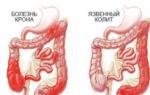
Regional (terminal) ileitis (Crohn's disease)
In 1932, Krohn, Gunzburg and Oppenheimer first described 13 cases of severe enteritis with diarrhea, abdominal pain, exhaustion, anemia, fever, infiltrate in the ileocecal region, complicated by stenosis and fistulas. Half of the patients had scars after appendectomy. Later, similar changes (granulomas) were described in the large intestine and even in the stomach.
Pathological anatomy. Most often, the terminal loop of the ileum is affected, from where the process continues in the proximal direction, less often caudally, moving to the colon, capturing the loops of the intestine that are richest in lymphatic elements. The affected segments are thickened, rigid, the serous membrane is covered with fat and fibrinous effusion. The adjacent mesentery is swollen, thickened, and the lymph nodes are enlarged. The process of the cecum is embedded in the commissures. There are perforations of the intestine into the mesentery, with the formation of fistulas. On resected intestinal loops, the wall is sharply thickened, the lumen is narrowed, there are ulcers, necrosis and mucosal hyperplasia. Histologically, nonspecific hyperplasia of the lymphatic tissue of the submucosal layer is noted. In the lymph nodes there are giant cells without caseation and Koch's bacilli. This lymphogranulomatous process is almost indistinguishable from tuberculosis.
It was hypothesized that this disease develops due to tuberculosis of the intestine by bovine bacilli with good resistance of the macroorganism, with blockage of the lymphatic vessels and secondary infection. Anatomical similarities with Beck's sarcoidosis were also pointed out.
It was experimentally proven that it is possible to reproduce a similar process in the dog’s intestines by introducing irritating substances (fine sand, talc) into the mesentery and lymph nodes, which clog the milky ducts. Subsequent intravenous administration of bacteria enhanced the development of the process.
The etiology and pathogenesis boil down to a combination of an unknown infection with blockage of the lymphatic vessels.
The history often indicates appendicitis and bacterial dysentery, as well as poor nutritional conditions.
Crohn's disease, also known as regional ileitis, is a chronic inflammation of the intestinal wall, most commonly the final part of the small intestine or colon. Inflammation affects the deep layers of the intestinal walls, and ulcers and abscesses can form. Ulcers can completely destroy the wall, creating abnormal passages (fistulas) into other parts of the intestine, other organs such as the bladder or the skin. Deep fissures may also develop in and around the anus. Inflammation can lead to thickening of the intestinal wall and eventually complete blockage of the intestine. Symptoms of Crohn's disease appear in the form of attacks that alternate with periods of normal well-being. Some people have only one or two attacks and then recover; For others, attacks recur throughout their lives.
Causes
. The cause of Crohn's disease is unknown. . Hereditary factors apparently play some role in the development of this disease. . According to some theories, the causes of the disease may be: viral or bacterial infectious diseases, autoimmune diseases, food allergies or lymphatic stagnation. Prevention. There is currently no known way to prevent Crohn's disease, but there are various treatments that relieve symptoms.Symptoms
1) ulcerative colitis syndrome with diarrhea and discharge of mucus and pus without obvious blood. The colon is affected only up to the descending loop, the sigmoid and rectum remain intact. However, in the future the process spreads caudally, so the operation must be performed before the onset of a late stage; 2) small intestinal obstruction syndrome; 3) pseudoappendicitis; 4) intestinal colic; 5) neurotic reactions. . Attacks of pain or cramping, usually around the belly button or lower right side of the abdomen. . Constant watery diarrhea. . Bleeding from the rectum or blood in the stool. . Anal fissures. . Nausea. . Fever. . Fatigue. . Loss of appetite and weight. . Complications in various organs and systems, such as joint pain due to arthritis, inflammation of the eyes and lesions on the skin.Diagnostics
. A medical history and physical examination are necessary. . A blood test may be required. . An x-ray of the small intestine may be performed. . A barium enema may be given. Barium creates a clear image of the large intestine. . A rectosigmoidoscopy (to look at the lower part of the large intestine) or colonoscopy (to look at the entire colon and part of the small intestine) may be done. . A biopsy of the tissue lining the colon is usually performed during a rectosigmoidoscopy or colonoscopy to distinguish Crohn's disease from ulcerative colitis.Treatment
. For mild attacks, patients can take over-the-counter anti-diarrhea medications and pain relievers. . Anti-inflammatory drugs such as sulfasalazine or corticosteroids may be recommended. . Antibiotics may be prescribed to treat secondary viral illnesses. . Enemas containing corticosteroids or aspirin-like medications may be used to combat internal inflammation. . Immunosuppressants may be prescribed for long-term use to suppress autoimmune activity. . Measures such as dietary changes, vitamin or mineral supplements, or vitamin B12 injections may be used to replace nutrients lost due to poor absorption. . Surgery may be required to clear blockages, fistulas, or abscesses in the rectum or intestines. . With severe and prolonged development of the disease, the damaged part of the intestine can be removed. . See your doctor if you experience symptoms of Crohn's disease (especially pain in the lower right abdomen, which may indicate appendicitis). . Call your doctor if you have black or bloody stools, a swollen belly, or a fever. Crohn's disease, also known as regional ileitis, is a chronic inflammationThe disease is complicated by abscesses, perforations, fistulas, and stenoses.
The course is long-term, cyclical, with relapses of diarrhea, anemia, exhaustion, and fever. Diarrhea is allergic in nature, without the presence of certain food allergens.
An objective examination reveals infiltration in the terminal loop of the ileum, general exhaustion, edema, dermatitis, anemia, and leukocytosis. Radiological signs are: “string sign” in the area of narrowed loops and a nipple-like filling defect in the cecum.
Regional ileitis must be differentiated from intestinal tuberculosis, lymphogranulomatosis, leukemia, lipodystrophy, and sarcomatosis.
Treatment in severe cases is only surgical (resection, bypass operations, opening of abscesses, suturing fistulas). Conservative treatment is possible only in the initial stages without dystrophy and stenosis, when the inflammatory process is still partially reversible. Treatment is the same as for chronic ulcerative colitis: blood and plasma transfusions, good nutrition, multivitamins, fresh yeast, large doses of calcium.
Pellarga and SPRU
Both diseases, in which one of the most severe symptoms is debilitating diarrhea, are considered vitamin deficiencies. Even if vitamin deficiency is not the only cause of diarrhea, it still plays a major etiological role.
Pellagra
In the etiology of pellagra, the main role is played by deficiency of nicotinic acid - exogenous, due to insufficient supply with food, or endogenous, due to poor absorption or increased destruction of this vitamin. Deficiency of vitamins C and B1 also plays a role.
The well-known triad of symptoms - diarrhea, dermatitis and dementia (the "three Ds") is not repeated so clearly in any other clinical syndrome. However, individual elements of the same clinical picture (“pellagroid”) are found in all types of severe eating disorders.
Diarrhea, which began as a consequence of vitamin deficiency, subsequently becomes the cause of progression of the disease (according to the mechanism of a vicious circle). Watery stools contain a lot of undigested food debris. Intestinal passage is accelerated throughout the entire tract. Skin changes (hyperkeratosis, hyperemia with transition to brown pigmentation, peeling, weeping blisters), as well as neuropsychic disorders, are the result of deep degenerative, often irreversible changes.
The most effective therapeutic agent is nicotinic acid, administered subcutaneously or intravenously in a 1% solution of 1-5 ml or orally 0.05-0.1 g two to three times a day. At the same time, ascorbic acid and thiamine in a 5% solution of 1-2 ml are injected in the same way.
Sprue
Sprue disease is known in two forms: tropical and endemic. Like pellagra, it manifests itself as a triad of clinical symptoms: diarrhea is combined with glossitis and anemia. The crimson-red tongue with smoothed papillae resembles Genter's tongue in Biermer's anemia. Diarrhea is accompanied by steatorrhea - abundant release of all fat fractions, mainly neutral fats, as in pancreatic insufficiency, but without simultaneous creatorrhoea.
Regular passage of fatty stools is described under the terms idiopathic steatorrhea, endemic (non-tropical) sprue, white diarrhea, Herter's disease or celiac disease of childhood. This disease should not be confused with various forms of symptomatic steatorrhea due to pancreatic disease or lipodystrophy (tuberculosis of the lacteal vessels, mesentery - Whipple's disease).
Pathological changes are expressed in lympho- and plasmacytic infiltration of the wall of the small intestines, atrophy, and less often in ulcerations of the mucous membrane and fibrosis of the submucosal membrane. The same changes, less pronounced, are also observed in the large intestine. The adrenal glands show some atrophy and a decrease in lipid content.
Along with essential forms of sprue, symptomatic, secondary ones due to mesenteric lymphosarcoma and Hirschsprung's disease are also noted.
The pathogenesis of sprue is not fully understood and, apparently, is not uniform. Biochemical disorders are reduced to a violation of the absorption of dietary fats and the phosphorylation process that occurs with the participation of hormones of the adrenal cortex. It is possible that a lack of secretion of bile acids by the liver plays a role. Recently, a certain role has been attributed to folic acid deficiency. The whole syndrome appears, therefore, as a hormonal and vitamin deficiency. However, some authors do not exclude the role of infection in the form of tuberculous lesions of the lymphatic system of the mesentery or typhoid fever suffered in early childhood.
The main etiological factor is considered to be inadequate food, one-sided, excessive carbohydrate nutrition, which, especially in hot climates, contributes to the development of fermentative dyspepsia. Endocrine insufficiency plays a role as a concomitant factor, in particular during pregnancy and lactation.
According to other authors, the main role in the etiology and pathogenesis of sprue belongs to a deficiency of external or internal Castle factor, as well as damage to the absorption capacity of the small intestines after severe infections (dysentery, typhoid fever, tuberculosis).
Fermentative (“gas”) dyspepsia leads in these patients to impaired absorption of fats, calcium, vitamins and antianemic substances in the affected small intestine. From here it is easy to explain all the main symptoms of the disease: fatty diarrhea, hypocalcemia with osteoporosis, anemia and a number of vitamin deficiencies (cheilosis, glossitis, hemeralopia, polyneuritis).
Symptoms
In the first place is diarrhea with the release of grayish-yellow pasty or liquid feces, an acrid or putrefactive odor. Feces contain 45-70% of ingested fat (instead of 6% during normal digestion), mainly in the form of fatty acids and soaps. The release of nitrogenous wastes is not increased. A regular symptom in the period of exacerbation is anemia, the nature of which is variable: in the period of exacerbation - hyperchromic-macrocytic, in the period of remission - hypochromic. With severe dehydration, anemia can be masked by thickening of the blood. Glossitis of the Genterov type, stomatitis and cheilosis are regular signs of vitamin deficiency. Less regular symptoms are polyneuritis of the beriberi type (I.A. Kassirsky) and funicular myelosis. With severe diarrhea, symptoms of deficiency of all other vitamins also appear.
Hypocalcemia is accompanied by symptoms of osteoporosis and muscle spasms with normal calcium levels in the urine. The basal metabolic rate is usually increased. Despite profuse diarrhea, there is polyuria with low specificity. weight of urine, refractory to pituitrin and adiurecrin, i.e. not of pituitary origin, but arising due to insufficient reabsorption in the tubules. The content of proteins, residual nitrogen, cholesterol and blood sugar is reduced. Liver function tests are normal. Rectoscopy does not reveal anything characteristic. An X-ray examination reveals smoothness of the contours of the small intestines, persistent barium retention in individual loops of the jejunum (“fake symptom”), spotty and pinnate relief, and sometimes a megacolon pattern. The patchiness of the relief apparently depends on the accumulation of small lumps of mucus in the small intestines, which can explain the disruption of absorption processes.
The disease usually lasts for many years. The first dysyeptic symptoms (diarrhea, bloating) are often noted in childhood. Women get sick more often than men. The course is always cyclical: long-term exacerbations are replaced by more or less light intervals, when all symptoms subside, even steatorrhea disappears against the background of a low-fat diet. Anemia does not go away completely, but becomes hypochromic.
The cyclical course of sprue is characterized by a change in symptoms during periods of exacerbations and remissions: the onset of remission may be accompanied by the appearance of glossitis, cheilosis and angular stomatitis. Glossitis is observed in 90% of cases and serves as a kind of transitional symptom (from relapse to remission and back). It is not curable by diet, niacin, or riboflavin, but may resolve spontaneously or with liver therapy. Anemia is also subject to dynamic fluctuations, passes with the onset of remission through the phases of macrocytic anemia, pure macrocytosis without anemia with a transition to (normalization of red blood. The outcome is often aplastic anemia.
One of the causes of anemia and cachexia is sodium deficiency, and to a lesser extent, chloride deficiency due to the loss of electrolytes in feces.
In the differential diagnosis, a number of related “diseases” should be taken into account. Lesions of the pancreas (severe pancreatitis, cystic fibrosis) occur with a normal nitrogen content in the stool, neutral fats predominate, urine diastasis is increased. Pancreatic cancer is characterized by severe pain and weight loss. Intestinal tuberculosis pulmonary history, lung damage and the presence of bacilli in the stool are important. With Addison's disease, skin pigmentation, hypotension, and a flattened sugar curve after exercise are observed. Tropical sprue occurs with persistent hyperchromic anemia, otherwise it is difficult to distinguish from endemic sprue and is treated with the same methods.
Pellagra does not cause hyperchromic anemia even in the period of exacerbation, but it is characterized by characteristic dermatitis and mental changes. Biermer anemia occurs with more pronounced hemolysis, normally colored stool, persistent gastric achylia, without cachexia. Among the changes in the nervous system, funicular myelosis is most often noted, and not polyneuritis, as with sprue.
Treatment. In the foreground is a diet aimed at influencing two main digestive disorders - steatorrhea and fermentative dyspepsia.
Some authors solve this problem by recommending a sharp restriction of carbohydrates and an increased supply of fats, which should replenish their loss in feces (10-15 eggs and 50 g of butter per day). However, most doctors put forward another principle of diet, which seems more justified: a sharp restriction of fats with a slightly reduced supply of carbohydrates, which, as experience shows, are better tolerated on a low-fat diet. Among carbohydrates, products with loose fiber (fruits, berries) are most recommended, while limiting starchy foods (bread, potatoes), which enhance fermentation processes in the intestines. During periods of exacerbation, sugar is sharply limited. It is necessary to increase the supply of proteins to 1.5-2 g per 1 kg of weight to combat fermentative dyspepsia and hypoproteinemia. Calcium is prescribed in large quantities both in the form of sour dairy products and as a medicine: intravenously 5% calcium chloride and orally gluconate and calcium carbonate. To increase its absorption, daily injections of 1 ml of parathyroidism are indicated.
The effect on anemia is achieved with the help of iron and vitamin B 12 supplements. Liver drugs are effective only when administered parenterally, in contrast to Biermer's anemia, when internal methods also help. Treatment of vitamin deficiency requires the use of all vitamins.
The effect of folic and folinic acid (10-15 times more effective than folic acid), as well as vitamin B 12, was studied mainly in tropical sprue. An increase in reticulocytosis and an improvement in the composition of red blood were noted in some patients treated during the period of remission. Folic acid potentiates the hematopoietic effect of vitamin B 12. Plasma transfusions help normalize blood protein levels. The use of pancreatin in doses up to 3.0 per day is indicated. Corticohormones and ultraviolet irradiation are recommended as general stimulants.
A complete cure for sprue is hardly possible. However, long-term remissions make it possible to restore relative working capacity for a long time. For illustration, we present excerpts from case histories.
The role of tuberculosis was reduced to the influence of a secondary aggravating factor, which was layered on the main background of vitamin deficiency. The factors of vitamin deficiency, dystrophy and secondary infection were closely intertwined, which made it difficult to identify the leading factor in pathogenesis and etiology, and only dynamic observation made it possible to clarify the sequence and relationship of individual elements of this complex nutritional disorder.
Chronic enterocolitis
This section of intestinal pathology presents the greatest difficulties in terms of nomenclature and classification. The terms “colitis” and “enterocolitis” refer primarily to inflammatory processes in the intestines, as opposed to purely functional conditions.
Naturally, the line between functional and organic suffering can be blurred in some cases, for example, when functional constipation is complicated by inflammatory reactions with the development of “constipative colitis.” The feedback relationship between functional disorders due to the primary organic process is also clear, i.e. colitis can occur with constipation, when both conditions mutually aggravate each other, according to the “vicious circle” mechanism. And yet, with all the closeness of organic and functional diseases, they can be studied separately in order to gain a clearer understanding of the pathogenesis of intestinal diseases.
An attempt by some authors to distinguish between two pathogenetic groups of colitis - infectious and alimentary - has not found wide recognition, since the first group can be considered as dysenteric, post-dysenteric, protozoal colitis, etc., and the second - as intestinal dyspepsia and dystrophic enterocolitis.
The widely used term “spastic colitis” is completely unsuitable from a clinical point of view. Under this mask, a variety of conditions are hidden: reflex colospasm due to certain diseases of the abdominal organs, vegetative reactions due to general neurosis, as well as any form of colitis accompanied by intestinal spasms. In each individual case, a detailed examination will be required to clarify the etiology. The diagnosis of “spastic colitis” is usually only a forced designation for all sorts of unclear intestinal diseases.
Syndromes of multiple bowel diseases
1) spastic syndrome with alternating constipation and diarrhea; 2) hyperkinetic syndrome with diarrhea and acceleration of the entire intestinal passage; 3) pain syndrome with normal or delayed stool, intestinal colic and severe pain in the intestinal tract; 4) toxic syndrome with symptoms of general intoxication (headaches, weakness, fatigue, sleep disturbances); 5) anemic syndrome due to intestinal bleeding and slow blood regeneration; 6) dystrophic syndrome due to severe enterocolitis with impaired absorption and utilization of food and multiple hypovitaminosis.The listed syndromes, although not associated with a specific etiology, give a typical pathogenetic imprint of the disease and help in the choice of differentiated therapy.
The study of the causes of chronicity of colitis deserves great attention. Most chronic colitis develops from acute forms, and this transition occurs either directly, without visible recovery, or imperceptibly, after an intermediate hidden stage of apparent, false recovery. The latter forms are more difficult to recognize, their etiological roots can remain hidden for a long time.
But there are also forms with such an imperceptible tendency towards a gradual, persistent course that one gets the impression of primary chronic colitis, occurring like chronic appendicitis. This includes many cases of intestinal giardiasis and trichomoniasis, and possibly other chronic infections, even bacterial dysentery, when the onset of the disease is not specified by anamnesis, the disease proceeds cyclically with alternating exacerbations and remissions over a number of years.
The cause of chronicity can often be accurately determined. With bacterial dysentery, it usually consists of delayed recognition, late initiation of treatment and insufficient radicality and duration of therapy using too weak doses, which contribute to the development of sulfonamide-resistant strains of bacteria. In other cases, for example, with invasion by Giardia and amoebas, these protozoa become encysted and disappear from the intestinal lumen; they take refuge in the submucosa or deep pockets and become inaccessible to specific drugs.
Finally, in a number of cases, the chronicity of the lesion is supported by weak body resistance, vitamin deficiency, anemia, dystrophy, which delays the fight against invasion for a long time and interferes with recovery. Resistance to infection may precede or develop as a result of the disease itself. In such cases, specific therapy usually turns out to be powerless and must be replaced by general restorative means of influencing the macroorganism, for example, blood transfusions, glucose with vitamins, enhanced nutrition, and climatic treatment.
Pathomorphology of enterocolitis
The old division into “superficial” and “deep” colitis (in other terminology - severe, ulcerative, parietal) does not cover the entire amount of morphological changes occurring in the intestinal wall during enterocolitis. In addition, a morphological sign does not always serve as an indicator of the severity of the disease. Only in the most severe, irreversible forms of colitis (amoeba, tuberculosis, dysentery) do we deal with deep lesions of all layers of the intestinal tube, up to penetrating and perforated ulcers, fistulas, stenoses, reticulosis, granules, tumor-like infiltrates.
In acute and subacute processes, the most common are the usual inflammatory changes (swelling, hyperemia, increased secretion of mucus, easy soreness and bleeding). Inflammatory exudate may contain leukocytes, plasma cells, erythrocytes, deflated epithelial cells, and soluble protein. In clinical conditions, these inflammatory changes are detected using laboratory and rectoscopic studies and partly according to X-ray examination of the relief.
In chronic processes, deeper lesions (wall rigidity, adhesive adhesions, stenosis and deformation of intestinal loops) are established during life mainly by radiology. These intravital diagnostic methods make it possible, along with a physical examination of the abdomen, to monitor the dynamics of anatomical changes, which are partially reversible with treatment.
Autopsy data also indicate a wide variety of morphological changes in the intestinal wall during colitis. The anatomical process in some cases occurs diffusely, in the form of “pan colitis,” often spreading to the small intestines. In other cases, it is nested, focal in nature, localized in certain parts of the intestinal tube. A specific topography is characteristic of the infiltrative-ulcerative form of intestinal tuberculosis with its favorite localization in the cecum (tumor ileo-coecalis), with a tumor-like infiltrate and caseous decay. Regional ileitis produces similar gross morphological changes in the terminal ileum, but without the participation of bacillary factor.
The histomorphology of colitis is characterized by exceptional diversity, an abundance of cytological variants (proliferation of lymphocytic, plasmacytic, reticuloendothelial elements according to the type of reticulocytosis). Individual layers of the intestinal wall are also affected differently: from hypertrophic and polypous changes to complete atrophy, then the intestine turns into a thin-walled tube with almost complete disappearance of all parenchymal elements, by analogy with atrophic gastritis. Unlike the pathology of the stomach, when in chronic gastritis inflammatory processes alternate or are combined with degenerative restructuring of the epithelium, in colitis inflammatory changes predominate. It is noteworthy, however, that in a number of cases there is a striking discrepancy between the clinical picture of dystrophy and the nature of the anatomical process, when instead of the expected thinning of the intestinal wall, hypertrophy and swelling of the folds of the mucosa are detected. Thus, in patient 3., who died from dystrophic enterocolitis, the section revealed hypertrophy of the intestinal wall with the proliferation of reticular elements of the submucosal layer.
The main anatomical forms of enterocolitis (catarrhal, follicular, diphtheritic and ulcerative-necrotic) do not exhaust the variety of morphological changes in the intestinal wall. Tuberculosis is characterized by caseous decay, and amoebiasis is characterized by the formation of granulomas (“amoebas”) with the possibility of malignancy, which is not characteristic of bacterial dysentery.
Ulcerative processes in the intestines often reflect the specific etiology of the disease. Thus, ulcers in dysentery, amoebiasis, tuberculosis, lymphogranulomatosis and cancer, regional ileitis and ulcerative colitis have a morphological imprint characteristic of this etiological factor. But there are also uncharacteristic changes characteristic of any chronic inflammatory process.
In bacterial dysentery, three morphological stages are described: catarrhal-diphtheritic, ulcerative and regenerative. As with all anatomical processes, in the intestine, especially with ulcerative lesions, changes in the peripheral autonomic nervous system are of great importance.
There is no doubt that damage to nerve endings and plexuses (Auerbach's, Meissner's, solar, aortic) occurs not only in ulcerative forms, but also in all other severe intestinal diseases, and possibly in functional dyspepsia, dyskinesias and anatomical anomalies such as megacolon and dolichosigma , in which denervated areas of the distal colon are observed, explaining movement disorders during the expansion of intestinal loops.
Lesions of the peripheral nervous system best explain trophic, vascular and motor disorders in all forms of colitis and serve as a link between functional and organic diseases.
Lesions of the lymphatic vessels and mesenteric nodes are important, causing severe disorders of fat absorption. This applies to various forms of mesenteric lymphadenitis (tuberculous, etc.), so-called intestinal lipodystrophy (Whipple's disease), regional ileitis (Crohn's disease), celiac disease (Herter's disease), etc.
Acute diarrhea, frequent passage of watery stools, is not a disease, but rather a sign of an underlying medical condition. As food passes through the digestive system, water is absorbed through the walls of the large intestine. Diarrhea and dehydration occur when fluid is not absorbed but remains in the intestines and is excreted in the feces. Although diarrhea usually clears up without treatment within two or three days, any resulting dehydration is a serious problem (especially among infants and older adults) and needs to be treated quickly.
Causes
Food poisoning due to various causes, including viruses or bacteria. . Viral infection of non-food origin. . Reaction to certain foods (for example, citrus fruits or beans). . A large number of artificial sweeteners found in foods, chewing gum and other products. . Alcoholic drinks. . Certain medications, including medications for hypertension, heart disease, and some antibiotics. . Infectious diseases such as traveler's diarrhea, typhoid fever, amoebiasis and bacillary dysentery (shigellosis). . Emotional stress and anxiety.
Symptoms
. Watery stool. . Increased bowel frequency. . Cramps and pain in the abdomen. . Signs of dehydration in young children: drowsiness; indifference; tight skin; glassy eyes; dry, sticky mouth and tongue; constant crying. . Eat cooked foods and fruits that you can peel yourself. Local water or raw food may contain bacteria that cause diarrhea. . Find ways to relieve emotional stress and try to avoid stress.Diagnostics
Diarrhea is easy to identify by its characteristic symptoms. . A laboratory stool test may be performed for persistent diarrhea. . A colonoscopy with a rectal biopsy can help diagnose diarrhea caused by viruses, bacteria, or inflammation.
Treatment
Prevent dehydration (especially important for older people and young children) by drinking a solution of one teaspoon of salt and four teaspoons of sugar in one liter of water. Measure accurately as too much salt can increase dehydration. Drink 0.5 liters of solution while diarrhea continues. . Do not take anti-diarrhea medications for the first few hours (diarrhea may be ridding your body of infection carriers or irritants). If work or other obligations require the use of anti-diarrhea medications, use one that contains loperamide (such as Imodium) or bismuth subsalicylate (Pepto-Bismol). . Reduce (or avoid) dairy, alcohol, and fiber-rich foods during your recovery. . For young children: While diarrhea persists, do not feed them milk. Instead, give them an electrolyte solution, which can be purchased at the pharmacy. If the diarrhea goes away within two days, start giving milk gradually over 24 hours. . Call your doctor if diarrhea persists for more than 48 hours or is accompanied by dizziness, severe cramping, a temperature higher than 38.3°C, or blood in the stool. . Contact your doctor if diarrhea occurs frequently. . Attention! Call a doctor immediately if a young child or elderly person shows signs of dehydration.
Prevention
Do not eat food if you think it has gone bad. . Avoid foods to which you are sensitive. . When traveling abroad, drink only bottled or boiled water.
Bacterial dysentery
Despite the long-known etiology, well-studied typical characteristics of different types of bacteria and rich epidemiological experience, bacterial dysentery continues to remain the focus of attention of doctors. The reasons for this interest are clear. Dysentery is still one of the most difficult infections to eradicate, since contact infections persist throughout the year almost everywhere. The complete elimination of dysentery is made difficult by the constant presence of small foci, mainly in the form of carriers and excretors of bacteria - people who are practically healthy, but have recently been ill or have not even been ill at all. In addition, a delay in recognizing and isolating patients with acute dysentery easily leads to dispersion of infection and epidemic outbreaks, which are much more difficult to eliminate than individual, sporadic cases. Dangerous epidemic foci are also maintained by unsanitary conditions, especially during the hot months of the year.
In the first days of the disease, positive cultures from mucous-bloody stools are observed in 85% of cases, from mucous stools - in 18%, and from stools uncharacteristic of dysentery - only in 9%.
Bacterial dysentery is a potentially dangerous and extremely easily transmitted infectious disease of the colon. Symptoms appear after an incubation period of one to four days and usually subside after 10 days. In severe cases, the illness can last up to six weeks, but in most cases the illness is mild. Dysentery is most common in children aged one to four years. It is common in overpopulated areas with poor sanitation and often occurs in epidemics; To stop the spread of the disease, sick people are isolated and quarantine is introduced in the area of the epidemic.
Symptoms
Initially, watery diarrhea. It can progress to diarrhea with mucus and blood. . Tension during bowel movements, accompanied by pain in the rectum. . Abdominal pain; pain all over the body. . Nausea and vomiting. . Fever. . Rapid dehydration and weight loss (young children and older adults are especially susceptible to dehydration).
Treatment
Electrolyte solutions (such as sodium and potassium) may be needed to prevent dehydration. In severe cases, they can be used intravenously. Until you see your doctor, stay hydrated by using sports drinks or a solution of one teaspoon of salt and four teaspoons of sugar per liter of water. It is important to prepare the solution correctly as too much salt can increase dehydration. Drink half a liter every hour while diarrhea persists.
Do not take over-the-counter anti-diarrhea medications unless your doctor recommends them. With the help of diarrhea, the body gets rid of infectious agents.
Although the infection mostly goes away on its own, antibiotics are often given to limit its spread. Medicines must be taken for the entire prescribed period.
Isolation from other people is necessary to prevent the spread of the disease.
See your doctor as soon as you notice signs of dysentery. The disease is dangerous and spreads extremely quickly, so you need to see a doctor as soon as possible.
Prevention
To prevent the spread of infection, wash your hands frequently with warm water and soap, especially after bowel movements or before eating. (Because dysentery has an incubation period of up to four days, you could be a carrier of the disease without knowing it.) When traveling abroad or in areas with poor sanitation, drink only bottled or boiled water or other bottled drinks, and eat only cooked foods and fruits that you can peel yourself.
Symptoms
Typical bacterial dysentery begins acutely, after a short, often hidden prodromal period of general malaise (corresponding to the phase of absorption of toxins in the intestines). The initial symptom is diarrhea with the release of loose stools, soon followed by tenesmus with the discharge of mucous-bloody films and cramping abdominal pain. Tenesmus reaches a frequency of 20-40 or more per day. The severity of the disease is determined by high temperature, frequency of tenesmus, general condition of the patient, activation of previous diseases (malaria, tuberculosis, hidden septic lesions). An aggravating factor is the age of the patients: dysentery is especially dangerous and has the highest mortality rate in infancy and old age.
The pathogenesis of diarrhea in dysenteric colitis is toxic, similar to malarial colitis. Late and inconsistent damage to the small intestine is explained by local immunity of the small intestinal mucosa.
Immunity to dysentery is usually very unstable and only rarely lasts 2-3 years. Hence the frequency of relapses, reinfections and chronic forms of the disease. Relative immunity still apparently occurs, since healthy people have a positive agglutination reaction, the diagnostic value of which, however, is small.
The entire course of the disease can be divided into three periods (not counting the prodromal period). The first period, lasting 2-3 days, is characterized by symptoms of acute irritation of the nerve endings of the distal colon with the release of first liquid feces, and then films of mucus, blood and pus, with a high temperature reaction. Depending on the severity, this stage is also marked by a number of general toxic symptoms (headache, drowsiness, fatigue). It is especially pronounced when infected with Grigoriev-Shiga-Kruse bacilli, which form endotoxins. In mild and atypical cases, tenesmus disappears within a few hours or may be absent altogether, making diagnosis difficult.
In the second period, there is a subsidence of tenesmus, a drop in temperature, the disappearance of general intoxication, the appearance of loose stools due to additional damage to the small intestines and the removal of primary colopasm, which delayed the passage of feces. This enterocolitis is already commonplace, uncharacteristic and can drag on for 2-3 weeks. An aggravating factor is gastric achylia, which developed due to a dysentery infection or preceded it. During this period, symptoms from the liver and biliary tract, activated by intestinal infection, may appear. Dysentery bacteria in feces disappear when acute symptoms are eliminated, but can linger until complete clinical recovery in deep encysted foci of intestinal folds.
The third period is marked by early complications in the form of pyelocystitis, nephrosis, hepatitis, polyneuritis, polyarthritis or arthromyalpy, general dystrophy. Irritation of the thyroid gland and interstitial medulla sometimes causes prolonged tachycardia and sweating. Toxic reactions from various organs and systems can drag on for many months. Post-dysenteric arthritis and neuritis, as well as general nutritional disorders, are particularly persistent.
In severe forms, symptoms of dehydration (cholerine type) develop in the form of dry tongue and skin, convulsions, collapse, cold extremities, and hypotension. We also had to observe a picture of real meningitis, which subsided within (several days.
In recent years, the general “regime” of dysenteric diseases has changed significantly. The Grigoriev-Shiga forms have become very rare, inferior in frequency to the Sonne and Flekener forms. The clinical picture has also changed significantly, in which it is not the previous acute hemorrhagic forms with tenesmus and multiple ulcers on the colon mucosa that predominate, but milder, erased forms of the disease. Severe complications, especially from the joints and nervous system, are also much less common.
In the blood picture in the acute period, the most constant is the band shift of neutrophils, which is almost never found in food infections (salmonellosis). There is often a slightly increased leukocytosis (ie 12,000-15,000). An increase in the number of leukocytes to 70,000 is observed only in the toxic form, often simultaneously with erythrocytosis, due to irritation of the reticuloendothelium and blood thickening. Increased lymphocytosis is observed less often. The toxic form, in addition to leukocytosis, is characterized by the phenomena of nutritional dystrophy, necrosis of the intestinal wall and general sepsis from a secondary purulent infection.
Complications of dysentery affect numerous organs and systems. Otitis, iridocyclitis, polyarthritis, pericolitis, mesenteric lymphadenitis, pancreatitis, and less commonly myocarditis, encephalitis, and paraplegia are observed. In addition, there are complications due to secondary infection in the form of bronchitis, pneumonia, and pleurisy. Children often have stomatitis, gingivitis, mumps, pyelocystitis, and nephritis. Sepsis may develop due to pyogenic and anaerobic infections, especially after intestinal perforation.
Nowadays, thanks to timely and vigorous therapy, these complications have become very rare.
Many patients, after suffering from dysentery, experience achylia of the stomach, usually of a transient nature. However, in some cases it is replaced by persistent anacid gastritis.
Diagnostics
Dysentery differs from ordinary infectious diarrhea in the presence of blood. . A physical examination and medical history are necessary. . Stool samples are taken for examination under a microscope and for culture of bacteria to confirm the presence of dysentery bacillus. . A blood test may be taken to look for abnormalities in ion concentrations or to determine anemia.
Bacteriological diagnosis turns out to be positive on average only in 40-50% of cases of infection, despite technical improvements (Zhagar et al.), and in chronic dysentery even much less often. An increase in the percentage of positive answers is possible only with 3-5 repeated tests on the same patient. Therefore, it is quite correct to propose that any acute intestinal disease that occurs against the background of an epidemic outbreak be considered dysentery, regardless of a negative stool test.
Naturally, technical defects in bacteriological research (remoteness of laboratories and their overload, incorrect taking of substrate, lack of high-quality media, etc.) necessitate clarification of clinical diagnostics. In typical cases, this is not difficult: an acute onset, an increase in temperature and, most importantly, characteristic tenesmus with the release of muco-bloody films - a fairly typical picture, almost never encountered in acute colitis of other etiologies.
However, this acute stage can be so short-lived or erased that by the time you first contact a doctor, the disease already gives the impression of banal enterocolitis, especially if the disease is associated with food intoxication.
Such cases often give rise to diagnostic errors when acute dysentery is visible. In these patients, during the initial stool examination, no dysentery bacteria are found; the patients are discharged with a diagnosis of “food intoxication,” but upon secondary hospitalization, one or another type of dysentery bacteria is already detected.
All this gives grounds for infectious disease specialists to consider every case of acute enterocolitis as dysentery and to demand the immediate isolation of such patients in special departments, not only against the backdrop of summer epidemics, but also under normal conditions, all year round. This strict approach is justified from an epidemiological point of view. But it also has a downside: a certain percentage of non-dysenteric patients end up in infectious diseases departments, which does not exclude the danger of contact nosocomial infection. . This often forces the less conscious population to hide intestinal disease from the district doctor. An even greater danger of such a one-sided approach to the etiology of intestinal diseases is that it reveals a whole series of colitis of a different nature, especially protozoal and salmonella.
A further aggravating factor is the hot summer season with an abundance of flies that transmit infections from sewage to humans, contaminating food and drinking sources, with the danger of infection from increased consumption of raw water and cold drinks, and contamination of food with dusty hands. It is clear that the fight against dysentery comes down mainly to issues of housing and food sanitation and hygiene - personal and mass.
In the first place is clinical diagnosis, taking into account the overall picture of the disease, which in typical acute cases is almost impossible to mix with any other disease. With more erased forms, the epidemiological situation, the multiplicity of diseases in limited foci, as well as anamnestic data on a previous similar illness and contact with dysentery patients are taken into account. In addition, the clinic has the last, decisive word in very frequent cases when bacteriological testing gives a negative result.
Clinical diagnosis is fairly firmly based on the typical picture of the disease: acute onset, headaches, fever, abdominal pain, tenesmus and the type of intestinal discharge. Milder forms occur under the guise of banal enterocolitis, gastroenteritis or food intoxication. There is a well-known parallelism between the clinical picture and the type of pathogen. Thus, Sonne sticks give a more violent onset with high but short-term fever, mild tenesmus and a predominance of symptoms of gastroenteritis. Flexner's dysentery occurs as ulcerative colitis with a more protracted febrile period. The Grigoriev-Shiga forms are more rare in our country; they are characterized by a severe course with pronounced intoxication. This clinical-bacteriological connection, however, is far from constant. The nature and severity of the clinical picture, as well as mortality, are also associated with the characteristics of each epidemic.
An important method is the bacteriological examination of stool or intestinal mucus taken through a proctoscope. We have already discussed the limitations of this method, which rarely gives more than 70% positive results even in the acute stage of the disease. One hundred percent positive answers are possible only in the fourth or fifth analysis, and then subject to the complete exclusion of medications. The first analysis at best gives 70%, the second - up to 84% of positive answers. There is even a proposal to increase the number of tests to nine. It is clear that such requirements in the context of the fight against dysentery are generally impossible to fulfill.
The following factors play a role in increasing the frequency of positive findings: the technique of taking the substrate, repeated, fairly frequent tests, proximity to the laboratory and the quality of the culture media. A single negative answer does not exclude bacterial infection. The early period of the study is also important, while the bacteria are still in the intestinal lumen and have not been eliminated or encysted in deep intestinal foci.
The accuracy of the bacteriological response is especially important not so much in the midst of an epidemic, when all cases of acute colitis can practically be considered as dysenteric, but in sporadic diseases, as well as in the general dynamics of observations of those who have recovered from acute dysentery. An accurate assessment of the moment of bacteriological recovery is very important. As a rule, this moment is determined by three consecutive negative tests over 5-7 days. Here, the research technique is especially important to avoid unnecessary delays in hospitalization due to the alternation of negative tests with positive ones. Even five negative consecutive tests were established for workers in food shops and childcare facilities.
Thus, bacteriological examination data are especially important for confirming the diagnosis of dysentery, establishing the timing of discharge from the hospital, and avoiding bacilli carriage as a source of new infections.
Serodiagnosis. A relative help for bacteriological research is the agglutination reaction (“dysenteric Vidal”). According to the literature, with Sonne type infection, this reaction is positive on the 6-10th day of illness in 50% of patients, on the 10-15th day - in 96 % (E.M. Novgorodskaya). According to other data (E.D. Ravich-Birger), agglutinins appear in the blood serum of 62% of dysentery patients on the 2nd day, in 73% - on the 3rd. The reaction is considered positive for Flexner cultures 1: 200, for Shiga cultures 1: 100. The precipitation reaction with hapten gives, according to E. D. Ravich-Birger, up to 60% positive results.
Scatological diagnosis is unreliable. The presence of a large number of leukocytes in the stool is by no means characteristic of a dysentery coprogram. The appearance of the discharge is typical only in the initial period of tenesmus. Later, during the period of enterocolitis with liquid or mushy feces, the coprogram no longer has anything characteristic.
Sigmoidoscopy for acute dysentery reveals catarrhal changes (swelling, intense hyperemia, slight soreness, bleeding of the mucous membrane), deposits of mucus and pus, and minor erosions. In later stages, as well as in chronic dysentery, characteristic ulcers of various shapes and sizes are noted. Ulcers form in areas of angiospastic and trophic defects and wall necrosis. The mechanism of their formation is predominantly neurotrophic, as in the case of damage to the gray tuberosity or peripheral sympathetic nerves (in animal experiments). In the rectum, oval-shaped ulcers with thickened edges and periulcerous hyperemia are especially common. The bottom of the ulcers is covered with purulent-fibrinous or mucous deposits. Deeper ulcers actively bleed and are covered with hemorrhagic clots. Necrotic changes around ulcers lead to infiltrative thickenings of the entire intestinal wall, but without developing into true granulomas and stenoses characteristic of amoebiasis. In a number of cases, copious discharge of pus in the form of real pyorrhea is described, which can cause purulent “cases” or “muffs” on the stool and lead to a state of septicopyemia. The factor of vitamin deficiency and dystrophy plays a role in the occurrence of ulcers, especially in chronic dysentery with a decline in general nutrition. Such ulcers in post-dysenteric stages have nothing characteristic and do not differ from other forms of trophic ulcers. Hence the great difficulty in differentiating dystrophic ulcerative colitis from dysenteric colitis. Anamnestic data on acute dysentery should speak in favor of the latter.
However, often only banal symptoms of proctosigmoiditis are observed (marbling, hyperemia, increased vascularization, swelling of the mucous membrane, mucus deposits), characteristic of acute colitis of any etiology, even non-infectious ones (for example, with hemorrhoidal or constipating proctitis). Therefore, basing the diagnosis of dysentery only on such nonspecific signs is extremely risky.
Timely diagnosis of acute bacterial dysentery allows for early isolation and effective therapy, localizing the epidemic focus and preventing the development of chronic forms, complications and bacilli carriage.
Differential diagnosis
Amoebas and aza are characterized by stool in the form of raspberry jelly or gelatinous lumps, funnel-shaped ulcers on the rectal mucosa and, of course, mobile amoebas or cysts in the feces. Regular examination of freshly obtained warm stool is necessary to exclude other forms of protozoal colitis. Colitis due to food toxic infections (hemorrhagic form) is difficult to distinguish from acute bacterial dysentery. However, even hemorrhagic forms of colibacillosis and salmonellosis usually occur with a picture of initial gastroenteritis, and not sigmoiditis, without tenesmus typical of dysentery. In children, acute hemocolitis can be caused by intussusception of the colon; in older people, tenesmus with bloody discharge is a common symptom of rectal cancer.
Chronic dysentery
The transition of an acute form of dysentery to a chronic form changes the “face” of the disease so much that it seems like a new disease, a new nosological unit. All signs and properties of the disease change: course, clinical picture, outcomes, response to treatment. Chronic forms, along with the associated carriage of healthy and recovered patients, are the main source of persistence of infection throughout the year and the reason for its difficult elimination.
The frequency of chronic forms depends on treatment tactics in relation to the acute period and, according to various sources, ranges from 25 to 75% of all acute dysentery diseases. The causes of chronicity lie mainly in the late and insufficiently vigorous treatment of the acute stage. In this case, bacteria resistant to sulfonamide drugs can develop, which take refuge in hermetic foci, in the deep folds of the intestinal mucosa.
Concomitant diseases, especially helminthiasis and protozoal infection, are aggravating factors. According to A.F. Bilibin, pure forms of chronic dysentery (without the participation of a second infection) occur only in 20% of cases. It should be noted that, thanks to vigorous anti-dysenteric therapy, the number of chronic cases has decreased significantly in recent years.
Chronic forms are especially common in early childhood. Duration of the disease up to a year occurs in 9% of cases, from 1 year to 5 years in 59%, over 5 years in 32%. Chronic dysentery is characterized by an alternation of true relapses (with activation of remaining bacteria) and exacerbations due to superinfection or eating errors. These options can be distinguished by determining the serological types of intestinal flora.
The transition to the chronic form occurs in some cases almost immediately after the acute stage, in others, more often, after an intermediate period of apparent recovery. All symptoms of colitis may disappear for several weeks or even months (the exact period is not established), after which a relapse occurs. In some cases, the relapse seems to repeat the picture of acute dysentery, in others it occurs less clearly, under the guise of banal enterocolitis.
Chronic dysentery is characterized by a sluggish, recurrent course, and exacerbations are usually associated not with a violation of the diet, but with an accidental new infection of the intestines, sometimes only with nervous irritations.
Diagnosis of chronic dysentery is much more difficult than in the acute form. The frequency of bacteriological data does not exceed 25-40% due to the difficulty of culturing bacteria from deep intestinal lesions. With erased forms, a precipitation reaction with a hapten sometimes helps.
Clinical diagnosis encounters certain difficulties due to the blurred picture of the disease. Characteristic dysentery symptoms in their original form, characteristic of the acute form, are relatively rare, and are inferior in frequency to the symptoms of banal enterocolitis. Due to diagnostic doubts, some experts consider all chronic diarrhea to be dysenteric. Progressive dystrophy allegedly speaks in favor of a specific etiology. Such statements, it seems to us, require great caution due to the abundance of similar clinical forms of other, non-dysenteric etiology (protozoa, salmonellosis, tuberculosis). The development of dystrophy is possible on any soil. The instability of the uniform (dysentery) position is obvious, if only in view of the frequent failure of antidysentery therapy. Only a detailed examination of patients, careful weighing of anamnestic and differential diagnostic data can clarify this complex problem and give a strong weapon into the hands of therapists.
The above does not at all deny the fact of the frequency of cases of chronic dysentery, which even many years after the onset of the disease is amenable to targeted treatment.
Treatment of dysentery
In the acute form of dysentery at a time when sulfonamides were not yet used, the most effective remedy was the early administration of a laxative, especially castor oil, for the vigorous evacuation of all parts of the intestines. Subsequently, bacteriophage was prescribed, and then tannalbin, bismuth, and chalk as astringents. Currently, we have at our disposal a large supply of sulfa drugs and antibiotics, which, with early and vigorous use, provide almost one hundred percent recovery from acute dysentery. It is recommended to use two sulfonamides simultaneously, combining their local action with a resorptive one and choosing the least toxic drugs. In first place are sulgin and phthalazole, and among the resorptive ones are norsulfazole and sulfodimezin. Dosage: 0.5 of each drug, in the first 2 days 6 times a day, on the 3-4th day - 4 times, on the 5-6th day - 2 times; total for the course 22.0-25.0.
Gramicidin enemas (50-100 ml of 0.4% solution every other day), a total of 6-10 enemas, as well as a decoction of eucalyptus, 15.0-300.0 tablespoons orally, are a good help. Painful tenesmus can be soothed with microenemas of chamomile or a weak solution of manganese.
For toxic forms, injections of bivalent serum against dysentery bacteria of the Flexner-Sonne species with desensitization according to Bezredka are indicated. For collapse - caffeine, camphor, infusion of saline with glucose.
After the first treatment stage, which stops the acute period of the disease, two more stages of treatment are carried out. The second stage prevents early relapses. Antibiotics are given in combination with sulfonamides. Levomycetin works best and is least toxic, starting with a loading dose of 1.0, then 0.5 five to six times a day, up to 15.0-20.0 per course.
The third stage prevents late relapses and is carried out against the background of practical recovery. The most effective combination of these two antibiotics with sulgin, as well as a course of treatment with biomycin (10.0-15.0 in single doses of 0.2).
In chronic dysentery, sulfonamides and antibiotics are less effective than in acute dysentery, having only a weak bacteriostatic effect on the secondary intestinal flora. The main goal of therapy is to influence the macroorganism to increase its resistance and reduce hypersensitivity. Phytoncides in the form of enemas from a decoction of garlic are less potent.
Treatment of complications of dysentery. The most persistent are infectious polyarthritis, second only to long-term physiotherapy (warm salt-pine, radon and sea baths, ion-galvanization, short-wave diathermy, etc.). For neuromyositis, injections of 5% thiamine chloride (vitamin B1, dibazol 0.005 each) are indicated. Local novocaine therapy in the form of subcutaneous injections of a 0.25% solution in the area of muscles and joints is also very effective.
Treatment of collapse, dystrophy and dehydration is carried out using conventional methods.
For children with chronic dysentery, special sanatoriums have been organized, where long-term restorative treatment courses are conducted.
Diet for dysentery
Main objectives of the diet:
1) fight against intoxication and starvation, 2) accelerated elimination of bacteria and toxins from the body, 3) mechanical sparing of the large intestine due to its ulcerative lesions.
The course of diet therapy is divided into several cycles.
First cycle, during the period of fever and acute intestinal irritation with tenesmus: broth, pureed vegetable soup, sweet tea with lemon, rosehip infusion, fruit jelly are given (except blueberry and blackcurrant, which make it difficult to recognize blood in the stool).
Second cycle, 3-4 days after normalization of temperature, cessation of tenesmus: vegetable purees, white crackers, softly cooked porridge with milk and half and half water, raw juices (100-200 ml).
Third cycle, after the appearance of fecal stool, but in the presence of enterocolitis with diarrhea: steamed chopped meat and fish dishes, stale white bread, mashed cottage cheese, cheesecakes, and yogurt are added.
Fourth cycle transitional to the general table, in the period of residual effects of enterocolitis: complete, varied food in a mechanically gentle form (vegetable soups, boiled meat and fish in pieces, compotes, baked apples, coffee with milk).
The calorie content of the table increases from 1500 to 3000 calories. During the third and fourth cycles, with prolonged diarrhea, two apple days are carried out: 4 times a day, 250 g of freshly mashed raw apples without adding other food.
In chronic dysentery, even more than in the acute period, it is necessary to observe the principle of adequate nutrition, with minimal mechanical sparing. The most important task of diet therapy for these forms of dysentery is to increase the body's defenses in the fight against prolonged toxic infection.
Due to the weariness and cyclical nature of the diet, the diet cannot be uniform; it must correspond to the stage of the disease. In the phase of sharp exacerbations, a diet is prescribed for the second or third cycle, with a rapid expansion to the fourth cycle. In the calm phase, it is necessary to provide all whole foods in various forms of preparation, an abundance of complete proteins (meat, fish, cottage cheese, kefir) and vitamins in the form of raw juices, yeast dishes, bran infusions and concentrates. It is necessary to limit only fresh milk, spicy seasonings, particularly coarse bulky dishes and foods such as fresh black bread, chops, and sauerkraut. It is advisable to prescribe fasting days (raw mashed apples). Any delay in mechanical sparing is not only not indicated, but is even harmful.
Along with measures for timely isolation of patients, home hospitalization is permitted.
Discharge from the hospital should be made after the disappearance of clinical manifestations, but not earlier than the 7th day from the onset of the disease. Employees of food enterprises, public catering networks, water supply systems, children's institutions, hospitals, sanatoriums, as well as children attending child care institutions, persons who have suffered acute intestinal infections, can be discharged after clinical recovery and after at least three negative bacteriological tests at intervals of 1-2 days.
Food poisoning
Intestinal diseases caused by the introduction of certain harmful substances with food are united into one general group: bacteria from the group of salmonella and cocci, toxic substances formed in infected, contaminated (contaminated) and spoiled products, and chemical poisons. The variety of etiological factors in these cases is associated with a single mechanism of infection directly through food and drink.
The main importance of this group of diseases is sanitary and hygienic. But clinically they are also of some interest, especially in their rarer, atypical forms, sometimes mistaken for acute appendicitis, dysentery, typhoid or septic condition.
The etiology of food poisoning is very diverse.
1. In terms of frequency, infection with bacteria from the Salmonella group ranks first. Of the 160 types in this group, 15 are pathogenic for humans. Typing is carried out by tests for the absorption of agglutinins and reactions to somatic, flagellar and viral antigens. The most common are Salmonella paratyphoid A, B and C, murine typhus (Breslau), swine cholera, Derby, Thompson, Newport, enteritis (Gertner). Infection occurs through meat (80% of cases of poisoning), especially cattle meat (50%), fish, milk, eggs, egg powder (mainly from duck eggs), potatoes, peas, and beans. 2. Coccal infections. 3. Botulism. 4. Intoxication, bacterial (endotoxins), nutritional (alkaloids, toxamines, ptomains) - from spoiled or poisonous foods (mushrooms, beans, potatoes). 5. Chemical poisons (heavy metals, arsenic, etc.). 6. Allergic diseases. 7. Psychogenic dyspepsia, mistaken for food poisoning.
Cases of epidemic food poisoning have been described, when up to 7 strains of staphylococci were isolated from the nasopharynx and feces of patients, as well as from food (salad, ham), which served as a source of poisoning.
The danger of foodborne diseases lies not only in the severity of the disease, but mainly in the possibility of mass outbreaks from certain local foci. The number of sick people sometimes reaches several hundred, and the severity of the disease may depend not only on the virulence of the pathogen, but also on the characteristics of the macroorganism. Food poisoning is most severe in children, the elderly and people weakened by previous diseases, such as gastritis, hepatitis, colitis, and psychoneurosis.
One should also take into account the element of seasonality, the influence of summer heat, an abundance of flies, slight contamination of food with dirty hands, increased thirst, drinking from contaminated sources, and increased sweating. A temporary decrease in gastric acidity in hot weather also plays a role, causing a weakening of resistance to foodborne infections. The undoubted fact of the sharp increase in these diseases in the hot months requires particularly strict sanitary and food control and increased personal and public hygiene during the hot season.
Symptoms
The most common diseases from the salmonellosis group deserve special attention. They include the following forms.
1. Gastroenterocolitis is trivial with the usual gastrointestinal symptoms (vomiting, diarrhea, intestinal colic). If these symptoms develop soon after poisoning, the disease progresses rapidly, but quickly ends, partly due to the regulatory mechanisms of self-cleaning of the body (vomiting and diarrhea). With a longer incubation period, toxic symptoms (headache, fever, cardiac weakness) have time to develop within 12-24 hours and the disease drags on, although it usually still retains a completely benign character.
2. Pseudodysenteric colitis is especially difficult to distinguish from bacterial dysentery against the background of an epidemic outbreak, when every case of enterocolitis, even without bloody diarrhea, should be epidemiologically regarded as true dysentery. The absence of a band shift is too unreliable a sign. The diagnosis of food poisoning becomes probable in case of mass infections from the same source. In sporadic cases, the diagnosis of dysentery is excluded in the presence of rapid disappearance of bloody discharge and repeated negative stool tests.
3. The septic form with prolonged intermittent fever and severe intoxication is most often caused by infection with Salmonella suipestifer or Cholerae suis.
4. The pseudotyphoid form is caused by infection with paratyphoid C and occurs under the guise of a “miniature” form of typhus, with an 8-10-day temperature curve, bloating, colic, mild diarrhea, and an enlarged spleen.
Similar enterocolitis can be caused by a number of other bacteria, especially Proteus bacillus, Escherichia coli, etc.
Treatment. Apart from such exceptional cases when recovery is impossible due to irreversible morphological changes in the parenchymal organs and nervous system, most food poisoning responds well to therapy aimed at quickly removing the infection from the body from food. The most urgent measures are lavage of the stomach and intestines, taking a laxative and a starvation diet. Symptomatically prescribed: stimulants, belladonna, glucose with insulin (for liver irritation), from the 2nd day sulfonamides in an antidysenteric dosage, but usually in a shortened course (3-4 days). A gradual expansion of the diet is carried out under the control of residual effects of enterocolitis and taking into account the need to prevent constipation - a common consequence of such a disease.
For some time, fresh milk, fatty meat dishes, canned food, spicy seasonings and snacks should be excluded from the diet. In the presence of gastric achylia, fried and stringy meat, fatty dishes and sauces are excluded. For residual dyspeptic symptoms, often associated with gastritis, warmed mineral waters (Essentuki, Slavyanovskaya) are prescribed, 1/2 cup half an hour before meals.
Prevention. Massive. prevention involves measures of sanitary and hygienic control over food enterprises and catering departments, including systematic medical and bacteriological control of service personnel. Personal prevention consists of following the rules of food hygiene (washing hands before eating, washing raw foods, cleanliness in food preparation, removing food waste, fighting flies). These precautions are especially important during the hot months of the year, when food poisoning tends to become more frequent.
Botulism
Botulism is caused by poisoning from Clostridium botufinum toxins. The first cases were described in connection with poisoning from sausages, and then from canned meat, fish, vegetables and fruits, mainly homemade, without the addition of acidic preservatives that destroy bacilli and their heat-stable spores. Botulism is considered the most dangerous form of food poisoning, causing up to 70% of deaths. Fortunately, this disease has become very rare in our country thanks to high sanitary and food technology and control. The severity of the disease depends not only on the high virulence of the infection, but also on the duration of the incubation period (6-36, on average 24 hours), during which toxins have time to have a depressing effect on the central nervous system. Early symptoms of gastroenteritis (vomiting, diarrhea) are not specific to botulism. They are only a consequence of a secondary food infection. Specific symptoms appear suddenly and are observed in the nervous and vascular systems in the form of severe adynamia, diplopia, nystagmus, mydriasis with a sluggish reaction to light, paralysis of the external and internal eye muscles. Bulbar symptoms include difficulty speaking, swallowing and breathing. Inhibition of the salivary glands causes dry mouth, along with increased secretion of sticky, thick mucus. Paralysis of the smooth muscles of the digestive tube leads to delayed gastric emptying, constipation and flatulence. Paralysis of the stomach with retention of eaten food in it can serve as a diagnostic sign based on the presence of botulism toxins in food debris.
The pulse is initially slow; subsequently, tachycardia develops, which, in combination with subnormal temperature, is one of the characteristic symptoms of botulism. Common symptoms include apathy and drowsiness, followed by periods of anxiety, agitation and insomnia. Consciousness is maintained until death, which occurs from paralysis of the vasomotor centers, often with symptoms of motor aphasia. Pneumonia is also a terminal symptom. Patients die within the first 3-6 days, sometimes as early as 48 hours after the onset of the disease.
Recovery is slow, and residual paresis of the skeletal and ocular muscles can last for several months.
In differential diagnosis, encephalitis and diphtheria paralysis must be excluded. Other severe food poisoning infections are characterized by high fever and a predominance of gastrointestinal symptoms, in contrast to botulism with its typical triad: a long latent period, hypothermia, and nervous symptoms. Wood alcohol poisoning is characterized by loss of consciousness, convulsions, and blindness.
Among the poisonous foods that cause intestinal damage, one should point out mushrooms (pale toadstool, alkaloid, muscarine), some types of fish (marinka), sprouted potatoes containing poisonous solanine, and powdered eggs contaminated with duck salmonellosis. All these products cause gastroenterocolitis. White bean poisoning (favism) causes hemoglobinuria, hemolytic jaundice, anuria, azotemia and hemorrhagic colitis. In Italy, fatal cases of poisoning of children with bean flour added to sausage liver have been described.
Regular cow's milk can cause mild gastroenteritis not only due to individual intolerance, but also in completely healthy individuals after drinking milk during late milking, when milk becomes “conditionally harmful.”
Poisoning by overwintered grain causes severe illnesses such as septic tonsillitis (alimentary-toxic aleukia).
Chemical poisons
Among the large number of chemical poisonings that make up the special subject of toxicology, we will point out only a few food-related sources.
Copper. The cause of poisoning is the use of poorly tinned kitchen utensils, in which toxic compounds of soluble copper and fatty acids are formed from contact with sour and fatty foods. In addition to the usual symptoms of gastroenteritis, thirst, blue-greenish vomiting, burning in the esophagus are characteristic, and late resorptive symptoms include tremors, anuria, and collapse.
Zinc. It is dangerous to eat liquid warm food cooked in galvanized pots or tanks (violation of the rules of culinary sanitation). The symptoms are the same as for copper poisoning.
Lead. One should keep in mind mainly chronic intoxication due to prolonged use of poorly tinned dishes. Symptoms: loss of appetite, weakness, anemia, gum fringes, stomatitis, lead colic, constipation, muscle paresis. Acute poisoning causes vomiting with white films and bloody diarrhea.
Arsenic. Rare cases of poisoning have been caused by eating confectionery and beer containing molasses contaminated with arsenic.
Cases of poisoning due to ingestion of sodium fluoride and anabasine sulfate, used as insecticides in the fight against kitchen pests, have been described.
Prevention of these poisonings falls within the scope of sanitary and food inspection. Individual acute diseases should be treated like any acute gastroenteritis.
Food allergies
Food allergies include sporadic cases caused by individual increased susceptibility to certain foods,
completely benign, but causing general and local pathological reactions. Allergic reactions occur partly in the digestive organs (gastroenteritis, hepatocholecystitis), partly in other body systems (chills, fever, rashes, swelling). Allergies can also explain isolated cases of poisoning by products that are well tolerated by most people, but cause reactions in particularly sensitive subjects.
Most food allergens belong to proteins, often animals (dairy, fish, meat), the rest belong to plant products (certain varieties of berries and vegetables).
Here is a list (by no means complete) of products that can cause allergic reactions (by frequency per 500 cases of food allergies), based on skin tests (according to Alvarez).
Onions (mostly raw)....... | |||
Pork. . . | |||
Milk, cream, ice cream........ | |||
Tangerines. . | |||
Apples (raw).... | Bananas. . . | ||
Boiled cabbage. . . , | |||
Chocolate....... | Oranges. . | ||
Radish........ | Sweets | ||
Tomatoes....... | Spices. . | ||
Cucumbers........ | |||
Eggs......... | |||
Fats, fatty seasonings | |||
Melon (cantaloupe). . . | Fruits. . . | ||
Beef....... | |||
Strawberry... ... | |||
Coffee......... | Polka dots. . . | ||
Lettuce... . . | Potato. . | ||
Dried beans..... | |||
Cauliflower. . . . | Chickens. . . |
In addition to those listed, there are 104 more types of food, the consumption of which causes diseases to occur less frequently.
Tuberculous diseases of the intestines
We will consider three main clinical forms of tuberculous intestinal diseases: the so-called tuberculotoxic dyspepsia, mesenteric lymphadenitis and ulcerative-infiltrative tuberculosis. They all have the same infectious etiology, but give a completely different clinical picture. Their pathogenesis is also different: in some cases, the primary focus is in the lungs, from where it spreads through the lymphatic tract into the intestinal wall, in others - in the lymph nodes of the mesentery in the absence of lung damage.
1. Tuberculous dyspepsia
The so-called tuberculous intestinal dyspepsia is very often observed in persons suffering from pulmonary tuberculosis. Dyspeptic syndrome consists of anorexia, gastric “discomfort” in the form of unpleasant sensations of heaviness, bloating, belching and general malaise after meals, bloating, and a tendency to diarrhea. Often these symptoms develop against the background of intensive fattening of patients in sanatoriums (excess fatty and sweet foods or excessive consumption of koumiss). On the part of gastric secretion there is a subacid or anacid state, in the feces there is an abundance of undigested muscle fibers, plant fiber, partly fat fractions, without admixture of inflammatory elements.
Dyspeptic symptoms are relatively mild, but pose a danger to the main process in the lungs, contribute to the deterioration of appetite and food absorption, and thereby reduce the body's defenses in the fight against infection.
Timely treatment quickly relieves dyspepsia. The first condition is to regulate your diet. Instead of excess nutrition, a regime sufficient to maintain normal body weight is established with physiological norms of proteins and carbohydrates, some restriction of fats (40-50 g per day), with fractional meals of mechanically chopped food. In addition to a light diet, pancreatin, adsorbents (infusion of chamomile, dill and mint), and mineral waters are prescribed.
In the absence of timely treatment, diarrhea becomes protracted, causing a deterioration in overall nutrition, which aggravates the process in the lungs and, in turn, aggravates the course of colitis. The main condition for success is the rapid restoration of appetite, which is impossible without a temporary reduction in total calorie content and the removal of excessively bulky and fatty foods.
Phthisiatricians are well aware that such a temporary decrease in caloric intake also serves as the best means of restoring and even increasing weight in case of deficiency.
2. Mesenteric lymphadenitis
Mesenteric lymphadenitis occurs with a unique clinical picture, knowledge of which helps to recognize the true nature and cause of disorders often mistaken for other diseases: chronic appendicitis, colitis, “gastroptosis,” “solar plexus neuralgia,” etc. Patients’ complaints are focused on the abdominal cavity. Women aged 15-35 years are most often affected, but among the patients we observed there were also men aged 20 to 50 years.
The main symptom is pain in the navel or in the sides of the abdomen, aggravated by walking, physical stress and after a heavy meal. The pain is not accompanied by tenesmus or the urge to defecate, but is relieved by lying down and after the passage of gas. The pain is not particularly intense, but is persistent and exhausts the patient. Bowel movement. delayed: stool of dense consistency, defecation occurs once every 2-3 days. Occasionally, constipation is replaced by one-day diarrhea. The body temperature remains at low-grade levels, and chills and night sweats are sometimes noted. The history often indicates tuberculosis diseases in the patient himself or his family members. The Pirquet reaction is sharply positive, sometimes with axillary lymphadenitis. The disease proceeds for a long time and is sluggish, with symptoms of general intoxication. in the form of mild anemia, headaches, nervous disorders (increased excitability, followed by lethargy and apathy, loss of ability to work, drowsiness, alternating with insomnia). Overall nutrition and weight may decrease significantly.
Diagnosis is based on a careful study of the clinical picture and medical history, but especially on careful palpation of the abdomen. In the absence of muscular protection and irritation of the peritoneum, palpation reveals well-localized painful areas in the right lower and left upper quadrants of the abdomen, i.e., along the mesentery of the small intestines (Sternberg's symptom). Unlike typhlitis and appendicitis, maximum pain is localized inward from the edge of the cecum, closer to the navel, as well as up and to the left of the navel. In some cases, dense calcified lymph nodes are palpated here, sometimes of considerable size, usually the size of a pea or a roasted nut. Pain increases when sliding palpation is applied perpendicular to the root of the mesentery, due to the displacement of the latter. But even without the presence of dense lymph nodes, strictly localized painful areas of the abdomen have diagnostic value.
Final confirmation of the correctness of diagnosis is provided by a successful course of treatment with streptomycin, PAS or ftivazid with irradiation of the abdomen with ultraviolet rays and enhanced nutrition.
Among the complications of the disease, stenosis of the digestive tract due to compression by dense packs of lymph nodes is of particular importance. Compression of the pyloric area easily gives rise to the diagnosis of cicatricial ulcerative stenosis, and the presence of a similar package of nodes in the ureter area suggests kidney stone disease, especially if there is a dense shadow on the radiograph. These diseases can be especially easily mixed in cases where both processes alternate with each other.
It should be emphasized that in approximately 30% of cases the disease is caused by mesadenitis of non-tuberculous etiology. The differences relate to a number of points. The history always includes one or another intestinal infection, and often current chronic enterocolitis as the cause of regional mesenteric lymphadenitis, without indicating tuberculosis of the lungs and with a negative Pirquet reaction. The clinical picture is dominated by symptoms of colitis in the form of alternating diarrhea and constipation. When palpating the abdomen, not only the mesentery area is painful, but also the intestinal tract.
However, despite these significant differences, the main role in the pathogenesis of the disease is not played by colitis, but by damage to the mesenteric lymph nodes. This is reflected in the low effectiveness of treatment for colitis. Pain, intestinal dysfunction and general toxic symptoms are inferior only to vigorous treatment of mesadenitis, that is, mainly to ultraviolet irradiation - artificial or, even better, natural at the climate stations of the Crimea. In recent years, we have successfully used the latest anti-tuberculosis drugs in a number of cases, although we were not sure of the specific etiology of the disease. The antibiotic effect of streptomycin in non-tuberculosis infections, as is known, cannot be excluded.
Treatment of non-tuberculous mesadenitis allows the therapist to influence the primary enterocolitis, and indirectly, the complicating mesadenitis. The main elements of treatment are sulfonamide drugs and a diet limited in carbohydrates and plant fiber.
3. Ulcerative-infiltrative intestinal tuberculosis
Before the advent of new specific drugs, this severe form of tuberculosis of the intestine was considered practically incurable. Now this situation has changed radically. The development of the intestinal process occurs 1-3 years after the onset of the process in the lungs.
Common data. Tuberculous lesions of the intestines are found in 67-72°/o people who died from pulmonary tuberculosis. According to other sources, these figures reach 90% or more, and according to histological studies - 84%. The fibrous-productive form of tuberculosis accounts for 40%, and the fibrous-caseous form accounts for 72% of intestinal lesions. According to Rubin, dissemination from the lungs to the larynx is observed in 50%, to the intestines - in 2/3 of cases. With simultaneous damage to the lungs and larynx in 75°/o patients, the intestines are also involved in the pathological process.
The intestinal process often aggravates the course of pulmonary tuberculosis due to infection of the lungs from intestinal ulcers and caseous lymph nodes of the mesentery, due to a decline in nutrition, hematogenous and lymphogenous dissemination of tuberculosis bacilli located in the portal vein and thoracic duct. However, D. A. Manucharyan also observed cases where extensive intestinal ulcers did not complicate the pulmonary process. In addition, cases of primary tuberculous ileotiphlitis without lung damage have been described. These observations are confirmed by our data. Particularly indicative of the pathogenesis of the tuberculosis process is its connection with the general trophism of the body. The infectious and trophic factors are closely intertwined here, as can be seen from various cases.
In both cases, the connection between colitis and dystrophy with the activation of pulmonary tuberculosis is clearly shown.
The clinical picture is initially a little characteristic: alternating diarrhea and constipation, pain in the ileocecal region and around the navel, low-grade fever. In the presence of pulmonary tuberculosis, these symptoms are suspicious of a possible specific process in the intestine; in the absence of pulmonary tuberculosis (one can think of toxicosis due to mesadenitis. For the diagnosis of intestinal tuberculosis, anamnesis (indication of tuberculosis), the presence of tuberculosis bacilli in sputum or feces, as well as the exclusion of other etiologies of colitis are crucial.
Intestinal tuberculosis develops progressively, starting with minor clinical symptoms, dyspepsia, and mucous diarrhea. Subsequently, an ulcerative process develops with progressive intoxication and miliarization. In a number of patients in previous years, before the use of antibiotics, such dynamics of the process brought the inevitable fatal outcome closer. Currently, the mortality rate of such cases is steadily falling.
Establishing etiology poses some difficulties; in particular, it is necessary to prove the presence of a specific process in the lungs.
Diagnostics. For intestinal tuberculosis, the presence of tuberculosis bacilli in the stool is not necessary, variable, and even inconclusive, since the bacilli can be ingested with sputum. Some authors, for example D. A. Manucharyan, attach importance to the Triboulet test for soluble protein in formalized feces (it is necessary to exclude accelerated evacuation). According to our data, this test is not specific. Equally unconvincing is Guafon’s data on the supposedly specific program containing an abundance of undigested starch and fiber.
Ulcerative tuberculosis is diagnosed only on the basis of the persistent presence of blood in the stool, high temperature and pulmonary process. Even the presence of ulcers on the rectal mucosa, visible during proctoscopy, is not necessary due to the frequent localization of the process in the proximal parts of the colon, inaccessible to proctoscopy.
In X-ray diagnostics the following are important:
1) Stirlin’s symptom - a filling defect in the ileocecal region (according to P. D. Tarnopolskaya “crescent defect”); 2) absence of haustradia in a certain segment of the large intestine; 3) shortening the size of the cecum and ascending colon; 4) rigid narrowing of the lumen of the same segment; 5) paradoxical retention of barium in the cecum while the remaining segment is released.
All of the listed signs relate to the localization of the process in the cecum, the most common, but not the only one. According to D. A. Manucharyan (summary data), the ileocecal region is affected in 92% of cases, the small intestines - in 70%, the colon - in 61%, the appendix - in 30%. The process usually begins from the ileocecal region.
X-ray signs of the small intestine are less characteristic and constant; they come down to a delay in the entry of barium into the cecum from 3-4 to 7-8 hours. Functional radiological symptoms are equally uncharacteristic: spasms, increased peristalsis, acceleration of passage, spastic defects. Indications of ulcerative colitis are pathognomonic for tuberculosis only in combination with other signs of this infection. More specific is the presence of ulcers of the small intestine, especially if an X-ray examination reveals a combination of spasms with expansion of the proximal loops, disconnection of individual loops, and clearness of their contours. The most constant sign is deformation of the ileocecal contours. Changes in the ileocecal region in the form of stasis, hyperperistalsis, unevenness of the lumen of the terminal loop of the ileum serve as radiological precursors of the ulcerative period of the disease.
(Clinical forms of intestinal tuberculosis are difficult to fit into a specific classification. Bonafe’s scheme is not very convincing, distinguishing latent forms without gastrointestinal symptoms, latent forms under the guise of “atonic dyspepsia”, pain syndrome (intestinal colic), (rare cases of early intestinal bleeding and diarrheal form All these forms are characterized by emaciation.
The anatomical classification of Glan-Albrecht-Aschof is equally unconvincing. It is based on the assumption that the intestinal process reflects the form of the pulmonary process (fibrous, ulcerative, infiltrative, exudative). On the contrary, morphological dissociation of pulmonary and intestinal processes is more often observed.
D. A. Manucharyan proposed a clinical and radiological “classification” close to Boyaafe’s scheme:
1) premorbid forms,
2) inperceptual forms (early and developed),
3) ulcerative forms (early and developed).
Symptoms of intestinal tuberculosis
1. General symptoms- emaciation, fever with temperature increases in the first 2 hours after eating up to 38°, depression and irritability of the nervous system. This overexcitability is especially great in cases of fermentative dyspepsia, a frequent companion to intestinal tuberculosis.
Anemia, sometimes masked by thickening of the blood from heavy sweats and diarrhea, is usually a late symptom of intestinal tuberculosis.
2. Local symptoms. Dyspeptic complaints, anorexia, nausea, heaviness after eating are especially characteristic of intestinal tuberculosis against the background of a calm pulmonary process; during periods of exacerbation of such, dyspepsia develops due to general intoxication. Appetite changes in waves, often in parallel with fluctuations in body temperature. Nausea is associated with spasm of the bauhinium valve or with toxic hepatopathy.
Pain syndrome is at the center of the clinical picture. The pain is of a different nature. They are localized in the transverse colon or are bilateral (in the sigmoid and cecum), often depending on irritation of the transverse colon by fermentation products from the cecum. In some cases, post-dinner pain predominates due to irritation of the transverse intestine, in others - late pain, 6-7 hours after eating. True intestinal colic is also observed from tension in the mesentery (with mesadenitis) or from muscle spasms with stenotic peristalsis. Diagnostic difficulties are presented by false appendiceal syndrome, which in the presence of pulmonary tuberculosis is very suspicious of tuberculous etiology. In such cases, the section reveals ileocecal tuberculosis with a healthy process or local fibrinous peritonitis. Particularly characteristic of intestinal tuberculosis is persistent persistence or a progressive increase in pain - in contrast to flying pain in patients with pulmonary tuberculosis and simple toxic dyspepsia.
3. Objective symptoms. The tongue usually remains clean. There is flatulence, often localized in the navel area. The nature of the stool is varied: from normal to dysenteric diarrhea. However, blood in the stool is observed less frequently than with nonspecific ulcerative colitis.
As a rule, diarrhea in severe pulmonary tuberculosis is associated with a specific process, but another etiology is also possible (chronic enterocolitis, intestinal amyloidosis in 12-13% of cases). According to some data, in sanatoriums for patients with pulmonary tuberculosis, nonspecific dyspepsia is observed 4-5 times more often than specific ones.
Nevertheless, in a patient with pulmonary tuberculosis, any diarrhea - constant or alternating with constipation - is always suspicious of a specific intestinal lesion. In some cases, there are characteristic constipation associated with the presence of a gas plug in the splenic angle due to dysfunction of the diaphragm. It is noteworthy that the severity of diarrhea is not directly related to the extent of intestinal ulceration: a single ulcer can cause severe diarrhea, and multiple ulcers can occur with normal stool.
As a result, the diagnosis of intestinal tuberculosis is often made on the basis of a number of data: anamnesis, active pulmonary process, degree of exhaustion, clinical and radiological signs.
Treatment. Despite the success of treatment with antibiotics and chemotherapy drugs, intestinal tuberculosis remains a very serious disease that requires vigorous, comprehensive treatment. The difficulties of therapy are associated not only with the variety of clinical symptoms. It is especially necessary to take into account the degree of lung damage and the danger of nutritional toxic dystrophy. When prescribing a diet, one should take into account the deficiency of proteins, vitamins and mineral salts associated with tuberculosis toxicoinfection and aggravated by the dystrophic condition. The principle of sparing should not be reduced to prescribing a low-calorie, deficit regimen. It is necessary to limit only carbohydrates, coarse plant fiber, and fats, especially if hepatopathy is often present. The protein diet should be at least 100 g, if possible 120-150 g per day, with an equal amount of animal (meat, eggs, cottage cheese) and plant proteins (legumes, oatmeal, stale white bread). The preferred fats are butter, sour cream and cream. Vitamins in the form of raw juices (100-150 ml per day), citrus fruits, apples, grapes, yeast. It is necessary to ensure high palatability of food with the help of various seasonings (green onions, lemon, sour cream, raw juices) and to combat anorexia, which is dangerous for the patient, by maximizing the variety of dishes.
For mesenteric lymphadenitis, the diet is based on the degree and form of intestinal disorders. So, in the presence of pain, flatulence and constipation, only foods that cause bloating (soft bread, fresh milk, cabbage and legumes) are excluded. It is necessary to eat greens, fruits, kaffir, curdled milk, raw juices along with normal quantities of meat and oil. In the presence of enterocolitis with diarrhea, carbohydrates, coarse fiber and fats, as well as stringy fried meat, are sharply limited, especially with gastric achylia.
Irradiation of the abdomen with ultraviolet rays, both artificial and especially in the form of climatic and heliotherapy courses in the Crimea, has a good analgesic effect on mesenteric pain.
Neuralgic pain from irritation of the mesentery is often relieved by injections of a 5% solution of vitamin B1, 1-2 ml intramuscularly.
The drugs of choice for various forms of intestinal tuberculosis are streptomycin (200,000 units 2-3 times a day) and ftivazid (0.3-0.5 three times a day). In milder outpatient cases, you can combine the use of one of the above drugs with PAS (2.0 four times a day). The latter drug requires careful use due to the possibility of side effects on the liver and intestines.
Intestinal infections are one of the most common diseases in the world. Their prevalence among the population is extremely high, both in the children's age group and in adults. When we talk about intestinal infection, we mean acute intestinal disease.
Acute intestinal infections (AI) are a group of acute human infectious diseases caused by various infectious agents (mainly bacteria), with a nutritional mechanism of infection, manifested by fever and intestinal syndrome with the possible development of dehydration and severe course in children and the elderly.
The incidence of intestinal infections in the world, and in particular in Russia, is quite high. Every year more than 500 million people get sick on the planet. The incidence rate in Russia reaches 400 or more cases per 100 thousand population. The structure of childhood morbidity and mortality suggests that acute intestinal diseases are in third place.
Causes of intestinal infections
The digestive tract consists of the oral cavity, pharynx, esophagus, stomach, small intestine (including the duodenum, jejunum, ileum), and large intestine. In the saliva of the oral cavity there is a substance - lysozyme, which has a bacteriostatic effect. This is the first protective barrier. The mucous membrane of the stomach has glands that produce gastric juice (consisting of hydrochloric acid and pepsin). Hydrochloric acid is the second barrier for pathogenic microorganisms that can die in it (however, this does not always happen). The mucous membrane of the small intestine is covered with numerous villi that participate in parietal digestion and perform protective and transport functions. In addition, the intestinal mucosa contains secretory immunoglobulin - IgA, which plays a role in the immunity of the human body.
The microflora inhabiting the intestines is divided into obligate (mandatory for presence in the intestines), which includes bifidobacteria, lactobacilli, E. coli, bacteroides, fusobacteria, peptococci. Obligate flora makes up 95-98% of all representatives. The function of obligate flora is protective due to competitive presence and participation in digestive processes. Another group of microorganisms inhabiting the intestines is called facultative (additional) flora, which includes staphylococci, fungi, opportunistic microorganisms (Klebsiella, streptococci, Proteus, Pseudomonas aeruginosa, clostridia and others). Additional flora can also participate in the digestion process through the production of certain enzymes, however, opportunistic flora with a certain growth can cause the development of intestinal syndrome. All other flora that enters from the outside is called pathogenic and causes an acute intestinal infection.
What pathogens can cause acute intestinal infection?
There are several types of intestinal infections depending on the etiology:
1. Intestinal bacterial infection: salmonellosis (Salmonellae enteritidis et spp.), dysentery (Shigellae sonnae et spp.), yersiniosis (Iersiniae spp.), escherichiosis (Esherihiae coli enteroinvasive strains), campylobacteriosis (enteritis caused by Campylobacter), acute intestinal infection caused by Pseudomonas aeruginosa ( Pseudomonas aeruginosa), clostridia (Clostridium), Klebsiellae (Klebsiellae), Proteus spp., staphylococcal food poisoning (Staphilococcus spp.), typhoid fever (Salmonellae typhi), cholera (Vibrio cholerae), botulism (botulinum toxin poisoning) and others .
2. AEI of viral etiology(rotaviruses, Norfolk group viruses, enteroviruses, coronaviruses, adenoviruses, reoviruses).
3. Fungal intestinal infections(usually fungi of the genus Candida).
4. Protozoal intestinal infections(giardiasis, amoebiasis).
Causes of intestinal infections
 The source of infection is a patient with a clinically pronounced or erased form of intestinal infection, as well as a carrier. The contagious period is from the moment the first symptoms of the disease appear and the entire period of symptoms, and for a viral infection - up to 2 weeks after recovery. Patients release pathogens into the environment with feces, vomit, and less often with urine.
The mechanism of infection is nutritional (that is, through the mouth). The routes of infection are fecal-oral (food or water), household, and for some viral infections - airborne. Most pathogens of acute intestinal infection are highly resistant in the external environment and retain their pathogenic properties well in the cold (in the refrigerator, for example). Transmission factors are food products (water, milk, eggs, cakes, meat, depending on the type of intestinal infection), household items (dishes, towels, dirty hands, toys, door handles), swimming in open water. The main role in the spread of infection is given to compliance or non-compliance with personal hygiene standards (washing hands after using the toilet, caring for the sick, before eating, disinfecting household items, allocating personal utensils and towels to the sick person, reducing contact to a minimum).
Susceptibility to intestinal infections is universal, regardless of age and gender. The most susceptible to intestinal pathogens are children and the elderly, people with diseases of the stomach and intestines, and people suffering from alcoholism.
Factors predisposing to the development of intestinal infection in children: bottle-fed children, premature babies; violation of the rules for introducing complementary foods without the necessary heat treatment; warm season (usually summer); various types of immunodeficiencies in children; pathology of the nervous system in the perinatal period.
Immunity after an infection is unstable and strictly type-specific.
General symptoms of acute intestinal infections
The incubation period (from the moment the pathogen enters until the first signs of the disease appear) lasts from 6 hours to 2 days, rarely longer.
Almost any intestinal infection is characterized by the development of 2 main syndromes, but to varying degrees of severity:
1. Infectious-toxic syndrome(ITS), which is manifested by a temperature ranging from subfebrile numbers (37º and above) to febrile fever (38º and above). In some infections there is no temperature at all (for example, cholera), and the absence of temperature or a slight short-term rise is typical for food poisoning (staphylococcal, for example). Fever may be accompanied by symptoms of intoxication (weakness, dizziness, body aches, nausea, and sometimes vomiting due to high fever). Often, an infectious-toxic syndrome is the beginning of an acute intestinal infection, lasting until the appearance of the second syndrome from several hours to a day, rarely longer.
2. Intestinal syndrome. Manifestations of intestinal syndrome may be different, but there are similarities in symptoms. This syndrome can manifest itself as a syndrome of gastritis, gastroenteritis, enteritis, gastroenterocolitis, enterocolitis, colitis.
Gastritis syndrome is characterized by the appearance of pain in the stomach (epigastric region), constant nausea, vomiting after eating and drinking water, and even a sip of liquid can cause it. Vomiting can be repeated, bringing short-term relief. It is possible to liquefy the stool over a short period of time, sometimes once.
Gastroenteritis syndrome accompanied by abdominal pain in the stomach and umbilical region, vomiting, and the appearance of frequent stools, first of a mushy nature, and then with a watery component. Depending on the cause of the occurrence, the color of the stool may change (greenish with salmonellosis, light brown with escherichiosis, for example), as well as mucus and undigested food debris may appear.
Enteritis syndrome characterized by the appearance of only stool disorders in the form of frequent watery stools. The frequency depends on the type of pathogen and the degree of its infectious dose reaching a particular patient.
Gastroenterocolitis syndrome manifested by vomiting and frequent loose stools, abdominal pain becomes widespread and almost constant, defecation becomes painful and does not bring relief, often with blood and mucus in the stool. Some acts of defecation with scanty mucous discharge.
Enterocolitis syndrome characterized only by severe pain along the entire perimeter of the abdomen, frequent stool mixed with scanty discharge.
Colitis syndrome is manifested by fights in the lower abdomen, mainly on the left, acts of defecation are painful, the contents are scanty with an admixture of mucus and blood, false urge to stool, lack of relief at the end of defecation.
Syndromes such as gastroenteritis, gastroenterocolitis and enterocolitis are characteristic of salmonellosis, enterocolitis and colitis - for dysentery, escherichiosis is accompanied by the development of gastroenteritis, enteritis is the leading syndrome of cholera, gastritis syndrome can accompany food poisoning, but it can also be gastroenteritis, viral intestinal infections occur more often in form of gastroenteritis.
Features of acute intestinal infection in children
More severe course of acute intestinal infection,
rapid development of symptoms of dehydration,
a higher proportion of viral intestinal damage than in the adult age group.
When an acute intestinal infection occurs, a child quickly develops dehydration and desalination of the body, resulting in high mortality; In addition, even opportunistic microorganisms are characterized by the ability to cause a severe process in the intestines of children.
Complications of acute intestinal infections
1) Dehydration (dehydration)– pathological loss of water and salts in an unnatural way (vomiting, loose stools). There are 4 degrees of dehydration in adults:
- 1st degree (compensated) – loss of body weight up to 3% of the original; 2nd degree (transitional) – loss of body weight 4-6% of the original; 3rd degree (subcompensated) – 7-9% of the original; 4th degree (decompensated) – more than 10% loss of body weight from the initial one.
In children, grade 3: 1 degree (weight loss up to 5% of the original), 2 degree (6-9%), 3 degree (algid) - more than 10% loss of body weight from the original.
In addition to weight loss, concerns about dry skin and mucous membranes, thirst, decreased skin elasticity, and hemodynamic disturbances (increased heart rate, decreased blood pressure). Thirst does not always happen: if there is a salt-deficient type of dehydration (this happens more often with repeated vomiting), then there may not be thirst. If the water deficiency type of dehydration is present, then thirst is the main symptom.
2) One of the manifestations of fulminant dehydration: dehydration shock with possible death. There is severe dehydration and hemodynamic disorders (critical drop in blood pressure).
3) Infectious-toxic shock: occurs against a background of high temperature, often at the onset of the disease, and is accompanied by high toxinemia (high concentration of bacterial toxins in the blood), serious hemodynamic disturbances and possible death.
4) Pneumonia(pneumonia).
5) Acute renal failure.
Differential diagnosis (non-infectious “masks” of intestinal infections)
At the stage of making a preliminary diagnosis of an acute intestinal infection, the doctor has to differentiate the intestinal infection from other conditions and diseases, the symptoms of which may also include vomiting and diarrhea (loose stools). An important role is played by a correctly collected medical history (medical history), in which it is necessary to describe in as much detail as possible the symptoms and the timing of their onset, the severity of complaints and their duration.
Gastroenteritis syndrome can accompany poisoning with mushrooms, heavy metal salts, and fish and shellfish poisons. Unlike infectious diarrhea, with the above poisonings there will be no ITS (infectious toxic syndrome) - neither fever nor symptoms of intoxication.
The syndrome of enterocolitis or colitis (with blood in the stool) occurs with UC (nonspecific ulcerative colitis), intestinal neoplasms, Crohn's disease, diverticular disease and others. Each of these conditions has other specific symptoms that characterize the disease. In particular, with Crohn's disease, diarrhea will be chronic, prolonged, cramping abdominal pain, weight loss, anemia. With UC - prolonged low-grade fever, prolonged diarrhea with blood, weight loss, pain in the lower left abdominal region, and others.
Most often, a practitioner must differentiate an acute intestinal infection from mushroom poisoning, ulcerative colitis, acute appendicitis, rectal cancer, thrombosis of mesenteric vessels, and acute intestinal obstruction.
If there is significant abdominal pain, especially in children, the first step should be to visit an emergency surgeon to rule out surgical pathology.
It is no secret that the appearance of frequent loose stools for most people is not a reason to see a doctor. Most try to use various drugs and methods to stop diarrhea and restore impaired health. At the same time, a simple (as it seems at first glance) intestinal infection can turn into a serious problem with long-term disability.
Symptoms that require you to see a doctor immediately:
1) early childhood (up to 3 years) and preschool age of the child;
2) elderly people (over 65 years old);
3) frequent loose stools more than 5 times a day in an adult;
4) repeated vomiting;
5) high fever with diarrhea and vomiting;
6) blood in the stool;
7) cramping pain in the abdomen of any localization;
8) severe weakness and thirst;
9) the presence of chronic concomitant diseases.
What absolutely should not be done if you suspect an acute intestinal infection:
If frequent loose stools appear, accompanied by abdominal pain and fever, then:
1) Painkillers should not be used. In the case of hidden symptoms of any surgical pathology (cholecystitis, appendicitis, intestinal obstruction and others), pain relief can complicate the diagnosis and delay the provision of timely specialized care.
2) You cannot independently use fixing agents (astringents) - such as immodium or loperamide, lopedium and others. In acute intestinal infection, the bulk of pathogen toxins are concentrated in the intestines, and the use of such drugs contributes to their accumulation, which will aggravate the patient’s condition. The course of intestinal infection will be favorable with timely emptying of intestinal contents along with pathogen toxins.
3) You cannot do enemas yourself, especially with hot water.
4) You cannot use heating procedures on the abdomen (a heating pad with hot water, for example), which certainly increases the inflammatory process, which will aggravate the patient’s condition.
5) If you have symptoms of an acute intestinal infection and suspect a surgical pathology, you should not hesitate and try to treat with improvised means (folk, homeopathic and others). The consequences of delaying seeking medical help can be very serious.
Laboratory diagnosis of acute intestinal infection
 A preliminary diagnosis is made after a clinical and epidemiological examination, which includes contact with the patient, possible cases of intestinal infection among the immediate environment, consumption of poor quality products, products without water treatment and heat treatment, failure to comply with personal hygiene rules, as well as symptoms of the disease ( the onset of the disease, the main symptoms characteristic of a particular infection).
Already at this stage, an unmistakable diagnosis is possible (for example, in the case of the outbreak nature of the disease and the presence of similar patients in the infectious diseases clinic, in the presence of specific symptoms - blood in the stool, false urge to stool, temperature during dysentery, for example; copious watery stools without odor or impurities , without fever - with cholera), due to which in some cases, after collecting all materials for laboratory testing, specific treatment is prescribed already at the stage of preliminary diagnosis.
An experienced doctor, in the presence of obvious symptoms, may suspect a certain intestinal infection and prescribe adequate treatment.
The final diagnosis is made after laboratory confirmation:
1) Bacteriological methods (seeding materials for research on special media and growing bacterial colonies). Materials can be feces, vomit, gastric lavage, food debris, water samples. Preliminary sowing and the result can be issued on the 2nd-3rd day.
2) Serological methods (detection of specific antibodies in the blood) ELISA, RNGA - paired blood sera are necessarily taken with an interval of 10-14 days.
3) PCR diagnostics in biological fluids (for example, L-form salmonella). The result is issued on the same day.
Instrumental diagnostic methods: sigmoidoscopy, colonoscopy, irigoscopy.
Treatment for acute intestinal infection
1. Organizational and routine measures. All young children are subject to hospitalization
age with any severity of intestinal infection due to the risk of rapid development of dehydration syndrome. Adults are hospitalized for moderate and severe forms of acute intestinal infection, as well as when it is impossible to isolate the patient (living in small families with a shared toilet, dormitories, closed organized institutions - orphanages, etc.). For the entire period of fever, bed rest, then semi-bed rest until the loose stools stop.
Diet therapy (table No. 4 according to Pevzder). In the acute period of the disease - slimy soups, weak meat broths, pureed lean meat, boiled lean fish, scrambled eggs, cereals, white stale bread and crackers, dry uneaten cookies, baked apples without peel.
Excluded: milk, seasonings, spices, smoked meats, canned food, garlic, green onions, radishes, alcohol. They are transferred to the general table carefully and gradually over 3-4 weeks. Products such as milk and refractory fats are poorly digested for another 3 months.
2. Drug treatment of acute intestinal infection.
1) Rehydration therapy(replenishment of fluid loss and detoxification of the body). It is carried out for any acute intestinal infection in 2 stages: 1) elimination of symptoms of dehydration at the moment, 2) replenishment of ongoing losses.
You can take the liquid orally (drinking regimen in the absence of vomiting and the urge to do so), as well as parenterally (intravenous infusions of solutions). How to calculate the volume of oral rehydration at home with 1 degree of dehydration and outpatient treatment: this is 30 ml/kg of body weight per day in an adult, and 30-50 ml/kg/day in children. You need to drink the liquid in small portions every 5-10-15 minutes, warm. These are solutions of rehydron, citroglucosolan, enterodez. Intravenous rehydration is carried out only in a hospital setting under strict control of water-salt metabolism indicators.
2) Pathogenetic and syndromic therapy.
- Antidiarrheal drugs: enterosorbents (polyphepam, white coal, Filtrum, Lactofiltrum, Enterosgel and others), smecta, bactisubtil, Helac-Forte.
- Probiotics (linnex, acipol, acylak, bion3, bifidumbacterin forte, bifiform, bifistim and many others),
- Intestinal antiseptics (intetrix, enterol, entero-sediv, intestopan, enterofuril)
- Enzymes (pancreatin, creon, ermital, micrazim, mezim and others).
- Antibacterial drugs of the fluoroquinolone group only prescribed by a doctor!
- Probiotics (linnex, acipol, acylak, bion3, bifidumbacterin forte, bifiform, bifistim and many others).
Rehydration therapy should be carried out at the first symptoms of an intestinal infection, and treatment with enterosorbents should also be started at the first symptoms. Intestinal antiseptics and antibacterial agents will not help with a viral infection, but they can be prescribed by a doctor until a definitive diagnosis is confirmed or to prevent a secondary bacterial infection. On the third day of treatment with antibacterial agents, be sure to start taking probiotics to restore intestinal microflora.
Prognosis of acute intestinal infection
The outcomes can be both favorable (recovery) and unfavorable (formation of chronic forms, carriage). In the children's age group, in 25% of cases, the outcome of intestinal infection can be the formation of gastrointestinal tract pathology in the form of pancreatic dysfunction, biliary tract disorders, intestinal dysbiosis, and functional dyspepsia.
Prevention of acute intestinal infection comes down to the following measures:
1) compliance with personal hygiene rules;
2) drinking boiled or bottled water;
3) washing vegetables and fruits before consumption with running water, and for small children - with boiled water;
4) thorough heat treatment of the necessary food before consumption;
5) short-term storage of perishable foods in the refrigerator;
6) do not accumulate garbage;
7) monitor the cleanliness of the home and the sanitary maintenance of the toilet room and bathroom.
Infectious disease doctor N.I. Bykova
Intestinal infection is a concept that unites many different diseases. The cause of their appearance is viruses and bacteria, as well as toxins that are produced during their life processes. Almost every person faces this problem at least once in their life. In order to avoid such unpleasant situations, it is necessary to understand the characteristics of the disease, the characteristics of infection and the emerging symptoms.
Intestinal infection combines about 30 diseases. They develop under the influence of pathogenic microorganisms. First of all, damage occurs to the digestive system.
Regardless of the type of microorganism that provoked the development of the disease, intestinal infections begin acutely and have general symptoms. Signs of general intoxication of the body appear, as well as special manifestations by which it is possible to recognize the causative agent of the problem.
Intestinal infections affect people of all ages, including infants. There are several different ways of infection. At risk are children, the elderly, and people suffering from chronic diseases or alcoholism.
Main causes of the disease
The cause of the disease is the entry of pathogenic microorganisms into the body.. This can happen in several ways:

- Contact with contaminated feces of rodents or insects on dishes or food.
- Violation of food storage standards. You cannot store raw meat or fish and fruits on one shelf of the refrigerator that will not undergo heat treatment before consumption.
- Insufficient heat treatment of products. Most pathogenic microflora die when heated above 70 degrees.
- Violation of temperature conditions for food storage. This is especially true for confectionery and sausages, dairy products, as well as prepared dishes. At room temperature, microorganisms enter the active reproduction phase.
- Consumption of low quality products that are contaminated with microorganisms.
- Drinking contaminated water.
In order to prevent infection, it is necessary to carefully monitor the quality of drinking water and food, as well as follow the rules for storing and preparing food.
If you are unsure about the safety of food, it is best to throw it away. Even the slightest signs of spoilage indicate that the entire product is contaminated and its consumption is hazardous to health.
What does the disease lead to?
Penetrating into the human body, bacteria have a negative effect on the mucous surfaces of the intestines and stomach. The inflammatory process starts. Its localization leads to the appearance of the following concomitant diseases: 
- Enteritis is a lesion of the small intestine.
- Gastritis is inflammation of various parts of the surface of the stomach.
- Colitis is a lesion of the walls of the large intestine.
- Enterocolitis - inflammation simultaneously affects several parts of the intestine.
- Gastroduodenitis is simultaneous damage to the duodenum and the surface of the stomach.
All of the above diseases are observed in acute form. Intestinal infection becomes a provoking factor.
Classification and pathogens
The classification of intestinal infections is based on the type of microorganism that caused the disease. Highlight: 
- Bacterial. The causative agents are bacteria. They can be pathogenic or opportunistic. The first include microorganisms that should not be contained in the human body. When infected, they immediately provoke the development of inflammation. Opportunistic bacteria live in the body of every person. But under certain conditions, they actively multiply, which triggers the inflammatory process. Penetration of dangerous microflora into the body occurs when basic hygiene is violated, as well as through the consumption of infected food and water.
- Viral. Inflammation occurs due to the activity of viruses. Infection occurs by oral, household or airborne droplets. The likelihood of such an infection occurring is much higher than a bacterial one. A person who has recovered from the disease also becomes a carrier of the virus. It is dangerous to others for two to three weeks after recovery.
- Protozoan. They are quite rare. Infection occurs by ingesting dirty water from infected bodies of water. Treatment is quite long and requires the use of specialized medications.
Depending on the type of disease, treatment programs may vary slightly. That's why, Before starting therapy, it is imperative to determine the pathogen.
Causative agents of bacterial infection
The names and types of intestinal infections often come from the name of the pathogen. Based on the frequency of infection, the list of bacterial diseases is headed by: 
- Escherichiosis. The disease is caused by the activity of Escherichia coli. Rapid damage to the intestines occurs. This problem is most often observed in young children.. The bacteria do not lose activity for several months.
- Dysentery. The cause of intoxication is the vital activity of bacteria belonging to the genus Shigella. As they die, a large amount of toxin is produced in the human body. Dysentery is spread by contact with an infected person or by drinking contaminated water or food.
- Typhoid fever . Microorganisms enter the human body with water and food. As the disease progresses, lesions in the intestine increase, ulcers and wall ruptures form. The danger of the disease is that its incubation period can reach two weeks.
- Salmonellosis. The causative agent is the bacterium Salmonella. Infection most often occurs after eating poor quality meat, butter, eggs or milk. It is difficult to bear in childhood. May lead to serious complications such as brain swelling or kidney failure.
- Cholera. The causative agent is Vibrio cholerae. During illness, severe dehydration occurs due to incessant diarrhea and vomiting. Deaths are common.
- Brucellosis. Brucella intoxication leads to damage not only to the gastrointestinal tract, but also to the musculoskeletal, reproductive and nervous systems. Most often, infection occurs after consuming low-quality dairy products. Microorganisms are not transmitted from person to person.
- Helicobacteriosis. Exposure to Helicobacter pylori leads to serious damage to the duodenum and other parts of the digestive system. Ulcers may form on the mucous walls.
- Botulism. This deadly disease is caused by botulinum toxin. Microorganisms multiply in the absence of oxygen. Therefore, home canned food prepared in violation of technology often becomes the source of infection.
- Staphylococcus. Caused by bacteria of the same name from the group of opportunistic pathogens. The slow progression of the disease is often confused with a cold. Incorrect treatment leads to complications.
Pathogens of bacterial intestinal infections multiply rapidly in the human body. If such diseases are treated incorrectly or untimely, serious complications may develop. Therefore, at the first symptoms, you should seek help from a doctor.
Viral infections
A viral infection of the gastrointestinal tract is no less dangerous than a bacterial one. There are several varieties of it: 
- Enteroviral. Observed in acute form. Primarily affected are the muscular and nervous systems, the heart.
- Enteral hepatitis A and E. Infection occurs when drinking poor-quality water, eating contaminated products or using dirty dishes.
- Rotavirus gastroenteritis. This disease is also called intestinal flu. Infection can occur through contact with a sick person. Therefore, when there is close contact between a large number of people, for example in a school or kindergarten, an epidemic often occurs.
Treatment of such diseases should be carried out under the mandatory supervision of a doctor.. Remember that self-medication can have an extremely negative impact on your health.
Protozoal infections
The following types of disease are distinguished:  Amebiasis is infection by amoebas. The colon is primarily affected. The development of abscesses in various internal organs is observed. Infection through water, food or interaction with a sick person.
- Toxoplasmosis. Caused by toxoplasma - microorganisms that live in the cells of the human or animal body.
- Giardiasis. The causative agent is Giardia. They settle in the small intestine of humans. The disease develops as enteritis. If not treated promptly, protozoa spread throughout the body..
- Balantidiasis. Caused by the vital activity of the ciliate balantidia. Accompanied by the development of ulcerative colitis.
The presence of microorganisms in the human body can be detected by analyzing urine, feces or vomit. Immunity to such diseases is not developed.
Symptoms of the disease
The main manifestations and symptoms of acute intestinal infections are similar. At first the disease may be asymptomatic. But more often the symptoms manifest themselves quite sharply. The first to appear are: 
- Pain in the abdomen that is paroxysmal in nature. The duration of one attack can reach four minutes.
- Decreased appetite.
- Diarrhea. This symptom must be stopped as soon as possible. Prolonged diarrhea often causes severe dehydration.
- Sleep problems.
- Skin rashes.
- Attacks of nausea, vomiting.
- Loud extraneous noises in the abdomen.
- Fatigue, drowsiness.
- Elevated temperature during acute intestinal infection occurs quite often.
Depending on the pathogen that started the disease, a number of specific symptoms are also identified. Intestinal infection syndrome is manifested by one of the following problems:
- Gastritis syndrome. Accompanied by pain localized in the stomach, incessant attacks of nausea and vomiting after each meal.
- Gastroenteric syndrome. Unpleasant sensations are concentrated in the navel area, vomiting appears, stool acquires a greenish tint, and may contain mucus or blood impurities.
- Enteric syndrome. Its main symptom is frequent watery stools, which are not accompanied by nausea or vomiting. Most often, this course of the disease is observed in cholera.
- Enterocolitic syndrome. Characterized by severe abdominal pain and frequent urge to defecate. Such symptoms are characteristic of dysentery or salmonellosis.
- Colitic syndrome. Pain sensations are localized in the lower abdomen. There are traces of mucus and blood in the stool. There may be a false urge to defecate.
Bacterial intestinal infection has symptoms of dehydration. This is a dangerous condition that, if left untreated, can be fatal.
Features of the course of the disease in childhood
Intestinal infections in childhood are quite common. The course of the disease is accompanied by vivid symptoms. Infection most often occurs through poor-quality drinking water, infant formula, dirty fruits and vegetables.. In some cases, a child becomes ill after contact with an infected person. Most often, children suffer from the following diseases: 
- Enterovirus.
- Salmonellosis.
- Rotavirus infection.
- Dysentery.
- Escherichiosis.
The first signs of an intestinal infection: pain in the abdomen, increased body temperature, and vomiting. Traces of mucus and blood are found in the stool. Dehydration is manifested by thirst, decreased urine output, and dry mucous membranes. If you remain in this state for a long time, weakness and excessive drowsiness appear.
If the baby's body temperature rises to 39 degrees, you should immediately consult a doctor. In addition to taking medications, treatment will include following a special diet.
What complications can the disease lead to?
If therapy was started untimely or the degree of intoxication was significant, complications may develop. Among them are: 
- Infectious-toxic shock. It appears a short time after infection. Its cause is an increased concentration of toxic substances in the body.
- Dehydration. Occurs due to prolonged diarrhea and vomiting. If dehydration reaches a critical point, a person may fall into a coma, followed by death. Signs of the problem include: prolonged absence of urination, dry mucous membranes, rapid heartbeat, low blood pressure, and discoloration of the skin.
- Acute renal failure. May develop under the influence of toxins or result from dehydration.
- Pneumonia. Often found in children. Develops against the background of partial dehydration.
If an intestinal infection leads to such complications, long-term complex treatment will be required.
What not to do if you suspect an intestinal infection
Often people, suspecting an infection in their intestines, try to cope with it on their own. Such self-medication cannot pass without leaving a trace and often leads to complications. Remember several activities that are prohibited for this disease: 
- Relieve pain with painkillers. This will make it difficult to make an accurate diagnosis and correctly develop a treatment program..
- Use fixatives without a doctor's prescription. During an acute infectious disease, toxins accumulate in the intestines. Diarrhea helps the body cleanse itself. Taking such medications at this moment will provoke an increase in the concentration of toxins, which will aggravate the course of the disease.
- Apply hot compresses to the stomach. Heat increases inflammation.
- Use folk or homeopathic remedies. Such techniques are permissible only as a complement to conservative treatment after consultation with the attending physician.
Only proper timely treatment with a preliminary medical examination can guarantee a successful recovery. Delay can threaten not only your health, but also your life.
Correct diagnosis
In order to accurately determine the diagnosis and prepare a treatment program, the following measures are necessary: 
- Anamnesis collection. The doctor should interview the victim about complaints. It is also necessary to find out the events that preceded the onset of symptoms. Then the specialist examines the patient and assesses his condition.
- A special rapid test will help identify a viral infection.. To carry it out, a test strip is dipped into the person’s stool. After 10 minutes, readings are taken. If the result is negative, testing for bacterial damage is necessary.
- Bacteriological research allows you to accurately determine the pathogen. It is carried out by culturing samples of stool and vomit on a favorable medium. After a few days, colonies of microorganisms form.
- Based on a blood test, a serological test is performed. It can be used to detect the presence of antibodies to microorganisms in the blood.
So simple and accessible techniques make it possible to identify pathogens with great accuracy. If signs of complications appear, the doctor will additionally prescribe an instrumental examination, for example, colonoscopy, irrigoscopy or sigmoidoscopy. They help determine the condition of the digestive organs.
Basic principles of treatment
People often wonder if an intestinal infection can go away on its own. Experts give a clear answer to this - no. The disease requires complex treatment. Otherwise, there is a high probability of developing serious complications. An individual therapy table is compiled for each patient. The principles of treatment are as follows: 
- Compliance with the pastel regime and a certain diet. During an exacerbation, it is recommended to consume liquid foods: vegetable soups, broths from lean meats, porridge. Boiled fish, steamed omelettes, baked peeled apples, and cookies without baking are also allowed. It is strictly forbidden to consume milk, smoked and fried foods, canned food, seasonings, onions, garlic, and alcoholic beverages. It is not recommended to drink milk for the next three months after recovery.
- Use of specialized medications. It includes the following areas:
- Treatment is primarily aimed at stopping the activity of pathogenic microorganisms. For this purpose, antibiotics and intestinal antiseptics are used. Antibiotics of the fluoroquinolone group are most often prescribed.
- At the same time, it is necessary to take medications to restore normal intestinal microflora. These include probiotics: Linex, Hilak Forte, Acipol and others.
- Rehydration therapy helps restore water-salt balance. In conjunction with sufficient fluid intake, intravenous infusions of certain solutions are used.
- It is possible to cleanse the body of toxins with the help of sorbents: Filtrum, Enterosgel, Smecta and others.
- Taking enzyme preparations: mezim, Creon, pancreatin helps to establish normal digestion.
In especially severe cases, the victim may need to be hospitalized in a medical facility. If a disease is detected that is dangerous to others, the patient is isolated. The disease must be treated under the supervision of specialists. The correct treatment regimen is developed after reliable identification of the pathogen.
Preventive actions
In order to minimize the likelihood of infection, you must adhere to the following recommendations: 
- Always practice good personal hygiene. Try to wash your hands as often as possible. If this is not possible, use antiseptics. These could be antibacterial wet wipes or a special hand gel.
- Use only bottled or boiled water. Never drink from unknown sources, much less water bodies.
- Wash all vegetables and fruits thoroughly in running water before eating. If you plan to feed children, then you need to wash the fruit in boiled water. It wouldn’t hurt to scald them with boiling water.
- Eat only well-cooked foods. This is especially true for meat and fish.
- Perishable foods must be stored in the refrigerator.
- Don't accumulate trash in your home. It can serve as a favorable environment for the growth of bacteria. Try to wet clean your home as often as possible. Pay special attention to the sanitary condition of the bathroom and toilet. High humidity also promotes the growth of bacteria.
Following these simple rules will help you forever forget about the problem of how to get rid of an intestinal infection. Always be attentive to your health and the products you consume.
Remember that at the first symptoms of intoxication you should immediately consult a doctor. Self-medication in this case is unacceptable.
“Wash your hands before eating” is not an advertising slogan or an empty phrase, but a norm of life. By ignoring it, eating unwashed fruit or spoiled canned food, a person deliberately exposes himself to the danger of contracting an intestinal infection. Nausea, vomiting and diarrhea are its mildest manifestations, at worst - many days of “rest” in the hospital or death. This article will tell you how it manifests itself intestinal infection in adults, treatment and signs. A list of antibiotics and folk remedies to combat it will be given.
First, you should decide what you mean by “intestinal infection.” After all, in fact, this is not one contagious disease, but a group. They have several things in common:
- All of them have a negative effect on the digestive tract.
- Infection occurs through infection, the pathogen enters through food.
Food poisoning is a mild disease; the list is completed by botulism, cholera, and typhoid fever, which can lead to mass death. The following infectious agents are distinguished:
- viruses (famous enteroviruses, rotaviruses);
- toxins (botulism);
- bacteria that cause cholera, salmonellosis and other diseases.
An urgent question that needs to be considered is how intestinal infection manifests itself in adults. The first period is asymptomatic; after the pathogens enter the incubation period begins, which lasts from 4 hours to 2 days.
The following symptoms of intestinal infection in adults (without fever) can be observed:

The symptoms of intestinal infection in adults with fever are the same, only the process is more severe. This is due to the fact that elevated temperature indicates inflammatory processes accompanying the course of the disease. Another unpleasant consequence that occurs with an intestinal infection is partial or complete dehydration. Most often this occurs with vomiting or frequent, loose stools. Dehydration leads to kidney failure, other complications, and even shock. It all depends on age and health status. Healthy adult men and women are less susceptible to dehydration than the younger generation or older people.
Acute intestinal infection in adults treatment
The above symptoms of intestinal infection manifest themselves differently in each specific case. For some people, everything is mild, accompanied only by nausea. In others, in more severe cases, there is a combination of several symptoms that indicate the presence of an acute intestinal infection. Treatment in adults, as in children, must begin immediately, without delay.
At the first stage, an accurate diagnosis is required, since the same symptoms are found in a number of other diseases:
- poisoning by non-food products - paints, chemicals, drugs;
- acute appendicitis (nausea, fever, abdominal pain);
- pneumonia;
- ectopic pregnancy (acute pain, blood in excrement).

Making diagnoses yourself is fraught with health risks. The best way to find out is to consult a doctor, for example, a therapist, or even better, an infectious disease specialist. It is also recommended to conduct a series of tests to find out the true cause of the disease. If your condition suddenly worsens, you need to call emergency medical help. If the disease progresses smoothly, its cause is determined, pills are prescribed, and treatment can begin. It should include the following components:
- Fighting germs.
- Fight against poisons released by microbes.
- Preventing dehydration.
- Restoration of intestinal microflora.
- Dieting.
Following all doctor's orders, taking medications, maintaining fluids and eating a healthy diet will help you quickly get back to work with minimal losses. 
This infection is popularly called intestinal flu. The main cause of the disease is rotaviruses, which enter the stomach from outside. The bacteria must pass through the stomach and reach the small intestine. Next, they penetrate the walls of the mucosa, which increases the motor activity of this organ.
The incubation period in adults lasts about five days, depending on the condition. But the first causes begin to appear much earlier than bacteria cause irritation of the mucous membrane. sometimes they pass easily. When treated with high-quality medications, after a few days you can begin your normal rhythm of life. In especially severe cases, the case ends in a hospital ward and a very long recovery period.
An intestinal infection can be easily treated at home and does not require complex medical procedures; the main thing is an integrated approach, including stopping the action of microbes and neutralizing existing toxins. In terms of efficiency, the leading positions are occupied by:

The fight against toxins takes place in different ways, because the body also strives to get rid of them as quickly as possible, hence vomiting and diarrhea. Experts advise doing gastric lavage (in the absence of vomiting), intestinal lavage, using an ordinary enema, which everyone has at home. The water-salt balance is easily restored when using a rehydron solution; you need to drink it often, but little by little. Restoring microflora is the work of modern, widely advertised “Linex”, “Lactobacterin”. By the way, according to doctors, lactobacilli have a positive effect on internal organs.

At the moment, as doctors admit, there are no effective treatments for rotavirus infection. Therefore, many sick people are in no hurry to consult a doctor and are looking for methods of treatment with folk remedies in adults. You can use natural absorbents, restore the water balance with the help of herbal teas, regular dried fruit compote, and diet.
Patients with fever should not rely on their own strength and medicinal plants. In this case, assistance should be provided in full and in a medical facility. The same applies to cases when severe vomiting begins and blood is present in the feces.
Antibiotics for intestinal infections in adults list of the best
Doctors unanimously say that antibiotics for infections in adults should be prescribed only by specialists. There can be no talk of self-treatment; in many countries they are sold only by prescription. The question of which antibiotics for intestinal infections are effective in adults cannot be answered either. Everyone has a different flow, the list of antibiotics is quite long - Rifaximin, Bancomycin, Neomycin, Ramoplanin, Cefix, all of them have a wide spectrum of action.

Rotavirus intestinal infection diet in adults
Often typing into a search engine the phrase “list of antibiotics for intestinal infections in adults,” the patient forgets that only an integrated approach is needed. In addition to antibiotics, rehydration (restoration of water balance) should be carried out. “Smecta” helps well, protecting the digestive system and removing toxins. It is possible to use antiviral and enzyme drugs.

In addition, diet in adults is included as one of the mandatory stages in the full treatment of rotavirus infection. Medical specialists name a list of foods that should be excluded from the diet during treatment:
- dairy;
- fried;
- high in salt and pepper;
- fruits (except fruit compotes without sugar).
The diet should be strict; porridge cooked in water, with a minimum salt content, and without oil is recommended. The best option is rice porridge with water. Because rice, when cooked, releases mucus, which envelops the walls of the digestive tract, preventing the introduction of bacteria and their reproduction.
Do you understand what intestinal infection is in adults, treatment and signs are not in doubt? Do you understand how to treat intestinal infections in adults at home? Leave your opinion or feedback for everyone on the forum.
Manifestation of intestinal flu
Intestinal flu in traditional medicine is officially called gastroenteritis. It still affects thousands of people. Both children and adults can develop intestinal flu, but children and older people are more susceptible to it.
The causative agent of this disease is a virus that can enter the body in several ways, but most often infection occurs through drinks and food, especially when consuming low-quality dairy products. Signs of this intestinal infection are often not detected immediately after infection.
Without diarrhea and other obvious manifestations, the latent period of development of intestinal flu can last from several hours to several days; this is the incubation period.
The main signs of the disease are pain in the right or left side, abdomen and stomach, inflammatory manifestations in the intestines, esophagus and stomach.
Moreover, the development of the disease is often accompanied by nausea and vomiting in children without fever or diarrhea. In some cases, influenza is either accompanied or leads to esophagitis and gastroduodenitis, colitis, enterocolitis, gastritis, pancreatitis, cholecystitis, enterovirus and rotavirus infections, appendicitis. A patient infected with a dangerous virus may experience a rise in temperature and a noticeable headache. If the temperature during intestinal flu rises to critical levels, you should immediately take an antipyretic drug.
The danger of intestinal flu is that noticeable dehydration of the body occurs due to frequent vomiting, therefore, treatment should be started at the first signs of the disease. Advanced intestinal flu leads to unpleasant consequences. It is necessary to contact the infectious diseases department for diagnostics and all necessary studies.
Medicines and nutrition for intestinal flu
For intestinal flu, special medications and antibiotics are prescribed - Hilak forte, Furagin, Furazolidone rehydron, Furadonin, Immodium, Enterofuril, Nifuroxazide. Still mineral water also helps a lot.
Prevention of the disease is simple - careful adherence to the rules of personal hygiene. Wash your hands before eating, after using the toilet, and before preparing food. Carefully study the expiration date of the products you use, because expired food very often serves as an impetus for the development of intestinal flu.
When treating intestinal flu, a special diet is used. Meat and meat products are excluded from the diet. Strong black tea and cheeses are desirable every day. In addition to citrus fruits, the consumption of grated fruits and vegetables is allowed.
A proven folk remedy against the flu is marsh cudweed and cyanosis root. Pour a tablespoon of dried cucumber into a glass of boiled water, leave for two hours and filter. The infusion is consumed warm three times a day, half a glass, half an hour before meals. After stool normalization, take a decoction of cyanosis roots. Grind the roots, take a tablespoon and brew with a glass of boiling water, simmer a little over low heat, cool and filter. Drink a tablespoon of the decoction before going to bed.