Meaning of flexor hallucis longus in medical terms. Flexor toes longus Muscle adductor hallucis, m. adductor hallucis
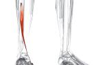
| Flexor hallucis longus | |
Flexor hallucis longus highlighted in red |
|
| Latin name |
Musculus flexor hallucis longus |
|---|---|
| Start | |
| Attachment |
distal phalanx of the big toe |
| Blood supply |
a. tibialis posterior |
| Innervation | |
| Function |
flexes the big toe |
| Catalogs | |
Flexor pollicis longus(lat. Musculus flexor hallucis longus ) - muscle of the lower leg of the posterior group.
Occupies the most lateral position, located on the posterior surface. Covers the tibialis posterior muscle (lat. Musculus tibialis posterior) .
It starts from the lower two-thirds of the fibula, the interosseous membrane and the posterior intermuscular septum of the leg. It goes down and turns into a long tendon that runs under the armor. retinaculum mm. flexorum and passes to the sole, lying in the groove between the talus and calcaneus. At this point, the tendon passes under the flexor digitorum longus tendon, giving it part of the fibrous bundles. It then moves forward and attaches to the base of the distal phalanx of the big toe.
Function
It flexes the thumb, and also, due to its connection with the tendon of the flexor digitorum longus, can act on the II, III and IV fingers. Like the rest of the posterior muscles of the leg, it produces flexion, adduction and supination of the foot. Strengthens the longitudinal arch of the foot.
Write a review of the article "Flexor hallucis longus"
Notes
| |||||||||||||||||||||||||||||||||||||||||||
Mobility of the foot is provided by different muscles, including the short extensor of the big toe, as well as other short and long muscles. The short muscles do not leave the area of the foot itself and are attached within it. The long muscles have their base in the lower leg and are attached to the foot. Thanks to the short and long muscles, extension of both the big and other toes occurs. The foot performs an important shock-absorbing and stabilizing function. The main movements that the foot makes are flexion and extension.
Anatomy of the foot
The muscles of the foot are divided according to their position into dorsal (or dorsal) and plantar. In addition, they can be lateral and medial. If we draw a conditional midline through the human body, then those areas that are closer to this line are called medial. The areas located closer to the outer edge are called lateral. The human foot can move in many directions. The following types of limb movement are distinguished:
- flexion/extension;
- abduction/adduction;
- pronation/supination.
The phalanges of the toes are also quite mobile. This is necessary to perform a stabilizing function and maintain balance. Their mobility is provided by the short extensor digitorum and a separate muscle related to the thumb. The extensor digitorum brevis muscle is a fairly wide and flat muscle that runs along the entire outer area of the foot. It attaches to the heel bone, then passes to the phalangeal region, where it branches into 3 tendons. In the upper part, these tendons unite with the extensor tendon of the thumb and are attached to the phalanges. This muscle is fed by the tibial artery, and innervation is provided by the peroneal nerve.
The plantar side has its own muscles, thanks to which movement of the phalanges and the foot as a whole becomes possible. These include the muscles that abduct and flex the phalanges of the legs, as well as the lumbrical and quadratus muscles.
Long muscles of the feet
The muscles involved in flexion and extension of the phalanges can also be long. At one end they are attached to the bones of the lower leg, and at the other to the phalanges of the legs. The flexor digitorum longus attaches to the tibia. Uniting with the quadratus muscle, the flexor longus is divided into 4 tendons, which are attached directly to the phalanges. Due to the fact that the flexor digitorum longus is attached to four phalanges at the same time, not only flexion movements become possible, but also movement in different directions.
There is also a separate muscle that is responsible for flexing the big toes. The flexor hallucis longus is attached at one end to the lower part of the fibula, and at the other end to the base of the big toe. The flexor hallucis longus muscle is the most powerful muscle on the back of the leg. In addition to ensuring the movement of the phalanx, it is needed to support the arch of the foot. The flexor digitorum longus is also necessary to bend the second and third phalanges, since its tendon is partially associated with the tendons of these fingers. In addition to flexion and extension of the foot, the flexor pollicis longus muscle is also needed for supination and adduction.
Long muscles are responsible for extending the phalanges of the legs. The extensor digitorum longus muscle is located on the outer side of the leg and is attached to a bone called the tibia. Then the extensor digitorum longus stretches along the shin and in the foot diverges into 5 branches, which are attached to the phalanges with the help of tendons. The extensor digitorum longus is involved not only in their extension, but also in extension of the limb.

Extensor hallucis longus
The extensor hallucis longus originates at the bottom of the fibula. It is attached to the base of the bones of the thumbs. The extensor pollicis longus is necessary not only for its movement, but also for the mobility of the limb.
The extensor pollicis longus muscle also provides supination and circular motion of the feet.
How to strengthen your feet
Strengthening these structures is important for our health. There is such a thing as a “leg core”. It contains small muscles that are essential for stabilizing the entire body. Thanks to them, shocks when running and walking are softened, and a stable body position is maintained. If these muscles are weakened, then the entire load will be distributed on the plantar fascia, which can lead to the development of plantar fasciitis. Moreover, a weak ligamentous muscle system leads to a gradual change in gait, which can cause problems with the knees, hip joints and even the spine.
To strengthen your feet, there are simple exercises you can do at home.
| Complex | Performance |
| Exercise No. 1. | For this exercise you will need a towel. Grab it with your toes and drag it across the room. Having reached the opposite wall of the room, use your feet to form a ball out of this towel. Then grab the fabric again and drag him to the other end of the room. Do this exercise with each foot. |
| Exercise No. 2. | This exercise is performed while sitting. To complete it, you will need small objects (for example, glass balls, dice, buttons). Grab an object from one pile with your feet and transfer it to another. Do the same with the other foot. |
| Exercise No. 3. | The exercise can be done while sitting at first. Over time, it is performed while standing on one leg. Place your foot on the floor in its normal position. Then pull your toes towards you, forming an arch with the arch of your foot. |
| Exercise No. 4. | Sit on the floor with your legs extended straight in front of you. Tighten your foot and arch it as if you were wearing a high-heeled shoe. Fix your leg in this tense position and slowly turn your foot towards you. |
 The positive effect of such home exercises occurs after 3-4 months. The main thing is not the duration of the exercises, but their regularity. After a few months, the muscles of the feet will become stronger and the arch will rise. Blood circulation will also improve and the sensitivity of the foot will increase, which is extremely important for developing stability.
Walk barefoot on grass, sand and pebbles more often, after making sure there are no foreign objects.
You need to pay special attention to the health of your feet. Fatigue and heaviness in the legs are perhaps the first signs that something is wrong with the legs. To prevent the development of many unpleasant diseases, it is necessary to adhere to some preventive recommendations.
- Avoid the “wrong” shoes. Start by throwing away your slippers. If you don't feel comfortable being barefoot, you can buy thick sports socks. When choosing shoes for everyday wear, pay attention to the quality of the shoes and the manufacturer. Make sure that it has a fairly dense (but not “wooden” backdrop). It’s good if the insoles in shoes have special instep supports or inserts.
- If you have extra pounds, you will have to get rid of them. The fact is that excess weight creates an additional and constant load on the feet, as a result of which they seem to “creep apart” and sag. This can cause the development of flat feet.
- To strengthen the muscles of the lower leg and feet, use a jump rope. If you have no contraindications, jumping rope will not only help make your muscles stronger, but will also increase the overall endurance of the body. In addition, when jumping, plaques on the walls of blood vessels are destroyed, which has an additional positive effect.
- Engage in general strengthening of the body. For this purpose, hardening, sunbathing and walking on grass or sand barefoot are suitable. Do not forget also about taking vitamin complexes, especially in the autumn-winter period.
The combination of simple exercises and recommendations has a significant positive effect on the entire body. Do not neglect these recommendations and remember that regularity and consistency in their implementation are the key to your health.
- FLEXOR in Medical terms:
(musculus flexor; syn. flexor) a muscle, the contraction of which causes flexion of any part ... - LONG in the Encyclopedic Dictionary:
, -th, -oe; -nen, -nna, -no and -no. 1. Having a large length, extension. D. sleeve. D. lane. D. guy (very... - FLEXOR
bender, bender, bender, bender, bender, bender, bender, bender, bender, bender, bender, ... - LONG in the Complete Accented Paradigm according to Zaliznyak:
long, long, long, long, long, long, long, long, long, long, long, long, long long, long, long, long, long, long, long, long, … - LONG in the Popular Explanatory Encyclopedic Dictionary of the Russian Language:
-aya, -oe; dl"inen, long"a, dl"inno and long"o, dl"inna and long"s 1) Having a large length, extension. Long descent. Long clearing. Dul... - LONG in the Thesaurus of Russian Business Vocabulary:
- LONG in the Russian Language Thesaurus:
‘lasting for a considerable time’ Syn: long, long, long (book), long-term (of.) Ant: short, short-term, ... - LONG in Abramov's Dictionary of Synonyms:
long, oblong, elongated. Prot. . See tall, long || have long... - FLEXOR
bender... - LONG in the Russian Synonyms dictionary:
tall, tall, tall, elongated, longest, longish, longish, long-haired, elongated, long-long, long-long, elongated, long, long, long, long-lasting, long-lasting, lanky, long-length, long-term, protracted, ... - FEET
pl. outdated Steps, … - FLEXOR in the New Explanatory Dictionary of the Russian Language by Efremova:
m. Muscle that flexes ... - LONG in the New Explanatory Dictionary of the Russian Language by Efremova:
adj. 1) a) Having a large length, extension (opposite: short). b) colloquial Tall (about a person). c) transfer decomposition Extensive, detailed, verbose. ... - FLEXOR
bender, ... - LONG in Lopatin’s Dictionary of the Russian Language:
- FLEXOR in the Complete Spelling Dictionary of the Russian Language:
flexor... - FLEXOR in the Spelling Dictionary:
bender, ... - LONG in the Spelling Dictionary:
long; cr. f. longinen, long, ... - LONG in Ozhegov’s Dictionary of the Russian Language:
longer than needed Sleeves are long. The skirt is long. long == long D. break. Long journey. long having great length... - FLEXOR
flexor, m. (anat.). The muscle that flexes the joints is the same as... - LONG in Ushakov’s Explanatory Dictionary of the Russian Language:
Long, long; long (long wrong), long, long. 1. Having a large length or extension, op. short. Long street. Long fence. Long... - FEET
feet pl. outdated Steps, … - FLEXOR in Ephraim's Explanatory Dictionary:
flexor m. Muscle that flexes ... - LONG in Ephraim's Explanatory Dictionary:
long adj. 1) a) Having a large length, extension (opposite: short). b) colloquial Tall (about a person). c) transfer decomposition Extensive, detailed,... - FEET
pl. outdated Steps, … - FLEXOR in the New Dictionary of the Russian Language by Efremova:
m. Muscle that flexes ... - LONG in the New Dictionary of the Russian Language by Efremova:
adj. 1. Having a large length, extension. Ant: short ot. decomposition Tall (about a person). Ott. trans. decomposition Extensive, detailed, verbose. 2. ... - FEET
pl. outdated Steps, … - FLEXOR in the Large Modern Explanatory Dictionary of the Russian Language:
m. Muscle that flexes ... - LONG in the Large Modern Explanatory Dictionary of the Russian Language:
adj. 1. Having a large length, extension. Ant: short ot. colloquial. High (about a person). ot. trans. colloquial. Spacious, ... - SHOES in the Encyclopedic Dictionary of Brockhaus and Euphron.
- SHOES* in the Encyclopedia of Brockhaus and Efron.
- LONG-LONG in the Russian Synonyms dictionary:
long, … - FOOT BONE FRACTURE in the Medical Dictionary:
- FOOT BONE FRACTURE in the Big Medical Dictionary:
Fracture of the talus - Causes: indirect trauma - falling from a height onto one's feet, sudden braking of a car while resting one's feet on it... - SYMPTOM OF THE THUMB in Medical terms:
(syn. ankylosing spondylitis symptom of the thumb) involuntary flexion and adduction of the first finger with passive extension of bent fingers II - V ... - FLEXOR OF THE FIFTH TOE SHORT in Medical terms:
(m. f. digiti quinti brevis, pedis, bna, jna) see List of anat. ... - FLEXOR OF THE FIFTH FINGER in Medical terms:
(m. digiti quinti manus, bna, jna) see List of anat. terms. ... - FLEXOR FOREARM RADIAL in Medical terms:
(m. antibrachii radialis) see List of anat. ... - FOREARM FLEXOR ULNA in Medical terms:
(m. antibrachii ulnaris) see List of anat. ... - FLEXOR TOE SHORT in Medical terms:
(m. f. digitorum brevis pedis, pna, bna, jna) see List of anat. ... - FLEXOR TOE LONG in Medical terms:
(m. f. digitorum longus pedis, pna, bna, jna) see List of anat. ... - FLEXOR OF THE FINGERS SUPERFICIAL in Medical terms:
(m. f. digitorum superficialis manus, pna, jna; f. digitorum sublimis, bna) see List of anat. ... - FLEXOR OF THE FINGERS OF THE HAND DEEP in Medical terms:
(m. f. digitorum profundus manus, pna, bna, jna) see List of anat. ... - FLEXOR OF THE LITTLE FINGER SHORT in Medical terms:
(m. f. digiti minimi brevis manus, pna) see List of anat. ... - FLEXOR TOE SHORT in Medical terms:
(m. f. digiti minimi brevis pedis, pna) see List of anat. ... - FLEXOR CARUS RADIALISM in Medical terms:
(m. f. manus radialis) see List of anat. ... - FLEXOR WRIST RADIAL in Medical terms:
(m. f. carpi radialis, pna, bna, jna) see List of anat. ... - FLEXOR WRIST ULNA in Medical terms:
(m. f. carpi ulnaris, pna, bna, jna) see List of anat. ... - FLEXOR ACCESSORY in Medical terms:
(m. f. accessorius, pna) see List of anat. ... - EXTERNAL FLEXOR OF THE SHIBITION in Medical terms:
(m. f. cruris externus) see List of anat. ... - IRRATIONAL FEET in the Literary Encyclopedia:
feet of ancient metrics, departing from their 581 normal duration. In ancient metrics, based on the alternation of long and short syllables, feet, ...
Flexion of the foot is carried out by the foot flexor muscles, which cross the transverse axis of the ankle joint, located behind it, on the posterior and lateral surfaces of the lower leg.
These muscles include:
1) triceps surae muscle;
2) plantar;
3) posterior tibial;
4) flexor hallucis longus;
5) flexor toes longus;
6) long fibula;
7) short fibula.
The triceps surae muscle has three heads. Two of them, the lateral and medial, make up the gastrocnemius muscle, and the third is the soleus. They all pass into one common calcaneal tendon (Achilles), which is attached to the heel bone. The origin of the gastrocnemius muscle is the medial and lateral femoral condyles. The soleus muscle begins from the posterior surface of the upper third of the body of the tibia and from the tendon arch located between the bones of the lower leg. The gastrocnemius muscle curves around the foot at the ankle joint. The soleus muscle, passing behind the ankle and subtalar joints, causes flexion of the foot. In addition, it plays an important role when standing, fixing the lower leg and preventing the body from falling forward.
The plantaris muscle starts from the lateral condyle of the femur and has a long tendon that passes into the calcaneal tendon, which is common with the previous muscles. This muscle is vestigial in nature (in 12% of cases it is absent) and cannot have a significant effect on movements in the ankle joint.
The tibialis posterior muscle originates from the posterior surface of the interosseous membrane and adjacent areas of the tibia and fibula. Passing under the medial malleolus, it attaches to the tuberosity of the scaphoid, to all the sphenoid bones and to the bases of the metatarsals. Its function is to flex the foot, adduct it and supinate it.
The flexor hallucis longus muscle is the strongest of all the deep muscles of the posterior surface of the leg. It starts from the lower part of the posterior surface of the tibia and the posterior intermuscular septum. On the plantar surface of the foot, this muscle passes between the heads of the flexor hallucis brevis and is attached to the plantar surface of the base of the stump of the distal phalanx of the big toe. Its function is to flex the big toe and the entire foot, as well as supination and adduction of the foot. Due to the fact that the tendon of this muscle partially passes into the tendon of the flexor digitorum longus, it also has some influence on the flexion of the second and third fingers.
The flexor hallucis longus plays an important role in holding the medial part of the longitudinal arch. The flexor toe longus arises from the posterior surface of the tibia and passes to the foot under the medial malleolus in a channel located under the flexor tendon retinaculum ligament. On the plantar surface of the foot, this muscle crosses the tendon of the flexor hallucis longus and, after joining the quadratus plantaris, it divides into four tendons that attach to the bases of the distal phalanges of the 2-5th fingers. The function of the muscle is to flex and supinate the foot, as well as flex the toes. The quadratus plantae muscle, attached to the tendon of this muscle, helps to “average” its action.
The peroneus longus muscle lies on the lateral surface of the peroneal foot. It originates from the head of the fibula, the fascia of the tibia, the lateral condyle of the tibia, and the lateral surface of the fibula. The tendon of this muscle curves around the lateral malleolus from below. Moving to the plantar surface, the muscle tendon runs along the groove located on the lower surface of the cuboid bone, reaches the medial edge of the foot and attaches to the tuberosity of the base of the plus bone, the 1st cuneiform bone and the base of the 2nd metatarsal bone. The peroneus longus muscle is involved in flexion, pronation and abduction of the foot. In addition, together with the tibialis anterior muscle, it forms a tendon-muscular loop that strengthens the transverse arch of the foot.
The peroneus brevis muscle originates from the lateral surface of the fibula a; intermuscular septum of the leg. The tendon of this muscle curves around the lateral head from below and is attached posteriorly to the tuberosity of the 5th metatarsal bone. The muscle provokes and abducts the foot.
Extension of the foot is carried out by extensor muscles that cross the transverse axis of the ankle and are located in front of it, on the front surface of the lower leg.
These muscles include:
1) anterior tibial;
2) extensor toes longus;
3) long extensor of the big toe.
The tibialis anterior muscle is adjacent directly to the lateral surface of the tibia, from which it originates. Going down, the muscle passes under the ligaments located in the ankle and ankle joint, reaches the medial cuneiform bone and the base of the 1st metatarsal bone and attaches to the medial edge of the foot. The muscle fixes the ankle joint and contributes not only to extension of the foot, but also to supination and adduction, although its participation in the latter movement is small.
The extensor toes longus originates from the upper end of the tibia, the head and anterior edge of the fibula, the interosseous membrane and the fascia of the leg. Moving to the foot, the muscle is divided into five tendons, four of which are directed to the 2nd, 3rd, 4th and 5th toes and are attached to their distal phalanges, and the fifth, called the third peroneal muscle, is attached to the base of the 5th metatarsal bone. The function of the extensor digitorum longus as a multijoint muscle is not only to extend the fingers, but also to extend the foot. Due to the fact that the fifth tendon of this muscle is attached to the lateral edge of the foot, it not only extends, but also somewhat armors the foot.
The extensor hallucis longus originates from the medial surface of the fibula and the interosseous membrane in the lower half of the leg. Attached to the base of the distal phalanx of the big toe, the muscle is an extensor not only of this toe, but of the entire foot. In addition, it promotes supination of the foot.
There are no special muscles involved in adducting the foot. This movement is carried out according to the rule of parallelogram of forces with simultaneous contraction of the tibialis anterior and tibialis minor muscles. The muscles involved in foot abduction are located on the lateral side of the vertical axis of the ankle joint. These include the peroneus brevis and peroneus longus muscles.
Pronation of the foot involves muscles located on the lateral side of the sagittal axis around which this movement occurs. The foot is armored by the peroneus longus, peroneus brevis and the third peroneus muscles. Of these, the peroneus longus muscle is the strongest.
Supination of the foot involves muscles that cross the sagittal axis around which this movement occurs, as well as those located medial to it. The foot is supinated by the tibialis anterior muscle and the extensor pollicis longus muscle. The alternating action of muscle groups passing near the joints of the foot and coming to it from the lower leg causes its circular movement.
The movements of the toes involve the muscles that move from the lower leg to the foot and the muscles of the foot itself. The muscles located on the plantar surface of the foot flex the toes, and the muscles located on the back of the foot extend them.
Related information.
Latin name lex - bend; digit - finger; longus - long.
The attachment of the tendons of this muscle to the four toes is similar to the attachment of the deep flexor digitorum.
Place of origin- The medial portion of the posterior surface of the tibia below the soleus line.
Place of attachment- Bases of the distal phalanges from the second to fifth toes.
Action- Flexes all joints of the four toes (allows the foot to rest firmly on the surface when walking). Participates in plantar flexion of the ankle joint and eversion of the foot.
Innervation- Tibial nerve L5, S1, (2).
Blood supply- Posterior tibial artery (from the popliteal artery).
Examples: walking (especially with bare feet on uneven surfaces). Standing on tiptoes.
Flexor hallucis longus / Musculus flexor hallucis longus

Latin name flex - bend; hallux - big toe; longus - long.
This muscle supports the medial longitudinal arch of the foot.
Place of origin- The lower two-thirds of the posterior surface of the fibula. Interosseous membrane. Adjacent intermuscular septum.
Place of attachment- Base of the distal phalanx of the big toe.
Action- Flexes all joints of the big toe and is an important muscle in the final thrust of the foot during walking. Participates in plantar flexion of the ankle joint and eversion of the foot.
Innervation
Blood supply
Basic functional movement- Examples: lifting the foot off the surface when walking (especially with bare feet on an uneven surface). Standing on tiptoes.
Posterior tibialis muscle / Musculus tibialis posterior

Latin name tibia - trumpet or flute/tibia; posterior - rear.
The tibialis posterior muscle is the deepest muscle in the back of the leg. It supports the arches of the feet.
Place of origin- Lateral part of the posterior surface of the tibia. The upper two-thirds of the posterior surface of the fibula. Most of the interosseous membrane.
Place of attachment- Tuberosity of the scaphoid. Using fibrous extensions to the supports of the talus, the three cuneiforms, the cuboid and the bases of the second, third and fourth metatarsals.
Action- He twists his foot. Participates in plantar flexion of the ankle joint.
Innervation- Tibial nerve L(4), 5, S1.
Blood supply- Peroneal artery through the posterior tibial artery (from the popliteal artery).
Basic functional movement- Examples: tiptoe position. Pressing the car pedals.
Soleus muscle / Musculus soleus

Latin name soleus is a form of flounder.
Part of the triceps surae muscle. The soleus muscle is named so because it is shaped like a fish. The calcaneal tendon of the soleus and gastrocnemius muscles is the thickest and strongest tendon.
Place of origin- Posterior surfaces of the head of the fibula and the upper third of the body of the fibula. Soleus line and middle third of the medial edge of the tibia. Arch of tendon between the tibia and fibula.
Place of attachment- Together with the tendon of the gastrocnemius muscle to the back of the heel bone.
Action- Plantar flexion of the ankle joint. The soleus muscle contracts frequently when the body is upright, which prevents falling forward at the ankle joint; i.e., it maintains the fulcrum in the area of the body’s center of gravity. The muscle maintains a vertical position.
Innervation- Tibial nerve L5, S1, 2.
Blood supply- Posterior tibial artery (from the popliteal artery). The gastrocnemius branches of the popliteal and peroneal arteries through the posterior tibial artery.
Basic functional movement- Example: standing on tiptoes.