All about eyelid ptosis: causes, diagnosis and treatment without surgery. Upper eyelid prolapse: types of ptosis and its causes Congenital ptosis of the upper eyelid
Ptosis (drooping) of the upper eyelid is an uncontrolled malfunction of the muscles that raise and lower the upper eyelid. Muscle weakness is expressed as a cosmetic defect, in the form of asymmetry in the size of the palpebral fissures, which develops into a lot of complications, up to loss of vision.
Patients of any age are susceptible to the disease, from newborns to pensioners. All methods of treatment, including the main operable therapy for ptosis, are aimed at increasing the tone of the muscles of the eye.

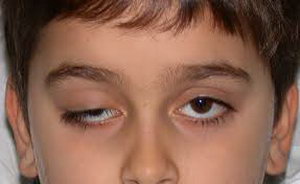
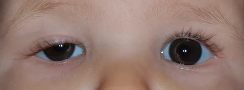
Blepharoptosis (drooping of the upper eyelid) is a pathology of the muscular apparatus, in which the eyelid partially or completely covers the iris or pupil, in advanced stages it completely covers the palpebral fissure. Normally, the right and left eyelids should cover no more than 1.5-2 mm of the upper edge of the iris. If the muscles are weak, poorly innervated or damaged, the eyelid ceases to be controlled and falls below normal.
Ptosis is a disease of the upper eyelid only, since the lower eyelid lacks the levator muscle responsible for lifting. There is a small Muller muscle, which is innervated in the cervical region and is capable of expanding the palpebral fissure only a couple of millimeters. Therefore, with paralysis of the sympathetic nerve, which is responsible for this small muscle in the lower eyelid, ptosis will be insignificant, completely invisible.

Physical overlapping of the visual field leads to the formation of a number of complications that are especially dangerous in childhood, when the function of vision is just being formed. Ptosis in a child leads to impaired development of binocular vision,.
All these complications are also typical for adults, but when they appear in an infant, they contribute to improper brain training to compare visual images. Subsequently, this will lead to the impossibility of correcting or restoring correct vision.
Classification and reasons
Muscle weakness can be acquired or congenital. Congenital ptosis of the upper eyelid is a disease of young children, its causes are underdevelopment or absence of the muscles that lift the eyelid, as well as damage to the nerve centers. Congenital ptosis is characterized by a bilateral lesion of the upper eyelid of the right and left eyes at the same time.
Watch an interesting video about the congenital form of the disease and methods of treatment:
Unilateral lesion is characteristic of acquired ptosis. This type of ptosis develops as a complication of another, more serious pathological process.
Classification of ptosis of the upper eyelid, depending on the cause of the appearance:
- Aponeurotic blepharoptosis - excessive stretching or relaxation of muscles, loss of tone.
- Neurogenic ptosis is a violation of the passage of a nerve impulse to control the muscle. Neurogenic ptosis is a symptom of CNS disease, the appearance of neurology is the first signal for an additional examination of brain structures.
- Mechanical blepharoptosis - post-traumatic muscle damage, tumor growth, scarring.
- Age-related - the natural physiological processes of aging of the body provoke weakening, stretching of muscles and ligaments.
- False blepharoptosis - observed with a large amount of skin folds.
Also, the causes of blepharoptosis in adults are:
- injuries, bruises, tears, eye injuries;
- diseases of the nervous system or brain: stroke, neuritis, multiple sclerosis, tumors, neoplasms, hemorrhages, aneurysms, encephalopathy, meningitis, cerebral palsy;
- paresis, paralysis, ruptures, muscle weakness;
- diabetes mellitus or other endocrine diseases;
- exophthalmos;
- a consequence of unsuccessful plastic surgeries, the introduction of Botox.
By stages:
- partial;
- incomplete;
- full.

Ptosis has 3 degrees, which are measured in the number of millimeters of distance between the edge of the eyelid and the center of the pupil. In this case, the patient's eye and eyebrows should be relaxed, in a natural position. If the location of the edge of the upper eyelid coincides with the center of the pupil, this is the equator, 0 millimeters.
Degrees of ptosis:
- The first degree is from +2 to +5 mm.
- The second degree is from +2 to -2 mm.
- Third degree - from -2 to -5 mm.
Symptoms of the disease
Ptosis of the eyelid is characterized by the main, most obvious visual symptom - it is a drooping with a partially or completely closed palpebral fissure. At an early stage of the disease, pay attention to the symmetry of the location of the eyelids of the right and left eyes relative to the edge of the cornea.
Other manifestations of blepharoptosis:
- decreased visual acuity of one of the eyes;
- fast fatiguability;
- the astrologer's position, when the patient has to throw his head back to get a clear image;
- double vision;
- the pathological eye stops blinking, this leads to;
- the formed pocket under the lowered eyelid contributes to the accumulation of bacteria, subsequently the development of frequent inflammations;
- double vision;
- unconsciously, the patient tries to lift the upper eyelid with the help of the superciliary arches or forehead muscles;
- gradual development of strabismus.

Diagnostics
Diagnosis is aimed at identifying the root cause of the disease, prescribing adequate treatment. Eyelid droop in the early stages is subtle, but it is an extremely important sign of the onset of the development of serious diseases, such as a brain tumor. Therefore, it is important for an ophthalmologist to find out whether ptosis is congenital or appeared suddenly. To do this, the patient is interviewed, an anamnesis is collected.
It happens that the patient did not notice the omission before or cannot say exactly when it appeared. In this case, it is necessary to conduct additional examinations to exclude all possible causes of the disease.
Stages of diagnosis of blepharoptosis:
- Visual inspection, measurement of the degree of ptosis.
- Measurement of acuity, visual field, intraocular pressure, examination of the fundus.
- Biomicroscopy of the eye.
- Measurement of muscle tone, symmetry of folds and blinking.
- Ultrasound of the eye, electromyography.
- Radiography.
- Head MRI.
- Checking for binocular vision.
- Examination by a neurosurgeon, neuropathologist, endocrinologist.
How to cure ptosis of the upper eyelid
Fighting ptosis is necessary only after finding out the cause. In the early stages, congenital pathology in the absence of visual impairment, a small cosmetic defect is recommended not to be treated, but to carry out comprehensive prevention.
Treatment of ptosis is divided into conservative and surgical. Conservative methods go well with homemade folk recipes.
For ptosis due to injury or nerve damage, it is recommended to wait about a year after the incident. During this time, effective treatment can restore all nerve connections without surgery or significantly reduce its volume.

What to do if the eyelid drooped after Botox
Botox (botulinum toxin) is a drug derived from botulinum bacteria that disrupts neuromuscular communication. As part of the drug, a neurotoxin, which in small doses, when applied locally, attacks and kills nerve cells in the muscles, due to which they completely relax.
When using the drug in the cosmetic industry, a complication of incorrect or inaccurate injection may be ptosis of the upper eyelid after Botox injection, the treatment of which is very long. Moreover, the first few procedures can be successful, but each subsequent one requires an increase in the amount of the drug, which can lead to an overdose, as the body learns to develop immunity and antibodies to botulinum toxin.
Remove omission (blepharoptosis) or difficult, but possible. The first option of the simplest non-surgical treatment is to do nothing or just wait. After about 2-3 months, the body will build additional lateral branches of the nerves, which will allow you to regain control over the muscle on its own.
The second method helps to speed up this process, for this physiotherapeutic procedures are actively used (UHF, electrophoresis, massage, darsonval, microcurrents, galvanotherapy), injections of prozerin, taking large doses of B vitamins, neuroprotectors. All this accelerates the restoration of innervation, contributes to the speedy resorption of the remnants of Botox.
Operation
Surgery to correct ptosis (drooping) of the upper eyelid is called blepharoplasty. The operation is indicated in the case of advanced ptosis with a violation of the quality of vision. The intervention is performed under local anesthesia on an outpatient basis. The rehabilitation period lasts about a month, during which the patient is observed by the operating surgeon.

There are many methods of operation, but the essence is the same - to shorten a relaxed muscle either by incision and removal of a part, or by folding it in half and flashing it. The cosmetic seam hides in a natural skin fold, and over time completely resolves.
The cost of the operation depends on:
- complexity of the operation;
- stages of ptosis;
- additional research;
- medical institution of your choice;
- the number of specialist consultations;
- the number of laboratory diagnostics;
- type of anesthesia;
- associated pathologies.
On average, the amount per operation varies from 20 to 60 thousand rubles. You can find out the exact figure directly at the reception, after examination by a specialist.
See in the video how the operation (blepharoplasty) takes place:
home treatment
Ptosis of the upper eyelid is treated conservatively at home. In non-surgical treatment, medicines, massage, alternative medicine, and physiotherapy are used.
Methods for the treatment of eyelid prolapse with folk remedies:
- a mask of raw chicken eggs with sesame oil is applied to the skin 1 time per day, washed off with warm water;
- lotions or warm compresses from infusions of chamomile, calendula, rosehip, black tea, birch leaves;
- applying "dry heat" with a cloth bag with overcooked sea salt;
- a potato mask made from grated raw potatoes is applied for 20 minutes once a day;
- a mask of honey with aloe pulp is applied 2 times a day.
Traditional drugs are used orally, mainly B vitamins, neuroprotectors, drugs that stimulate growth, as well as the regeneration of nerve tissues, which enhance the nutrition of nerve cells. Everything is prescribed individually and depends on the stage, form, cause of ptosis.
Physiotherapy:
- vacuum massage for ptosis of the upper eyelid;
- electrophoresis;
- warming up;
- myostimulation by currents.
All procedures and preparations must be clarified and coordinated with your attending ophthalmologist. The information on the site is for informational purposes only, do not use it as a guide to action.
In addition, we invite you to watch a video about ptosis. Elena Malysheva will tell you in detail about the disease and how to deal with it.
Article content: classList.toggle()">expand
Ptosis of the eyelid is a pathology of the location of the upper eyelid, in which it is lowered down and partially or completely covers the palpebral fissure. Another name for the anomaly is blepharoptosis.
Normally, the eyelid should overlap the iris by no more than 1.5 mm. If this value is exceeded, they speak of pathological drooping of the upper eyelid.
Ptosis is not only a cosmetic defect that significantly distorts the appearance of a person. It interferes with the normal functioning of the visual analyzer, as it interferes with refraction.
Classification and causes of eyelid ptosis
Depending on the moment of occurrence, ptosis is divided into:
- Acquired
- Congenital.
Depending on the degree of eyelid drooping, it happens:
- Partial: covers no more than 1/3 of the pupil
- Incomplete: covers up to 1/2 pupil
- Full: The eyelid completely covers the pupil.
The acquired variety of the disease, depending on the etiology (causes of ptosis of the upper eyelid), is divided into several types:

As for cases of congenital ptosis, it can occur due to two reasons:
- Anomaly in the development of the muscle that lifts the upper eyelid. May be associated with strabismus or amblyopia (lazy eye syndrome).
- Damage to the nerve centers of the oculomotor or facial nerve.
Ptosis symptoms
The main clinical manifestation of the disease is drooping of the upper eyelid., which leads to partial or complete closure of the palpebral fissure. At the same time, people try to strain the frontal muscle as much as possible so that the eyebrows rise and the eyelid stretches up.
Some patients, for this purpose, throw their heads back and take a specific posture, which in the literature is called the astrologer's posture.
A drooping eyelid prevents blinking movements, and this leads to the appearance of soreness and overwork of the eyes. A decrease in the frequency of blinking causes damage to the tear film and development. Infection of the eye and the development of an inflammatory disease can also occur.
Features of the disease in children
 In infancy, ptosis is difficult to diagnose. This is largely due to the fact that most of the time the child sleeps and is with his eyes closed. You need to carefully monitor the facial expression of the baby. Sometimes the disease can be manifested by frequent blinking of the affected eye during feeding.
At an older age, ptosis in children can be suspected by the following signs:
- While reading or writing, the child tries to throw his head back. This is due to the limitation of visual fields when lowering the upper eyelid.
- Uncontrolled muscle contraction on the affected side. This is sometimes mistaken for a nervous tic.
- Complaints about rapid fatigue after visual work.
Cases of congenital ptosis may be accompanied by epicanthus(overhanging skin folds over the eyelid), damage to the cornea and paralysis of the oculomotor muscles. If the child's ptosis is not corrected, it will lead to the development and decrease in vision.
Diagnostics
To diagnose this disease, a simple examination is enough. To determine its degree, it is necessary to calculate the MRD indicator - the distance between the center of the pupil and the edge of the upper eyelid. If the eyelid crosses the middle of the pupil, then the MRD is 0, if higher - then from +1 to +5, if lower - from -1 to -5.A comprehensive examination includes the following studies:
- Determination of visual acuity;
- Determination of fields of view;
- Ophthalmoscopy with the study of the fundus;
- examination of the cornea;
- Study of the production of lacrimal fluid;
- Biomicroscopy of the eyes with assessment of the tear film.
It is very important that during the determination of the degree of the disease the patient is relaxed and does not frown. Otherwise, the result will be unreliable.
Children are examined especially carefully, since ptosis is often combined with amblyopia of the eyes. Be sure to check visual acuity according to Orlova's tables.
Ptosis treatment
Elimination of ptosis of the upper eyelid can only be after determining the root cause
Treatment of ptosis of the upper eyelid is possible only after determining the root cause. If it has a neurogenic or traumatic nature, its treatment necessarily includes physiotherapy: UHF, galvanization, electrophoresis, paraffin therapy.
Operation
As for cases of congenital ptosis of the upper eyelid, it is necessary to resort to surgical intervention. It is aimed at shortening the muscle that lifts the eyelid.
The main stages of the operation:

The operation is also indicated if the upper eyelid is still lowered, after the treatment of the underlying disease.
After the intervention, an aseptic (sterile) bandage is applied to the eye and broad-spectrum antibacterial drugs are prescribed. This is necessary to prevent infection of the wound.
The medicine
A droopy upper eyelid can be treated conservatively. To restore the functionality of the oculomotor muscles, the following therapies are used:

If the upper eyelid has drooped after an injection of botulinum toxin, then it is necessary to instill eye drops with alfagan, ipratropium, lopidine, phenylephrine. Such drugs contribute to the contraction of the oculomotor muscles and, as a result, the eyelid rises.
You can speed up the lifting of the eyelid after Botox with the help of medical masks, creams for the skin around the eyelids. Also, professionals recommend massaging the eyelids daily and visiting a steam sauna.
Exercises
A special gymnastic complex helps to strengthen and tighten the oculomotor muscles. This is especially true of involutional ptosis, which arose as a result of natural aging.
Gymnastics for the eyes with ptosis of the upper eyelid:

Only with regular performance of a set of exercises for ptosis of the upper eyelid, you will notice the effect.
Folk remedies
Treatment of ptosis of the upper eyelid, especially at the initial stage, is possible at home. Folk remedies are safe, and there are practically no side effects.
Folk recipes to combat ptosis of the upper eyelid:

With regular use, folk remedies not only strengthen muscle tissue, but also smooth out fine wrinkles.
Amazing results can be achieved with the complex use of masks and massage. Massage technique:
- Treat your hands with an antibacterial agent;
- Remove makeup from the skin around the eyes;
- Treat the eyelids with massage oil;
- Perform light stroking movements on the upper eyelid in the direction from the inner corner of the eye to the outer. When processing the lower eyelid, move in the opposite direction;
- After warming up, lightly tap the skin around the eyes for 60 seconds;
- Then continuously press on the skin of the upper eyelid. Do not touch the eyeballs;
- Cover your eyes with cotton pads soaked in chamomile extract.
Photo of ptosis of the upper eyelid


Ptosis of the upper eyelid- age-related changes in muscles and tissues caused by gravity, but pathology can occur against the background of diseases of the nervous, endocrine system, often develops with injuries and tumor processes in the body, can be congenital, hereditary.
The drooping of the upper eyelid is most often an age-related change.
Upper eyelid drooping symptoms
Blepharoptosis (ptosis)- an ophthalmic disease, characterized by the drooping of the upper eyelid below the border of the iris by 2 mm or more, the disease begins its development with muscle abnormalities.
How the disease manifests itself:
- brow arches lose their bend;
- the head is slightly thrown back;
- eye irritation, frequent conjunctivitis;
- eyes get tired quickly even with minimal stress;
- fuzziness of the image;
- dry eye syndrome.
Against the background of the impending century, a functional decrease in visual acuity almost always occurs.
Ptosis classification
Blepharoptosis is congenital and acquired, depending on the origin, severity of the pathology, ptosis is divided into several types. ICD code 10-H 02.4, congenital ptosis - Q 10.0.
Types of pathology:
- aponeurotic- occurs when the muscle is stretched and weakened, lifting the upper eyelid, develops after plastic contouring;
- neurogenic - a consequence of diseases of the nervous system, accompanied by a narrowing or expansion of the pupil, one eyelid is noticeably lower than the other;
- myogenic - develops with myasthenia gravis, the disease is transmitted to the child from the mother;
- mechanical - the result of scars on the eyelid, getting into the eye of a foreign object;
- false ptosis - skin folds create the impression of an impending century;
- oncogenic - a consequence of tumor processes.
Children are more often diagnosed with congenital dystrophic and non-dystrophic ptosis, a neurogenic form of the disease.
Degrees of ptosis
As the disease develops, the eyelids droop more and more, close the eye, with senile ptosis, the pathology is accompanied by a number of additional symptoms.
1 degree of ptosis - partial, the pupil is closed by 1/3
- With the first degree of age-related ptosis, folds form on the upper eyelid, circles and bags under the eyes, eyebrows rise, and a nasolabial triangle stands out.
- At stage II, deep folds lie between the eyebrows, many small wrinkles form around the eyes, the eyelid drops to the very eyelashes.
- At stage III, all signs intensify, the eyelids hang down, close the eye, the condition and appearance of the skin worsens.
Before and after semi-ptosis surgery
Before and after partial ptosis surgery
Causes of ptosis
Congenital ptosis develops due to an underdeveloped muscle of the upper eyelid, or its complete absence, often accompanied by amblyopia, strabismus.
Causes - hereditary pathologies, intrauterine abnormalities.
Reasons for the development of acquired ptosis:
- pathologies of the nervous system, which are accompanied by paresis, paralysis;
- injuries of the organs of vision, ophthalmic operations;
- endocrine pathologies;
- long-term use of hormonal drugs;
- stretching in the area of \u200b\u200bthe connection of the muscle of the upper eyelid with the tendon;
- age-related omission of tissues due to gravity;
- the presence of neoplasms in the brain, eye socket.
Eyelid drooping occurs for age-related reasons, as well as for a number of serious diseases.
Congenital ptosis is most often bilateral, the acquired form of pathology is diagnosed in one eye.
Ptosis in children
Eyelid prolapse in a child occurs due to birth injuries, nerve tumors, hemangioma, partial paralysis.
Causes of congenital ptosis:
- the appearance of the third fold;
- genetic underdevelopment of the palpebral fissure;
- dystrophic myasthenia - a severe autoimmune disease;
- paralysis of the third pair of cranial nerves;
- the Marcus-Gunn phenomenon - the eyelids involuntarily rise when the masticatory muscles move;
- neuroblastoma.
The child's eyelid is almost closed - complete ptosis
The acquired form of pathology in children develops in violation of the functions of the thymus gland, after eye injuries.
In newborns, it is difficult to recognize ptosis, one of the main signs is frequent blinking during feeding.
Which doctor should I contact?
Diagnosis and treatment of ptosis and, if necessary, surgery, consultation.
Diagnostics
To diagnose ptosis, a simple examination is enough to determine the degree of pathology, the doctor measures the length between the edge of the upper eyelid and the center of the pupil.
Stages of a comprehensive examination:
- examination of the cornea;
- analysis of the functions of the lacrimal gland;
- adrenaline test;
- assessment of visual acuity;
- measurement of intraocular pressure;
- eye biomicroscopy;
- X-ray, CT scan of the eye;
- MRI of the brain.
The topography of the brain will make it clear in which area the problem is, but such a diagnosis is not always carried out.
During the determination of the degree of ptosis, you can not frown, strain - this will distort the measurement results.
Methods for eliminating ptosis
After identifying the root cause of eyelid drooping, the correct treatment is selected. Complex therapy consists of traditional and non-traditional methods.
Treatment without surgery
Effective methods for I, II degree, if the procedures are carried out regularly and correctly, it will take 3-6 months to eliminate the lowered upper eyelid.
Gymnastics
A special set of exercises strengthens, tightens the eye muscles, gymnastics helps well with senile ptosis.
Exercises:
- Fix your eyes on the object, make slow circular movements of the eyes in a clockwise direction. Repeat 7 times.
- Look up, open your mouth, blink frequently. Perform the exercise for 30 seconds, gradually increase the time to 3-4 minutes.
- Close your eyes, at the expense of 5 open wide, look forward. Repeat 7-8 times.
- Open your eyes, gently press your fingers to your temples, slightly stretch the skin, blink frequently for 30 seconds.
- Eyes closed, slightly stretch the skin near the outer corners of the eye. Overcoming resistance, lift the eyelids as high as possible. Perform 5 repetitions.
- Tilt your head back, close your eyes, fix the position at the expense of 10.
Eye exercises for ptosis
You need to perform gymnastics in the morning and in the evening, with regular exercises, the effect is noticeable after 2-4 weeks.
Massage for blepharoptosis
Massage in combination with gymnastics and folk remedies allows you to achieve noticeable improvements in the impending century.
Procedure steps:
- Remove makeup from skin.
- Treat hands with antiseptic preparations.
- Apply hypoallergenic massage oil to the skin of the eyelids.
- To warm up the skin with light stroking movements, move along the upper eyelid from the inner corner to the outer edge.
- Movements on the lower eyelid should be carried out in the opposite direction.
- With skin pads, tap the skin around the eyes for 1 minute.
- Press on the skin of the upper eyelid for a count of 5, repeat after a short break. Perform the exercise 5-7 times.
Eyelid massage with ptosis will give a tangible and visible effect
After the end of the session, apply a compress of chamomile or green tea to the eyes, lie down for 5 minutes.
How to remove ptosis folk remedies
Alternative medicine is used to prevent and treat the initial stage of ptosis.
Simple Recipes:
- Mix in equal parts chamomile inflorescences, cornflower, green tea leaves, 1 tbsp. l. collection, pour 250 ml of boiling water, leave in a sealed container for 30 minutes, strain. Pour the infusion into ice molds, freeze, wipe the skin around the eyes every morning.
- Mix 30 g of chopped fresh parsley and birch leaves, 1 tsp. collection, pour 220 ml of water, simmer over low heat for 5 minutes, strain. Moisten cotton pads in the decoction, apply to the eyes for 15 minutes 3-4 times a day.
- Beat the egg yolk, add 3-4 drops of sesame or olive oil, apply the mixture on the upper eyelid. Wash off after 20 minutes, sessions are carried out daily for 2-3 weeks.
- Grate raw potatoes, put in the refrigerator for 20 minutes, make a compress on the eyes. After a quarter of an hour, remove the mass with warm water.
Folk remedies help to tighten the drooping eyelid, cope with wrinkles, circles and bags under the eyes.
Medications
Drug treatment for ptosis is ineffective, at the initial stage of the disease, agents based on apraclonidine are prescribed - Cytoflavin, Clonidine, B vitamins, these drugs cause contraction of the eye muscles.
B vitamins contract muscles
The introduction of Botox preparations- after the injection, muscle paralysis occurs, the upper eyelid rises. The duration of the procedure is no more than 20 minutes, discomfort rarely occurs, after the injection the skin is treated with antiseptics, the recovery period takes 7-8 days.
The effect of the operation is noticeable after 2 weeks, lasts for 6-12 months. The method is suitable for eliminating the manifestations of partial and incomplete ptosis.
Ptosis - often occurs after Botox with improper administration of the drug in order to eliminate wrinkles, but within 4 weeks the problem disappears on its own.
Other correction methods
Conservative methods of treatment help to restore the function of the damaged nerve, they are used in the neurogenic form of pathology.
Treatment methods:
- UHF therapy - a gentle effect on the cornea with an electromagnetic field with a high frequency, after the procedure, muscle spasms disappear, blood circulation and patency of nerve impulses improves;
- galvanotherapy– damaged areas are affected by low voltage direct current, destroyed nerves and muscles are restored, metabolic processes are normalized;
- paraffin therapy- under the influence of heat, metabolic processes in tissues are accelerated, muscles are gradually strengthened;
- ultraphonophrese with drugs, the action of which is aimed at improving the functioning of the eye muscles, enhancing the synthesis of collagen, elastin;
- laser therapy- one of the best methods of treatment, after 2 weeks, metabolic processes normalize, muscle function improves, swelling disappears;
- myostimulation - the effect of electrical impulses on the muscles and nerve endings, which leads to the strengthening of the fibers.
Exposure to the eye by an electromagnetic field
Duration of conservative therapy- 6 months, if there is no noticeable improvement, the person is recommended to undergo surgery.
Physiotherapy is contraindicated in diseases of the heart, blood vessels, severe forms of hypertension, mental disorders, epilepsy, during an exacerbation of infectious pathologies.
Operation
To correct a drooping eyelid, several surgical methods are used, each of them has its own advantages and disadvantages.
Classic operation
Surgery is performed in severe forms of congenital ptosis, during the operation, the muscle that lifts the eyelid is shortened. The average cost is 15–25 thousand rubles.
Photos before and after blepharoplasty
Operation steps:
- The surgeon makes an incision in the area of the upper eyelid.
- The muscle or tendon of the eyelid is brought into the incision.
- A small piece of muscle is excised.
- The imposition of cosmetic sutures, after resorption they are not visible, there are no scars.
After surgery, a sterile bandage is applied to the eye, antibiotics are prescribed to avoid infection.
With ptosis in a child, surgery is done only after 3 years - before this age, the organ of vision is actively formed. But if the chest has a tilting of the head, the functions of the lacrimal glands are disturbed, then the defect is removed immediately by the surgical method.
The modern method of treating ptosis, refers to the methods of plastic surgery, is suitable for eliminating the age-related form of pathology. The average cost is 28–38 thousand rubles.
Before and after blepharoplasty
- Procedure steps:
- The marker indicates the location of the incision.
- Perform local anesthesia.
- Laser cuts are made along the markings - under the influence of high temperatures, fat, old, damaged cells are destroyed.
- Suturing.
- Applying an antiseptic ointment, fixing the incision site with a band-aid.
For blepharoplasty, hospitalization is not required, after completing all the necessary manipulations, you can go home.
Possible Complications
If left untreated, ptosis develops amblyopia, vision rapidly deteriorates. With complete unilateral ptosis, they give the III group of disability with a bilateral form of the disease - II.
Possible complications after surgery are conjunctivitis, photophobia, sometimes there is eversion of the eyelid, slight asymmetry.
What to do to prevent ptosis
To avoid the appearance or recurrence of blepharoptosis, it is enough to follow simple rules of prevention.
How to prevent the development of ptosis:
- timely treat all diseases that can provoke drooping of the eyelid;
- use protective goggles for hazardous work;
- do not try to remove a foreign body in the eye yourself;
- after 30 years, start doing gymnastics, massage to strengthen the eye muscles, use high-quality cosmetics with a lifting effect;
- be attentive to the choice of a clinic, a plastic surgeon if Botox is necessary.
A sedentary lifestyle can lead to any disease - move
Smoking, alcohol, junk food, a sedentary lifestyle negatively affects the state of the body as a whole, and the eye muscles in particular
The fall of the supraorbital skin fold is called ptosis of the upper eyelid. This disease is acquired or congenital and does not depend on age criteria. In most cases, the problem is solved after the operation, but there are other ways to eliminate the defect that do not involve surgical intervention.
What is ptosis of the upper eyelid
Ptosis or drooping (drooping) of the upper eyelid is a congenital or acquired defect. It can be unilateral (one eyelid drooping) or bilateral (both eyelids fall). The severity of the defect depends on the degree of ptosis:- First degree. The upper eyelid is partly drooping. The eye is covered by a maximum of 33%.
- Second degree. Significant drop in the upper eyelid. The visible area of the eyeball varies between 33-66%.
- Third degree. Due to the total drooping of the eyelid, the pupil is completely covered. Zero visibility.
- First stage. Visual changes are subtle. However, the facial muscle weakens, and folds, circles, and bags form around the eyes.
- Second stage. There is a formation of a clear border between the eyes and the cheek.
- Third stage. It is characterized by noticeable changes in the eye area, when the upper eyelids are literally pulled over the pupils. The look is dull, inexpressive and sad. The effect of a frowning person or a glance from under the brows is created.
- Fourth stage. Due to the deepening of the nasolacrimal groove, both the eyelids and the corners of the eyes are subject to drooping. Facial changes add several years to a person's age. He starts to look older.
It is possible to talk about ptosis of the upper eyelid only if the distance between its edge and the border of the eye iris exceeds 1.5 mm.
Causes of ptosis of the upper eyelid
The reason for the fall of the upper eyelid is directly related to some features of the defect. There are the following types of ptosis:
- acquired;
- congenital.
1. Aponeurotic. Pathology of structures regulating eyelid elevation. The muscle fibers responsible for raising the eyelid were stretched or pulled apart. The occurrence of the disease is due to age-related changes in the body. So the elderly are mostly at risk.
2. Neurogenic. It is caused by paralysis of the nerve fibers responsible for the motor function of the eyes. This phenomenon is due to a number of factors associated with impaired activity of the nervous system:
- multiple sclerosis;
- stroke;
- brain tumor;
- abscess of substance in the cranium.
- the presence of tumor-like structures in the eye;
- obtaining by ingestion of foreign bodies;
- the course of the process of eye scarring;
- ruptures of the eye areas.
5. False. Ptosis is provoked by the following deviations:
- strabismus;
- excess eyelid skin.

Congenital ptosis is caused by the following factors:
- Underdevelopment or absence of a specific muscle controlling the lifting of the upper eyelid.
- Blepharophimosis. A rare genetic anomaly characterized by a vertical/horizontal shortening of the slit of the eye due to chronic conjunctivitis or fused eyelid margins.
- Marcus-Gunn syndrome, palpebromandibular syndrome. The system that regulates the lifting of the eyelids is not functioning due to the affected brain stem and is sometimes complicated by strabismus or amblyopia. However, when opening the mouth, chewing, jaw vibrations, involuntary movements of the eyelids occur in the direction of increasing the palpebral fissure.
Signs of ptosis
Eyelid drooping is accompanied by various symptoms. The most common signs of ptosis are:- visually lowered upper eyelid;
- everted border of the eyelids outside;
- small eye, short palpebral fissure;
- massive falling skin fold at the upper edge of the eyelids;
- close-set eyes;
- increased eye fatigue;
- frequent redness, pain, irritation of the mucous membrane of the eye;
- reduced visual function;
- feeling of sand in the eyes;
- pupil constriction;
- split in the eyes;
- no blinking;
- the habit of moving your eyebrows, tilting your head back to raise your lowered eyelid;
- sometimes strabismus;
- inability to completely cover the eye.

In rare cases, ptosis is accompanied by the following symptoms:
- myasthenia gravis, feeling weak and tired in the afternoon;
- myopathy, partial drooping of the eyelids due to weakened muscle structures;
- involuntary lifting of the eyelids due to movement of the jaw, mouth;
- palpebral violation of the eyelids, omission of the upper and eversion of the lower, narrowing of the palpebral fissure;
- Bernard Horner's syndrome, simultaneous drop of the eyelid, retraction of the eye and constriction of the pupil.
Signs of drooping eyelids can serve as a diagnostic criterion in establishing the cause of ptosis and prescribing treatment.
Eyelid prolapse (ptosis) and its treatment (video)
What is ptosis of the eyelid, its characteristic features. Identification of the signs and causes of the fall of the century. Performing diagnostic procedures. Seek help from an ophthalmologist. essence of surgery.
Diagnostics
Measures to examine the fall of the upper eyelid include the following procedures:- measurement of the vertical length of the upper eyelid;
- determination of muscle tone;
- revealing the symmetry of the folds of the eyelids in the process of blinking;
- conclusion of a neurologist;
- assessment of bioelectric muscle potential (electromyography);
- eye ultrasound;
- eye x-ray;
- brain MRI;
- determination of the degree of strabismus;
- binocular vision test;
- study of the optical abilities of the eye (autorefractometry);
- perimetric eye diagnostics;
- eye convergence.
Treatment without surgery
The non-surgical therapeutic approach implies a set of procedures aimed at restoring the working capacity of the muscle and nerve structures responsible for the movement of the upper eyelid. The technique for treating ptosis consists of the following steps:- UHF therapy local / local;
- galvanotherapy;
- gymnastic exercises;
- professional massage procedures (self-massage is possible);
- drug therapy aimed at restoring nerve / muscle tissue.

Botox therapy is prescribed by a doctor after a thorough diagnosis and in the absence of an allergy to drugs.
The essence of Botox therapy is as follows:
- Photographing and signing the agreement before the procedure.
- The patient sits down in a chair.
- Places marked for injections are treated with antiseptic agents.
- Within 5-7 minutes, Botox injections are made with a predetermined concentration of the drug.
- After the introduction of Botox through an insulin syringe, the puncture sites are again treated with an antiseptic.
- The total duration of therapy is 15 minutes.
- You can leave the place of the procedure after 30 minutes.
- requires a stay in an upright position for at least 4 hours;
- lifting weights and bending is prohibited;
- avoid any touching at the injection sites;
- refusal to take alcoholic beverages;
- exclude contact with hot objects (compress) and stay in hot rooms (sauna, bath);
- bans are valid for a week, and the effect is noticeable after 2 weeks.

In case of complications after Botox therapy, the following therapeutic measures are prescribed:
- the use of eye drops containing phenylephrine, alphagan, lopidine and ipratropium to stimulate muscle contractions that move the upper eyelid;
- the use of creams, eye masks of corrective and tightening action;
- active eyebrow massage;
- daily saunas with steam.
In the absence of a positive result after non-surgical treatment of ptosis, the question arises of surgical intervention.
Operation
Plastic correction of the eyelid (blepharoplasty) implies the absence of the following contraindications:- poor blood clotting;
- inflammatory processes of any nature;
- very weak immunity;
- hypertonic disease;
- AIDS;
- diabetes mellitus category 2/3;
- cardiac, renal pathologies;
- disruption of the endocrine system;
- serious mental/neurological abnormalities.
Thanks
The site provides reference information for informational purposes only. Diagnosis and treatment of diseases should be carried out under the supervision of a specialist. All drugs have contraindications. Expert advice is required!
What is ptosis?
The term "ptosis" is translated from Greek as "omission". Most often in medicine, the word "ptosis" refers to the drooping of the upper eyelid, shortening the full name of this pathology - blepharoptosis. However, in some cases, the phrases "breast ptosis", "buttock ptosis", etc. are also used, denoting the omission of the corresponding organs.Most of this article is devoted specifically to blepharoptosis, which, according to a long tradition, is called simply ptosis. Points 8, 10, 12 deal with facial ptosis, breast ptosis, and buttock ptosis.
So, blepharoptosis, or just ptosis- pathology of the organ of vision, which is characterized by the drooping of the upper eyelid below the upper edge of the iris by 2 mm or more. The disease occurs due to a violation of the innervation of the muscles of the upper eyelid or its developmental anomalies.
Reasons for the development of ptosis
 Ptosis can be congenital or acquired.
 congenital ptosis most often it is bilateral. It occurs due to the absence or underdevelopment of the muscle that lifts the upper eyelid. This happens for several reasons:
- hereditary diseases;
- anomaly of intrauterine development of the fetus.
Acquired ptosis is usually unilateral and occurs due to a violation of innervation levator(muscle that lifts the upper eyelid). Acquired ptosis in most cases is one of the symptoms of common diseases. The main reasons for its occurrence:
- acute and subacute diseases of the nervous system, which lead to paresis or paralysis of the levator;
- stretching of the aponeurosis of the muscle (the place where the muscle passes into the tendon) and its thinning.
Types of ptosis (classification)
Acquired ptosis has its own classification and subspecies, which directly depend on the causes that caused the pathological condition of the muscle.Aponeurotic ptosis, in which the muscle is stretched and weakened, is divided into:
- Involutional (senile, senile) ptosis occurs against the background of general aging of the body and, in particular, the skin. Occurs in older people.
- Traumatic ptosis occurs due to damage to the aponeurosis of the muscle as a result of trauma or after an ophthalmic operation. Moreover, postoperative ptosis can be both transient and stable.
- Ptosis caused by long-term use of steroid drugs.
- Injuries that affect the nervous system.
- Acute infectious diseases of the nervous system of viral or bacterial etiology.
- A number of neurological diseases, such as stroke, multiple sclerosis, and others.
- Diabetic neuropathy, intracranial aneurysms, or ophthalmoplegic migraine.
- The defeat of the sympathetic cervical nerve, which is responsible for lifting the eyelid. This is one of the signs of Horner's oculosympathetic syndrome. The remaining symptoms of this condition are enophthalmos (retraction of the eyeball), miosis (narrowing of the pupil), dilator pathology (radially located muscle of the pupil) and dyshidrosis (impaired sweating). In children, this syndrome can lead to heterochromia - irises of different colors.
mechanical ptosis occurs as a result of a rupture or scar on the upper eyelid, the presence of a scar in the area of \u200b\u200bthe internal or external adhesion of the eyelids, and also due to the ingress of a foreign body into the eye.
False ptosis (pseudoptosis) has several reasons:
- excess skin folds of the upper eyelid;
- hypotension of the eyeball (decrease in elasticity);
- endocrine unilateral exophthalmos.
Anophthalmic ptosis manifested in the absence of the eyeball. In this state, the upper eyelid does not find support for itself and falls.
Ptosis also varies in severity:
- 1st degree(partial ptosis) - the pupil is closed by the eyelid by 1/3;
- 2nd degree(incomplete ptosis) - the eyelid closes the pupil by 2/3;
- 3rd degree(complete ptosis) - the pupil is completely closed by the upper eyelid.
Ptosis symptoms

- A drooping eyelid in one or both eyes;
- sleepy facial expression;
- permanently raised eyebrows;
- thrown back head ("stargazer pose");
- strabismus and amblyopia (functional decrease in visual acuity), as a result of ptosis;
- irritation of the eye, which can lead to the development of an infectious process;
- the inability to close the eye completely, for this you have to make additional efforts;
- increased eye fatigue;
- diplopia ("doubling" in the eyes).
Diagnostics
In order to correctly prescribe therapy, the doctor must first of all establish the cause of ptosis and its type - congenital or acquired, since the method of treatment - surgical or conservative - depends on this.Their effect is based on the relaxation of the muscle that is responsible for lowering the eyelid. In this case, the upper eyelid rises, and the field of view is normalized.
Before the procedure, the doctor must collect complete information about the patient - injuries, illnesses, medications taken. Finds out the presence of allergies and cases of ptosis in the family.
When there are no contraindications, the exact cause of ptosis has been established and a scheme for its treatment has been developed, you can proceed to the procedure. But before that, the patient must be informed about the method, photographed and signed with him consent to treatment.
The concentration of the drug is determined by the doctor during the examination. Subcutaneous or intradermal injections are made with disposable insulin syringes.
The procedure lasts 5-6 minutes, injections are practically painless. The patient is in a comfortable cosmetic chair. Before the procedure, the skin of the eyelids is disinfected, after which the doctor necessarily outlines the injection sites with dots.
At the end of the procedure, the upper eyelid at the injection sites is treated with an antiseptic. The patient is under the supervision of a doctor for another 20-30 minutes.
After the treatment, the patient must follow several recommendations:
- three to four hours after the procedure, be only in an upright position, you can not bend over and lift weights;
- you can not massage and knead the injection site;
- do not drink alcoholic beverages;
- the injection site should not be exposed to high temperatures, that is, bandages and warm compresses should not be applied, all visits to the sauna, bath and solarium should not be postponed, since the effect of the treatment may decrease or disappear.
At the moment, Botox treatment is an excellent alternative to surgery. This technique allows patients to cope with partial or incomplete ptosis of the upper eyelid.
Ptosis after Botox
Although Botox injection is used to treat ptosis of the eyelids, the same procedure, if performed insufficiently, can aggravate existing ptosis or even cause it (if Botox is injected, for example, to smooth out wrinkles).
However, the appearance of ptosis (or an increase in its degree) after Botox is not considered a serious complication requiring treatment. Approximately one month after the Botox injection, the resulting ptosis disappears spontaneously.
Surgery
 Surgery is necessary when conservative treatments have not given the desired result, and Botox therapy is not suitable.
 It is especially important to eliminate ptosis in a child, since at this time his posture and organ of vision are being formed, and in case of refusal of treatment, various complications may occur. Moreover, the sooner ptosis is diagnosed and cured, the better.
Treatment of congenital ptosis consists in shortening the muscle that lifts the upper eyelid, and acquired - in shortening the aponeurosis of this muscle.
The operation is performed under local or general anesthesia and lasts from 30 minutes to an hour. The wound is sutured with cosmetic sutures, so the scars are practically invisible. The stitches are removed after a week.
After the operation, an aseptic dressing is applied to the wound, which is removed after 2-4 hours. Soreness of the wound is not expressed, so most often patients do not need analgesics.
The operations themselves are conditionally divided into three groups:
- Hess operation, in which the function of the levator (the muscle that lifts the upper eyelid) is transferred to the frontal muscle with the help of hemming; this operation is performed only with paralysis of the levator and superior rectus muscles;
- Mote method- the function of the levator is enhanced by the superior rectus muscle, if it is not paralyzed; the operation is technically complex, so many cosmetology clinics do not undertake it;
- Everbush operation- duplication (formation of a fold) on the aponeurosis (tendon) of the levator; this is the most common method of surgical treatment of ptosis, especially its modification - the Blaszkowicz operation.
1. To raise the upper eyelid, it is necessary to resect (excise) the muscle; with a shortened muscle, the eyelid will not spontaneously fall; for this, a small incision is made, a small part of the muscle and skin is removed, after which everything is sewn together with cosmetic sutures. Before use, you should consult with a specialist.


