Cholecystitis in cats. Liver diseases in cats, their manifestation and therapy
The liver is a biological filter of a living organism, which takes part in the processes of metabolism, digestion, blood circulation, as well as in maintaining the constancy of the internal environment.
Harmful substances of any origin (toxins, toxic substances, gas, allergens, etc.) enter the liver cells with the blood, where they are neutralized. In some cases, the liver cannot completely neutralize the poisons, and then the inflammatory process develops.
Among liver diseases in cats, there are: hepatosis, cirrhosis, cholelithiasis, cholecystitis.
The main signs of liver disease in cats
If you suspect a liver disease in a cat, it should be taken to the veterinarian.The liver is the organ, the disease of which is not manifested by specific symptoms. Usually, indirect signs or syndromes indicate liver pathology, among which are:
- dyspeptic syndrome (vomiting, violation of the process of defecation, more often diarrhea);
- jaundice - staining of the mucous membranes and subcutaneous tissue in yellow;
- skin itching;
- change in the color of urine to brown, and feces to gray or clay;
- ascites - an increase in the volume of the abdomen due to the accumulation of fluid in the abdominal cavity;
- hepatic coma;
- an increase in the size of the liver - its protrusion through the abdominal wall;
- pain in the liver, which is called hepatic colic;
- numerous hemorrhages in the skin and subcutaneous tissue;
- anemia;
- poor blood clotting.
A characteristic sign of any liver disease in cats, which can be detected by visual examination, is jaundice.
Jaundice is a disease of the liver and bile ducts; can be of mechanical, parenchymal and hemolytic origin.
- Obstructive jaundice occurs during blockage of the bile ducts, as a result of which bile stops flowing into the intestines and indigestion occurs.
- With damage to the liver and its cells, parenchymal jaundice develops. During the inflammatory process, liver cells cannot utilize the harmful substances formed in the intestines, and they accumulate in the bloodstream. There are signs of cholemia (blood contains bile components), as well as severe itching.
- Hemolytic jaundice results from the breakdown of red blood cells during an infection or invasion.
Brief description of feline liver diseases
Hepatitis and hepatosis
Inflammation of the liver, which can occur in acute and chronic form.
The causes of hepatitis are:
- Poisoning by poisons of plant and mineral origin.
- The action of toxic agents that enter the blood as a result of infectious or allergic processes.
- An overdose of drugs that can accumulate in the body.
Signs: jaundice, staining of the mucous membranes, conjunctiva, skin yellow.
In some cases, the temperature may rise, appetite may be lost, thirst may increase, develop or, while the feces have a grayish-yellow color, in more severe cases of the disease, convulsions are observed.
Diagnosis is put on the basis of clinical signs of the disease, laboratory tests of blood and urine for bilirubin.
Treatment hepatitis (hepatosis) in cats depends on the cause that caused it. First of all, it is necessary to put the animal on a diet in which there would be no fatty foods. On the first day of treatment, hunger, after which the cat is fed with cereals, after a week, minced meat is gradually introduced into the diet.
Basic principles of therapy:
- the use of B vitamins and preparations containing choline (Essentiale, etc.);
- the appointment of antispasmodic drugs to reduce pain and eliminate congestion in the liver (cholestasis);
- antibiotic therapy;
- drip infusion of saline in case of signs of dehydration;
- injections of glucose with vitamin C to relieve intoxication;
- antihistamines, prednisolone help eliminate allergic reactions.
Cirrhosis of the liver of cats
Growth of connective tissue and changes in the structure of the organ.
The reasons: hepatitis in the past, prolonged intake of toxic substances in the body, lack of B vitamins and protein in the diet, infectious diseases of viral and bacterial origin.
Signs: loss of appetite, diarrhea, conjunctival hemorrhages, jaundice, accumulation of fluid in the abdomen and an increase in its volume, liver compaction, palpable on palpation, shortness of breath, disruption of the heart.
Diagnosis: put on the basis of anamnesis, clinical manifestations of the disease, laboratory tests of blood, urine and ultrasound.
Treatment: glucocorticoids, calcium and potassium preparations to restore hematopoietic function, choleretic and diuretic drugs, vitamin therapy (mainly vitamins A, E, C, B).
Cholelithiasis
A rare disease of cats, characterized by the formation of stones in the gallbladder, as well as the ducts of the liver.
The reasons: the presence of hepatitis in history, congestion in the liver, impaired patency of the bile ducts, lack of vitamin A.
signs: pain in the liver, jaundice, indigestion, fetid feces, sometimes.
Diagnosis it is quite difficult to put, it is required to conduct laboratory blood tests, as well as ultrasound.
The liver is a vital organ in the animal body that performs a large number of different physiological functions.
The liver serves as a filter in the body, it is involved in the processes of digestion of food, metabolic functions and blood circulation, in addition, it ensures the normal state of the internal environment.
Various harmful elements, such as toxins and allergens, enter the liver with blood and are neutralized there. Sometimes the liver fails to completely neutralize the poison, in which case the inflammatory process begins.
In cats, as in humans, there are problems with the liver, such as hepatitis, gallstone disease, cirrhosis, hepatosis, cholecystitis.
Signs of liver disease
With liver pathologies, there are no specific symptoms. But indirect signs speak of a violation of the liver:
- Vomit;
- Skin itching;
- Jaundice, in which the mucous membranes and skin turn yellow;
- Darkening of urine to brown;
- Modification of faeces to a gray color;
- Hepatic coma;
- Bloating as fluid collects in the peritoneum
- An increase in the size of the liver so that it extends beyond the abdominal wall;
- Anemia;
- Hepatic colic - pain syndrome;
- Violation of blood clotting.
For each of the above diseases, there is a certain sign that is noticeable during an external examination of the animal. This pathology can be parenchymal, mechanical, and also hemolytic in nature.
The liver performs a huge function in the life of any organism, including cats. Like any other organ, the liver is prone to various diseases..
Obstructive jaundice is a consequence of clogging of the bile ducts. With this problem, bile does not penetrate the intestines, as a result of which the digestive process is upset.


Parenchymal jaundice is formed when liver cells are damaged. The developed inflammatory process does not allow liver cells to cope with the utilization of harmful substances that form in the intestines, and they are collected in the bloodstream. With this pathology, symptoms of cholemia and unbearable itching appear.
Hemolytic jaundice is formed against the background of the breakdown of red blood cells due to invasion or infection.
Types of liver disease in pets
Hepatosis and hepatitis
This pathology is associated with inflammation of the liver. Hepatitis in cats can be chronic or acute. The following reasons can provoke the development of hepatitis:
- Infections and allergies, as a result of which toxic agents enter the bloodstream;
- Poisoning with mineral or vegetable poisons;
- Drug intoxication.
The main symptom of hepatitis is yellowing of the skin, mucous membranes and yellow color. The animal may:
- Loss of appetite;
- Increase in body temperature;
- open diarrhea;
- constipation;
- Increase thirst;
- Feces become grayish-yellow.
- In severe cases, the cat has seizures.
Diagnosis of hepatitis in a cat by examining the pet and performing urine and blood tests for the amount of bilirubin.
Treatment methods depend on the cause that provoked the pathology. First you need to put the cat on a diet: the diet should not contain fatty foods. The first day the animal should starve, then they switch to cereals, and after 7 days minced meat is added.
The cat is prescribed the following medications:
- Vitamin therapy of group B;
- Medications containing choline;
- Antispasmodics that reduce pain, as well as eliminate congestive processes in the liver;
- Antibiotic therapy;
- Antihistamines to eliminate allergies;
- Injections of glucose with vitamin C, eliminating intoxication;
- Drops of saline solution if the animal is dehydrated.
Cirrhosis of the liver
With this problem, the connective tissue grows and the structure of the organ is deformed. The following factors provoke the development of liver cirrhosis in cats:
- Transference in the past of hepatitis;
- Prolonged poisoning of the body with toxins;
- Infectious diseases;
- Lack of vitamin B and protein in food.
The following signs indicate the development of this pathology in a cat:
- Loss of appetite;
- Jaundice;
- Diarrhea;
- accumulation of fluid in the abdomen;
- An increase in the volume of the liver;
- Heart problems;
- Dyspnea.
The veterinarian makes a diagnosis based on symptoms, an ultrasound of the liver, and also prescribes blood and urine tests.


If cirrhosis of the liver is detected in a cat, the following therapy is prescribed:
- Glucocorticoids;
- Diuretic and choleretic agents;
- Vitamin therapy A, B, C, E;
- Preparations containing potassium and calcium, normalizing hematopoietic function.
Cholelithiasis
This pathology in cats develops quite rarely. Stones form in the gallbladder and liver ducts of a cat. Causes of gallstone disease:
- transmission of hepatitis;
- Obstruction of the bile ducts;
- congestion in the liver;
- Vitamin A deficiency.
The following signs indicate the development of gallstone disease in a cat:
Disorder of the digestive process;
- Jaundice;
- Increase in body temperature;
- Strong smell of faeces.
Diagnosis can be difficult, and ultrasound and laboratory blood tests should be performed.
Therapy is to eliminate the symptoms of the disease. During treatment, the animal is prescribed painkillers. In some cases, it will not be possible to do without surgery.
Cholecystitis in cats
This pathology occurs when a cat has inflammation of the gallbladder. Provoke the development of cholecystitis can:
gallstones;
With cholecystitis, a cat alternates between diarrhea and constipation, while she experiences pain in the liver area.
It is not easy to make a diagnosis of cholecystitis in a cat; for this, anamnesis data, symptoms, and blood tests are taken into account.
Therapy is based on the following actions:
- The diet is made up of feed that is quickly and easily digested;
- The abdominal cavity is heated with a heating pad, but the heating pad cannot be used for purulent processes;
- Choleretic agents are prescribed;
- Antibiotic therapy is performed;
- Physiotherapy is being done.
Preventive measures
Preventive measures for liver disease in cats are limited to preventing the formation of infections and infestations. It is necessary to vaccinate a cat in a timely manner. The diet should consist of high-quality feed, with a content of vitamins and protein. Do not allow poisonous substances to enter the pet's body.
If you find an error, please highlight a piece of text and click Ctrl+Enter.
Sourced from www.merckmanuals.com
The liver performs several functions in the cat's body. The liver has a large margin of safety, is capable of regeneration and has functional reserves, which provides it with a certain protection against irreversible damage. However, the liver is susceptible to diseases related to its role in metabolism - detoxification and storage of various toxic compounds.
Symptoms of liver disease can vary. These include loss of appetite, nausea, stomach ulcers, diarrhea, fever, blood clotting problems, jaundice, bloating, excessive urination and thirst, changes in liver size, weight loss, and sometimes gastrointestinal bleeding.
Hepatic encephalopathy- a neurological syndrome that is observed in many liver diseases. Symptoms of hepatic encephalopathy in cats include dizziness, aimless movement, weakness, incoordination, blindness, excessive salivation, aggression, dementia, and seizures.
Ascites- a disease in which fluid accumulates in the abdominal cavity of a cat. In liver disease, a combination of high blood pressure in the liver and a disorder of salt and water metabolism leads to ascites. Edema can be controlled with diuretics (drugs that reduce the amount of water excreted in the urine), suction of fluid through a special needle, or a combination of these methods.
Hepatic lipidosis in cats.
Hepatic lipidosis refers to common liver diseases in cats. Excess accumulation of fat (triglycerides) in the liver causes liver failure. The cause is not yet known, but it has been observed that the disease is associated with a period of poor appetite (from several days to several weeks), especially in obese cats. Factors that cause loss of appetite include a change in diet (leading to weight loss) or other stressful events such as moving, travel, death of other animals, or change of owner. Hepatic lipidosis may be associated with metabolic diseases (eg, diabetes mellitus) or with digestive disorders that cause loss of appetite.
Symptoms of hepatic lipidosis in cats vary widely, including severe weight loss (over 30 - 40% of body weight) due to loss of appetite, vomiting, lethargy, and diarrhea. Usually there are signs of hepatic encephalopathy, rarely bleeding (more often in the later stages of the disease). Yellowing or pallor of the mucous membranes, excessive salivation, enlarged liver, worsening of the general condition of the cat while maintaining fat in the abdominal cavity are often observed.
Treatment of hepatic lipidosis in cats is generally supportive until the underlying disease is found. To eliminate dehydration, cats are given fluids. It is important to restore the diet as soon as possible, so sometimes veterinarians prescribe appetite stimulants for cats. However, more often a feeding tube is required. When the cat is able to eat, a balanced protein-rich, high-calorie diet is prescribed. In cases where the cat shows signs of hepatic encephalopathy, the diet should, on the contrary, be low in protein. At first, the cat is fed often, but in small doses. If the disease is detected on time and treatment is started without delay, and the primary disease has been identified (and it is curable), the prognosis for recovery is considered good.
Inflammatory liver disease in cats.
Inflammatory liver disease is the second most common disease in cats. The two most common diseases are Cholangiohepatitis(acute and chronic) and .
Cholangiohepatitis in cats.
Cholangiohepatitis is an inflammation of the bile ducts (ducts connected to the gallbladder) that pass into the cat's liver. Cats with cholangiohepatitis may also suffer from other digestive disorders, such as inflammatory bowel disease or pancreatitis.
Acute (short-term) cholangiohepatitis often associated with bacterial, fungal, or protozoal infection, less often with liver fluke infection. Symptoms are usually short-lived and include fever, liver enlargement, abdominal pain, jaundice, lethargy, vomiting, poor appetite, and weight loss. Treatment consists of fluids to relieve dehydration and a long course of antibiotics (3 to 6 months) to clear the infection. If there is an obstruction between the liver and gallbladder, surgery is required to restore normal functions.
Chronic (long-term) cholangiohepatitis may be a type of acute cholangiohepatitis, an immune-mediated disease, and also a disease caused by some severe infections - feline infectious peritonitis, leukemia, toxoplasmosis, or flukes. Chronic cholangiohepatitis is more common in Persian cats than in other cat breeds. The most common symptom is bloating and jaundice, as well as inflammation of the lymph nodes. Other signs coincide with those of acute cholangiohepatitis. The disease can progress to cirrhosis of the liver (the end stage of the disease). Treatment is with fluid infusions, antibiotics, and other medications prescribed by a veterinarian. Treatment with corticosteroid drugs is often prescribed to eliminate the immune part of the disease. At the initial stages, many cats respond well to treatment, others experience relapses, some unfortunately do not help and the cats die.
Lymphocytic portal hepatitis in cats.
Lymphocytic portal hepatitis is an inflammatory liver disease not associated with cholangiohepatitis. The cause of the disease is not clear, but is likely related to a violation of the immune functions of the cat's body. The disease is more common in cats suffering from hyperthyroidism. Symptoms include loss of appetite, weight loss, frequent vomiting, diarrhea, lethargy, and fever. About half of cats with lymphocytic portal hepatitis have liver enlargement. Treatment with antibiotics and immunosuppressive agents has had mixed success, so the veterinarian adjusts treatment based on the most up-to-date information on the cat's condition.
The impact of toxic substances on the liver of a cat.
Because the liver is involved in drug metabolism, some drugs can cause liver dysfunction in cats. Specific signs and effects are associated with specific drugs and their doses. In many cases, the veterinarian in prescribing treatment must take into account the potential danger of the drug to the liver and observe the cat for any signs of a decrease or change in liver function. Substances that are toxic to the liver include heavy metals, certain herbicides, fungicides, insecticides, rodent poisons, aflatoxins (produced by fungi), some fungi, and blue-green algae.
If a cat has accidentally taken an overdose of medication, or is not responding well to the prescribed dose, or has ingested poisonous substances, seek medical attention immediately. If necessary, the veterinarian will take action to reduce the absorption of toxic substances. Depending on the situation, the cat is induced to vomit, activated charcoal is administered, the stomach is lavaged, or an appropriate antidote is administered. Any information related to a possible toxin can help the doctor choose the right treatment.
Portosystemic shunts in cats.
Portosystemic (or portocaval) shunts are congenital defects of the liver. However, in some cases, they can form as a result of certain diseases - in such cases they are called acquired shunts. Symptoms of portosystemic shunts in cats include extreme thirst, vomiting, and diarrhea. There is usually an accumulation of fluid in the abdominal region (ascites). Treating the underlying disease, along with applying a special tape around the caudal vena cava (to slightly raise blood pressure outside the liver, reducing shunting), may be beneficial for some cats.
Liver infections in cats.
Feline infectious peritonitis cause viruses. Infection leads to extensive inflammation in the abdominal region, including the liver, blood vessels (vasculitis). Common symptoms are jaundice, abdominal effusion (fluid buildup), vomiting, diarrhea, and fever.
Most cases of fungal infections that cause liver dysfunction occur in coccidioidomycosis and histoplasmosis. If the liver is affected, symptoms include abdominal distention, jaundice, and an enlarged liver. Coccidioidomycosis is treated with antifungal drugs (course - from 6 to 12 months), but relapses sometimes occur. Histoplasmosis is also treated with antifungal agents. The prognosis of treatment depends on the severity of the disease and may be poor.
Some diseases affecting the endocrine glands can cause liver problems in cats. Among such diseases are diabetes mellitus and hyperthyroidism.
Cats with diabetes are at an increased risk of developing hepatic lipidosis, as diabetes increases the metabolism and mobilization of lipids, including some water-soluble fats and fat-like chemicals, which are sources of fuel for the body. However, when too many lipids accumulate in the liver, its function is impaired. This problem can sometimes be corrected with insulin substitutes.
In cats with hyperthyroidism, the liver has elevated levels of certain enzymes and, in some cases, excessive amounts of bilirubin (a yellow bile pigment). These cats have jaundice. The level of enzymes in the liver almost always returns to normal after the underlying disease is cured.
Hepatocutaneous syndrome in cats.
Hepatocutaneous syndrome(Hepatocutaneous syndrome) is a rare, chronic, progressive and usually fatal dermatological disease. Often, at the same time, cats have diabetes mellitus. Typical signs are the formation of crusts and skin disorders on the feet, ears, around the eyes of the cat. Cats also show poor appetite, weight loss, and lethargy. Treatment may include antifungals and antibiotics to treat skin infections, zinc and vitamin supplements, a high protein diet, insulin to control diabetes, and cleansing of damaged skin. Unfortunately, treatment is currently ineffective and the prognosis for recovery is unlikely to poor.
Liver cysts in cats.
Cysts in the liver may be acquired (usually solitary) or present at birth (usually multiple). Congenital polycystic liver disease is more common in Persian cats. Cysts often go unnoticed, but sometimes they begin to enlarge, causing abdominal distention and other signs of illness such as lethargy, vomiting, and increased thirst. Sometimes the veterinarian can find painless masses in the abdominal region. Cysts are identified using x-rays or ultrasound, but a biopsy is needed for accurate diagnosis. Surgical removal of cysts usually gives a positive result.
Liver cancer in cats.
Tumors that start in the liver (primary tumors) are much less common in cats than tumors that have spread to the liver from other parts of the body. Primary tumors are more common in cats over 10 years of age and can be either benign or malignant. Metastatic liver tumors are much less common in cats than in dogs. Tumors that form in the liver are of the types spreading from the pancreas, intestines, and also renal cell carcinomas. Metastatic tumors usually occur in multiple locations.
Cats with liver tumors usually refuse food and are inactive. Seizures can develop due to hepatic encephalopathy, low blood sugar, or cancer spreading to the brain. Physical examination may show enlargement of the liver or abdomen. The mucous membranes may be pale (due to bleeding or anemia caused by chronic renal failure) or icteric. A biopsy is performed to confirm the diagnosis. If a single lobe of the liver is affected, surgical removal is recommended. Chemotherapy may work for some types of cancer. For primary tumors in which several lobes of the cat's liver are affected, it is unfavorable, since effective treatment is not yet available.
Hepatic amyloidosis in cats.
Amyloid is a protein that has an irregular structure. Such a protein causes damage by crowding out normal cells. Amyloidosis is a hereditary disease of Abyssinian, Siamese and Oriental cats. Some cats may not show symptoms of amyloidosis, while others may experience decreased appetite, increased thirst and urination, nausea, jaundice, and liver enlargement. Affected cats may become unconscious and have pale mucous membranes due to liver ruptures and subsequent bleeding. Amyloidosis is diagnosed by identifying amyloids in liver biopsy samples. Amyloidosis is a progressive disease, the prognosis of treatment is poor, especially if the disease is detected late.
Gallbladder disease in cats.
The liver secretes bile, a substance that aids in digestion, the absorption of fats, and the elimination of certain harmful substances from the body. Bile is stored in the cat's gallbladder and enters the duodenum through the bile duct.
Jaundice (yellow tint visible on the skin, mucous membranes, and eyes) is often the primary sign of gallbladder and bile duct disease in cats. The exception is gallbladder cancer, which is not accompanied by jaundice.
Bile duct obstruction in cats.
Obstruction of the bile ducts often caused by diseases of the pancreas. Tumors, inflammation, or fibrosis of the pancreas can compress the ducts. Diagnosis is based on laboratory tests, X-ray and ultrasound images, indicating problems with the cat's pancreas. Successful treatment of pancreatitis often resolves bile duct obstruction. If the blockage remains, surgery may be needed to connect the gallbladder to the intestines. Obstruction is often caused by stones that form in the gallbladder, in most of these cases the cat's gallbladder has to be removed. Cancer of the pancreas, bile ducts, liver, intestines, and lymph nodes can also cause blockage. A biopsy is required to confirm the diagnosis. Surgical treatments for cancer may provide a temporary effect, but recovery is not possible.
Inflammation of the gallbladder in cats.
Inflammation of the gallbladder(cholecystitis) is usually due to bacterial infections that start in the intestines and travel up the bile ducts or are carried in the blood. Loss of appetite, abdominal pain, jaundice, fever, and nausea are common symptoms of gallbladder inflammation in cats. When inflammation occurs, the cat may go into shock.
The inflammation can also spread to the surrounding branches of the bile ducts and the liver. Diagnosis is confirmed by bacterial culture biopsy and analysis of tissue samples. Treatment is usually by removing the gallbladder and giving a course of antibiotics to clear the infection. If treatment is started in a timely manner, the prognosis is favorable; in the later stages of the disease, the prognosis worsens.
Gallbladder stones in cats.
Stones in the gallbladder rarely cause disease in cats, they usually form with inflammation of the bile ducts. Symptoms include vomiting, jaundice, abdominal pain, and fever. Treatment consists of removing the stones and prescribing appropriate antibiotics.
Description
The gallbladder lies on the abdomen, attached securely to the liver, and serves as a storage container for bile, a fluid that is essential for the digestion of food in the stomach and intestines. The bile ducts transport bile from the liver to the gallbladder and small intestine.
The liver, with the help of special cells, hepatocytes, secretes bile. In a healthy animal, all of these components of the digestive system work in tandem. Violation of the functioning of any of them leads to negative consequences from which the internal organs of the pet suffer.
Inflammation of the gallbladder can sometimes be caused by stones in the gallbladder or in its ducts. In severe cases, the disease can lead to rupture of this organ, followed by severe inflammation of the bile ducts (peritonitis). It will require complex therapy, which combines surgical and medical methods of treatment.
There is no direct relationship with the breed, sex or age of pets. However, gallbladder cancer in dogs tends to occur in middle and old age. In addition, dogs suffer more from an enlarged liver than from gallbladder cancer, which can prevent bile flow and can be attributed to inflammation of the gallbladder.
Symptoms
A disease such as cholecystitis in a cat can be indicated by:
- sudden loss of appetite;
- lethargy;
- vomit;
- abdominal pain.
Yellow eyes and yellowed gums with simultaneous fever indicate developing jaundice. The animal may go into shock due to infection and reduced blood volume. This is indicated by:
- frequent breathing,
- abnormally low body temperature (hypothermia);
- pale or gray gums;
- weak but rapid pulse.
The reasons
One or more factors can lead to inflammation of the gallbladder or its ducts. These include:
- Violation of the functions of the muscles of the gallbladder. It worsens bile in the cystic ducts or gallbladder, which irritates the walls of this organ;
- Restriction of blood supply to the walls of the gallbladder;
- Too sensitive and reactive bile ducts;
- Consequences of abdominal surgery - operations of the abdominal cavity;
- Injury to the abdomen, the consequences of which lead to an increase in the internal sensitivity of one or more internal organs, including the liver and gallbladder;
- Some of the most common intestinal disorders;
- Bacteria of the Escherichia coli group (E.coli strain). They are a normal part of the intestinal bacterial flora, protect it from harmful bacteria, but sometimes cause cholecystitis.
Emphysematous cholecystitis - one of the varieties of pathology - is a complex disease. It is classified as a complication of acute inflammation of the gallbladder. It is characterized by the presence of gas in the wall of this organ and indicates the development of diabetes. This condition is associated with traumatic restriction of blood flow to the gallbladder and its acute inflammation.
Diagnostics
Laboratory conditions are required to determine the disease. In addition to the anamnesis, analyzes of urine, feces, and blood are studied, and other diagnostic methods are used, depending on the first results of studies of the cat's body. The problem is complicated by the fact that the symptoms of cholecystitis indicate other diseases, namely:
- pancreatitis;
- Diffuse or biliary peritonitis (inflammation of the mucous membrane of the biliary tract or surrounding tissues);
- Gastroenteritis with secondary inflammation of the stomach and intestines extends to the bile ducts;
- Stones in the gallbladder;
- Cholangiohepatitis - inflammation in the gallbladder and liver;
- Destruction of liver cells or abscess in this organ;
- blood poisoning;
- Metastatic cancer (growing or spreading tumor);
- Accumulations on thickenings of bile in the bladder itself.
Your veterinarian may order an ultrasound or MRI to make a diagnosis. A clear image of the internal system will confirm or refute the assumptions previously put forward by the doctor, narrowing the range of possible diseases.
For intestinal disorders in a cat, a veterinarian will order tests to confirm or ignore bacterial infections. It is also important for him to detect impaired blood supply to the gallbladder.
Treatment
If the cat's condition is not severe, the detected pathological processes are not life-threatening, the treatment is carried out on an outpatient basis. Therapy includes antibiotics for cholecystitis in cats or drugs to dissolve gallstones, as well as vitamins B1. Inpatient care is not required if you do not need to resort to surgical interventions.
Restoring fluid and electrolyte balance to normal, and frequent monitoring is important in the early phase of treatment when therapy is aimed at stabilizing the cat. Other methods of therapy are the introduction of intravenous fluids, plasma. Whole blood transfusion is used for bleeding animals that have lost a lot of blood.
In some cases, the veterinarian may consider surgery to remove part of the gallbladder (resection) necessary. If the cat has involuntary urination due to a weakened urination function, atropine is prescribed. It not only slows down these secretions, but also prevents nervous excitement, slows down the cat's heart rate.
Symptoms
The first thing to alert is lethargy and loss of appetite in the animal. This does not yet indicate cholecystitis in dogs, such signs appear in the vast majority of diseases.
The animal may develop a fever and vomit. You need to pay attention to urine and feces. In the first, the amount of such a bile pigment as bilirubin increases, in color it looks like orange juice. Stercobilin does not stop getting into the feces, it becomes pale. Other obvious symptoms of cholecystitis in dogs include:
- indigestion;
- Profuse diarrhea.
If the stool remains dark, this may indicate that the bile ducts have retained patency, which is generally positive. Otherwise, such a symptom may indicate an exacerbated enteritis (inflammatory lesion of the large and small intestine), which caused cholecystitis. That is, bleeding occurs in the intestines, the animal needs urgent medical care.
Acute cholecystitis is manifested by severe pain localized on the right side of the animal's abdomen, just under the dog's ribs. Palpation of this area causes the corresponding reaction of the pet. The skin in the abdominal region of the patient is stretched, with a deep breath the pain intensifies, which indicates an acute inflammatory process.
The reasons
Feeding a dog “from a common table” leads to various ailments and diseases of the gastrointestinal tract. The dog receives substances that its body is not able to process and assimilate. Fatty, fried foods disrupt the digestive system.
When feeding with industrial feeds, the development of the disease is also possible. Low-quality feed does not contain useful substances, but only cheap and harmful components are present.
With any type of feeding, a lack of vitamin A in the dog's diet can lead to cholecystitis.
Other causes of the disease:
- Violations in the segments of the spine in the lower back and sacral region, due to which the position of the internal organs, in particular, the pancreas, is weakened;
- Excess in the diet of carbohydrates, grains and cereals.
- Deficiency of minerals and nutrients.
- Worm invasion.
- Infectious diseases.
Diagnostics
In addition to the history, urine and stool tests, the veterinarian relies on the results of biochemical blood tests, which can often indicate liver failure. At the stage of determining the disease, differential diagnosis is carried out. For example, inflammation of the pancreas, which caused cholecystitis, is indicated by an increase in:
- aminotransferase (AST) and alanine aminotransferase (ALT) activities;
- concentration of total bilirubin in the blood.
A help in the diagnosis of the disease is the determination of the activity of amylase and lipase in the blood serum. But these indicators may indicate other diseases associated with diseases of the internal organs, for example, the pancreas, such as:
- glomerulonephritis;
- liver necrosis;
- lymphomas and adenocarcinomas of the small intestine.
In addition, increased lipase activity has been noted in dogs after administration of glucocorticoids.
Treatment
In acute inflammation of the gallbladder, conservative treatment is used. It consists in the exclusion of all factors that irritate the bladder. A strict diet plays an important role. The veterinarian prescribes painkillers and antibiotics for cholecystitis in a dog.
But such treatment can be used for a short time, because there is a risk of gallbladder rupture. Doctors adhere to the rule that if signs of acute inflammation do not go away within 48 hours, surgery is necessary. The gallbladder is removed. Often resort to minimally invasive surgical intervention using laparoscopy. The instrument is inserted through a small incision to remove the bladder. The animal quickly recovers, restriction in movement requires only 3-5 days.
If the cause of cholecystitis is in stones, they are removed by ultrasound. For this, special means are used, which are introduced through the endoscope into the bile ducts. So you can get rid of small stones. Larger ones can be moved to the intestines after a muscle incision in the nipple area. Although there is a possibility of complications, 90%. such procedures are completed successfully.
- Prevention of the disease is as follows:
- Cleansing the body and eliminating excess toxins from food, switching to natural dog food;
- Replenishing nutritional deficiencies with nutritional supplements;
"Fasting" from time to time in the dog. During this period, the organs of the digestive system rest. Most dogs can fast even for several days, but it is enough to observe it for 24 hours.
The main goal of therapy and prevention is to cleanse the body and strengthen it.
D.E. Mitrushkin. Veterinary clinic "Biocontrol", Clinic for experimental therapy N.N. Blokhin RAMS
Keywords: bile, gallstones, cholelithiasis, bile duct, cholelithiasis, gallbladder, cholecystolithiasis, liver, hepatic ducts
Abbreviations: ALT- alanine aminotransferase, CT- CT scan, breast cancer- mammary cancer, ultrasound- ultrasound procedure, SW- alkaline phosphatase, ECG– electrocardiogram
Introduction
Bile is a secret that is constantly produced in the liver and enters the intrahepatic bile ducts, which, merging, form the right and left extrahepatic ducts located near the gates of the liver. These ducts join to form the common hepatic duct, which becomes the common bile duct, which empties into the duodenum. Bile enters the gallbladder (a reservoir for storing bile) from the common bile duct through the cystic duct and from there, as needed, is ejected back into the common bile duct.
Cholelithiasis (cholelithiasis, from Greek chole - bile and lithos - stone) is a metabolic disease of the hepatobiliary system, characterized by the formation of gallstones in the gallbladder (cholecystolithiasis), less often in the intrahepatic bile ducts (hepatic cholelithiasis) or common bile duct (choledocholithiasis) .
Cholelithiasis is a rare disease in dogs and cats. Even its presence in animals is often asymptomatic, and before the introduction of ultrasound into veterinary practice, it was more often detected only at autopsy. The main reason for the formation of gallstones is a violation of the functional state of the liver (due to hepatitis, hepatosis or cirrhosis) and, as a result, a change in the physicochemical properties of bile (dyscholia). The formation of gallstones is associated with a violation of the metabolism of the main components of bile - cholesterol, phospholipids (lecithin, etc.), bile acids, bile pigments (bilirubin, biliverdin) and inorganic salts. Cholesterol in the composition of bile in healthy animals due to cholesterol-retaining factors (bile acids and phospholipids) remains in a dissolved state. With the above pathologies of the liver, the amount of these two cholesterol-retaining factors falls below a critical level and favorable conditions are created for the formation of colloidal solutions of cholesterol with the formation of thick heterogeneous bile (the initial or prestone stage of cholelithiasis) with further crystallization of cholesterol and the formation of stones. The formation of these stones may also be associated with increased secretion of cholesterol.
The predisposing factors of gallstone disease include the presence of pathology (stenosis, tumor, adhesion, atrophy, dyskinesia, hypertrophy, etc.) of the biliary tract or gallbladder, leading to stagnation of bile (cholestasis) both in the liver and in the gallbladder. Getting into the stagnant bile of microorganisms or trematodes creates the most favorable conditions for cholelithiasis, because. at the same time, mucus and dead epithelial cells are added to the stagnant bile. Obesity, hemolytic anemia, irrational feeding, insufficient exercise, hereditary factors, etc. are also considered risk factors for stone formation.
Stones in the intrahepatic bile ducts in animals and humans are much less common than in the gallbladder or in the extrahepatic bile ducts. This is due to the fact that the bile in the gallbladder is the most concentrated and the tendency to precipitate is manifested in it in the first place. In addition, bile in the intra- and extrahepatic bile ducts is constantly moving (flowing), and in the gallbladder it is at rest for a certain time.
Gallstones in composition, appearance differ sharply from each other. Their chemical composition mainly includes three substances - cholesterol, calcium bilirubinate and calcium carbonate.
There are three main types of gallstones:
- cholesterol stones. They are mainly made up of cholesterol. As a rule, single, yellowish-white color, soft consistency. If the stones are in the bladder for a long time, then they can be encrusted with calcium salts and become combined;
- pigment stones. Consist of calcium bilirubinate, cholesterol and bile acids. Most common in dogs. They are always multiple, black in color with a shiny surface, faceted. More often loose consistency. Their appearance is associated with an excess of bile pigments, which are formed, in particular, in diseases accompanied by hemolysis;
- combined (cholesterol-pigment-lime) stones. They include all three components in various proportions, and the color and consistency of the stones depend on the predominance of one of them. Cholesterol gives a yellowish tint, calcium bilirubinate - black-brown, calcium carbonate - white. Combined stones are always multiple. Their surface is usually smooth, the shape is irregular, rarely rounded. If there are few stones and they are large enough, articular surfaces are formed between them, as it were, slightly concave on one stone and, accordingly, convex on the next one.
In the presence of any stones, there is a possibility of developing acute and chronic calculous cholecystitis, although inflammatory processes of the gallbladder are rare in cholesterol and pigment stones.
Small gallbladder stones in chronic cholecystitis with cystic duct dilation can migrate from the bladder and, depending on their size, slip into the duodenum, get stuck in the cystic duct, common bile duct, or rise into the hepatic ducts. The stone can act as a valve that blocks the flow of bile into the duodenum or gallbladder. In the latter case, the bubble collapses first, then the absorption of bile and edema of the organ wall. If the outflow of bile from the gallbladder is disturbed, the bladder overflows with bile, blood circulation in it is disturbed as a result of compression of the supply vessels and destructive changes develop in the wall of the organ. In the presence of stones in the ducts, stones in the bladder or liver are constantly found. Isolated choledocholithiasis, apparently, does not happen. If stones are found in the ducts and there are no stones in the bladder or liver, it can be assumed that all the stones passed into the ducts.
A streamlined bile duct stone may not cause clinical symptoms and morphological changes in the ducts, gallbladder, and liver. But more often the presence of a stone in the duct leads to serious consequences. First of all, the development of mechanical (cholestatic, obstructive, subhepatic) jaundice is possible. With incomplete obturation, there may be intermittent jaundice, expansion of the biliary tracts lying above and hypertrophy of their walls. Stagnation of bile extends to the intrahepatic bile ducts, with prolonged obstruction, secondary biliary cirrhosis of the liver, cholangitis develops. Complete obstruction of the bile ducts causes the development of a symptom complex of acute obstructive jaundice, which is characterized by cholemic syndrome and acholia syndrome.
The cholemic syndrome develops due to the entry into the systemic circulation of the main components of bile against the background of cholestasis (leading to increased pressure in the overlying biliary tract, stretching and increased permeability of the bile capillaries or their rupture). Clinical manifestations of cholemia are jaundice (deposition of bilirubin gives the mucous membranes and sclera a characteristic icteric color), anorexia, vomiting, dehydration, pain on palpation of the right hypochondrium (due to spasm of the smooth muscles of the gallbladder and bile ducts), bradycardia and pruritus (due to increased bile acid levels in the blood). In a biochemical blood test, high levels of total bilirubin, ALT, alkaline phosphatase and cholesterol are determined; in the study of coagulogram - a decrease in the rate of blood clotting; with a clinical blood test, moderate or severe leukocytosis (with a shift to the left) or anemia is possible.
The cessation of the flow of bile into the intestines (acholia syndrome) leads to discoloration of feces, steatorrhea, dysbacteriosis and intestinal autointoxication.
Description of clinical cases of gallstone disease
During the first half of 2009, three cases of cholelithiasis were registered in patients of the Biocontrol clinic. In three animals (a Cornish Rex cat, a miniature poodle and a Yorkshire terrier), the owners' complaints at the initial visit were associated with other pathologies (pyometra, convulsive syndrome, breast cancer and cough), and cholelithiasis was detected during examination and further treatment of the underlying disease with a concomitant disease. . In all three cases, the diagnosis was confirmed by pathological and anatomical examination.
Clinical case 1. An 11-year-old Cornish Rex cat was admitted to the clinic with owners complaining of purulent discharge from the loop, periodic vomiting of bile and anorexia during the day. An animal diagnosed with pyometra underwent supravaginal ovariohysterectomy. 12 days after the operation, the animal was admitted in a critical condition. Body temperature 32.0 o C, pale mucous membranes, lethargy, anorexia, vomiting of bile, convulsions, hard breath sounds during auscultation.
Clinical blood test: leukocytes - 32.8 thousand / μl; erythrocytes - 7.28 million / μl; hemoglobin - 101 g/l, hematocrit - 35.7%; platelets - 58 thousand / μl.
Biochemical blood test: glucose - 1.98 mmol / l; bilirubin - 9.9 µmol/l; ALT - 599 U / l; AST - 237 U / l; urea - 10.4 mmol/l; creatinine - 190 µmol/l; pancreatic amylase - 1734 U / l.
An ultrasound scan of the animal revealed many hyperechoic inclusions in the liver and gallbladder. On the same day, the cat underwent an exploratory laparotomy, in which the animal underwent cholecystotomy with the removal of stones. During the operation, the animal went into cardiac arrest.
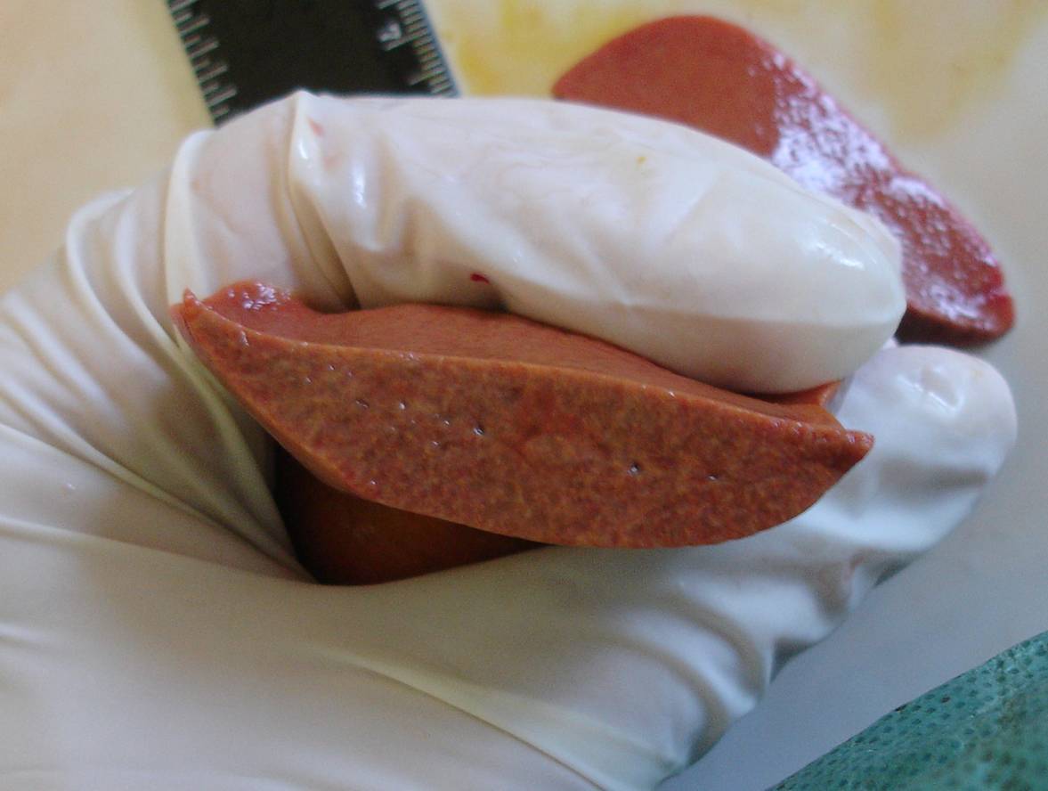
Pathological and anatomical examination revealed a sharp edema, acute inflammation of the liver (Fig. 1); hepatic cholelithiasis (Fig. 2); interstitial nephroso-nephritis; severe fibrosis of the pancreas; myocardial edema; lung atelectasis.
Rice. 1. Microphoto. Histological section of the liver. Severe edema, leukocyte infiltration. Stained with hematoxylin and eosin, vol. ×40, approx. ×10

BUT

B

AT

G
Rice. 2. Macro photo. Hepatic cholelithiasis. Many combined yellow and dark green stones in the intrahepatic bile ducts. The stones are easily “squeezed out” with slight compression of the liver, dense consistency (Fig. A, B, C). On the cut of the stone, the layered structure and color change are clearly visible (shown by an arrow in Fig. D)
Clinical case 2. A dog, a miniature poodle breed, female, 17 years old, was admitted to the clinic with complaints of the owners of convulsive syndrome during the day. On clinical examination, the general condition of the animal is severe. Body temperature 40 o C. Mucous membranes cyanotic pink. On the ECG - single extrasystoles. Pain on palpation of the abdominal wall. Ultrasound revealed parietal hyperechoic round formations up to 0.3 cm in diameter in the gallbladder cavity, diffuse changes in the liver, and signs of chronic nephritis.
Clinical blood test: leukocytes - 23.5 thousand / μl; erythrocytes - 6.08 million / μl; hemoglobin - 128 g/l; hematocrit - 40.2%; platelets - 752 thousand / μl.
Biochemical blood test: glucose - 2.0 mmol/l; bilirubin - 0.9 µmol/l; ALT - 50 U / l; AST - 182 U / l; urea - 7.9 mmol / l; creatinine - 78 µmol/l; pancreatic amylase - 559 U / l.
The animal was placed in the hospital of the clinic, where it received infusion therapy. The dog had epileptiform seizures for 15-30 seconds every 2 hours. On the 4th day of treatment, due to the extremely serious condition of the animal, at the request of the owners, it was euthanized.
Pathological and anatomical examination revealed: massive intracerebral hemorrhage in the right frontal lobe of the brain, moderate internal hydrocephalus (Fig. 3); edema, plethora, fatty degeneration, perivascular sclerosis of the liver (Fig. 4); cholecystolithiasis (Fig. 5); macronodular cirrhosis of the body and head of the pancreas; bilateral macrofocal nephrosonephritis with cirrhosis and polycystic; myocarditis; a combination of emphysema, pneumosclerosis and congestive plethora of the lungs; hemosiderosis of the spleen.

Rice. 3. Macro photo. Frontal section of the brain. Massive intracerebral hemorrhage in the right parietal lobe of the brain (shown by arrow), moderate hydrocephalus

Rice. 4. Microphoto. Histological section of the liver. Edema, plethora, fatty degeneration, perivascular sclerosis of the liver. Stained with hematoxylin and eosin, vol. ×40, approx. ×10


Rice. 5. Macro photo. Cholecystolithiasis. Multiple pigment stones up to 4 mm in diameter (shown by an arrow in Fig. A) in an unchanged gallbladder, loose consistency, crumbling under moderate pressure (Fig. B).
Clinical case 3. A 5-year-old female Yorkshire terrier dog was admitted to the clinic with complaints from the owners about a neoplasm of the mammary gland (noticed 6 months ago) and cough for 3 months, aggravated after exercise. A clinical study revealed: breast cancer stage II, mucous membranes are cyanotic, the tracheal reflex is sharply positive, breathing is clean, vesicular. Ultrasound showed hyperechoic contents in the lumen of the gallbladder (Fig. 6), bilateral nephrolithiasis, diffuse changes in the liver. X-ray examination: an increase in the right heart, collapse of the trachea.
BUT 
B 
Rice. Fig. 6. Ultrascanogram of the gallbladder in transverse (a) and longitudinal (b) sections. Hyperechoic contents in the lumen of the gallbladder (shown by arrow)
The animal was treated in the clinic for 4 months: a course of radiation therapy followed by a regional mastectomy and three courses of chemotherapy. The condition worsened after the end of the course of chemotherapy: persistent pancytopenia, epileptiform seizures, gastrointestinal bleeding.
Due to the extremely serious condition of the animal, at the request of the owners, it was euthanized.
Pathological and anatomical diagnosis: severe internal hydrocephalus (Fig. 7), fatty degeneration of the liver (Fig. 8, 9), cholecystolithiasis (Fig. 10), right ventricular cavity thrombosis, grade III tracheal collapse, bilateral nephrolithiasis, pinpoint hemorrhages in the thin and thick sections of the intestine.

Rice. 7. Macro photo. Segmental section of the brain. Enlargement of the ventricles of the brain
Rice. 8. Macro photo. Fatty degeneration of the liver. Yellowish organ on section

Rice. 9. Microphoto. Fatty degeneration of the liver. Numerous fat droplets in the cytoplasm of hepatocytes, creating a fine mesh pattern. Stained with hematoxylin and eosin, vol. ×40, approx. ×10
BUT 
B 
Rice. 10. Cholecystolithiasis. Pigmented stones of the gallbladder in Fig. A are shown by arrows. Stones of a loose consistency, crumble from moderate pressure (Fig. B)
Discussion and Conclusions
Gallstone disease is a rare disease in dogs and cats that is often asymptomatic. In most cases, the pathology is concomitant with the development of the underlying disease. Only in one of the three clinical cases described by us can we say that cholelithiasis was the main disease of the animal.
The main etiological factor of the pathology, both according to the veterinary literature and according to the above clinical cases, is liver pathology. Among the animals studied by us with cholelithiasis, severe liver damage was confirmed (histologically) in all three cases. It represented both fatty degeneration and hepatitis or perivascular cirrhosis.
Severe pathologies of the kidneys (intermediate nephrosonephritis, nephrosonephritis with cirrhosis and polycystosis, and nephrolithiasis, identified in each individual case) and pancreas (fibrosis or cirrhosis of the organ, which we found in two out of three cases) may indicate a possible correlation of cholelithiasis with deficiency of these organs. It should be noted that in all three cases, the disease was detected in females, and according to numerous data in the medical literature, the disease has a sexual predisposition (stones are 3-4 times more common in women).
Changes in hematological and biochemical parameters that appear when obstructing the biliary tract with stones, leading to cholestasis, are more often manifested by leukocytosis and an increase in liver parameters.
The main instrumental method for studying the disease is ultrasound or CT, which allows to detect the presence of stones, their size, quantity, localization and, to a certain extent, structure.
In the presence of stones in the gallbladder, the main method of treatment is cholecystotomy with the extraction of stones, and in case of serious pathology of the gallbladder - cholecystectomy. It is gaining distribution in veterinary practice to restore the outflow of bile by applying various anastomoses between the biliary system and the duodenum (cholecystoduodenostomy).
Bibliography
1. Kaliteevsky P.F. Macroscopic differential diagnosis of pathological processes. Moscow, "Miklosh", 1993. p. 221-226.
2. Lyutinsky S.I. Pathological physiology of animals. M.: KolosS, 2005. p. 351-352.
3. Fingers M.A. Pathology: a course of lectures. Volume 2. M., "Medicine", 2007. p. 287-289.
4. Savoysky A.G., Baimatov V.N., Meshkov V.M. pathological physiology. M.: KolosS, 2008, p. 409-411.
5. Buote N. J, . The surgical treatment of cholelithiasis in cats: a study of nine cases. J Am Anim Hosp Assoc. 2002, 38(3): 290-6.
9. Fahie M.A., Martin R.A. Extrahepatic biliary tract obstruction: A retrospective study of 45 cases (1983–1993). J Am Anim Hosp Assoc. 1995, 31: 478–481.
10. Heidner G.L , Campbell K.L . Cholelithiasis in a cat. J Am Vet Med Assoc. 1985, 15; 186(2): 176-7.
11. Kirpensteijn J., Fingland R.B., Ulrich T, Sikkema D.A., Allen S.W. Cholelithiasis in dogs: 29 cases. J Am Vet Med Assoc. 1993, 202: 1137–1142.
12. Neer M.T. A review of disorders of the gallbladder and extrahepatic biliary tract in the dog and cat. J Vet Intern Med 1992; 6:186–192.
13. Rege R.V., Prystowsky J.B. Inflammatory properties of bile from dogs with pigment gallstones. Am J Surg. 1996; 171(1): 197–201.
14. Strombeck D.R., Guilford W.G. Small Animal Gastroenterology, 2nd ed. Davis, California: Stonegate Publ, 1990, p. 686–689.
15. Wolf A.M. Obstructive jaundice in a cat resulting from choledocholithiasis. J Am Vet Med Assoc. 1984, 1; 185(1): 85-7.
Summary
D.E. Mitrushkin. Cholelithiasis in dogs and cats. The frequency of cholelithiasis in dogs and cats is rare and often is subclinical, but can result in clinical signs such as icterus, anorexia, vomiting, dehydration, abdominal pain, bradycardia, skin itch and acholia. Values for bilirubin total, alanine aminotransferase, alkaline phosphatase, cholesterol and white blood cells are higher than normal at obstructive cholelithiasis. In this article three cases of cholelithiasis were presented. In all three cases we found expressed histopathological changes of the liver, pancreas and kidneys. It is suggested that the pathology of these organs might have contributed to gallstones formation. The main method of treatment of disease is cholecystotomy, however, cholecystectomy is indicated if damage of gall bladder is severe.


