The structure of the pelvic bones of a woman. Female pelvis (bone pelvis) Female pelvic bone
The female pelvis is a volumetric cavity in which the genitals are located, and an important function is to ensure the flow of the birth process. The bones of the female pelvis have significant differences from the male. What are the sex differences in the pelvis?
The structure of the pelvis of a woman
The anatomy of a woman's pelvis undergoes modifications, starting from the birth of a girl and throughout the stages of growing up. In a born girl, its location is vertical, it is rather narrow, the entrance has an oval shape. In the process of growing up, it acquires a different shape and size of the pelvic bones.
Formation depends on a number of reasons:
- genetic features;
- external factors;
- rickets;
- infectious pathologies (for example, poliomyelitis);
- physical activity;
- injuries of the spine, legs.
The female pelvis is an articulation of several types of bones and ligaments between them. Muscle fibers of the spinal column and legs are attached to them.
Large pelvis
It is located in the upper part of the pelvic joint. Along its edges are the ilium, behind are the lumbar vertebrae, and in front of the anterior abdominal wall. The value may vary from the tension of the muscular apparatus of the abdominal press.
In terms of volume, a large pelvis can differ significantly from a small one. Judging by the size of the large, doctors conclude about the volume of the small, which is very important for determining the process of childbirth in women. Will it be natural childbirth or caesarean section. Quite often there are cases of the impossibility of independent childbirth due to the peculiarities of the anatomy of the pelvis in a woman.
Small pelvis
This is the bone structure of the birth canal. It consists of the upper plane, the bone cavity and the lower opening.
What is the small pelvis formed by:
- Posteriorly represented by the sacrum and coccyx.
- On the sides of the sit bones.
- In front of the symphysis and pubic bones.
- Between the two basins there is a border - an unnamed line.
- The female pelvis is represented by two systems.
reproductive system

This may include the following authorities:
- the uterus and its neck;
- two fallopian tubes;
- two ovaries;
- vagina;
- labia.
In addition, large vessels and nerves are located in the pelvic area.
excretory system
Includes the following organs:
- bladder;
- rectum.
The musculature of the excretory system includes longitudinal and circular muscle fibers.
Types of bones
The pelvis of a woman, like men, performs a supporting function and distributes the weight of the body on the legs, which contributes to the motor activity of a person.
Bones of the female pelvis:
- two pelvic (or nameless) bones;
- coccygeal;
- sacral.
Ligaments connect all kinds of bones.
Pelvic bone
In children under 15 years old, the innominate bone is a movable joint of three main bones:
- Ischial. It consists of two branches and a body. At the end of the upper branch is the ischial tubercle. The lower one is directed down and forward. It has an ischial spine.
- The pubic bone is represented by two branches: lower and upper. There is also a body. There is a comb on the top branch.
- Iliac. It consists of a wing and a body. At the top of the wing is a crest.
The pelvic bones grow together and become monolithic only after the child reaches 17-19 years of age.
coccygeal bone
The bone is represented by several rudimentary vertebrae. Usually there are 4 or 5 of them. It performs a supporting function and distributes the load on other structures of the pelvic joint. In the birth process, he can deviate a little back, to facilitate the process of giving birth to a child.
sacrum
These are 6 sacral vertebrae, monolithically interconnected. They are then joined into one bone. The sacrum looks like a massive triangle. In its upper part, the sacrum is connected to the 5th lumbar vertebra, and to the coccyx from below. In children, the components of the sacrum are interconnected by cartilage, while complete ossification and its transformation into a monolithic structure occurs at the age of 24-26.
Types of forms of the female pelvis
Anatomy is represented by four forms:
- Gynecoid. Not very deep cavity, oval entrance. The subpubic angle is 900. This is the ideal shape for the delivery of a woman. Usually women of average height and the same physique have this form.
- Android. More in line with the male form. It is characterized by a heart-shaped entrance, a funnel-shaped cavity. The pelvic outlet is compressed. The subpubic angle is less than 900. This form is often found in short women, their physique is quite dense.
- Anthropoid. The fifth lumbar vertebra is connected to the sacrum. The subpubic arch is large. This form is inherent in tall women. As a rule, natural childbirth proceeds without problems.
- Platipelloidal. The cavity is shallow. The angle is more than 900. The process of childbirth with this form proceeds normally.

The shape of the pelvis can be examined using x-rays.
male pelvis
It is usually smaller than the female by about 1.7 cm. The difference in size may depend on several reasons, for example, age, type of posture of a person.
Its cavity includes the following organs:
- bowel loops;
- appendix.
The pelvic inlet in men is narrower than in women, the coccyx is slightly less forward. Lymphatic, large blood vessels are also located here.
Differences between male and female
The anatomical features of the female pelvis differ from the male in a number of ways.
The difference between the female pelvis and the male pelvis begins to appear during the period of growing up of boys and girls.
The anatomy of the pelvic joint is quite complex. Violation of its integrity entails negative consequences. Such as dysfunction, lameness.
FEMALE pelvis from an obstetric point of view.
The bone pelvis consists of two pelvic bones, sacral and coccygeal bones, which are firmly connected through cartilaginous layers and connections.
The pelvic bone is formed from the fusion of three bones: longitudinal, ischial and pubic. They join in the region of the acetabulum.
The sacrum consists of 5-6 fixedly connected vertebrae, which merge into one bone.
The coccygeal bone consists of 4-5 underdeveloped vertebrae.
The bony pelvis in the upper section is open forward. This part is called the greater pelvis. Bottom part- this is a closed bone formation - the small pelvis. The border between the large and small pelvis is the terminal (nameless) line: in front - the upper edge of the symphysis and pubic bones, from the sides - arcuate lines of the ilium, behind - the sacral protrusion. The plane between the large and small pelvis is the entrance to the small pelvis. The large pelvis is much wider than the small one, it is bounded laterally by the wings of the ilium, behind by the last lumbar vertebrae, and in front by the lower part of the anterior abdominal wall.
All women undergo a measurement of the large pelvis. There is a relationship between the sizes of the large and small pelvis. By measuring the large pelvis, we can draw conclusions about the size of the small one.
Normal dimensions of the female pelvis:
- distantia spinarum - the distance between the anterior upper bones of the longitudinal bone - 25-26 cm;
- distantia cristarum - the distance between the distant points of the iliac crests - 28-29cm;
- conjugata externa - (external conjugate) - the distance from the middle of the upper edge of the symphysis to the upper corner of the Michaelis rhombus (measurements are made with the woman lying on her side) - 20-21 cm.
Rhombus Michaelis- this is an expansion of a depression in the sacral region, the limits of which are: from above - a fossa under the spinous process of the fifth lumbar vertebra (supracrine fossa), from below - points corresponding to the posterior superior iliac spines. The average length of a rhombus is 11 cm, and the diameter is 10 cm.
Diagonal conjugate- the distance from the lower edge of the symphysis to the most protruding point of the promontory of the sacral bone is determined during vaginal examination. With normal pelvic dimensions, it is 12.5-13 cm.
The size of the true conjugate (the direct size of the entrance to the small pelvis) is determined by subtracting 9 cm from the length of the outer conjugate or subtracting 1.5-2 cm from the length of the diagonal conjugate (depending on the Solovyov index).
Solovyov index - the circumference of the wrist-carpal joint, divided by 10. The index allows you to have an idea of \u200b\u200bthe thickness of a woman's bones. The thinner the bones (index = 1.4-1.6), the greater the capacity of the small pelvis. In these cases, 1.5 cm is subtracted from the diagonal conjugate and the length of the true conjugate is obtained. With the Solovyov index
I, 7-1.8 - subtract 2 cm.
Pelvic tilt angle - the angle between the plane of the entrance to the small pelvis and the horizon is 55-60 °. Deviations in one direction or another can adversely affect the course of childbirth.
The height of the symphysis is normally 4 cm and is measured with the index finger during vaginal examination.
The pubic angle - with normal pelvic dimensions is 90-100 °.
Small pelvis is the bony part of the birth canal. The posterior wall of the small pelvis consists of the sacrum and the coccyx, the lateral ones are formed by the ischium, the anterior one is formed by the pubic bones and the symphysis. The small pelvis has the following sections: entrance, cavity and exit.

In the pelvic cavity, a wide and narrow part is distinguished. In this regard, four planes of the small pelvis are determined:
1 - the plane of the entrance to the small pelvis.
2 - the plane of the wide part of the pelvic cavity.
3 - the plane of the narrow part of the pelvic cavity.
4 - the plane of the exit from the pelvis.
The plane of the entrance to the small pelvis passes through the upper inner edge of the pubic arch, the innominate lines and the top of the promontory. In the plane of the entrance, the following dimensions are distinguished:
- Direct size - the distance from the sacral protrusion to the point that protrudes most on the upper inner surface of the symphysis - this is an obstetric, or true conjugate, equal to 11 cm.
- Transverse size - the distance between the distant points of the arcuate lines, which is 13-13.5 cm.
- Two oblique dimensions - from the iliosacral junction on one side to the iliopubic tubercle on the opposite side of the pelvis. They are 12-12.5 cm.
The plane of the wide part of the cavity of the small pelvis passes through the middle of the inner surface of the pubic arch, on the sides through the middle of the acetabular cavities and behind - through the connection between the II and III sacral vertebrae.
In the plane of the wide part of the small pelvis, there are:
- Direct size - from the middle of the inner surface of the pubic arch to the junction between II and III sacral vertebrae. It is equal to 12.5 cm.
- The transverse dimension passes between the midpoints of the acetabulum. It is equal to 12.5 cm.
The plane of the narrow part through the lower edge of the pubic junction, on the sides - through the gluteal spines, behind -
through the sacrococcygeal junction.
In the plane of the narrow part they distinguish:
1. Direct size - from the lower edge of the symphysis to the sacrococcygeal junction. It is equal to II, 5 cm.
2. The transverse dimension between the distant points of the inner surface of the ischial spines. It is equal to 10.5 cm.
The plane of exit from the small pelvis passes in front through the lower edge of the symphysis, from the sides - through the tops of the gluteal tubercles, from behind - through the crown of the coccyx.
In the plane of exit from the small pelvis, there are:
1. Direct size - from the top of the coccyx to the lower edge of the symphysis. It is equal to 9.5 cm, and when the fetus passes through the small pelvis, it increases by 1.5-2 cm due to the deviation of the tip of the coccyx of the presenting part of the fetus.
2. Transverse dimension - between the distant points of the inner surfaces of the ischial tuberosities; it is equal to 11cm.
The line connecting the midpoints of the direct dimensions of all planes of the pelvis is called the leading axis of the pelvis, and has the form of a forward concave line. It is along this line that the leading point passes through the birth canal.
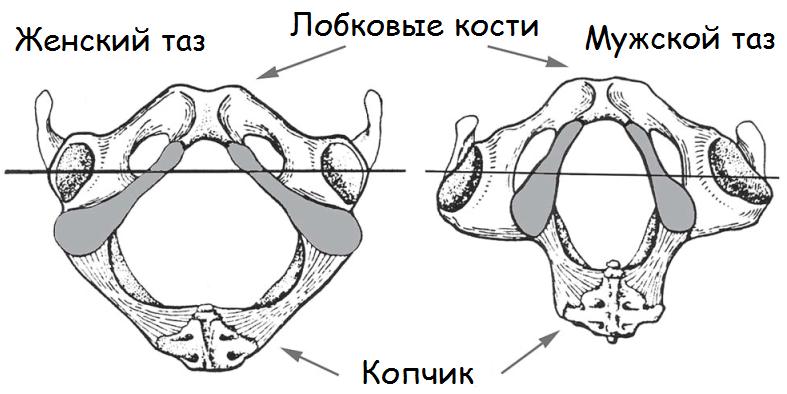
The main differences between the female pelvis and the male:
- The bones of the female pelvis are thin and smooth compared;
- The female pelvis is relatively wider, lower and larger in volume;
- The wings of the ilium in women are more developed, so the transverse dimensions of the female pelvis are larger than the male ones;
- The entrance to the small pelvis of a woman has a transverse oval shape, and in men it has the shape of a card heart;
- The entrance to the small pelvis in women is larger and the pelvic cavity does not narrow down funnel-shaped, as in men;
- The pubic angle in women is obtuse (90-100 °), and in men it is acute (70-75 °);
- The pelvic tilt is greater in women (55-60°) than in men (45°).
The bone pelvis is a strong container for the internal hollow organs and their surrounding tissues. The woman's pelvis forms the birth canal through which the fetus is born.
Differences between the female pelvis and the male pelvis begin to emerge during puberty and become distinct in adulthood.
The bones of the female pelvis are thinner, smoother and less massive than the bones of the male. The female pelvis is lower, wider and larger in volume. The sacrum in women is wider and less concave than in men. The symphysis in women is shorter and wider. The cape of the sacrum protrudes less forward. The entrance to the small pelvis in women is more extensive and has a transverse-oval shape with a notch in the region of the cape of the sacrum, while in men it resembles the shape of a card heart due to the sharp protrusion of the cape. The pelvic cavity in women is more extensive and in shape approaches the cylinder, curved anteriorly. In men, the pelvic cavity is smaller, and it narrows downwards in a funnel-shaped manner. The exit of the pelvic cavity in a woman is wider, since the distance between the ischial tubercles is greater. The pubic angle is wider (90-100) than in men (70-75). And the coccyx protrudes anteriorly less than in the male pelvis
Thus, we can conclude that the female pelvis is more voluminous and wider, but less deep than the male.
Pelvic bones
The pelvis is made up of four bones: two pelvic bones, the sacrum, and the coccyx.
Pelvic (nameless) bone
(os coxae ). Up to 16-18 years, it consists of three bones connected by cartilage: iliac, ischial and pubic. After ossification, the cartilages fuse together to form the innominate bone.
Ilium (
os ilium ) has two parts: body and wing. The body makes up a short, thickened part of the bone and participates in the formation of the acetabulum.
Large pelvis dimensions
Knowing the size of the external pelvis is very important in obstetrics, since the size of the small pelvis is judged by its size. The measurement is made with a tazometer. There are four sizes: three transverse and one straight.
Distantia spinarum- distance between the anterior superior iliac spines.It is usually equal to 25-26 cm.
Distantia cristarum- the distance between the most distant points of the iliac crests. Usually it is 28-29 cm.
Distantia trochanterica -distance between the greater trochanters of the femur. It is 30-31 cm.
Conugata externa -external conjugate i.e. straight size of the pelvis. The woman is laid on her side, the underlying leg is bent at the knee and hip joints, the overlying one is pulled out. The button of one branch of the tazomer is set in the middle of the upper outer edge of the symphysis, the other end is pressed against the supra-sacral fossa, which is located between the spinous process of the 5th lumbar vertebra and the beginning of the middle sacral crest (the supra-sacral fossa coincides with the upper angle of the sacral crest). Normally, it is 20-21 cm .
Conugata vera-true conjugate. To determine it, it is necessary to subtract 9 from the outer conjugate, then we get the true size. The difference between the true and external conjugates depends on the thickness of the sacrum, symphysis and soft tissues, so the difference does not always correspond exactly to 9 cm. Or 1.5-2 cm is subtracted from the size of the diagonal conjugate.
Conugata diagonalis -- diagonal conjugate - this is the distance from the lower edge of the symphysis to the most prominent point of the promontory of the sacrum. It is determined by vaginal examination. With a normal pelvis, it is 12.5-13 cm.
Taking into account the danger of pelvic presentation of the fetus in the mother's pelvis, timely diagnosis, hospitalization of a woman in an obstetric hospital at 35-36 weeks of gestation is very important. fetus in the mother's pelvis by caesarean section. Indications for the operation of turning the fetus on the leg are: full disclosure of the cervix and discharge of water during examination or in a timely manner. The operation of turning the fetus on the leg is performed under deep anesthesia. Categorical contraindications for this operation are: premature, early discharge of amniotic fluid and incomplete opening of the cervix.
It forms a generic the channel through which the fetus moves. Unfavorable conditions for intrauterine development, diseases suffered in childhood and inpuberty, can lead to disruption of the structure and developmentpelvis. The pelvis can be deformed as a result of trauma, tumors, various exostoses. Differences in the structure of the female and male pelvis begin to appear during puberty and become pronounced in adulthood. The bones of the female pelvis are thinner, smoother and less massive than those of the male. pelvis. The plane of the entrance to the small pelvis in women has a transverse-oval shape, while in men it has the shape of a card heart (due to the strong protrusion of the cape).
Anatomically, the female pelvis is lower, wider and larger in volume. The pubic symphysis in the female pelvis is shorter than the male. The sacrum in women is wider, the sacral cavity is moderately concave. The pelvic cavity in women approaches the cylinder in outline, while in men it narrows downwards in a funnel-shaped manner. The pubic angle is wider (90-100°) than in men (70-75°). The coccyx protrudes anteriorly less than in the male pelvis. The ischial bones in the female pelvis are parallel to each other, and converge in the male.
All of these features are very important in the process of childbirth. The pelvis of an adult woman consists of 4 bones: two pelvic, one sacral and one coccygeal, firmly connected to each other,
Pelvic bone, or nameless (os coxae, os innominatum), consists up to 16— 18 years of 3 bones connected by cartilage in the area of the acetabulum(acetabulum): iliac (os ileum), sciatic (os ischii) and pubic (os pubis ). After puberty, cartilage fuses together and a solid bone mass is formed - the pelvic bone.
On the ilium distinguish between the upper section - the wing and the lower - the body.At the place of their connection, an inflection is formed, called arcuate or be-zymyanny line ( linea arcuata, innominata ). On the ilium follows from-mark a number of protrusions that are important to the obstetrician. Upper thickenedwing edge - iliac crest ( Crista Iliaca ) - has an arcuatecurved shape, serves to attach the broad muscles of the abdomen. Spere-di it ends with the anterior superior iliac spine ( spina iliaca anterior superior ), and behind - the posterior superior iliac spine ( spina iliaca posterior superior ). These two spines are important in determining the size of the pelvis.Ischium forms the lower and posterior thirds of the pelvic bone. She isconsists of a body involved in the formation of the acetabulum, and a branchischium. The body of the ischium with its branch makes an angle, openty anteriorly, in the region of the angle, the bone forms a thickening - the ischial tuberosity(tuber ischiadicum ). The branch goes anteriorly and upwards and connects with the lowerher branch of the pubic bone. On the back surface of the branch there is a protrusion - ischial spine (spina ischiadica). On the ischium, there are two tenderloin: greater ischial tenderloin ( incisura ischiadica major ), located below the posterior superior iliac spine, and a small sciatic notch ku (incisura ischiadica minor).
Pubic, or pubic, bone forms the anterior wall of the pelvis, consists of the bodyand two branches - the upper ( ramus superior ossis pubis) and lower (ramus inferior ossis pubis ). The body of the pubis forms part of the acetabulum. Togetherconnection of the ilium with the pubis is the iliopubic elevation ( eminentia iliopubica).
The upper and lower branches of the pubic bones are connected to each other in frontthrough cartilage, forming a sedentary joint, a half-joint ( symphysis ossis pubis ). The slot-like cavity in this connection is filled with liquid andincreases during pregnancy. The lower branches of the pubic bones formyut angle - pubic arch. Along the posterior edge of the superior ramus of the pubisthe pubic ridge stretches ( crista pubica ), passing backwards into linea arcuata of the ilium.
Sacrum(os sacrum ) consists of 5-6 vertebrae fixed to each other, the size of which decreases downwards. The sacrum has the shape of acone. The base of the sacrum is turned upward, the apex of the sacrum (narrowpart) - downwards. The anterior surface of the sacrum has a concave shape; on itthe junctions of the fused sacral vertebrae are visible in the form of transverserough lines. The posterior surface of the sacrum is convex. Along the midlinepass the spinous processes of the sacral vertebrae fused together.First sacral vertebra connected to V lumbar, has a protrusion - sacral cape (promontorium).
Coccyx (os coccygis ) consists of 4-5 fused vertebrae. He connectsusing the sacrococcygeal articulation with the sacrum. In braid connections the pelvis has cartilaginous layers.
The female pelvis from an obstetric point of view
There are two parts of the pelvis: the large pelvis and the small pelvis. border between them is the plane of entry into the small pelvis.
The large pelvis is bounded laterally by the wings of the ilium, behind -last lumbar vertebra. In front, it has no bony walls.
The pelvis is of the greatest importance in obstetrics. Through the small pelvisthe birth of the fetus is going on. There is no easy way to measure the pelvis.At the same time, the dimensions of the large pelvis are easy to determine, and based on them you can judge the shape and size of the small pelvis.
The small pelvis is the bony part of the birth canal. Form andthe size of the small pelvis is very important during childbirth and the definition of tactics for their management. With sharp degrees of narrowing of the pelvis and its deformationyah, childbirth through the natural birth canal becomes impossible, and women well, delivery by caesarean section.
The posterior wall of the small pelvis is made up of the sacrum and coccyx, the lateral ones arefarther bones, anterior - pubic bones with l circumferential symphysis. Top-The lower part of the pelvis is a solid bone ring. In the middle andlower thirds of the wall mscarlet pelvis is not continuous. In the lateral sections there are large and small sciatic foramen ( foramen ischiadicum majus etminus), limited respectively by large and small ischial notches (incisure ischiadica major et minor) and withviscous ( lig. sacrotuberale, lig. sacrospinale ). The branches of the pubic and ischial bones, merging, surroundobturator opening ( foramen obturatorium ) shaped like a triangle with rounded corners.
In the small pelvis, an entrance, a cavity and an exit are distinguished. In the pelvic cavity, excretelyayut wide and narrow parts. In accordance withthis in the pelvis distinguish four classical planes ( rice. one ).
The plane of the entrance to the small pelvis anteriorly bounded by the superior margin of the symphysis andthe upper inner edge of the pubic bones, from the sides - arcuate linesiliac bones and behind - sacral promontory. This plane is shapedtransversely located oval (or kidney-shaped). It distinguishes three size (rice. 2): straight, transverse and 2 oblique (right and left). Straight size is the distance from the upper inner edge of the symphysisto the sacral cape. This size is called the true or obstetric conjugates (conjugata vera) and equal 11 cm.
In the plane of the entrance to the small pelvis, tea still anatomical conjugate (conjugata anato - mica ) - the distance betweenthe upper edge of the symphysis andsacral cape.The value of the anatomical conjugate is11.5 cm. P about the pepper size - the distance between the most distant parts of the du-curved lines. He co-sets 13.0-13.5 cm. plane dimensions entrance to the small pelvisrepresent the distance betweendu sacroiliacarticulation of one sideny and the iliac-pubic eminence of the oppositefalse side. Rightthe oblique dimension is determinedfrom the right sacro-under-iliac joint, le-exit - from the left. These sizes Rs range from 12.0 to 12.5 cm .
 The plane of the wide gas-ti cavity of the small pelvis from the front it is limited by the middle of the inner surface of the symphysis, from the sides - by the middle of the plates covering the acetabular cavities, from behind - by the junction of the II and III sacral vertebrae. In the wide part of the bands of the small pelvis, there are
2 sizes: straight and transverse. Straight size— distance between the junction of AND and III sacral vertebrae and the middle of the inner surface of the symphysis. It is equal to 12.5 cm. The transverse size is the distance between the midpoints of the inner surfaces of the plates covering the acetabulum. It is equal to 12.5 cm. Since the pelvis in the wide part of the cavity does not represent a continuous bone ring, oblique dimensions in this section are allowed only conditionally (13 cm each).
The plane of the narrow gasti of the pelvic cavity bounded in front by the lower edge of the symphysis, laterally by the awns of the ischial bones, and behind by the sacrococcygeal articulation.
In this plane, 2 sizes are also distinguished. Direct size - distance gap between bottom edgesymphysis and sacrococcygealjoint. He is equal 11.5cm. Cross dimension - the distance between thetyami ischial bones. He is 10.5 cm.
Plane of exit from the small pelvis( rice. 3 ) is limited in front by the lower edge of the pubic symphysis, from the sides - by the ischial tubercles, from behind - by the tip of the coccyx. Direct size - dis- standing between the bottom edgesymphysis and apex of the cop-chica. It is equal to 9.5 cm.the passage of the fetus through the birth canal (through the plane of exit from the small pelvis)due to protrusion of the coccyxposteriorly, this size is increasedshrinks by 1.5-2.0 cm and becomesnew equal to 11.0-11.5 cm. Cross dimension - the distance between the internal surfaces of the gray- personal bumps. It is equal to 11.0 cm.
When comparing the dimensions of the small pelvis in different planes, it turns out that in the plane of the entrance to the small pelvis, the transverse dimensions are maximum, in the wide part of the cavity of the small pelvis, the direct and transverse dimensions are equal, and in the narrow part of the cavity and in the plane of exit from the small pelvis, the direct dimensions are larger than the transverse ones.
 In obstetrics, in some cases, a system is used parallel Goji planes( rice. four ). The first, or upper, plane (terminal) passes through the upper edge of the symphysis and the border (terminal) line. The second parallel plane is called the main one and passes through the lower edge of the symphysis parallel to the first. The fetal head, having passed through this plane, does not encounter significant obstacles in the future, since it has passed a solid bone ring. The third parallel plane is the spinal plane. It runs parallel to the previous two through the ischial spines. The fourth plane - the exit plane - runs parallel to the previous three through the top of the coccyx.
All classical planes of the small pelvis converge in the direction of the anterior (symphysis) and fan-shaped diverge backwards. If you connect the midpoints of all the direct dimensions of the small pelvis, you get a line curved in the form of a fishhook, which is called wire axis of the pelvis. It bends in the cavity of the small pelvis, corresponding to the concavity of the inner surface of the sacrum. The movement of the fetus through the birth canal occurs in the direction of the wire axis of the pelvis.
Angle of inclination of the pelvis - this is the angle formed by the plane of entry into the small pelvis and the horizon line. The value of the angle of inclination of the pelvis changes when the center of gravity of the body moves. In non-pregnant women, the angle of inclination of the pelvis is on average 45-46 °, and the lumbar lordosis is 4.6 cm (according to Sh. Ya. Mikeladze).As pregnancy progresses, lumbar lordosis increases due to the displacement of the center of gravity from the region of the II sacral vertebra anteriorly, which leads to an increase in the angle of inclination of the pelvis. With a decrease in the lumbar lord dose, the angle of inclination of the pelvis decreases. Up to 16-20 weeks. pregnancy in the setting of the body, no changes are observed, and the angle of inclination of the pelvis does not change. By the gestational age of 32-34 weeks. lumbar lordosis reaches (according to I. I. Yakovlev) 6 cm, and  the angle of inclination of the pelvis increases by 3-4°, amounting to 48-50° ( rice. 5 ). The magnitude of the angle of inclination of the pelvis can be determined using special devices designed by Sh. Ya. Mikeladze, A. E. Mandelstam, as well as manually. When a woman is positioned on her back on a hard couch, the doctor holds her hand (palm) under the lumbosacral lordosis. If the hand passes freely, then the angle of inclination is large. If the hand does not pass, the angle of inclination of the pelvis is small. It is possible to judge the magnitude of the angle of inclination of the pelvis by the ratio of the external genitalia and thighs. With a large angle of inclination of the pelvis, the external genital organs and the genital gap are hidden between the closed thighs. With a small angle of inclination of the pelvis, the external genital organs are not covered by closed hips.
You can determine the value of the angle of inclination of the pelvis by the position of both iliac spines relative to the pubic joint. The angle of inclination of the pelvis will be normal (45-50°) if, in the horizontal position of the woman's body, the plane drawn through the symphysis and the superior anterior iliac spines is parallel to the plane of the horizon. If the symphysis is located below the plane drawn through these spines, the angle of inclination of the pelvis is less than normal.
A small angle of inclination of the pelvis does not prevent the fixation of the fetal head in the plane of the entrance to the small pelvis and the advancement of the fetus. Childbirth proceeds quickly, without damage to the soft tissues of the vagina and perineum. A large angle of inclination of the pelvis often presents an obstacle to fixing the head. Incorrect insertion of the head may occur. In childbirth, injuries of the soft birth canal are often observed. By changing the position of the body of a woman in labor during childbirth, it is possible to change the angle of inclination of the pelvis, creating the most favorable conditions for the advancement of the fetus through the birth canal, which is especially important if a woman has a narrowing of the pelvis.
The angle of inclination of the pelvis can be reduced by raising the upper body of the lying woman, or in the position of the body of the woman in labor on her back, bring the legs bent at the knee and hip joints to the stomach, or put a polster under the sacrum. If the polster is under the lower back, the angle of inclination of the pelvis increases.
In the human body there is a small and a large pelvis. The article will focus on the anatomy of the small pelvis in women and talk a little about their male structure.
The small pelvis is an anatomical space, which is limited by the bones, located in the lower part of the abdominal cavity. It is important to remember that women have many anatomical features of the structure of the pelvic organs.
The difference is due to the fact that men and women have different sex organs. The rectum and bladder are common to both sexes. In men, the pelvic cavity also includes the internal reproductive organs - the seminal vesicles and the prostate gland (see the diagram of the internal and external organs of men in the figure below).
An interesting fact is that men have a smaller structure. This is explained by the fact that women are able to bear a fetus. No wonder women's hips are wider, more massive than men's.
The location of the pelvic organs of men
The organs of the pelvic area in women, what is included and how are they located
Let's define the female organs of the small pelvis, what is it:
- Vagina;
- Uterus;
- The fallopian tubes;
- ovaries;
- Musculature;
- Bladder;
- Rectum.
This is a list of female pelvic organs. The layout of the internal organs is shown in the following picture.

The structure of the pelvic organs of women
All of them are located close to each other, tightly in contact. Therefore, inflammation often moves from one to the next.
- The vagina (vagina) is a muscle shaped like a canal or tube. The average length is 8-10 centimeters. The vagina plays an important role during conception, the birth of a child. Its important feature is the ability to strongly stretch, which allows the baby to be born.
- The bladder is located under the uterus, above the vagina. Its main function is the accumulation of urine and its preservation before the urination process.
- Through the rectum, feces, toxins, toxins, all waste products of the body are excreted. The coccyx supports her.
- The musculature of the pelvic area is represented by the pelvic floor, which is a collection of muscles. They consist of two layers: deep and superficial.
Thanks to muscle fibers, all organs of this area in women are stably supported. In case of weakening, it is recommended to train these muscles using the wumbling technique and Kegel exercises for women.
The structure of the female reproductive system
The striae of female intimate organs is presented in the diagram in the following photo.

First come the large and small labia, which protect the vagina from foreign microorganisms, bacteria and aggressive external environment. Next comes the vagina itself, and behind it the cervix, which connects the vagina to it.
The uterus is the main organ of the female reproductive system. It is to it that a fertilized egg is attached, which grows and turns into a full-fledged embryo. The fallopian tubes run from the ovaries to the uterus.
The ovaries are an important part of the female reproductive system. In them, eggs mature, the parameters of the menstrual cycle are regulated, and hormones are released: estrogen, progesterone. The egg travels through the fallopian tubes to the uterus.
Effective methods for examining female organs inside the small pelvis
Most often, ultrasound is used to examine the reproductive female organs and identify pathological processes - short ultrasound. It allows you to most accurately consider each of them, making the correct diagnosis. This method is also effective for confirming pregnancy.
Most often, the attending physician (gynecologist or therapist) refers to this procedure due to the following complaints: pain in the lower abdomen, menstrual irregularities, delays, heavy or scanty bleeding, with pathological vaginal discharge, with suspicion of malignant or benign formations, cysts, endometriosis. And also ultrasound helps to identify kidney stones. This type of diagnosis is used for diseases of the genitourinary tract.
Ultrasound examination successfully solves all these issues without harm to a woman's health.
Usually this procedure is prescribed for 5-11 days of the menstrual cycle. There are varieties of ultrasound. Preparation for the diagnosis also varies. Many girls and women are afraid of undergoing this procedure, but it is completely painless.
An important role in the detection of diseases is considered a gynecological examination. The doctor conducts an external examination of the genital organs (vagina, small and large labia), anus. Then, using a special tool (mirror), he examines the cervix. If there are complaints, the gynecologist takes a smear for research to identify the cause of some unpleasant symptoms.
If malignant and benign tumors are suspected, magnetic resonance imaging is used. This is the most accurate way to diagnose this type of disease.

How to maintain the health of the female pelvic organs?
- In the presence of pathologies every six months, or even more often. If you experience pain, discharge, pain, difficulty urinating, menstrual irregularities, you should visit a specialist as soon as possible to identify the cause of the problem. Pass a gynecological examination, take tests, a smear, if necessary, undergo a deeper diagnosis - ultrasound, MRI, x-rays, etc.
- Next, you need to improve your lifestyle: get used to a proper balanced diet, drink plenty of water, walk more often in the fresh air, avoid stress, negativity, sleep 8-9 hours a day, and rest in a timely manner.
- Also, girls and women are not recommended to lift weights, have excessively heavy loads.
- Be sure to remember! Do not sit on cold ground, concrete, etc. This provokes the appearance of inflammation in a feminine way.
Another important rule is not to have promiscuity. If there are no children in the plans, be sure to use contraception. To prevent pregnancy and sexually transmitted diseases, condoms are most effective. Abortions deal a blow to women's health, sometimes irreparable, and change the whole course of a woman's future life because of the murder of a man who was destined to live.
You should always dress according to the weather, do not wear light clothes in the cold, avoid hypothermia.
Naturally, personal hygiene plays an important role in the health of the reproductive system. It is best to wash 2 times a day, change clothes daily.


