What is bronchoscopy and why is it done? Bronchoscopy for lung diseases - what is it. Feelings after bronchoscopy
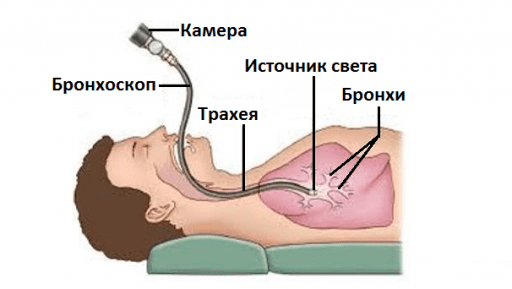
Bronchoscopy- a method of examining the mucous membranes of the trachea and bronchi using a special device - a bronchoscope. A tube equipped with lighting equipment and a video camera is inserted through the larynx into the respiratory tract. This modern equipment provides an accuracy of over 97%, which makes it indispensable in the diagnosis of various pathologies: chronic bronchitis, recurrent pneumonia, lung cancer.
A bronchoscope is often used for medical purposes. To do this, it is additionally equipped with a surgical set of instruments, biopsy forceps, and laser equipment.
History of the use of bronchoscopes.
The first bronchoscopic examination was carried out in 1897. The procedure was painful and traumatic, so cocaine was used for pain relief. For the first 50 years, the bronchoscope was used to remove small foreign bodies from the bronchi.
Early models were equipped with an external light source. The light bulb, with the help of a system of mirrors and lenses, transmitted a beam of light into the bronchi, thanks to which the doctor saw all the changes in the airways.
The first models of the bronchoscope were incomplete. They injured the respiratory system and caused serious complications. The first rigid (hard), but safe for patients, device was invented in 1956 by Friedel. The flexible fiberoptic bronchoscope was introduced in 1968. After 10 years, electronic technologies have made it possible to magnify the image tenfold and get a detailed picture of changes in the lungs.
What is bronchoscopy
Bronchoscopy- Examination of the respiratory tract. The term comes from two Greek words: "examine" and "windpipe". Myself bronchoscope- This is a special optical system for examining the mucous membrane of the larynx, trachea and bronchi to their second branch. It is a system of flexible or rigid tubes with a diameter of 3-6 mm and a length of about 60 cm.Modern bronchoscopes are equipped with photo and video equipment, as well as a cold light lamp, which are placed at the end of the tube. The image is displayed on the monitor screen, where it can be enlarged tenfold. In addition, it is possible to save a record that will be needed in the future to compare and evaluate the dynamics of the pathological process.
Purpose of bronchoscopy. Bronchoscopy is performed not only for the diagnosis of diseases of the respiratory system. With the help of a bronchoscope, you can perform a number of medical procedures:
- removal of foreign bodies from the bronchi
- cleansing of pus and thick mucus
- washing and administration of solutions of antibiotics, glucocorticoids, mucolytics, nitrofurans
- taking tissue samples for biopsy
- expansion of the lumen of the bronchi
- removal of small tumors
How is a bronchoscopy performed?
- The study is carried out in a specially equipped endoscopic room, where the same conditions of sterility are observed as in the operating room. The procedure is supervised by a doctor who has received special training in the study of the bronchi.
- Atropine sulfate, Eufilin, Salbutamol are injected subcutaneously or in the form of aerosols. They have a bronchodilator effect and contribute to the unimpeded advancement of the bronchoscope.
- The study is carried out in a sitting position or lying on your back. In this case, you can not stretch your head forward and arch your chest so that the device does not injure the respiratory mucosa.
- With the introduction of a bronchoscope, it is recommended to breathe often and superficially, this inhibits the gag reflex.
- The bronchoscope is inserted through the nostril or through the mouth. At the moment of deep inspiration, the tube is passed through the glottis. Then, with rotational movements, it is deepened into the bronchi. The tubes are much thinner than the airways, so they do not interfere with breathing.
- During the examination, pressure may be felt in different parts of the respiratory system, but you will not experience pain.
- The study begins with an examination of the larynx and glottis, then the trachea and bronchi are studied. The thin bronchioles and alveoli of the lungs remain inaccessible due to their small diameter.
- During the procedure, the doctor can take a piece of tissue for a biopsy, remove the contents of the bronchi, rinse them with a medicinal solution, take swabs for research, etc.
- After the procedure, the feeling of numbness remains for half an hour. It is not recommended to smoke and eat for 2 hours, so as not to provoke bleeding.
- Sedatives used to reduce anxiety reduce the reaction rate. Therefore, driving is not recommended for 8 hours.
- For some time it is recommended to stay in the hospital. Medical staff will monitor your condition to rule out complications.
The rule of thumb is that local anesthesia is used when examining with a flexible bronchoscope, while general anesthesia is required when using rigid models.
- Local anesthesia. For anesthesia, a 2-5% solution of lidocaine is used. It causes numbness of the palate, feeling of a lump in the throat, difficulty in swallowing, and mild nasal congestion. Anesthesia will also help suppress the cough and gag reflexes. When introduced through the bronchoscope tube, the mucosa of the larynx, vocal cords, trachea and bronchi are gradually sprayed with an anesthetic spray.
- General anesthesia. This procedure is recommended for children and people with an unstable psyche. The patient is put into a state of medicinal sleep and he will feel absolutely nothing.
Types of bronchoscopy
Modern bronchoscopes are divided into two groups: flexible and rigid. Each of the models has its own advantages and scope.
Indications for bronchoscopy
 Indications for bronchoscopy
- signs of disseminated pathological processes on X-ray (small foci, cysts, cavities)
- suspected tumor of the trachea or bronchi
- suspected presence of a foreign body
- prolonged shortness of breath (with the exclusion of bronchial asthma and heart failure)
- hemoptysis
- multiple lung abscesses
- cysts in the lungs
- chronic inflammation of the bronchi of an unexplained cause
- recurrent pneumonia
- abnormal structure and expansion of the bronchi
- finding out the causes of bronchial asthma
- collection of contents to determine the sensitivity of the flora to antibiotics
- preparation for lung surgery
| Pathology | Signs of this disease, which can be detected by bronchoscopy |
| Tuberculosis | Infiltrates of dense consistency. Limited pale pink edematous areas rising above the bronchial mucosa. In the later stages of the disease, they become red, loose, covered with bleeding erosions. |
| Constriction of the bronchi. The lumen becomes narrow, slit-like, due to swelling of the mucous membrane of the respiratory tract | |
| Fistulas - holes in the wall of the bronchi | |
| Endobronchitis - inflammation of the bronchial mucosa | Mucosal swelling |
| Vessels in the mucosa are poorly visible | |
| Thinning of the bronchial mucosa. It is red and bleeds easily on contact. | |
| In the hypertrophic form of the disease, the mucosa is evenly thickened. Bronchial lumen narrowed | |
| Profuse discharge of pus | |
| cystic fibrosis | Violation of the tone of the membranous part of the trachea and bronchi - narrowing of the lumen by more than 1/2 of the diameter |
| Bleeding of the bronchial wall | |
| Accumulations of thick mucus | |
| Cancer - exophytic tumors growing in the lumen of the bronchus | Well-circumscribed neoplasms on a broad base |
| Contours are wrong | |
| The surface is bumpy, covered with bleeding erosions, foci of necrosis (necrosis) | |
| Color from white to bright red | |
| The mucosa around the tumor may be unchanged or hyperemia (redness) appears in the form of flames | |
| Cancer tumors with infiltrating growth | On the wall of the bronchus, a smooth infiltrate, thickening |
| Edges can be sharp or blurry | |
| The surface is smooth or rough, covered with purulent plaque | |
| Pale pink to bluish color | |
| The mucosa around is reddened, covered with a yellowish purulent coating, erosion occurs on its surface | |
| The cartilaginous base of the bronchus is not visible due to mucosal edema | |
| Bronchial lumen is significantly narrowed | |
| Cancerous tumors growing around the bronchi (peribronchial) | Protrusion of the bronchus wall or narrowing of its lumen due to a growing tumor |
| Thickening of bronchial spurs (at the site of division of the bronchi) | |
| The mucous membrane is not changed | |
| Bronchial wall is hard and edematous | |
| foreign body | The lumen of the bronchi is completely or partially blocked by a small foreign body |
| If the object has been in the body for a long time, then it becomes overgrown with fibrin | |
| The mucosa around the foreign body is edematous and reddened | |
| Bronchiectasis | Cylindrical or sac-like dilatations of the bronchial lumen |
| Thinning of the walls of the bronchi, erosion, which can cause bleeding | |
| Accumulation of thick purulent sputum in an expanded area as a result of a violation of the drainage function of the bronchi | |
| Congenital malformations of the tracheobronchial tree | Areas of expansion or narrowing in the bronchi |
| Thinning of individual sections of the bronchi | |
| Cavities filled with air or liquid | |
| Fistulas in the walls of the bronchi | |
| Bronchial asthma | Swelling of the bronchial mucosa and other signs of endobronchitis |
| Swelling of the walls of the bronchial tree | |
| Copious discharge of a clear clear fluid without admixture of pus | |
| The color of the mucosa from pale with a bluish tinge to bright red |
Preparation for bronchoscopy
What tests should be done before bronchoscopy?- X-rays of light . The picture will indicate which parts of the lungs you need to pay special attention to during bronchoscopy.
- Electrocardiography. This method will help to identify the risk of developing complications from the heart.
- Coagulogram- blood clotting test
- Gas level dissolved in blood (oxygen, carbon dioxide and nitrogen)
- Urea level in blood
 How to prepare for a bronchoscopy?
- During the preliminary conversation, tell the doctor about allergies to medications, chronic diseases (heart failure, diabetes mellitus) and medications taken (antidepressants, hormones, anticoagulants). If any medicines are not recommended to be taken, the doctor will inform you about this.
- Tranquilizers (Elenium, Seduxen) will help reduce anxiety on the evening before the study. They can be combined with sleeping pills (Luminal) to fully relax before the study.
- The last meal should be no later than 8 hours before the procedure. This is the prevention of food debris entering the respiratory tract during bronchoscopy.
- It is forbidden to smoke on the day of the study.
- In the morning before the procedure, it is necessary to clean the intestines. To do this, you can use an enema or glycerin suppositories.
- It is recommended to empty the bladder immediately before the procedure.
- If necessary, sedatives may be administered immediately prior to the procedure to reduce anxiety.
You must have a towel with you for the examination, as short-term hemoptysis is possible after the procedure. If you suffer from bronchial asthma, then do not forget the inhaler.
Preparation for bronchoscopy of people with pathology of the cardiovascular system
Bronchoscopy is contraindicated in patients with the following pathologies:
- cardiac arrhythmias above the third degree
- an increase in lower (diastolic) blood pressure over 110 mm Hg
- myocardial infarction less than 6 months ago
- normalization of the heart rhythm (Ritmonorm, Nebilet)
- taking beta-blockers that improve the nutrition of the heart muscle (Carvedigamma Celiprolol)
- lowering blood pressure (Anaprilin, Monopril, Enap)
- taking sedatives, tranquilizers (Phenazepam, Mebikar)
- taking Heparin and Aspirin to prevent blood clots
There is a small risk of complications after bronchoscopy (bleeding, infection). It is important not to miss their symptoms and consult a doctor in a timely manner. You should be alert:
- prolonged hemoptysis
- unusual wheezing
- fever, chills.
Contraindications for bronchoscopy
 Currently, doctors are reducing the number of contraindications to bronchoscopy. But in some pathologies, the examination can do more harm than good.
 - Stenosis of the larynx and trachea II and III degree. A sharp narrowing of the lumen makes it difficult to insert a bronchoscope and can cause respiratory failure.
- Respiratory failure III degree. It is accompanied by a sharp narrowing of the bronchi. Therefore, during the study, the risk of damage is high.
- Acute period of bronchial asthma. Carrying out the procedure at this moment can increase bronchospasm and aggravate the patient's condition.
- Aortic aneurysm. Nervous stress and manipulation of the bronchoscope can cause an aneurysm to rupture.
- Myocardial infarction and cerebral infarction (stroke), transferred less than six months ago. Stress and vasospasm and some lack of oxygen during the procedure can cause a second episode of circulatory failure.
- Blood clotting disorder- minor damage to the bronchial mucosa can cause life-threatening bleeding.
- Intolerance to anesthesia drugs– the risk of developing severe allergic reactions that can cause suffocation.
- mental illness: schizophrenia, epilepsy, condition after traumatic brain injury. Stress and a decrease in the concentration of oxygen in the blood can trigger an attack of seizures.
- acute infectious diseases
- monthly
- an attack of bronchial asthma
- in the second half of pregnancy
Bronchoscopy and Bronchography (Video)
Bronchoscopy in children, indications, contraindications, benefits and risks, dangerous or not
Children also undergo bronchoscopy, and there are a lot of indications for this procedure. It is clear that it is difficult for parents to decide to give their permission for such manipulation for their child. But there are situations when bronchoscopy cannot be replaced by anything, and the life of the baby depends on this method of diagnosis or treatment.Indications for bronchoscopy in children:
 What does bronchoscopy give us in the treatment of tuberculosis?
1. Appointment of adequate therapy to relieve edema and bronchospasm
(Ventolin, Berodual, Eufillin, glucocorticoids, Spiriva, and so on), as a result, an increase in the effectiveness of anti-tuberculosis therapy;
2. diagnosis of tuberculosis
in difficult cases in children and adults;
3. detection and dynamic monitoring of bronchial tuberculosis;
4. receiving biopsy material
for histological examination;
5. identification of chemoresistant forms
tuberculosis;
6. spreading of atelectasis
lungs;
7. bronchial control
before the operation (safety of anesthesia, determination of the forthcoming volume of surgical intervention, and so on) and after it;
8. removal of bronchial granulations,
resulting from bronchial tuberculosis;
9. stop pulmonary hemorrhage
and hemoptysis by plugging a bleeding blood vessel;
10. washing out of caseous masses
from the bronchi;
11. removal of bronchial fistulas
from lung tissue affected by tuberculosis, intrathoracic lymph nodes;
12. sanitation of the bronchial tree
with chronic purulent diseases of the bronchi, after pulmonary bleeding;
13. insertion into the bronchi
anti-tuberculosis and other drugs, antibiotics.
Bronchoscopy with biopsy, how is it performed?
Bronchial biopsy is necessary in the diagnosis of many diseases, the most relevant of which is lung and bronchial cancer. A bronchus biopsy can only be performed using bronchoscopy or during a full-fledged operation on the chest cavity.It is almost impossible to make a diagnosis of bronchial cancer without a biopsy, because the symptoms of this disease are quite common in other pathologies of the bronchopulmonary system (cough, shortness of breath, fever, chest pain, and so on).
What is a biopsy?
 Biopsy
- taking tissues or cells for further research, which is carried out during the life of the patient. The resulting material is called biopsy or biopsy material.
Biopsy
- taking tissues or cells for further research, which is carried out during the life of the patient. The resulting material is called biopsy or biopsy material.
How is the biopsy material examined?
1. Histological examination of biopsy material- Examination of tissues under a microscope. In this case, it is possible to determine which process damaged the normal tissue of the bronchus, the composition and condition of the cells of the obtained material, and the immune response to this process. Such a study is carried out by pathologists or pathologists. A biopsy can be performed urgently, during a bronchoscopy or lung surgery. At the same time, the pathologist is in the operating room in order to immediately answer the question: cancer or not cancer. And if the histological picture is typical for cancer, surgeons on the spot decide on the removal of the neoplasm and further surgical tactics. This study allows you to establish a diagnosis with an accuracy of 95%.
2. cytological method- examination of cells under a microscope. For this study, not a part of the affected tissue is taken, but a smear, scraping or washings of the bronchi from the altered surface of the bronchial mucosa. This type of study is a screening one, it is carried out almost every time bronchoscopy is performed. The result of a cytological study allows us to identify cancer cells, cells of the immune system, which indicate what kind of inflammatory process is in the bronchus.
3. Microbiological method of biopsy study- detection of microorganisms in the tissue of the altered bronchus, which led to the development of bronchial pathology. This method is relevant in cases of suspected tuberculosis, when the causative agent of tuberculosis is not detected in sputum during various research methods. To do this, the biopsy preparation is subjected to additional histochemical examination (staining by various methods). In some forms of tuberculosis, conventional histology does not give a picture typical of this disease (miliary, HIV-associated tuberculosis, etc.), so it is important in this situation to identify the pathogen itself.
How is a bronchial biopsy performed?
In principle, the preparation and technique for performing bronchoscopy with biopsy does not differ from conventional endoscopic bronchoscopy. If any formation is detected, the doctor is obliged to take a biopsy material.
Biopsy material can be taken in different ways:
1.
Biting off suspicious tissue with special forceps,
2.
Brush biopsy
- taking biopsy material using a special scarifier brush, this biopsy method is relevant when examining bronchi of a smaller caliber, where the forceps do not pass.
It is very important to take the material correctly so that the histological examination is informative.
In addition to bronchial biopsy, bronchoscopy can also be used to take lung tissue. In this case, the bronchoscope is brought to the segmental bronchus, then a special catheter is inserted through it and advanced directly into the neoplasm, where the biopsy material is taken, all this under the control of fluoroscopy.
The patient does not feel the moment of taking a biopsy, it is painless. After such a procedure, short-term hemoptysis is often observed.
When taking a large amount of material, the surgeon applies stitches to prevent bleeding from the damaged blood vessel.
Bronchoscopy photo, what do diseased bronchi look like?

This is what healthy bronchi look like on bronchoscopy.

And in this photo is a picture of bronchoscopy for lung cancer (central cancer).

And such changes are characteristic of bronchus tuberculosis

With the help of bronchoscopy, the trachea is also examined. The photo shows the results of bronchoscopy for a benign tumor of the trachea.

Removal of a foreign body from the respiratory tract.

And this is what the bronchi look like in chronic obstructive pulmonary disease (COPD) - the most common disease of the respiratory system of heavy smokers.
Tracheobronchoscopy (full name of the procedure) is a modern therapeutic and diagnostic method for visualizing the internal surfaces of the trachea and bronchi.
The examination is performed with a special optical device - a fiberoptic bronchoscope. In essence, this is a multifunctional endoscope, which consists of a flexible cable with a light source and a video / camera at the end and a control knob with an additional manipulator.
Indications for bronchoscopy
The decision to conduct a bronchoscopy is made by a pulmonologist. He also determines the volume and frequency of the examination, taking into account the preliminary diagnosis and the age of the patient.
Bronchoscopy is prescribed in the following cases:
- Blackouts (disseminated foci) on x-rays;
- Suspicion of oncology;
- Suspicion of the presence of a foreign body;
- Chronic shortness of breath, not associated with diseases of the cardiovascular system or bronchial asthma;
- Hemoptysis;
- Abscesses or cysts in the lungs;
- Prolonged recurrent pneumonia;
- Protracted inflammatory processes in the bronchi;
- Bronchial asthma (to determine the cause);
- Abnormal expansion or narrowing of the lumen of the bronchi;
- Control of the condition of the organs of the upper and lower respiratory tract before and after surgical treatment.
Manipulations that can be additionally performed during the procedure:
- selection of pathological contents to determine sensitivity to antibiotics;
- biopsy - sampling of biomaterial for histological analysis;
- the introduction of a contrast agent, necessary for other diagnostic procedures;
- removal of foreign bodies;
- washing the bronchi from pathological contents (sputum, blood);
- targeted administration of drugs (directly into the area of inflammation);
- elimination of abscesses (foci with purulent contents) by drainage (liquid suction) and the subsequent introduction of antibacterial drugs into the inflamed cavity;
- endoprosthetics - the installation of special medical devices to expand the lumen of abnormally narrowed airways;
- determining the source of bleeding and stopping it.
Bronchoscopy is performed even for newborns, but in this case it is performed to examine only the upper respiratory tract and only under general anesthesia.
Contraindications
There are also a number of contraindications to this procedure, absolute of which are:
- stenosis of the larynx and trachea 2 and 3 degrees;
- respiratory failure of the 3rd degree;
- exacerbation of bronchial asthma.
These three conditions are associated with a risk of bronchial damage when the endoscope is inserted.
- Aortic aneurysm - nervous overexertion of the patient and manipulations with the endoscope can provoke a rupture of the aneurysm.
- Heart attack and stroke less than 6 months old;
- Blood clotting disorders;
- Mental illnesses (schizophrenia, psychosis, etc.). Stress and an acute lack of oxygen during the procedure can significantly worsen the patient's condition, causing another attack of the disease.
- Individual intolerance to painkillers. The reaction to them can provoke an allergy in any degree of its manifestation, up to the most severe - anaphylactic shock and suffocation.
Of the relative contraindications - conditions in which it is desirable to postpone the procedure to a later date, are:
- acute course of infectious diseases;
- menstrual bleeding (due to reduced blood clotting during this period);
- asthma attack;
- 2-3 trimester of pregnancy.
However, in cases for resuscitation (emergency), bronchoscopy is performed regardless of the presence of contraindications.
Preparation for bronchoscopy
Before bronchoscopy, it is necessary to undergo a number of diagnostic studies:
- lung radiography,
- ECG (electrocardiogram),
- blood tests (general, for HIV, hepatitis, syphilis),
- coagulogram (blood clotting)
- and others according to indications.
You can take light sedatives the night before;
Dinner should be at least 8 hours before the procedure;
On the day of the study, smoking is prohibited (a factor that increases the risk of complications);
Bronchoscopy is performed strictly on an empty stomach;
In the morning, make a cleansing enema (prevention of involuntary bowel movements due to increased intra-abdominal pressure);
If necessary, the doctor will prescribe mild sedatives on the day of the procedure. Patients with asthma should have an inhaler with them.
For people suffering from cardiovascular pathology, preparation for bronchoscopy is carried out according to an individually developed program.
Methodology

The duration of bronchoscopy is 30-40 minutes.
Bronchodilators and painkillers are administered subcutaneously or by spraying to the patient, which facilitates the passage of the tube and eliminates discomfort.
The position of the patient's body - sitting or lying on his back.
A bronchoscope is inserted through the mouth or nasal passage.
In the process of moving to the lower sections, the doctor examines the internal surfaces of the trachea, glottis and bronchi.
After the examination and the necessary manipulations, the bronchoscope is carefully removed, and the patient is sent for some time to the hospital under the supervision of the medical staff (to avoid complications after the procedure).
Feelings after bronchoscopy
Feelings of numbness, a lump in the throat and nasal congestion will persist for up to 30 minutes. At this time and after another hour, it is not recommended to smoke and take solid food. Also, doctors do not advise driving a car on this day, since the introduced sedative drugs can disrupt concentration.
Deciphering the results of the study takes only 10-15 minutes, since the image from the video / camera on modern devices is of very high quality. The specialist has the opportunity to view the picture on the computer monitor in real time and print it on paper. The result of bronchoscopy is evaluated by a pulmonologist, and then, if required, he also prescribes a course of treatment for the patient.
Possible Complications
The risk of negative consequences, although minimal, is possible. Therefore, you should immediately consult a doctor if the following symptoms are noticed:
- hemoptysis for a long time;
- pain in the chest;
- audible wheezing;
- feeling of suffocation;
- nausea and vomiting;
- rise in body temperature.
These symptoms may be signs of pneumothorax, bronchial damage, bronchospasm, pneumonia, allergies, bleeding, etc.
Bronchoscopy is considered relatively safe, the most modern and most informative diagnostic procedure. Timely and high-quality conduct of the procedure, competent interpretation of the results of the study allow us to establish the correct diagnosis with an accuracy of 100% and prescribe adequate treatment. Or to refute the assumptions about the presence of the disease, thereby avoiding medical errors and keeping the patient healthy, and sometimes even life.
Bronchoscopy (synonym: tracheobronchoscopy) is a method of examining the inner surface of the trachea and bronchi using special optical devices - bronchoscopes. Bronchoscopy can be both diagnostic and therapeutic. During diagnostic bronchoscopy, physicians monitor the condition of the lungs and bronchi. Therapeutic is carried out to remove foreign bodies or pathological contents of the bronchi, and this method can also be used to administer drugs.
Types of bronchoscopy:
- Rigid (rigid) bronchoscopy is performed using a rigid bronchoscope. This procedure allows you to detect foreign bodies in the respiratory tract, it is also used for bleeding of the respiratory system. Rigid bronchoscopy is performed under anesthesia.
- Flexible bronchoscopy is performed using an elastic fiber bronchoscope. This procedure is the most common, as it does not require anesthesia. It is performed under local anesthesia. Flexible bronchoscopy allows inspection of the inside of the upper airways.
Indications for diagnostic bronchoscopy:
- tuberculosis;
- suspected lung cancer;
- lung atelectasis;
- more than 5 years of experience as a smoker;
- hemoptysis;
- obstructive pulmonary disease;
- persistent cough with no apparent cause;
- suspected lung infections;
- pathological changes revealed as a result of X-ray examination of the lungs - nodules, seals, inflammatory processes.
Indications for therapeutic bronchoscopy:
- removal of foreign bodies from the respiratory tract;
- removal of a neoplasm blocking the airways;
- installation of a stent in one of the airways with compression by the tumor.
Absolute contraindications:
- myocardial infarction, transferred less than six months ago;
- intolerance to drugs that are used for local anesthesia;
- violation of the heart rhythm;
- acute stroke;
- stenosis of the larynx and / or trachea;
- hypertonic disease;
- exacerbation of bronchial asthma;
- cardiovascular or pulmonary heart failure;
- pain syndrome in the abdominal cavity;
- neuropsychiatric diseases (schizophrenia, epilepsy, etc.);
- condition after traumatic brain injury;
- the serious condition of the patient in the case when the clarification of the diagnosis will no longer affect the treatment.
Inflammatory changes in the bronchi
Inflammatory changes in the bronchi are among the most common manifestations of lung diseases detected during bronchoscopy. Assessment of inflammatory changes is based on the study of the state of the mucous membrane, as well as the nature and amount of bronchial secretions. Depending on the prevalence of inflammatory changes, endobronchitis can be unilateral or bilateral, diffuse or limited.
There are 3 degrees of intensity of inflammation. In the first of them, the mucous membrane of the bronchi is pale pink, covered with mucus, does not bleed, the tracheal bifurcation crest is sharp, the cartilaginous rings are embossed. In the second case, the mucous membrane is bright red, thickened, sometimes bleeding, the secret on it is mucous or mucopurulent, the interbronchial spurs are thickened, which makes it difficult to examine the peripheral bronchi, the cartilaginous rings are poorly differentiated. In the third degree, the mucous membrane of the trachea and bronchi is purple-bluish, thickened, bleeds easily, covered with a purulent secret, the crest of the bifurcation of the trachea is thickened. Cartilaginous rings are not differentiated. The mouths of the lobar bronchi are sharply narrowed due to mucosal edema. The abundance of secretion requires continuous aspirations.
Since bronchoscopy makes it possible to judge only about the endobronchial manifestations of the inflammatory process, the conditional term "endobronchitis" is used to a certain extent when describing inflammatory changes. Depending on the bronchoscopic picture, several types of endobronchitis can be distinguished. With catarrhal endobronchitis, signs of inflammation of the mucous membrane are found in the form of hyperemia, some swelling, friability, increased bleeding in the absence of data for its thickening or thinning. Atrophic endobronchitis is characterized by thinning, dryness of the mucous membrane. The cartilaginous pattern is enhanced, the interbronchial spurs are pointed, hyperemia is often uneven - in the form of injection of superficial vessels or redness in the intercartilaginous spaces, while maintaining a pale pink color over the cartilaginous rings. In hypertrophic endobronchitis, the mucous membrane is thickened, the cartilaginous pattern is smoothed, the interbronchial spurs are dilated, the bronchial lumen is not sharply, evenly narrowed. With pronounced changes, the cartilaginous pattern is not differentiated, the narrowing of the lobar bronchi increases and reaches a degree when examination of the segmental mouths becomes difficult or impossible. The leading symptom of purulent endobronchitis is abundant purulent secretion. Purulent endobronchitis in most cases is the result of a suppurative process in medium-sized bronchi inaccessible to endoscopy (bronchiectasis) or in intrapulmonary cavities (lung abscess). More rare forms of endobronchitis are fibro-ulcerative, hemorrhagic and granulating.
Tracheobronchial hypotonic dyskinesia
Tracheobronchial hypotonic dyskinesia is a violation of the elastic properties of the walls of the bronchi as a result of dystrophic changes in the supporting elements, accompanied by an increase in their respiratory mobility up to a complete subsidence on exhalation. With a sharp degree of hypotonic dyskinesia, expiratory collapse (collapse) of the walls of the trachea and main bronchi is observed, sometimes detected even with calm breathing.
Stenosis of the trachea and bronchi
Stenosis of the trachea and bronchi arise due to the growth of tumor tissue, inflammatory changes, cicatricial deformity, compression from the outside. Bronchoscopy allows you to establish the localization, degree and nature of tracheobronchial stenosis. Conventionally, three degrees of narrowing are distinguished: I - by 1/8 of the lumen, II - by 1/2 of the lumen, III - by more than 2/3 of the lumen. In cases of stenosis due to a tumor of the bronchi, bronchoscopy reveals the growth of tumor tissue, usually coming from one of the bronchial walls (endobronchial form), or an uneven, most often concentric narrowing of the bronchial lumen with infiltration of the mucosa (peribronchial form). With inflammatory narrowing, the lumen of the bronchus retains the correct rounded shape. In cases where stenosis is due to the formation of granulations, multiple papillomatous growths are visible, sometimes resembling endobronchial tumor growth. With cicatricial stenosis, the lumen of the bronchus has an irregular shape, whitish strands are often visible, deforming the bronchial wall. The condition of the mucous membrane can be different - from normal to severe inflammatory changes. Compression stenoses are manifested by bulging or convergence of the walls of the bronchi, their lumen becomes rounded oval or slit-like. As with cicatricial stenosis, the condition of the mucous membrane may be different. To clarify the cause of the narrowing of the trachea and bronchi, especially if a tumor nature is suspected, it is necessary to perform a biopsy and histological confirmation of the diagnosis.
Foreign bodies of the bronchi
Foreign bodies of the bronchi are easily detected and removed during bronchoscopy performed in the first hours after their aspiration, when there are no secondary inflammatory changes in the bronchial tree. If the entry of foreign bodies into the bronchi goes unnoticed, they usually lead to a severe inflammatory process distal to the site of obstruction, are often complicated by abscess formation in the lung parenchyma, and lead to the development of bronchiectasis. Long-term foreign bodies of organic origin (bone, tree bark, ear, nut shell, etc.) in the bronchial tree, as a rule, cause the growth of granulation tissue at the point of contact with the bronchial wall. After removal of the foreign body, it is necessary to make a biopsy from the altered area of the bronchus wall, since in some cases a malignant tumor may develop in this area. Foreign bodies of inorganic origin, even with a long stay, rarely lead to an abundant growth of granulation tissue, their detection and removal during bronchoscopy is usually easier.
Broncholithiasis (stone formation)
Broncholithiasis (stone formation) rarely occurs in the lumen of the bronchus. In most cases, lime is deposited in the lymph node adjacent to the bronchus as a result of necrotic inflammation, usually of tuberculous etiology. The penetration of the calculus into the lumen of the bronchus is preceded by bulging of the bronchial wall and the formation of a bedsore. Broncholitis can be in the lumen of the bronchi (endobronchial stone) or remain partially embedded in the bronchial wall (intramural stone). Bronchoscopy for broncholithiasis reveals bronchial obstruction with a grayish-yellow stone.
Hemoptysis and pulmonary hemorrhage
Bronchoscopy allows you to clarify the source of bleeding and helps in the diagnosis of the pathological process underlying the occurrence of complications. Bronchoscopic examination plays a leading role in identifying such causes of hemoptysis as benign and malignant tumors of the tracheobronchial tree, broncholithiasis, bronchial foreign bodies, and others. The possibilities of bronchoscopy in clarifying the source of bleeding increase if the study is performed against the background of ongoing hemoptysis. With profuse pulmonary bleeding, this is associated with a certain risk, and the study should be carried out under conditions that provide the possibility of emergency surgical intervention on the lungs.
When interpreting endoscopic data, it must be taken into account that the main lesion is often localized in smaller bronchial branches and the lung parenchyma. Clarification of the reasons underlying the changes in the bronchial tree requires, in addition to bronchoscopy, the use of radiography, bronchography and other research methods.
Norms
A normal tracheobronchial tree is endoscopically characterized by a clearly defined cartilaginous pattern, a pink color of the mucous membrane, and a regular rounded shape of the bronchial lumen. In the region of the membranous part of the trachea and main bronchi, it is often possible to distinguish longitudinal grooves formed as a result of contouring of muscle bundles. Interbronchial spurs are even, with narrow ridges. There is no bronchial secretion. Respiratory mobility of the walls of the trachea and bronchi is relatively small. Their clearance, even with forced breathing and coughing, does not decrease by more than 1/3.
Sometimes, for patients with broncho-pulmonary diseases, doctors prescribe a diagnostic and treatment procedure called lung bronchoscopy. What it is, why bronchoscopy is done, what such manipulation gives and what it shows, you will learn from this material.
What is lung bronchoscopy
The word "bronchoscopy" came to us from the Greek language, and translated into Russian literally means "look at the bronchi." Bronchoscopy in pulmonology is one of the methods of endoscopic (internal) examination of the state of the respiratory organs and the conduct of medical procedures in them.
The method consists in introducing into the bronchi through the throat under anesthesia a special device - a bronchoscope. Modern bronchoscopic equipment makes it possible to make a diagnosis with almost 100% accuracy.
The price of this examination in Russia varies widely (from 2,000 to 30,000 rubles) and depends on the city and clinic.
Bronchoscopy opens up wide opportunities for the diagnosis and treatment of pathologies of the broncho-pulmonary system of various origins:
- recurrent bronchitis;
- chronic pneumonia;
- tuberculosis;
- lung cancer.
Leading clinics in Israel
 A modern bronchoscope is a tube equipped with:
- camera or video camera - the latter is used when video bronchoscopy is prescribed , allowing you to view the result of the study on the screen;
- lighting equipment (lamp and cable);
- control handle;
- tools for removing foreign objects and for surgical manipulations.
The image of the internal mucous membrane of the bronchi and lungs, obtained using a bronchoscope, is shown on the monitor. It is possible to enlarge the photo many times over. Video and photographs can be saved, as they may be useful in the future for comparison with new results and evaluation of the effectiveness of the therapy.
Rigid bronchoscopy and bronchofibroscopy: what are the differences
The bronchoscope tube can be either rigid or flexible. The rigid instrument is ideal for bronchoscopy in the following situations:
- instability of the patient's psyche;
- the presence in the airways of cicatricial or tumor growths that create an obstacle to the flexible tube;
- the need for rapid resuscitation (for example, rescuing a drowned person).
Flexible devices are called bronchofibroscopes. They are used to examine the most distant and narrow branches of the bronchi, as well as to eliminate small foreign bodies. Bronchofiberscopes can be used both independently and as a flexible telescope together with devices equipped with "hard optics". Such a device, due to its small diameter, can be used to treat broncho-pulmonary diseases in children.
The procedure carried out with the help of a flexible bronchoscope is called bronchofibroscopy, or bronchial fibroscopy.
It allows you to study in more detail, down to the smallest detail, the internal state of the lower branches of the bronchi. The course of treatment with bronchofibroscopy can be carried out on an outpatient basis, without placing the patient in a hospital.
 The role of bronchofibroscopy in bronchial rehabilitation
Sanitary bronchofibroscopy plays a very important role in the treatment of broncho-pulmonary purulent diseases. It consists in washing the bronchial tree with a disinfectant solution. During aspiration (“suction”) of the pathological contents of the bronchi through the nose, the patient can independently cough up and spit out sputum, as a result of which the liquid secret is completely removed from the lower respiratory system.
Bronchofibroscopy is what can replace intrabronchial infusions using a nasal catheter or laryngeal syringe (bronchofilling) performed to sanitize the bronchi. Unlike bronchofillers, bronchofibroscopy allows not only to inject medicinal solutions deep into the bronchi, but also to perform a thorough cleansing of the bronchial tree from pus and mucus.
Advantages of bronchofibroscopy over hard examination
With pathological changes in the deep and narrow sections of the bronchial tree, the use of bronchofibroscopy is justified, because:
- flexible devices allow you to examine the respiratory organs to a much greater depth than rigid assembly bronchoscopes.
- using a flexible bronchofibroscope, it is possible to perform eye-controlled targeted biopsy of bronchial segments that are inaccessible to a rigid tube.
- targeted introduction of a catheter or biopsy forceps into the mouth of a small bronchus is much easier to do with a flexible and thin instrument.
- minimizes the risk of accidental injury to the walls of the bronchi.
- This procedure does not require general anesthesia - local anesthesia is sufficient, which minimizes side effects.
Why is a lung bronchoscopy necessary?
 Bronchoscopy of the lungs comes to the rescue during medical and diagnostic manipulations. A timely and high-quality diagnostic study, a competent interpretation of its results, allows not only to assess the state of the broncho-pulmonary system, but also to carry out therapeutic procedures inside the bronchial tree that cannot be performed in any other way.
Most often, this examination is carried out with suspicion of the presence of an oncological process in the respiratory tract and to extract foreign objects.
Such an internal examination (bronchial endoscopy) will also be appropriate in the following cases:
- persistent cough;
- hemoptysis;
- bleeding of unknown etiology;
- the need to evaluate the results of ongoing treatment;
- examination of the neoplasm and determination of its growth rate;
- bronchial burns with hot steam or chemicals.
Bronchoscopy of the lungs allows you to perform some medical and diagnostic manipulations:

Indications
Bronchoscopy of the lungs is prescribed and carried out by a pulmonologist, who, taking into account the age and the alleged diagnosis of the patient, decides on the depth of the examination and the need for repeated procedures. The same doctor deciphers the results, and, if necessary, prescribes treatment.
Indications for bronchoscopy in adults:
- prolonged, recurrent inflammatory processes in the lungs and bronchi.
- foreign object in the airways.
- dark areas in the lungs on x-ray.
- suspected malignancy.
- bronchial asthma (detection of its cause).
- purulent abscesses in the lungs and bronchi.
- hemoptysis or bleeding from the respiratory tract.
- persistent shortness of breath for an unknown reason.
- abnormal narrowing of the lumen of the bronchi, making it difficult to breathe.
- monitoring the results of the treatment.
How is a bronchoscopy done?
 Bronchoscopy of the lungs is performed under general or local anesthesia. It is performed by a pulmonologist in a specially equipped room for endoscopic procedures, under sterile conditions. How long the procedure lasts depends on the purpose of its implementation, but usually the duration of all manipulations does not exceed 35-45 minutes.
Do you want to know the cost of cancer treatment abroad?
* Having received data on the patient's disease, a clinic representative will be able to calculate the exact price for treatment.
, depending on the purpose of the procedure, administered through the mouth or through the nose. The advancement of the apparatus beyond the glottis is carried out during a deep breath of the patient. With smooth rotational movements, the doctor carefully inserts the tube into the trachea, and then into one of the bronchi, examining these organs along the way. With the introduction of a bronchoscope, the patient can breathe freely, since the device tube has a much smaller diameter than the airway lumen.Bronchoscopy of the lungs is performed with the patient lying down or half-sitting. For the free passage of the bronchoscope through the respiratory tract, a bronchodilator (Salbutamol, Atropine sulfate, Eufillin) is administered subcutaneously or by aerosol method to the patient.
During the advancement of the apparatus into the bronchi, the patient is asked to breathe often and shallowly. This breathing prevents possible vomiting. To avoid accidental damage to the airways during the procedure, do not move the head or chest. Since the study is performed under anesthesia, the person does not feel pain. The patient may feel only slight pressure in the chest.
After completing the examination or carrying out therapeutic measures, the tube is also carefully removed with rotational movements. The patient must lie down in the hospital for several hours to be monitored by medical workers.
Side effects and sensations after the procedure
Although lung bronchoscopy is not the most pleasant procedure, it usually does not cause any complications for the patient. After this examination, a person may have a feeling of a foreign body in the throat, hoarseness of the voice and nasal congestion, which pass before the end of the day.
 On the day of the procedure, it is not recommended:
- take solid food;
- smoke;
- drink alcohol;
- drive.
However, one cannot ignore the possibility of complications during the procedure or after it:
- bronchospasm;
- swelling of the larynx;
- trauma of the bronchial walls;
- bleeding;
- allergic reaction to administered medications;
- pneumonia.
You should immediately consult a doctor if, after a bronchoscopy, you find at least one of the following symptoms:
- chest pain;
- feeling of lack of air;
- hemoptysis;
- increase in body temperature;
- nausea and vomiting;
- rales heard by the patient and others.
Bronchoscopy should be used as the most informative, modern and relatively safe method for diagnosing diseases of the lower respiratory tract, which makes it possible to make a correct diagnosis with high accuracy and prescribe appropriate treatment. Or, on the contrary, to refute suspicions about the presence of a serious pathology, thereby avoiding a fatal medical error and saving the patient's health, and sometimes life.
Related videos
Pulmonology is an extensive branch of medicine that studies diseases and pathologies of the human respiratory system. Pulmonologists are engaged in the development of methods and measures for the diagnosis of diseases, prevention and treatment of the respiratory tract.
When diagnosing diseases of the respiratory system, the patient is first of all examined externally, the chest is probed and tapped, and also carefully listened to. And only then pulmonologists can resort to instrumental research methods:
- spiriography (measurement of respiratory volumes of the lungs);
- pneumotachography (registration of the volumetric flow rate of inhaled and exhaled air);
- bronchoscopy;
- beam research methods;
- thoracoscopy (examination of the pleural cavity using a thoracoscope);
- radioisotope research.
Most of the procedures are unfamiliar to ordinary people without medical education, so quite often you can meet questions like - how is bronchoscopy done? What is it, in general, and what to expect after the procedure?
General information
First of all, you should understand what a bronchoscopy is. In short, lung bronchoscopy is an instrumental examination of the mucous membranes of the trachea and bronchi using a bronchoscope.
For the first time, this method was resorted to back in 1897. The manipulation was painful and seriously injured the patient. Early bronchoscopes were far from perfect. The first rigid, but already safer for the patient, device was developed only in the 50s of the twentieth century, and doctors met with a flexible bronchoscope only in 1968.
Modern devices are equipped with LED lamps and have the ability to display photos and videos. The main working tube is inserted through the larynx into the airways.
There are two groups of modern devices:
- Fiber Bronchoscope (flexible)- excellent for diagnosing the lower parts of the trachea and bronchi, where a rigid device cannot penetrate. FBS bronchoscopy can be used even in pediatrics. This model of the bronchoscope is less traumatic and does not require the use of anesthesia.
- Rigid bronchoscope- is actively used for medicinal purposes that cannot be carried out with a flexible device. For example, expand the lumen of the bronchi, remove foreign objects. In addition, a flexible bronchoscope is inserted through it to examine the thinner bronchi.
Each group has its own strengths and specific applications.
In pediatric practice, bronchoscopy is most often used to remove foreign objects from the respiratory tract.
Appointment of the procedure and indications for use
Bronchoscopy is performed not only for the purpose of diagnosis, but also to perform a number of therapeutic procedures:
- taking a biopsy for histological examination;
- excision of small formations;
- extraction of foreign objects from the bronchi;
- cleansing of purulent and mucous exudate;
- achieving a bronchodilator effect;
- washing and administration of drugs.
Bronchoscopy has the following indications:
- X-ray revealed small foci and pathological cavities in the lung parenchyma, filled with air or liquid content.
- Malignancy is suspected.
- There is a foreign object in the airways.
- Prolonged shortness of breath, but not against the background of bronchial asthma or cardiac dysfunction.
- With tuberculosis of the respiratory system.
- Hemoptysis.
- Multiple foci of inflammation of the lung tissue with its decay and the formation of a cavity filled with pus.
- Sluggish chronic pneumonia of unknown nature.
- Malformations and congenital lung diseases.
- Preparatory stage before surgery on the lungs.
In each individual case, doctors use an individual approach when prescribing such a manipulation.
Preparation for the procedure
Preparation for bronchoscopy involves the following steps:
- There should be a thorough preliminary conversation between the doctor and the patient. The patient should report any allergic reactions, chronic diseases, and medications taken on a regular basis. The doctor is obliged to answer all questions of concern to the patient in a simple and accessible language.
- Eating on the eve of the procedure should not be 8 hours in advance, so that food debris does not enter the respiratory tract during manipulation.
- For a good rest and reduce anxiety on the eve of the patient, it is recommended to take sleeping pills in combination with a tranquilizer before going to bed.
- In the morning on the day of the procedure, it is recommended to cleanse the intestines (enemas, laxative suppositories), and empty the bladder just before the bronchoscopy.
- Smoking on the day of the procedure is strictly prohibited.
- Before starting the procedure, the patient may be given a sedative drug to reduce anxiety.

Patients with tuberculosis often undergo bronchoscopy in order to control the course of the disease and carry out therapeutic measures.
In addition, a number of diagnostic measures should be taken in advance:
- X-rays of light;
- clinical blood test;
- coagulogram;
- blood gas analysis;
- analysis for the content of urea in the blood.
Since after the procedure a short spitting of blood is expected, the patient should have a towel or napkins with him. And for those who suffer from bronchial asthma, it is important not to forget the inhaler.
They do bronchoscopy of the lungs in a special room for various endoscopic manipulations. There must be observed strict rules of asepsis. The procedure must be performed by an experienced doctor who has undergone special training.
Bronchoscopy is carried out as follows:
- Bronchodilators are administered subcutaneously or in aerosol form to the patient in order to expand the bronchi for unhindered passage of the bronchoscopic instrument.
- The patient sits or takes a supine position on his back. It is important to ensure that the head does not stretch forward, and the chest does not arch. This will protect against injury to the mucosa during the insertion of the device.
- From the moment the procedure begins, frequent and shallow breathing is recommended, so it will be possible to reduce the gag reflex.
- There are two ways to insert the bronchoscope tube - nose or mouth. The device enters the respiratory tract through the glottis at the moment when the patient takes a deep breath. To delve into the bronchi, the specialist will make rotational movements.
- The research is being carried out in stages. First of all, it is possible to study the larynx and glottis, and then the trachea and bronchi. Thin bronchioles and alveoli are too small in diameter, so it is unrealistic to examine them.
- During the procedure, the doctor can not only examine the respiratory tract from the inside, but also take a biopsy, extract the contents of the bronchi, perform therapeutic lavage or any other necessary manipulation.
- Anesthesia will be felt for another 30 minutes. After the procedure, you should refrain from eating and smoking for 2 hours so as not to cause bleeding.
- It is better to remain under the supervision of medical personnel for the first time in order to timely identify complications that have arisen.
How long the procedures will last depends on what purpose is pursued (diagnostic or therapeutic), but in most cases the process takes from 15 to 30 minutes.
During the procedure, the patient may feel squeezing and lack of air, but he will not experience pain. Bronchoscopy under anesthesia is done in the case of using rigid models of the bronchoscope. And also it is recommended in children's practice and people with an unstable psyche. Being in a state of medicinal sleep, the patient will not feel anything at all.

Bronchoscopy is the only way to take a lung biopsy without resorting to open surgery.
Contraindications and consequences
Despite the fact that the procedure is very informative and in some cases it is indispensable, there are serious contraindications to bronchoscopy:
- Significant reduction or complete closure of the lumen of the larynx and trachea. In these patients, insertion of the bronchoscope is difficult and breathing problems may occur.
- Shortness of breath and cyanosis of the skin can indicate a sharp narrowing of the bronchi, therefore, the risk of damage to them increases.
- Asthmatic status, in which the bronchioles swell. If you carry out the procedure at this moment, then you can only aggravate the already serious condition of the patient.
- Saccular protrusion of the aorta. In the process of bronchoscopy, patients experience severe stress, and this, in turn, can lead to rupture of the aorta and severe bleeding.
- Recent heart attack or stroke. Manipulations with a bronchoscope cause stress, and hence vasospasm. In addition, there is some lack of air in the process. All this can provoke a repeated case of a serious illness associated with circulatory disorders.
- Problems with blood clotting. In this case, even minor damage to the respiratory mucosa can provoke life-threatening bleeding.
- Mental illness and condition after traumatic brain injury. The bronchoscopy procedure can cause seizures due to stress and lack of oxygen.
If the procedure was performed by an experienced specialist, then the consequences of bronchoscopy will be minimized, however, they take place:
- mechanical obstruction of the airways;
- perforation of the bronchial wall;
- bronchospasm;
- laryngospasm;
- accumulation of air in the pleural cavity;
- bleeding;
- temperature (feverish condition);
- entry of bacteria into the blood.
If, after bronchoscopy, the patient experiences chest pain, unusual wheezing, fever, chills, nausea, vomiting, or prolonged hemoptysis, then he should urgently seek help from a medical institution.