Pelvic Doppler ultrasound what. Doppler examination of the uterine vessels. The procedure and possible complications
If vascular disturbances are suspected, as well as obvious signs of pathology, Doppler ultrasound of the pelvic organs is prescribed. This procedure is a type of ultrasound and provides reliable diagnostic results. Moreover, it is almost impossible to make an accurate diagnosis without such an ultrasound examination of blood vessels and organs.
What is Doppler ultrasound?
Doppler is a device that measures sound waves. They are reflected from moving objects, which is very useful for examination if vascular disorders are suspected. The same method is used for diagnosis during pregnancy.
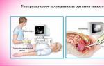
The processed information is transmitted to the monitor in the form of a picture or graph, which is studied by a specialist. Based on the data obtained, it is possible to accurately determine which and exactly where blood flow disturbances occur. Using Doppler ultrasound, the specialist determines:
- presence of blockages in blood vessels;
- movement of blood flow;
- structure of blood vessels;
- flow direction.
Additionally, using pelvic ultrasound, you can understand what the diameter of the vessels is.
Survey methods
 Scanning of veins, arteries and vessels is carried out using 3 methods:
- Abdominal study. The sensor is placed on top of the abdomen. The ultrasound ultrasound method is used in the last stages of pregnancy, as well as in girls who are not sexually active. For men, abdominal diagnostics are prescribed if bladder problems are suspected.
- Transvaginal examination. Performed exclusively on women, the technique provides reliable and complete information about the condition of the vessels of the genital organs. Transvaginally, you can place the sensor closest to the organ in which inflammation has occurred.
- Transrectal diagnosis. In this case, the sensor is installed in the rectum. It is prescribed for the diagnosis of the prostate, as well as for the purpose of clarifying the results of ultrasound examination after an abdominal examination in women. Despite the intervention in the rectum, the technique does not cause any discomfort, since the sensor used is very thin.
The examination method is determined by the doctor after an initial consultation. They use different scanning methods: duplex or triplex.
With a duplex study of vessels and veins, you can accurately determine the size of the circulatory system, determine the throughput of veins and arteries, and also determine the direction of blood flow. With triplex, or color scanning, the speed of blood flow is determined thanks to the bright image.
Pros and cons of Dopplerography
Ultrasound of the pelvic veins in women and men with Doppler is done quickly and safely. The data obtained is reliable, but it is sometimes not enough to make a diagnosis. Sometimes additional examination methods are required.
Indications for ultrasound examination
 In men and women, Doppler ultrasound is prescribed in the following general cases:
- varicose veins of the lower extremities;
- compressive pain in the lower abdomen that occurs when standing in an upright position for a long time;
- “intermediate lameness” - a person cannot walk short distances without stopping, he experiences severe burning pain associated with impaired arterial blood flow - most often under the influence of atherosclerosis;
- sharp pain in the hips, legs or lower abdomen associated with blockage of the lumen of blood vessels;
- a feeling of cold, as well as impaired sensitivity of the lower extremities, which is associated with a lack of nutrients and oxygen.
In gynecology, Doppler ultrasound of the pelvic vessels in women is prescribed to study the condition of the uterus, ovaries and fallopian tubes. In men, the technique is used to determine the condition of the prostate, seminal vesicles and cords. Very often, poor blood circulation in these areas leads to the development of diseases.
Indications for women
 Non-pregnant women undergo the procedure if they have persistent pain in the lower abdomen and menstrual irregularities. It is necessary to undergo an examination if pregnancy does not occur within 12 months of sexual activity without contraception. An examination is also required for tumors in the uterus and bladder, miscarriages and miscarriage.
The indication is a change in the thickness of the walls of organs located in the pelvis. If a woman has difficulty urinating, she is prescribed a Doppler ultrasound.
During pregnancy, a woman should undergo ultrasound examination in the following cases:
- the fetus is developmentally delayed;
- pregnancy occurs with 2 or more fetuses;
- routine examinations at 20-25 and 30-32 weeks;
- rapid weight gain by the embryo;
- Rh conflict;
- incorrect position of the fetus before birth;
- insufficient amount of amniotic fluid;
- large fruit size;
- malnutrition of the child;
- pathologies of the placenta.
If a woman suffers from vascular diseases, she must undergo ultrasound examination before and during pregnancy without fail.
Prescribed for men
In addition to suspected prostate diseases, ultrasound examination is recommended for erectile dysfunction, as well as suspected infertility, or if there is an established diagnosis. Any compaction in the testicles, changes in size are a direct indication for ultrasound.
Possible contraindications to Doppler ultrasound
The main contraindication is the presence of burns and wounds on the surface through which the examination is performed. Transvaginal scanning is prohibited if a woman has inflammatory diseases of the vagina. Ultrasounds are not performed during menstruation and in the last weeks of pregnancy. Rectal examination is prohibited if there are cracks or other abnormalities in the structure of the rectum. Transvaginal ultrasound is not allowed if you are allergic to latex.
Preparation for the procedure
 No specific preparation is required for ultrasound of the pelvic organs with or without CD. To avoid false scan results, you should not smoke 8 hours before diagnosis. It is not recommended to take any drinks that affect vascular tone: coffee, alcohol, energy drinks, strong tea, various extracts of exotic fruits.
In rare cases, experts recommend not eating or drinking liquids if a transrectal or vaginal examination is planned. A full bowel or bladder can worsen the ultrasound result and prevent it from being fully performed.
Technique of the procedure
During an abdominal examination, the patient should remove outer clothing and lower his pants or skirt slightly. An ultrasound is performed on the patient in a supine position, next to the machine.
If transvaginal testing is performed, the skirt, underwear or trousers must be completely removed. An ultrasound is performed in an obstetric chair. If there is no gynecological chair in the office, the woman lies down on the couch, bends her knees and moves them slightly to the side.
To perform a transrectal ultrasound, the patient is placed on the left side (less often on the right) and asked to bend his knees. Then a rectal sensor is inserted, having previously lubricated it with a special gel. A special agent is also applied to the sensor to ensure good contact with the skin and a clear image. The gel prevents dry slipping. During the examination, the doctor can change the device settings to increase or decrease power.
The examination process takes a maximum of 30 minutes, if the clinical situation is very complex, then 30-40 minutes. After the procedure, it is enough to remove the remaining gel, get dressed and get the results.
Decoding the results
When studying blood vessels using ultrasound, the resistance index and blood circulation rate are determined. There are several degrees of impairment. For example, when examined during pregnancy, they will look like this:
- 1A – normal blood flow in the vessels of the uterus and good nutrition of the placenta;
- 1B – blood circulation is partially disrupted at the placenta-fetus stage, but the uterus is fully supplied;
- 2 – blood flow in the uterus is noticeably impaired;
- 3 – pathological disturbance of blood flow in the uterus and fetal organs.
Ultrasound examination of veins now occupies one of the first places in terms of accuracy among other diagnostic methods, as it is fast, comfortable, safe and has no contraindications.
Purpose of ultrasound examination of the pelvic veins
Basically, it is prescribed for varicose veins. Most often it affects women of childbearing age. This type of ultrasound is also very important in gynecology, since the state of the vascular system can be used to judge the presence of inflammation, thrombosis in the arteries and veins of the pelvis, the condition of the female genital organs, differential diagnosis of neoplasms, assessment of fetal development and the functioning of the uterus during pregnancy.
Ultrasound examination of the pelvic veins is also prescribed to patients if they experience symptoms such as pain, a feeling of heaviness in the lower abdomen, or have uncharacteristic discharge or bleeding in the middle of the menstrual cycle. Ultrasound of the pelvic veins is aimed at studying the structure of its circulatory system, the condition of arteries and veins, the functioning of venous valves, the direction and intensity of blood flow and the completeness of blood supply to organ tissues.
Indications for undergoing ultrasound examination of the pelvic veins
Ultrasound of the pelvic veins It is advisable to undergo at least once a year for all women, as well as men suffering from urological diseases. Persons at risk should undergo an ultrasound examination at least twice a year, namely:
. people with a history of diabetes mellitus,When cholesterol levels increase,
Heavy smokers
Women who have had a pregnancy with complications, as well as difficult childbirth,
Patients suffering from varicose veins of the lower extremities or hemorrhoids,
It is imperative that those who have chronic venous congestion in the pelvic organs due to the fact that their occupation requires lifting excessively heavy things, having to sit or stand for long periods of time should undergo an ultrasound examination of the pelvic veins on a regular basis.
People suffering from genital dysfunctions.
It is better not to leave a visit to the medical center for later, because varicose veins of the small pelvis threaten complications such as thrombosis. In addition, chronic venous congestion can cause consequences in the form of disturbances in the functioning of other genital organs and lead to infertility, the risk of miscarriage during pregnancy or miscarriage.
Features of ultrasound examination of the pelvic veins
As a rule, Doppler technique is used to examine the pelvic veins. It is based on the fact that the ultrasonic signal is reflected from the formed elements of the blood and thereby gives an idea of their condition and the direction of movement through the veins and arteries. Sensors may have different transmission frequencies. The doctor chooses the most suitable one, depending on the need for a more in-depth study. The diagnostician also chooses the method of conducting an ultrasound examination of the pelvic veins - transabdominal or transvaginal.
The ultrasound diagnostic procedure itself is quite simple and does not take much time. During the transabdominal method of examination, the patient should take off his trousers or skirt and tights. The study area is covered with a special gel that does not allow air to pass through. The doctor moves a sensor over the skin. Signals from it are transmitted to the monitor of the ultrasound scanner, which displays a picture of what is happening. With the transvaginal method, a condom is placed on the sensor, which is treated with gel and inserted into the patient’s vagina. Upon completion of the ultrasound examination of the pelvic veins, the patient receives in his hands:
. research protocol with a full transcript of the results,Medical opinion,
Photos or videos saved on a convenient medium,
And if necessary, consult a phlebologist or other specialist about further diagnostic tests or treatment methods.
Where is the best place to undergo an ultrasound examination of the pelvic veins?
Our medical center provides the services of specialists in a wide variety of fields of practical medicine. An ultrasound examination of the pelvic veins may be required for diagnosis by a gynecologist, urologist, therapist, angiosurgeon and other specialists for differential diagnosis, since varicose veins can often be masked or combined with other gynecological or urological diseases. Determining an accurate diagnosis provides an opportunity to begin treatment immediately. Research may be required in cases where:
. pathology of the vascular parenchyma of the pelvic veins is revealed,There is a violation of the integrity of the venous wall,
Disturbances in the functioning of their valve apparatus,
Suspicion of the presence of parietal or floating thrombi,
Deep vein dysfunction,
When there is a risk of thrombosis or bleeding.
Experienced phlebologists at our medical center will help you choose the optimal treatment method for varicose veins of the small pelvis. It all depends on the severity of the disease. If in its initial period it is quite possible to get by with conservative treatment (exercise of therapeutic and general physical education, taking venotonic medications and wearing compression stockings), then if the diagnosis is delayed, surgical intervention may well be required.
Dopplerography- one of the methods of ultrasound examination (ultrasound). This method is based on the Doppler effect - a change in the frequency of ultrasonic waves when they are reflected from moving objects. Doppler ultrasound is used to diagnose blood vessels. In this case, the formed elements of blood act as moving objects.
Indications for Dopplerography of pelvic vessels
Among the blood vessels diagnosed using ultrasound are the pelvic vessels. It should be recalled that the small pelvis is limited in front by the pubic fusion, in the back by the sacrum, and on the sides by the pelvic bones. The pelvic organs include:
- Bladder with ureters
- Rectum
- In men - the prostate gland, spermatic cords and seminal vesicles
- In women - the uterus, ovaries, vagina.
In women - the uterine and ovarian arteries, through which blood flows to the uterus and its appendages.
In men - the inferior vesical artery (supplies the prostate), venous plexuses of the prostate, veins of the spermatic cord, arteries and veins of the penis.
Dopplerography of these vessels makes it possible to diagnose the following diseases:
- Congenital anatomical anomalies of the uterus and appendages
- Some inflammatory diseases of the uterus and ovaries
- Pipe welds
- Uterine fibroids
- Tumors of the body of the uterus and ovaries
- Prostate adenoma
- Prostate abscess
- Varicocele - dilation of the veins of the spermatic cord
- Congenital and acquired deformities of the penis.
Dopplerography technique of pelvic vessels
Dopplerography of the vessels of the pelvic organs, unlike standard ultrasound of internal organs, does not require special preparation, with the exception of some hygienic measures. The procedure itself, like all ultrasounds, is short-term and painless - the doctor moves the sensor along the lower part of the abdominal wall, and an image is displayed on the monitor screen indicating all the parameters being studied. This allows the doctor to make a conclusion immediately during the diagnostic process.In medicine, to obtain more detailed information about the internal organs and the ongoing pregnancy, pelvic ultrasound with Doppler is used, which makes it possible to identify disorders in the early stages.
For pregnant women, scanning is prescribed:
- with rapid growth or delayed development of the embryo;
- attachment of more than 1 fertilized egg;
- during the recommended studies at 20 and 30 weeks;
- if the Rh factor does not match;
- with a non-physiological position of the fetus for childbirth;
- for kidney and liver diseases.
Ultrasound scanning for men is carried out:
- for disorders in the testicles;
- problems with childbirth;
- injury;
- inflammation of the genital organ;
- mental or physical retardation;
- blood flow disturbance.
Women who are not pregnant are referred for an ultrasound examination due to:
- the presence of a tumor formation in the uterus or bladder, changes in the thickness of the walls of these organs;
- disruptions in the menstrual cycle;
- pain;
- infertility;
- fetal loss.

For prevention, Doppler testing of the pelvic vessels must be performed once a year. Doppler ultrasound is painless, does not require intervention in internal organs, and is harmless to the health of doctors and patients.
Preparation
When undergoing an ultrasound examination, you must follow the recommendations to obtain accurate results.
Requirements:
- Dopplerography of the pelvic organs is done on the 7th day of the cycle.
- Men can pass at any time; there is no need to prepare.
- If the examination is through the abdominal wall, then for 3 days they eat a diet without gas-forming products. The bladder should be full. Drink about 1 liter of water. They try to cleanse the intestines. You can take Mezim, activated carbon or Espumisan in advance.
- During a transvaginal examination, the bladder must be emptied.
- Using barium for x-rays some time before the ultrasound may interfere with the accurate results.
- No preparation is required for pregnant women.
If the rules are followed, the specialist will establish a more accurate diagnosis and identify violations at the initial stage.
Contraindications
There are cases when research is not recommended. These include:
- Ultrasound on the surface of the abdomen (transabdominal) is not performed for wounds or fresh burns at the study site;
- in case of inflammatory diseases of the vagina, menstruation, late pregnancy, transvaginal duplex scanning is not performed;
- In case of diseases of the rectum, intrarectal examination is contraindicated;
- Latex allergy is a contraindication to transvaginal ultrasound.

Ultrasound is used many times to monitor the dynamics of the pathological process.
How they do it
One of the types of ultrasound examination for accurate and early diagnosis of diseases of the genitourinary system associated with circulatory disorders, as well as during pregnancy, is pelvic ultrasound.
Duplex scanning of pelvic vessels in women
Ultrahigh-frequency sound waves are used for research - from 2 to 29 MHz. The range that the human ear can detect is 1000 times smaller.
Using a special device with double scanning capability, the pelvis is examined. High-frequency signals reflected from internal organs and recorded by a sensor are recorded. Using modern technologies, the results are analyzed. The monitor displays a two-dimensional image.
The Doppler method provides color reproduction in research. The result of the work performed is a cartogram or ultrasound examination with color flow. The sound wave is reflected from the blood elements moving through the vessels. The sensor detects their speed and direction. The monitor displays color mapping of the circulatory system.
During the examination, the patient is placed on a couch. The device, lubricated with gel, is passed over the abdomen or inserted into the vagina (a disposable condom is first placed on the sensor) or into the rectum. The cartogram is reflected on the monitor.
The study lasts about 20 minutes.
During pregnancy
An ultrasound is performed either along the abdominal wall or inside the vagina.
Starting from 3-5 weeks, the fertilized egg can be detected transvaginally. Transabdominal early pregnancy is determined at 6-7 weeks. It is difficult to diagnose an ectopic pregnancy, since blood clots can be mistaken for a fertilized egg.

Using ultrasound, you can determine the period, sex and number of embryos. You can listen to the heartbeat from 6 weeks, and from 8 weeks you can determine where the placenta is forming.
Research is carried out at least 3 times. Ultrasound for genetic pathologies is performed at 9-13 weeks. At 16-20, the sex is determined, the development of organs is monitored (deviations of the heart and brain are identified).
The correct position of the fetus, size, weight, blood flow of the uterus, placenta, vascular picture of the pelvis, and the amount of amniotic fluid are determined at 30 weeks. All this is necessary to prepare for childbirth.
For men
Ultrasound with Doppler is used to diagnose male organs (testicles, blood vessels, veins, etc.).
Ultrasound allows you to determine neoplasms, changes in the structure of the testicles, dilated veins, etc. It is carried out with a special device. They use reflected sound waves to determine the movement of blood through the vessels.
The man sits on the couch, lifts the organ and covers it with a towel. The doctor runs a sensor lubricated with gel over the skin. The received signals are transmitted to the device. On the monitor, the doctor receives information about the condition of the patient’s blood vessels. Such a study provides the most accurate indicators.
There is also a transrectal examination of the prostate. The device is inserted into the rectum to determine the blood flow of the male organ. The doctor examines the results to prescribe appropriate treatment.
Transabdominal, transvaginal and transrectal
There are several ways to perform an ultrasound:
- Transabdominal. A sensor is moved over the skin of the abdomen. Information is displayed on the computer. The method is suitable for virgins, men, and women in the third trimester.
- Transvaginally. Inserted inside the vagina. Using this method, you can obtain more detailed information about the state of blood circulation in a woman’s internal genital organs.
- Transrectal. An ultra-thin sensor is inserted into the patient's rectum, causing no discomfort. They determine prostate diseases for males and clarify the diagnosis for girls who are not sexually active.

A doctor directs you to each type of ultrasound.
Decoding the results
At the end of the study, a protocol is issued, which is signed by a clinician (proctologist, gynecologist). After collecting anamnesis and studying the conclusion, the doctor prescribes treatment.
Ultrasound shows whether the organs correspond to normal sizes, their echogenicity, whether they are located correctly, the presence of neoplasms, cysts, inflammations, pregnancy, the location of the spiral, etc.
The specialist describes the diagnostic result, compares the data with the norm, and identifies deviations indicating the presence of the disease.

Thick walls of the uterus and tubes indicate the presence of cancer. Cysts or fibromas are visualized as round formations. Polycystic disease is manifested by changes in the size of the ovaries and uterus. Fibroids or endometriosis are detected when echogenicity changes. With varicose veins, incompetence of the venous valves, narrowing and expansion of the lumen of blood vessels, and altered blood flow speed will be visible.
At the end of the ultrasound, the result is given on a disk or in the form of screenshots with a written transcript.
What is the price
Ultrasound scanning is available at diagnostic centers and clinics. Prices in different clinics vary from 650 to 2300 rubles. depending on the type of procedure.
Ultrasound scanning of the vessels of the pelvic organs is a method of ultrasound examination of veins and arteries, widely used to determine various vascular pathologies and diagnose many gynecological and andrological diseases.
Duplex and triplex scanning of blood vessels is a completely safe study that can be used repeatedly without harm to health.
You can undergo an ultrasound scan of the vessels of the pelvic organs in St. Petersburg for a fee at our MART clinic.
Indications for ultrasound examination of pelvic organs vessels
The pelvic organs in women include the bladder, uterus, its appendages, cervix, ovaries; in men - bladder, prostate gland, spermatic cords, seminal vesicles. The normal functioning of these organs largely depends on their high-quality blood supply, and any changes in blood circulation are usually associated with pathological complications.
Indications for duplex/triplex scanning of the vessels of the pelvic organs are:
- infertility in men and women,
- miscarriage,
- suspicion of the presence of tumor formations,
- urinary disorders,
- erectile dysfunction in men.
How is ultrasound examination of the vessels of the pelvic organs performed?
Ultrasound scanning of the vessels of the pelvic organs is performed in the lying position on the couch. Depending on the indications, scanning can be performed using several methods: transabdominal (through the anterior abdominal wall), transvaginally (through the vagina) in women and transrectally (through the rectum) in men.
The average duration of the procedure is 30 minutes.
When is an ultrasound scan of the vessels of the pelvic organs done?
For men, ultrasound examination of the vessels of the pelvic organs can be done at any time. For women, the test is usually performed on days 5–7 of the menstrual cycle.
What will an ultrasound scan of the vessels of the pelvic organs show?
During duplex/triplex scanning of the vessels of the pelvic organs in men, the doctor examines the vessels of the prostate and prostatic plexus, in women - the uterine, ovarian, spiral arteries, and endometrial vessels of the uterus. In addition, the iliac and arcuate veins and arteries are examined in all patients.
The examination allows you to study the structure of blood vessels, their patency, the condition of the vascular wall, as well as various indicators of blood flow.
Using duplex/triplex scanning, you can identify atherosclerotic changes in blood vessels, blood clots, compression of blood vessels, and also diagnose:
- ovarian cysts and tumors,
- uterine fibroids,
- volumetric formations in the wall and cavity of the uterus,
- some inflammatory diseases of the uterus and ovaries,
- varicose veins of the pelvis,
- varicocele,
- prostatitis,
- prostate abscess,
- prostate adenoma,
- prostate cancer,
- congenital anomalies.
In our ultrasound center you can urgently perform an ultrasound scan of the vessels of the pelvic organs.
Preparation for ultrasound examination of the vessels of the pelvic organs
Special preparation for examination of pelvic vessels is usually not required. In case of research using the transrectal method, it is necessary to cleanse the intestines before the procedure.
Where can I do an ultrasound scan of the vessels of the pelvic organs in St. Petersburg?
You can get a high-quality duplex/triplex scan of the vessels of the pelvic organs at the MART multidisciplinary clinic. In our clinic, examinations are carried out by ultrasound diagnostic specialists with extensive experience. The scan is performed using a modern expert-level ultrasound machine, which allows one to examine the structure of even the smallest vessels and the blood flow in them.
Sign up for the MART medical center in St. Petersburg