What is parenchymal jaundice, the causes of its occurrence and methods of treatment in adults. Parenchymal jaundice: causes, symptoms and treatment Parenchymal jaundice is characterized by
Good day, dear readers!
In today's article we will look at a disease such as jaundice, as well as its types, causes, symptoms, diagnosis, treatment, folk remedies, prevention and discussion of jaundice. So…
What is jaundice?
Jaundice (Gospel's disease, Latin icterus)– a collective name for a group of symptoms, the common feature of which is the coloring of the skin, mucous membranes and sclera in a yellow color.
The intensity of the staining depends on the cause and pathogenesis, and can vary from pale yellow to orange, sometimes even changing the color of urine and feces. The main cause of jaundice is excessive accumulation of bilirubin in the blood and various tissues of the body, which is usually facilitated by the presence of diseases of the liver, gall bladder and biliary tract, blood and other pathologies. However, these are the causes of true jaundice, but there is also false jaundice, which is caused by excess levels of carotenes in the body, which enter it when eating large quantities of carrots, pumpkin, beets, oranges or certain medications.
People often mean jaundice, but this is an incorrect statement, because Yellowing of the skin and other parts of the body with hepatitis is only a symptom.
Development of jaundice
Bilirubin– bile pigment, which is brown crystals and is one of the main components of bile. Formation occurs as a result of the breakdown of certain proteins (hemoglobin, myoglobin and cytochrome) in the spleen, liver, bone marrow and lymph nodes.
After fulfilling its function in the body (antioxidant), bilirubin is excreted through the biliary tract and then excreted in feces and urine.
The development of jaundice, depending on the pathology in the body, is mainly due to 3 reasons:
- hemolytic or suprahepatic jaundice - characterized by increased production of bilirubin, with excessive destruction of red blood cells;
- obstructive or subhepatic jaundice - characterized by the presence of obstacles in the gastrointestinal tract (cholelithiasis and others), due to which bilirubin in the bile cannot freely enter the intestine and is then excreted from the body;
- parenchymal or hepatic jaundice - characterized by a violation of the metabolism of bilirubin in liver cells and its binding to glucuronic acid, which is usually promoted by various liver diseases (hepatitis and others).
As we have already said, there is also false jaundice (pseudojaundice), which is characterized by yellowing of only the skin and is caused by excessive accumulation of carotenes in the body, which usually occurs with long-term consumption of large quantities of carotene-containing products - carrots, pumpkin, beets, as well as quince, picrine acids and some other drugs.
When bilirubin accumulates in the body in large quantities, circulating through the blood, it reaches almost all parts of the body, and due to its color, it appears externally as yellowness of the skin, mucous membranes and sclera.
Also, liver failure may develop against the background of Gospel disease.
 Causes of jaundice can be:
- Excessive destruction of red blood cells;
- Impaired metabolism of bilirubin in liver cells, as well as its binding to glucuronic acid;
- The presence of obstacles to the normal circulation of bile.
The following factors contribute to the above reasons:
- Various diseases and pathological conditions - hepatitis, erythropoietic porphyria, ineffective erythropoiesis, primary shunt hyperbilirubinemia, infectious mononucleosis, cholestasis, ovalocytosis, thalassemia, liver cancer, abnormalities in the development of the gastrointestinal tract;
- Mechanical damage to the gallbladder, biliary tract and other gastrointestinal organs;
- Liver poisoning, narcotic drugs, medications;
- Infection of the body with various;
- The appearance of complications during surgical treatment of the body.
The causes of false jaundice (pseudojaundice, carotene jaundice) are usually the consumption of carotene-containing foods - carrots, beets, pumpkin, oranges, as well as quinine, picric acid and others.
Jaundice is classified as follows:
 1. True jaundice, in which yellowing is caused by bilirubin. Divided into the following types:
Hepatic jaundice (parenchymal)— the development of the disease is caused by massive damage to liver cells (hepatocytes), due to which it cannot adequately convert bilirubin and excrete it into bile. Parenchymal jaundice is considered one of the most popular liver diseases. The causes of parenchymal jaundice are usually hepatitis, cirrhosis of the liver, liver cancer, leptospirosis, infectious mononucleosis, liver poisoning with alcohol or drugs. The main symptoms of hepatic jaundice are orange-reddish skin color, pain in the right hypochondrium, spider veins, “liver palms”, itchy skin, enlarged liver. Can be divided into the following types:
- hepatocellular jaundice;
- cholestatic jaundice;
- enzymopathic jaundice.
Hemolytic jaundice (prehepatic)– the development of the disease is due to the increased formation and destruction of bilirubin, due to which the liver is not able to remove it from the body in a timely manner and bilirubin (direct and indirect) is absorbed back into the blood. The causes of hemolytic jaundice are usually anemia, extensive heart attack, intoxication with arsenic, hydrogen sulfide and various drugs, for example, sulfonamides. In addition, as a symptom, hemolytic jaundice can occur against the background of Addison-Birmer disease, lobar, hepatitis.
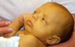
Jaundice in newborns is classified into a separate group, which is called - neonatal jaundice. The main reasons are the infant’s body systems that are not fully formed, which is why immature liver cells cannot bind bilirubin in a timely manner and remove it from the body.
Symptoms of neonatal jaundice appear immediately after the baby is born, and in some cases, when the amount of indirect bilirubin in the blood is extremely high, the baby needs treatment as it is a neurotoxic poison. If treatment is not carried out, indirect ruby can contribute to damage to the cerebral cortex and subcortical nuclei. Neonatal jaundice can be divided into the following types:
Physiological jaundice– symptoms are observed mainly in premature infants, whose enzyme system is not yet fully formed;
Kernicterus– characterized by increased accumulation of indirect bilirubin in infants, which can damage the cerebral cortex;
Conjugation jaundice– the development of the disease is caused by a violation of the uptake of indirect (unbound) bilirubin or the process of its binding in the liver with glucuronic acid, due to which ultra-high doses of bilirubin, which poisons the body, accumulate in the blood. More often it is a hereditary disease that develops against the background of congenital hypothyroidism, Gilbert syndrome or Crigler-Nayar syndrome.
Pregnane jaundice (breast milk jaundice)– yellowing of the baby’s skin is caused by an increased amount of the hormone prenandiol in the body, which passes along with breast milk from mother to child, which helps to delay the excretion of bilirubin. Treatment is based on limiting breastfeeding for several days, using formula milk.
2. False jaundice, in which yellowing is caused by excess carotene content in the body.
Diagnosis of jaundice
Diagnosis of jaundice includes:
- Analysis of urine;
- Determination of bilirubin content in the blood;
- liver, pancreas, biliary tract;
- Fibroesophagogastroduodenoscopy (FEGDS);
- Endoscopic retrograde cholangiopancreatography (ERCP);
- Relaxation duodenography;
- Percutaneous transhepatic cholangiography;
- Splenoportography;
In some cases, a liver biopsy may be required.

Treatment of jaundice
 begins with an accurate and differential diagnosis of pathology, and includes:
1. Drug treatment.
2. Physiotherapeutic procedures;
3. Diet
4. Surgical treatment
The choice of treatment methods and specific medications directly depends on the type of jaundice, its cause, and the presence of concomitant diseases.
1. Drug treatment
Important! Before using medications, be sure to consult your doctor!
Treatment of jaundice caused by anemia includes taking drugs based on, and - “Ferbitol”, “Ferrum Lek”, “Ektofer”, “Gemostimuli”, “Tardiferon”, “Ferroplex”.
Treatment of jaundice caused by impaired liver function due to its damage by infection or toxic substances includes taking drugs that restore and protect its cells - hepatoprotectors - “”, “Legalon”, “Silymarin”, “Gepabene”, “Galstena”, “Tsinariks”, “Allohol”, “Sibektan”, “Bonjigar”, “Dipana”.
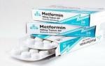
Treatment of jaundice caused by hepatitis, depending on the type of hepatitis virus, treatment with antiviral drugs is prescribed - alpha interferons (Alfaferon, Interferon), nucleoside analogues (Adefovir, Lamivudine), Ribavirin, Sofosbuvir , Ledipasvir, Velpatasvir, Daclatasvir, Paritaprevir, Ombitasvir and others.
After suppressing the infection, in order to remove it and its waste products from the body, detoxification therapy is prescribed, which includes taking Albumin, Atoxil, glucose solution (5%), Ringer-Locke solution (5-10%), Enterosgel "
To normalize liver function, additional intake of vitamins - and cocarboxylase is also useful.
Treatment of jaundice caused by disruption of the normal circulation of bile - due to the presence of stones in the gallbladder and bile ducts (cholelithiasis), therapy is prescribed aimed at removing foreign tumors from the body, which includes taking ursodeoxycholic acid (Ursonan, Ursodex ", "Exhol"), chenodeoxycholic acid ("Henosan", "Chenofalk", "Henochol"), herbal remedies (sandy immortelle extract), as well as drugs that stimulate contraction of the gallbladder, and, accordingly, the expulsion of stones from oneself and their further removal from the body - “Zixorin”, “Liobil”, “Holosas”. For the same purposes, ultrasound, laser, shock wave or surgical methods for removing stones may be prescribed.
Ursodeoxycholic acid also helps accelerate the recovery of liver cells.
For jaundice of autoimmune etiology, immunosuppressants are used - Methotrexate, Prednisolone, Azathioprine.
Treatment of jaundice in infants may include blood transfusion - an infusion of donor Rh-negative blood is given through the baby's umbilical vein.
Treatment of jaundice in adults may also include blood transfusion - red blood cells are infused intravenously, in combination with hormones (glucocorticosteroids - Dexamethasone, Prednisolone) and anabolic steroids (Retabolil).
In case of Gospel disease, caused by the presence of bacterial diseases, antibiotic therapy is carried out. The choice of antibacterial drug depends on the type of bacteria. In combination, they may prescribe additional probiotics.
If the immune system is weakened due to an infectious disease, immunostimulants may be prescribed - Vilozen, Zadaxin, Thymogen.
Symptomatic therapy
To alleviate the course of the disease, symptomatic therapy is prescribed.
For nausea and vomiting, use “ ”, “Pipolfen”, “ “.
For pain in the right hypochondrium, painkillers can be prescribed - “Baralgin”, “Dicetel”, “Duspatalin”, “No-shpa”, “Odeston”, “Papaverine”.
For severe skin itching, antihistamines are used: “Diazolin”, “”, “”, “Diphenhydramine”.
For increased anxiety, nervousness, and insomnia, sedatives are prescribed: “Valerian”, “Tenoten”.
2. Physiotherapeutic procedures
Physiotherapeutic treatment of jaundice includes the use of the following therapy methods:
- Plasmapheresis;
- Phototherapy;
- Ultrasound on the liver area;
- Inductothermy;
- Iontophoresis with solutions of iodine, novocaine or magnesium sulfate;
- Diathermy.
3. Diet for jaundice
A diet for jaundice of any etiology includes a ban on the consumption of foods such as fatty, fried, pickles, spicy, smoked foods, canned food, animal fats, onions, radishes, and alcoholic beverages.
Limit consumption - carrots, citrus fruits, pumpkin, carbonated drinks, strong coffee or tea, chocolate.
It is necessary to emphasize the consumption of foods enriched with vitamins and, especially, vitamins A, C, E, B9, B12 and other B vitamins, as well as iron and manganese.
What can you eat if you have jaundice: The following foods are healthy: legumes, nuts, grain bread, oats, buckwheat, dried apricots, fish, poultry, eggs, dairy products, green vegetables, cabbage, zucchini. Freshly squeezed vegetable and fruit juices are beneficial.
Meals are fractional, in small portions.
4. Surgical treatment
Surgical treatment for jaundice depends on its cause, and may include:
- Endoscopic papillosphincterotomy – aimed at removing stones from the gallbladder and biliary tract;
- Percutaneous transhepatic cholangiostomy;
- Percutaneous transhepatic microcholecystostomy;
- Endoprosthetics of bile ducts;
- Microcholecystostomy using the Seldinger method;
- Liver transplantation.
 Important!
Before using folk remedies for treating jaundice, be sure to consult your doctor!
Important!
Before using folk remedies for treating jaundice, be sure to consult your doctor!
Milk thistle. 3 tbsp. spoons of crushed milk thistle seeds, pour 500 ml of boiling water, place the product in a water bath and simmer until half of the broth has evaporated, then let the broth cool, strain it and take 1 tbsp. spoon 30 minutes before meals, 3 times a day, for 1-2 months.
Aloe. Mix 380 g of adult, 630 g and 680 ml of Cahors, then infuse the product in a dark, cool room for 15 days. You need to take the mixture before breakfast, during the first 5 days, 1 teaspoon, and then 1 tbsp. spoon until the product runs out.
Jaundice is a polyetiological syndrome characterized by yellowing of the skin, sclera and other tissues, due to an excessive amount of bilirubin in the blood and its excessive accumulation in tissues. Depending on the cause of the increase in the level of bilirubin in the blood, there are two main types of jaundice: true jaundice and false jaundice (pseudojaundice).
False jaundice (pseudojaundice) is an icteric discoloration of the skin due to the accumulation of carotenoids in it during prolonged and abundant consumption of strongly colored vegetables and fruits (carrots, oranges, pumpkins), and also occurs when ingesting certain medications (acriquine, picric acid and some other drugs). With pseudojaundice, the level of bilirubin in the blood does not increase and the mucous membranes do not stain (the sclera of the eyes remains white).
True jaundice- a symptom complex that is characterized by icteric discoloration of the skin and mucous membranes, caused by the accumulation of excess bilirubin in the tissues and blood. The intensity of jaundice depends on the blood supply to the organ or tissue. Initially, a yellow coloration of the sclera is detected, and somewhat later - the skin. Accumulating in the skin and mucous membrane, bilirubin, in combination with other pigments, colors them light yellow with a reddish tint. Subsequently, bilirubin is oxidized into biliverdin, and jaundice acquires a greenish tint. With prolonged existence of jaundice, the skin becomes blackish-bronze in color. Thus, examination of the patient allows us to resolve the issue of the duration of jaundice, which is of great differential diagnostic importance. In case of jaundice, it is very important to establish the ratio of the concentrations of direct and indirect bilirubin.
- Direct bilirubin is a water-soluble fraction that gives a direct Van den Bergh reaction (with Ehrlich's diazoreagent) and consists mainly of conjugated (bound) bilirubin (monoglucuronide and diglucuronide).
- Indirect bilirubin is a fat-soluble fraction that gives an indirect Van den Bergh reaction (with Ehrlich's diazoreagent after pretreatment with ethanol or methanol) and is represented mainly by unconjugated (unbound, free) bilirubin.
- Direct and indirect bilirubin together make up the so-called total bilirubin. The normal level of total bilirubin in serum is 5-17 µmol/L (0.3-1 mg%).
If, when performing the Van den Berg reaction, 80-85% of serum bilirubin turns out to be indirect, then the patient is considered to have indirect hyperbilirubinemia. If direct bilirubin in the serum is more than 50%, direct hyperbilirubinemia is considered. The appearance of bilirubin in the urine (bilirubinuria) is a consequence of hyperbilirubinemia. Urine with bilirubinuria is colored from bright yellow to dark brown; when shaken, its foam turns yellow. Bilirubinuria is observed only with direct hyperbilirubinemia, since indirect bilirubin does not pass through the glomerular membranes.
Depending on the mechanism of bilirubin metabolism disorder, the following are distinguished:
- suprahepatic (prehepatic, hemolytic) jaundice, where the erythropoietic system is primarily affected and hyperproduction of bilirubin, in the vast majority of cases, is associated with increased breakdown of red blood cells;
- hepatic (hepatocellular, parenchymal) jaundice, where liver cells (hepatocytes) are primarily affected and hyperbilirubinemia is associated with impaired metabolism and transport of bilirubin within liver cells;
- subhepatic (posthepatic, obstructive, obstructive) jaundice, where the extrahepatic bile ducts are primarily affected and hyperbilirubinemia is caused by difficulty or blockade of extrahepatic bilirubin transport.
In all types of jaundice, hyperbilirubinemia is the result of an imbalance in the dynamic balance between the rate of formation and excretion of bilirubin.
Prehepatic jaundice
Prehepatic (prehepatic, hemolytic) jaundice develops as a result of intensive breakdown of red blood cells (or their immature precursors) and excessive production of indirect bilirubin. In the blood of patients, the amount of unconjugated bilirubin increases to 40-50 µmol/l (3.5-5 mg%). A normally functioning liver cannot metabolize all of the bilirubin produced, which is water-insoluble and does not pass through the kidney filter. The content of stercobilinogen sharply increases in feces, and urobilinogen is detected in urine.
Intensive destruction of red blood cells. These phenomena occur with hyperfunction of cells of the reticuloendothelial system (primarily the spleen), with primary and secondary hypersplenism. A typical example of hemolytic jaundice is various hemolytic anemias, including congenital ones (microspherocytosis, etc.).
Immune hemolytic anemia develops under the influence of antibodies on red blood cells:
- hapten anemia - caused by the fixation on red blood cells of antigens foreign to the body - haptens (drugs, viruses, etc.) with antibodies formed in response to the combination of a hapten with a protein of the body;
- isoimmune anemia - associated with the entry into the body of a newborn of maternal antibodies directed against the child’s red blood cells (if the child and mother are incompatible for the Rh factor and much less often for antigens of the AB0 system).
- autoimmune anemia - caused by the appearance in the body of antibodies against its own red blood cells;
In hemolytic anemia, the formation of indirect bilirubin is so great that the liver does not have time to convert it into conjugated (direct) bilirubin. The causes of hemolytic jaundice can also be various other factors leading to hemolysis: hemolytic poisons, absorption of decay products of extensive hematomas into the blood, etc. Jaundice may be more pronounced in liver diseases with impaired function.
In practice, the diagnosis of hemolytic jaundice is easier than others. With hemolytic jaundice, the skin acquires a lemon-yellow color, the jaundice is moderate, and there is no itching. With severe anemia, pallor of the skin and mucous membranes is determined against the background of existing jaundice. The liver is of normal size or slightly enlarged. The spleen is moderately enlarged. In some types of secondary hypersplenism, severe splenomegaly may be detected. Urine is dark in color due to increased concentrations of urobilinogen and stercobilinogen. The urine reaction to bilirubin is negative. The feces are intensely dark brown in color, the concentration of stercobilin in it is sharply increased. Blood tests show an increase in the level of indirect bilirubin, the concentration of direct biliburin is not increased. Anemia, as a rule, is moderate; reticulocytosis is possible in the blood of patients. ESR is slightly increased. Liver tests, blood cholesterol are within normal limits. The level of serum iron in the blood is increased.
Prehepatic jaundice develops not only as a result of increased breakdown of red blood cells, but also when bilirubin conjugation in the liver is impaired, which leads to excessive production of indirect (unconjugated) bilirubin. A typical example is hereditary pigmentary hepatosis.
Pigmented hepatoses- benign (functional) hyperbilirubinemia - diseases associated with hereditary disorders of bilirubin metabolism (enzymopathies), manifested by chronic or intermittent jaundice without pronounced primary changes in the structure and function of the liver and without obvious signs of hemolysis and.
Gilbert's syndrome- the most common form of hereditary pigmentary hepatosis, which is detected in 1-5% of the population and is inherited in an autosomal dominant manner. In Gilbert's syndrome, liver function is normal; it is distinguished from hemolysis by the absence of anemia or reticulocytosis. The only deviation from the norm is a moderate increase in unconjugated bilirubin in the blood. The syndrome is detected in young people, probably lasts a lifetime, and is accompanied by vague, nonspecific complaints.
Meulengracht syndrome until recently, it was considered almost synonymous with Gilbert's syndrome, which was even called "Gilbert-Meulengracht syndrome." However, it was later proven that these are different syndromes with similar symptoms. Common to the two syndromes are a decrease in the level of bilirubin when activators of microsomal liver enzymes are prescribed, the age of onset of the disease, the intermittent nature of jaundice, the level of bilirubin in the blood no more than 80-100 µmol/l due to the unconjugated fraction, clinical manifestations in the form of jaundice of the skin and mucous membranes, dyspepsia , asthenia. But with Meulengracht's syndrome, there is only an isolated decrease in the activity of UDPGT, and the hepatocyte membrane, unlike Gilbert's syndrome, is actively involved in the uptake of bilirubin. Treatment is similar to the treatment of Gilbert's syndrome; phenobarbital is effective.
Dubin-Johnson syndrome- a rare pigmentary hepatosis with an autosomal dominant type of inheritance. Clinical manifestations usually develop in men 20-30 years of age. The pathogenesis is based on a violation of the excretion of pigment from hepatocytes, which leads to regurgitation of bilirubin. A feature of this syndrome is a change in the color of the liver: it becomes green-gray or brown-black. Histologically, a dark pigment located peribiliary is found - melanosis of the liver, which develops due to a disturbance in the metabolism of adrenaline. The liver structure remains normal. Pigment deposition also occurs in the spleen. Jaundice in patients is usually constant, periodically increasing, without skin itching or (rarely) with slight itching, pain in the right hypochondrium with periodic intensification like biliary colic, severe dyspeptic symptoms, fatigue, poor appetite, low-grade fever. In rare cases, the disease may be asymptomatic. The liver is usually moderately enlarged, sometimes there is splenomegaly. Often combined with. Diagnosis is based on the detection of conjugated and unconjugated hyperbilirubinemia up to 100 µmol/l in the blood (due to deconjugation and reflux of bilirubin into the blood), and bilirubinuria in the urine. A moderate increase in aminotransferases is possible in the blood serum. Alkaline phosphatase levels are usually unchanged, but a moderate increase is possible. Typically there is a delay or complete absence of contrasting of the gallbladder and bile ducts during oral or intravenous cholecystography. The bromsulfalein test has been changed: a late increase in the dye content in the blood is characteristic (after 2 hours). The content of coproporphyrins may be increased in the urine. Deterioration usually occurs during pregnancy or taking oral contraceptives. The prognosis is favorable, the disease does not affect the life expectancy of patients.
Rotor syndrome is a familial pigmentary hepatosis with an autosomal dominant type of inheritance. The pathogenesis is similar to that of Dubin-Johnson syndrome, but the defect in bilirubin excretion is less pronounced and there is no deposition of dark pigment. Signs of fatty degeneration are found in hepatocytes. Jaundice often appears in childhood and can be chronic or intermittent. It most often develops in boys during puberty. Symptoms are similar to Dubin-Johnson syndrome. Patients often complain of increased fatigue, pain in the right hypochondrium, decreased appetite, and dyspepsia. The liver is slightly enlarged. Hyperbilirubinemia is detected in the blood up to 100 µmol/l (direct and indirect bilirubin levels are equally increased). Bilirubinuria occurs. During an exacerbation, there may be an increase in the level of aminotransferases and alkaline phosphatase. The content of coproporphyrins is increased in the urine. The bromsulfalein test has been changed, but a repeated late increase in the level of dye in the blood, as with Dubin-Johnson syndrome, does not occur. During cholecystography, the gallbladder is contrasted. During liver biopsy, pigment accumulation is rarely detected; small-droplet fatty degeneration is more typical, mainly along the bile capillaries. The prognosis is favorable.
Crigler-Najjar syndrome- a rare pigmentary hepatosis with an autosomal recessive type of inheritance, characterized by jaundice and severe damage to the nervous system. It occurs with equal frequency in boys and girls. Hyperbilirubinemia is a consequence of impaired conjugation of bilirubin with glucuronic acid in the liver, caused by the absence or significant deficiency of the enzyme uridine diphosphate glucuronyltransferase ( UDFGT). There are two variants of the syndrome:
- Type 1 - complete absence of UDFGT, due to which the glucunization reaction of bilirubin does not occur and indirect bilirubin accumulates in the body, causing a severe clinical picture of the disease. The level of unconjugated bilirubin in the blood is above 200 µmol/l. There is a rapid accumulation of bilirubin in the nuclei of the gray matter of the brain, causing severe toxic damage. Bilirubin encephalopathy (kernicterus) occurs, resulting in the development of convulsions, opisthotonus, nystagmus, athetosis, muscle hypertension, and retardation in physical and mental development. Manifestation occurs in the first hours of life, and patients often die during the first year of life from kernicterus. No changes in the liver (biochemical, histological) were detected. A test with phenobarbital does not give a result (phenobarbital induces the activity of UDPGT, but due to the absence of this enzyme, the drug has no point of application).
- Type 2 - UDFGT is present in the body, but in small quantities (no more than 20% of the norm). Manifestation occurs somewhat later - from several months to the first years. The manifestations are similar to type 1 syndrome, but less severe, since UDFGT is present in hepatocytes, although its activity is significantly reduced. The level of unconjugated bilirubin in the blood does not reach 200 µmol/l. The phenobarbital test is positive. The life expectancy of patients with type II syndrome is higher than that of patients with type I syndrome and depends on the severity of the disease. Bilirubin encephalopathy occurs very rarely (with intercurrent infections or under stress).
Lucy-Driscoll syndrome- a rare variant of hereditary hyperbilirubinemia. It can be very severe and lead to the death of the newborn. The disease occurs in children in the first days of life, but only in those who are breastfed. Severe hyperbilirubinemia develops, and bilirubin encephalopathy is possible. The pathological process is based on a violation of bilirubin conjugation, which is caused by the presence of a UDPGT inhibitor in mother's milk, so stopping breastfeeding leads to recovery.
Aagenes syndrome(Norwegian cholestasis) is manifested by impaired liver function due to hypoplasia of its lymphatic vessels with the development of cholestasis. The disease manifests itself more often in the neonatal period (from birth to the 28th day of life) or in childhood (usually up to 10 years), subsequently in adults it takes on an intermittent course (with periodic subsidence and exacerbations).
Byler's syndrome (malignant familial cholestasis) is an extremely rare variant of genetically determined hyperbilirubinemia. Develops in the first week of a child's life. The formation of periportal fibrosis and proliferation of the bile ducts, due to which cholestasis develops, are important in pathogenesis. Violation of the flow of bile acids into the duodenum leads to impaired absorption of fats, promotes steatorrhea, weight loss, and deficiency of fat-soluble vitamins (A, D, K, E). The disease occurs with severe jaundice (bilirubin in the blood reaches 300 µmol/l due to direct bilirubin), hepatomegaly and splenomegaly. The prognosis is unfavorable.
Primary hyperbilirubinemia- a very rare disease associated with excessive formation of early labeled bilirubin in the bone marrow. The cause is considered to be premature destruction of immature red blood cell precursors in the bone marrow, i.e. ineffective erythropoiesis. In peripheral blood, red blood cell destruction occurs at normal rates. Clinically, the disease manifests itself as compensated hemolysis.
Hepatic jaundice
Hepatic (parenchymal) jaundice develops with various lesions of the liver parenchyma (acute and chronic liver diseases with necrosis of part of the hepatocytes, infectious mononucleosis, toxic drug and alcoholic liver damage) as a result of infectious or toxic damage to the liver cells and disruption or complete cessation of their functioning. Caused by disturbances in the metabolism, transport and uptake of bilirubin in hepatocytes and bile ducts (cytolytic syndrome).
Parenchymal jaundice also occurs when bile is retained in the smallest intrahepatic ducts (intrahepatic cholestasis), when the clinical picture of obstructive jaundice develops, but there is no obstruction outside the liver. This condition is observed with certain types of hepatitis, liver, and also with drug intoxication. Bile pigments penetrate into the lymphatic vessels and blood capillaries between affected and partially dying hepatocytes, and their content in the blood increases. Most of this bilirubin reacts directly and is excreted in the urine, turning it dark. A smaller amount of bile pigments enters the intestines than usual, so in most cases the stool is light-colored. Urobilinogen, synthesized in the intestine, is absorbed, but the affected liver cells are not able to break it down into bile pigments. Therefore, the amount of urobilinogen in the blood and urine increases.
In acute viral hepatitis, exposure to alcohol, drugs, chemicals, mushroom poisoning, sepsis, mononucleosis, leptospirosis, hemochromatosis, prolonged complete obstruction of the bile ducts is observed. The liver reacts to the effects of viruses, poisons, and drugs with cytolytic or.
Intrahepatic cholestasis develops with hepatitis of various etiologies: viral (viruses A, C, G, cytomegalovirus, Epstein-Barr virus), alcoholic, drug-induced, autoimmune. In acute viral hepatitis, the prodromal period lasts 2-3 weeks and is manifested by a gradual increase in jaundice (with a reddish tint) against a background of weakness, fatigue, loss of appetite, nausea, vomiting, and abdominal pain.
The liver is affected by various drugs: psychotropic (chlorpromazine, diazepam), antibacterial (erythromycin, nitrofurans, sulfonamide), antidepressants (carbamazepine), hypoglycemic (chlorpropamide, tolbutamide), antiarrhythmic (ajmaline), immunosuppressants (cyclosporine A), anthelmintics (thiabendazole). When you stop taking the drug, recovery can be long - up to several months or even years, but in some cases, liver damage progresses with the development of cirrhosis (nitrofurans). Intrahepatic cholestasis is observed with amyloidosis, hepatic vein thrombosis, congestive and shock liver.
Due to damage to hepatocytes, their function of capturing free (indirect) bilirubin from the blood, binding it with glucuronic acid to form non-toxic water-soluble bilirubin glucuronide (direct) and releasing the latter into the bile capillaries is reduced. As a result, the bilirubin content in the blood serum increases (up to 50-200 µmol/l, less often - more). However, not only the content of free but also bound bilirubin (bilirubin glucuronide) increases in the blood due to its reverse diffusion from the bile capillaries into the blood vessels during dystrophy and necrobiosis of liver cells. A icteric discoloration of the skin and mucous membranes occurs.
The symptoms of parenchymal jaundice are largely determined by its etiology. Hepatic jaundice is characterized by saffron-yellow, reddish skin color (“red jaundice”). Initially, the icteric color appears on the sclera and soft palate, then the skin becomes colored. Itching of the skin appears, but less pronounced than with obstructive jaundice, since the affected liver produces less bile acids, the accumulation of which in the blood and tissues causes this symptom. With prolonged jaundice, the skin may acquire a greenish tint (due to the conversion of bilirubin deposited in the skin into biliverdin, which has a green color).
Usually, the activity of aldolase and aminotransferases, especially alanine aminotransferase, increases in the blood, and other liver tests are changed. Urine acquires a dark color (the color of beer) due to the appearance of bound bilirubin and urobilin in it. Feces lighten or become discolored due to a decrease in the content of stercobilin in it. The ratio of the amount of stercobilin excreted in feces and urobilin bodies in urine (an important laboratory criterion for differentiating types of jaundice), which is normally 10:1-20:1, is significantly reduced in hepatocellular jaundice, reaching 1:1 in severe lesions.
Pathological processes in the liver are often accompanied by a decrease in the flow of bile into the duodenum due to a violation of its formation, excretion and/or excretion. The liver is enlarged, painful on palpation. Hemorrhagic syndrome and mesenchymal inflammation syndrome are often observed. The presence of the latter indicates sensitization of immunocompetent cells and the activity of the reticulohistiolymphocytic system. It manifests as hyperthermia, polyarthralgia, splenomegaly, lymphadenopathy and erythema nodosum.
The course of jaundice depends on the nature of the liver damage and the duration of the damaging effect. In severe cases, liver failure may occur. The final diagnosis of viral hepatitis is made on the basis of serological and immunological studies. Liver puncture biopsy and laparoscopy reveal signs of hepatitis or.
Subhepatic jaundice
As a result of mechanical obstruction of the main bile ducts, partial or complete obstruction of the biliary tract occurs, which leads to the development of extrahepatic cholestasis. With cholestasis, a decrease in the canalicular flow of bile, hepatic excretion of water and/or organic anions (bilirubin, bile acids), accumulation of bile in the liver cells and biliary tract, and retention of bile components in the blood (bile acids, lipids, bilirubin) are observed.
The amount of direct bilirubin in the blood plasma increases, which is excreted in the urine and turns it dark brown (the color of beer). There is no bile in the intestines, the feces are discolored, which is due to the lack of stercobilin in them. Urobilinogen is not formed in the intestines, so it is also absent in the urine. Bile acids can also enter the blood, and the content of cholesterol and alkaline phosphatase increases in the plasma.
The accumulation of bile acids causes damage to liver cells and increased cholestasis. The toxicity of bile acids depends on the degree of their lipophilicity and hydrophobicity. Hepatotoxic include chenodeoxycholic acid (primary bile acid synthesized in the liver from cholesterol), as well as lithocholic and deoxycholic acid (secondary acids formed in the intestine from primary ones under the influence of bacteria). Bile acids cause hepatocyte apoptosis—programmed cell death. Long-term cholestasis (over months and years) leads to the development of biliary cirrhosis.
Clinical symptoms are determined by the duration of extrahepatic cholestasis. Manifested by jaundice, discolored feces, itchy skin, impaired fat absorption, steatorrhea, weight loss, hypovitaminosis A, D, E, K, xanthomas, hyperpigmentation of the skin, the formation of biliary cirrhosis of the liver (liver failure).
Skin itching and jaundice are observed with significant impairment of the excretory function of liver cells (more than 80%) and are not always early signs of cholestasis. Itchy skin significantly worsens the quality of life of patients (even to the point of suicide attempts). Itching of the skin is believed to be associated with the retention of bile acids in the skin with subsequent irritation of the nerve endings of the dermis and epidermis. A direct relationship between the severity of skin itching and the level of bile acids in the serum has not been established.
Deficiency of bile acids in the intestines leads to impaired absorption of fats, promotes steatorrhea, weight loss, and deficiency of fat-soluble vitamins (A, D, K, E).
- Vitamin D deficiency contributes to the development of osteoporosis and osteomalacia (with chronic cholestasis), manifested by severe pain in the thoracic or lumbar spine, spontaneous fractures (especially of the ribs) with minimal trauma, and compression fractures of the vertebral bodies. The pathology of bone tissue is aggravated by impaired absorption of calcium in the intestine.
- Vitamin K deficiency (necessary for the synthesis of coagulation factors in the liver) is manifested by hemorrhagic syndrome and hypoprothrombinemia, which is quickly eliminated with parenteral administration of vitamin K.
- Symptoms of vitamin E deficiency (cerebellar ataxia, peripheral polyneuropathy, retinal degeneration) are observed mainly in children. In adult patients, vitamin E levels are always reduced, but there are no specific neurological symptoms.
- When liver reserves of vitamin A are depleted, dark adaptation disorders (night blindness) may develop.
The severity of steatorrhea corresponds to the level of jaundice. The color of stool is a reliable indicator of the degree of obstruction of the biliary tract (complete, intermittent, resolving).
Long-term cholestasis contributes to the formation of stones in the bile ducts (cholelithiasis). In the presence of stones or after operations on the bile ducts, especially in patients with hepatic-intestinal anastomoses, bacterial cholangitis is often associated (classic Charcot triad: pain in the right hypochondrium, fever with chills, jaundice).
Skin xanthomas are a common and characteristic marker of cholestasis. These are flat or slightly raised above the skin formations of yellow color with a soft consistency. They are usually located around the eyes (in the upper eyelid - xanthelasma), in the palmar folds, under the mammary glands, on the neck, chest, and back. Xanthomas in the form of tubercles can be located on the extensor surface of large joints, in the buttocks area. There may even be damage to nerves, tendon sheaths, and bones. Xanthomas are caused by lipid retention in the body, hyperlipidemia and lipid deposition in the skin as a result of their impaired metabolism. Skin xanthomas develop in proportion to the level of serum lipids. The appearance of xanthomas is preceded by a long-term (more than 3 months) increase in serum cholesterol levels of more than 11.7 µmol/l (450 mg%). When the cause of cholestasis is eliminated and cholesterol levels are normalized, xanthomas may disappear.
In the blood plasma, the level of all bile components, especially bile acids, increases. The concentration of bilirubin (conjugated) increases during the first 3 weeks and then fluctuates, maintaining an increasing trend. As cholestasis resolves, it decreases gradually, which is associated with the formation of bialbumin (bilirubin bound to albumin). In the peripheral blood, target-like red blood cells may appear (due to the accumulation of cholesterol in the membranes and an increase in cell surface area). In the terminal stage of liver damage, the cholesterol level in the blood decreases. The increase in transaminase activity, as a rule, is not as significant as that of cholestasis markers (alkaline phosphatase, 5-nucleotidase, γ-glutamyl transpeptidase). At the same time, with acute obstruction of the main ducts, the activity of AST and ALT can be 10 times higher than normal (as in acute hepatitis). Sometimes the activity of alkaline phosphatase may be normal or reduced due to the lack of cofactors of this enzyme (zinc, magnesium, B12).
The results of clinical and biochemical studies for intrahepatic and extrahepatic cholestasis may be similar. Sometimes extrahepatic obstruction is mistaken for intrahepatic cholestasis and vice versa.
- Extrahepatic cholestasis develops with mechanical obstruction of the main extrahepatic or main intrahepatic ducts.
- Intrahepatic cholestasis develops in the absence of obstruction of the main bile ducts. Any pathological process within the liver (with damage to hepatocytes and/or bile canaliculi) can be accompanied by cholestasis (hepatocellular or tubular). In some cases, the etiological factors of cholestatic liver damage are known (drugs, viruses, alcohol), in others they are not (primary biliary cirrhosis, primary sclerosing cholangitis).
Mechanical obstruction with the development of biliary hypertension is supported by abdominal pain (with stones in the ducts, tumors) and a palpable gallbladder. Fever and chills are symptoms in patients with bile duct stones or biliary strictures. The density and tuberosity of the liver upon palpation reflect advanced changes or tumor damage to the liver (primary or metastatic).
If an ultrasound examination reveals a characteristic sign of mechanical blockage of the bile ducts - suprastenotic dilatation of the bile ducts (biliary hypertension) - cholangiography is indicated. The method of choice is endoscopic retrograde cholangiopancreatography. If this is not possible, percutaneous transhepatic cholangiography is used. Both methods allow simultaneous drainage of the biliary tract in case of obstruction, however, with the endoscopic approach there is a lower incidence of complications. With endoscopic retrograde cholangiopancreatography, it is possible to perform sphincterotomy (to remove stones). The diagnosis of intrahepatic cholestasis can be confirmed by a liver biopsy, which is performed only after excluding obstructive extrahepatic cholestasis (to avoid the development of biliary peritonitis). If primary liver cancer is suspected, detection of α-fetoprotein in the blood plasma.
Consequences of hyperbilirubinemia
In most cases, hyperbilirubinemia does not cause severe problems. Excessive accumulation of bilirubin in the skin causes an icteric discoloration, but unlike bile acids, the level of which also increases in cholestasis, bilirubin does not cause itchy skin. However, indirect bilirubin, if not bound to albumin, can cross the blood-brain barrier.
In some conditions (eg, physiological jaundice of newborns, Crigler-Nayjar syndrome type I and type II), the level of indirect bilirubin can exceed 340 μmol/L (20 mg%), as a result it penetrates into the brain tissue, causing bilirubin encephalopathy (kernicterus). ) and persistent neurological disorders. The risk of bilirubin encephalopathy increases in conditions accompanied by high levels of indirect bilirubin, in particular with hemolysis, hypoalbuminemia, acidosis, as well as with high levels of substances in the blood that compete with bilirubin for binding sites with albumin (fatty acids, some drugs).
To reduce the level of indirect bilirubin in the blood, you need to eliminate these factors or stimulate its excretion in the bile.
Basic principles of treatment
Since jaundice is a syndrome that accompanies various diseases, it should be treated symptomatically, focusing on treating the underlying disease.
Etiotropic therapy. If the cause of jaundice is known, then etiotropic treatment is carried out: treatment of viral hepatitis, removal of stones, tumor resection, withdrawal of hepatotoxic drugs, deworming, surgical, endoscopic restoration of bile drainage (balloon dilatation of strictures, endoprosthetics, biliodigestive anastomoses).
Diet. Limiting the consumption of neutral fats (up to 40 g per day for steatorrhea), medium-chain triglycerides (up to 40 g per day).
Enzyme preparations. Creon is prescribed, which is the gold standard among this group of drugs.
Fat-soluble vitamins.
- Vitamins are prescribed internally: K - 10 mg/day, A - 25 thousand IU/day, D - 400-4000 IU/day.
- Vitamins are administered intramuscularly: K - 10 mg per month, A - 100 thousand IU 3 times a month, D - 100 thousand IU per month.
- For hypovitaminosis D, replacement therapy is prescribed at a dose of 50 thousand IU orally 3 times a week or 100 thousand IU intramuscularly once a month (higher doses can also be used). If serum vitamin D levels are not controlled, the parenteral route of administration is preferable to the oral route. For severe bone pain, slow intravenous administration of calcium is prescribed (calcium gluconate 15 mg/kg for several days), if necessary, in repeated courses. Vitamins are indicated for the prevention of hypovitaminosis and hepatic osteodystrophy in jaundice and prolonged cholestasis. It is necessary to take calcium supplements of 1.5 g per day and stay in scattered rays of sunlight for the synthesis of vitamin D.
Hepatoprotection. The drug of choice for non-obstructive cholestasis in many cases is ursodeoxycholic acid (UDCA). It makes up 0.1-5.0% of the total pool of bile acids and is non-toxic. When treated with Ursofalk, Ursosan, the proportions of the constituent parts of bile shift towards a sharp predominance of UDCA over other bile acids. Action of ursodeoxycholic acid:
- has a membrane-stabilizing and hepatoprotective effect, protecting hepatocytes from the influence of damaging factors;
- has immunomodulatory activity;
- reduces the severity of immunopathological reactions in the liver;
- reduces the formation of cytotoxic T-lymphocytes;
- reduces the concentration of bile acids toxic to hepatocytes (cholic, lithocholic, deoxycholic, etc.);
- inhibits the absorption of lipophilic bile acids in the intestine (apparently due to a competitive mechanism), increases their fractional turnover during the hepatic-intestinal circulation;
- induces choleresis with a high content of bicarbonates, which leads to an increase in the passage of bile and stimulates the excretion of toxic bile acids through the intestines;
- replacing non-polar bile acids, UDCA forms non-toxic mixed micelles;
- By reducing the synthesis of cholesterol in the liver, as well as its absorption in the intestine, UDCA reduces the lithogenicity of bile, reduces the cholate-cholesterol index, promotes the dissolution of cholesterol stones and prevents the formation of new ones.
UDCA is absorbed in the small intestine by passive diffusion and in the ileum by active transport. The maximum concentration in blood plasma after oral administration is reached after 0.5-1 hour. 96-99% binds to blood plasma proteins. The therapeutic effect of the drug depends on the concentration of UDCA in bile. About 50-70% of the total dose of the drug is excreted in the bile, in the intestine it is partially broken down to lithocholic acid, which, during enterohepatic circulation, enters the liver and is retransformed into cheno- and UDCA. The optimal dose of UDCA is 10-15 mg/kg per day. The drug is taken for a long time.
Treatment of itchy skin. Use phenobarbital and rifampicin very carefully until the effect is achieved and taking into account the toxic, sedative effect. Cholestyramine and cholesterol, which bind pruritogens in the intestinal lumen, are effective for itching. The drugs are prescribed in a short course in minimal doses, taking into account the possible deterioration in the absorption of fat-soluble vitamins. There is evidence of the effectiveness of opiate antagonists (nalmefene, naloxone), serotonin receptor antagonists (ondansetron), histamine H1 receptor antagonists (terfenadine), as well as S-adenosyl-L-methionine (heptral), which is involved in the detoxification of toxic metabolites and increases the level of cysteine, taurine, glutathione. For refractory itching, plasmapheresis and phototherapy (ultraviolet irradiation) are used.
Treatment of hereditary pigmentary hepatoses. Depending on the syndrome, various treatment methods are used.
- In the treatment of Crigler-Nayjar syndrome type 1, phototherapy, bloodletting, exchange transfusions of blood, albumin, plasmapheresis, liver transplantation, and genetic engineering are used. Phenobarbital is ineffective. Phototherapy helps destroy bilirubin in tissues. Frequent phototherapy sessions (up to 16 hours a day) can prolong the life of patients - the method is effective in 50% of cases, it can be performed on an outpatient basis. However, even with a good effect of phototherapy, kernicterus may develop during the first two decades of life. Therefore, phototherapy should be considered as preparation for liver transplantation. Liver transplantation fundamentally improves the prognosis of the disease, as it helps normalize bilirubin metabolism. Bloodletting, exchange transfusions, and plasmapheresis, which are used to reduce the level of bilirubin in the blood, are less effective.
- For Crigler-Najjar syndrome type 2, phenobarbital and phototherapy are quite effective.
- Treatment for Dubin-Johnson syndrome and Rotor syndrome has not been developed.
- The main treatment for Gilbert's syndrome and Meulengracht's syndrome is phenobarbital. Its effectiveness is explained by the fact that the drug induces the activity of UDPGT, promotes the proliferation of the smooth endoplasmic reticulum, and increases the pool of Y- and Z-ligands. The disadvantages of phenobarbital are sedation, distortion of the metabolism of drugs excreted in the form of glucuronides, and stimulation of the metabolism of steroid hormones. Flumacinol (zixorin) also has the property of inducing UDPGT activity. The drugs galstena and citrarginine are also prescribed.
Jaundice is a pathological condition that is accompanied by a characteristic yellowing of the skin tissue and sclera of the eyes. However, it is not an independent disease - it is only a symptom. According to statistics, the most common form is parenchymal jaundice, which develops against the background of inflammatory processes in the liver tissue.
Of course, patients faced with a similar problem are looking for additional information. What is a disease? What are the main signs of parenchymal jaundice? How is the disease treated and what dangers is it associated with? The answers to these questions are important to many.
Parenchymal jaundice: causes
As already mentioned, the development of such a pathology is associated with impaired liver function. Parenchymal (or hepatic) jaundice may appear against the background of the following diseases:
- viral infection, in particular hepatitis types A, B, C, D and E;
- oxygen starvation of hepatocytes, which is most often observed against the background of liver sepsis;
- an autoimmune form of hepatitis, in which the immune system begins to attack its own cells;
- poisoning of the body with toxic substances of industrial and household origin, alcohol or drugs;
- cirrhosis of the liver.
It is worth noting that during the diagnostic process it is important to determine the cause of jaundice - this is the only way to get rid of the disease.
Parenchymal jaundice: pathogenesis and symptoms

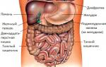
To begin with, it is worth understanding some features of the structure and functioning of the liver. This organ consists of parenchyma and stroma (connective tissue capsule), which supports the structure. The functional particle of the liver is the hepatocyte cell. Grouping together, these cells form lobules in which bile is actually produced. For its synthesis, so-called indirect bilirubin (yellow pigment) is required. The formed bile enters the duodenum through the bile ducts, where it participates in the digestive process.
If for one reason or another the liver stops capturing pigment, total bilirubin in the blood, or rather, its level increases sharply. This causes the staining of the sclera and skin tissue.
Jaundice skin is not the only symptom of this pathology. Against the background of liver damage, other signs appear:
- Many patients complain of skin itching, which is associated with the synthesis and accumulation of bile acids.
- Nodular rashes often appear on the skin.
- You may notice darkening of the urine, but the stool becomes discolored.
- Often, against the background of the disease, body temperature rises, nausea and vomiting appear.
- Patients are concerned about pain in muscles and joints.
- Symptoms include a constant feeling of heaviness in the right hypochondrium. Palpation of the liver is accompanied by pain.
The clinical picture may be supplemented by problems with sleep, weakness, and irritability.
Hepatocellular jaundice and its features

This form of pathology can occur with liver cirrhosis and acute hepatitis, but its most common cause is the use of medications and toxic agents. Against the background of the disease, there is a sharp increase in the level of bilirubin in the blood along with a decrease in cholesterol and hemoglobin. The appearance of a bright yellow color of the skin and sclera and the formation of a rash are characteristic.
Already a few days after the onset of the pathological process, a significant enlargement of the liver is observed. Other symptoms include fever and chills, joint pain, and general weakness. The stool becomes liquid. Patients complain of frequent nosebleeds.
Enzymopathic form of the disease

Enzymopathic jaundice develops against the background of decreased activity of enzymes that are responsible for the uptake and release of bilirubin. In most cases, the condition is associated with hereditary defects in enzyme synthesis.
It is worth saying that this is a relatively mild form of jaundice that can be easily treated. On the other hand, the disease is sometimes difficult to diagnose, since the clinical picture is blurred and the symptoms are mild.
Cholestatic jaundice: clinical picture

This form of pathology is based on a violation of the exchange of bile components. The level of bilirubin in the blood increases significantly. Along with this, a decrease in the permeability of the walls of bile capillaries is observed.
This condition can be provoked by cirrhosis, chronic forms of hepatitis, as well as benign cholestasis. In addition, the disease develops while taking potent drugs, including hormonal drugs, Chlorpropamide, Aminazine and others.
Stages of development of the disease
Parenchymal jaundice develops in three stages, each of which is accompanied by a number of features:
- At the initial stage, there is a decrease in enzyme activity and the rate of conversion of indirect bilirubin into direct form. The membranes of hepatocytes begin to be damaged.
- The second stage is characterized by an increase in the permeability of liver cell membranes. In addition, the patient complains of pain in the side, which is associated with compression of the capillaries.
- At the last stage, the process of converting indirect bilirubin is completely disrupted. Severe pain appears in the right side, the urine changes color.
In the absence of timely medical care, the patient develops liver failure. That is why you should seek help immediately after the first signs appear.
Diagnostic measures

It is extremely important to make a timely diagnosis of parenchymal jaundice. Diagnostics includes several points:
- A general examination already provides enough information, because yellowness of the sclera and skin, pain on palpation in the right hypochondrium are very characteristic symptoms.
- A general and biochemical blood test is also performed (makes it possible to determine whether total bilirubin is increased), laboratory testing of urine and feces.
- Additionally, the patient may be prescribed PCR and ELISA. Such studies help identify specific antibodies and genetic material of viruses in the blood, which makes it possible to accurately determine the presence and nature of the infection.
- An ultrasound and sometimes a computed tomography scan are also performed to determine the extent of liver damage and the presence of concomitant diseases.
- If cirrhosis is suspected, a biopsy is needed.
Effective treatments

When the first symptoms appear, you should consult a doctor. The sooner therapy is started, the faster it will be possible to get rid of the disease and prevent possible consequences.
When drawing up a treatment regimen, the doctor primarily focuses on the cause of jaundice. For example, if yellowing of the sclera and skin is associated with a viral infection, then patients are prescribed drugs containing interferon (for example, Alfaferon, Viferon). For autoimmune liver damage, immunosuppressive drugs are used, in particular Prednisolone and Azathioprine.
If we are talking about toxic liver damage, then specific antidotes can be used. For example, in case of an overdose of paracetamol, N-acetylcysteine is used, and in case of poisoning with ethylene glycol or methyl alcohol, ethanol is used.
Since bilirubin accumulates in the body, the patient needs detoxification. This process includes intravenous administration of saline and glucose, as well as the administration of enterosorbents and colloidal medications.
To restore normal liver function, hepatoprotectors are used, for example, Heptral and Silymarin. Taking B vitamins and ascorbic acid is indicated. Additionally, medications containing amino acids can be taken. Since a deficiency of vitamin D and calcium is often observed against the background of jaundice, patients are prescribed calcium gluconate tablets, as well as sessions of ultraviolet irradiation.
It is important to follow a proper diet during treatment. You need to completely stop drinking alcohol. If the patient takes hepatotoxic drugs, the doctor replaces them with safer analogues.
Nutrition for jaundice
An important part of therapy is proper nutrition. Parenchymal jaundice requires a special diet. For example, at the first stage of development of the disease, the patient is recommended to eat a sugar-fruit diet. This type of nutrition helps cleanse the body of toxins while simultaneously saturating it with carbohydrates.
If we are talking about the second stage, then dairy products are introduced into the patient’s diet, as well as fresh juices and fruit infusions. In more severe cases (stage III), doctors recommend eating only low-calorie foods and minimizing the amount of table salt. Fried foods are contraindicated - foods can only be steamed.
List of possible complications
In most cases, hepatic jaundice responds well to treatment. With proper treatment, you can get rid of the disease while avoiding complications. However, the disease in pediatric patients is accompanied by a decrease in blood supply to tissues, which can affect the further development of the nervous system (occasionally parenchymal jaundice causes a slowdown in mental development).
If therapy was started already at the third stage, then irreversible changes in the structure and functioning of the liver and gall bladder are possible. A sharp and persistent increase in bilirubin levels affects the functioning of the entire body, causing severe intoxication and damage to the nervous system. That is why in no case should such a problem be left unattended - it is important to seek help in time and strictly follow all the doctor’s instructions.
Jaundice is a symptom characterized by yellow discoloration of the skin and whites of the eyes.
Jaundice develops as a result of the accumulation of a substance called bilirubin in the blood and tissues of the body.
Signs of jaundice
The most common signs of jaundice are:
- yellowing of the skin, eyes and mucous membranes of the nose and mouth;
- pale color of stool (including white feces);
- dark urine (the color of beer or tea).
Classification of jaundice
- Prehepatic (hemolytic) jaundice - occurs as a result of increased accumulation of bilirubin in the blood, which the liver does not have time to process. This happens when there is excessive breakdown of red blood cells - hemolysis, for example, with sickle cell anemia (a congenital disease when red blood cells have an irregular shape and are quickly destroyed) or various poisons enter the bloodstream, causing the destruction of red blood cells.
- Hepatic (parenchymal) jaundice is a violation of the permeability or destruction of liver cells, resulting in excess bilirubin entering the blood. This happens with hepatitis, Gilbert's syndrome, cirrhosis, etc.
- Subhepatic (mechanical) jaundice - occurs when there is a violation of the flow of bilirubin from the liver into the intestine along with bile. It happens with gallstones or tumors.
Who is susceptible to the disease
Hepatic and subhepatic jaundice is more common in older and middle-aged people than in young people. Prehepatic jaundice can occur in people of all ages, including children.
You can avoid the appearance of jaundice with a healthy lifestyle. For example, you can maintain a normal body weight, avoid drinking too much alcohol, and minimize the risk of contracting hepatitis.
Treatment of jaundice
Treatment for jaundice in adults and older children will depend on what disease is causing it.
Jaundice of newborns
Babies are often born with symptoms of jaundice. At a very early age, the child’s mechanisms for removing bilirubin are not yet fully formed.
In general, newborn jaundice is not a cause for concern. It usually goes away without treatment within two weeks.
If the yellow discoloration of the baby’s skin persists longer and is accompanied by other alarming symptoms, contact a neonatologist (including, you can call the maternity hospital where your baby was born) or a pediatrician. With our service you can quickly or in your city.
Causes of jaundice
The cause of jaundice is an increased level of bilirubin in the blood, which penetrates into the soft tissues, giving them a characteristic yellow color.
Any disease that interferes with the passage of bilirubin from the blood into the liver and its removal from the body can cause jaundice.
What is bilirubin?
Bilirubin is a breakdown product of red blood cells (erythrocytes). It is delivered with the blood to the liver, from where, as part of bile, bilirubin enters the gallbladder, and then into the intestines.
In the digestive system, bacteria convert bilirubin into urobilin and stercobilin, which are excreted from the body in urine and feces. It is bilirubin that colors urine yellow and stool dark brown.
Classification of jaundice
According to the mechanism of development of bilirubin retention in the body, three types of jaundice are distinguished:
- Prehepatic (hemolytic) jaundice - occurs when there is excessive breakdown of red blood cells - hemolysis, which leads to the accumulation of bilirubin in the blood, which the liver does not have time to process. This happens, for example, with sickle cell anemia or various poisons entering the bloodstream, causing the destruction of red blood cells.
- Hepatic (parenchymal) jaundice is a violation of permeability or destruction of liver cells, resulting in excess bilirubin entering the blood. This happens with hepatitis, Gilbert's syndrome, cirrhosis.
- Subhepatic (obstructive) jaundice - occurs when something blocks the normal flow of bilirubin from the liver into the intestines along with bile. This happens with gallstones or tumors.
The causes of each type of jaundice are described below.
The causes of prehepatic jaundice are usually associated with excessive destruction of red blood cells, called red blood cells, which is called hemolysis. For example, the following diseases can lead to hemolysis:
- malaria is a blood-borne infectious disease spread by malarial mosquitoes, often found in tropical regions;
- sickle cell anemia is a congenital change in the shape of red blood cells, causing them to become brittle; in Russia - very rare, more common in dark-skinned people;
- thalassemia is a genetic disease similar to sickle cell anemia, which also accelerates the destruction of red blood cells;
- congenital non-hemolytic jaundice is a rare genetic disease in which the enzymes necessary to move bilirubin from the blood to the liver are missing;
- hereditary spherocytosis is a rare genetic disease that shortens the life cycle of red blood cells.
The causes of hepatic jaundice are usually associated with damage to hepatocytes - liver cells:
- viral hepatitis - hepatitis A, B and C;
- alcoholic liver disease - when the liver is damaged as a result of alcohol abuse;
- drug use and side effects of medications - ecstasy use and paracetamol overdose;
- leptospirosis is a bacterial infection that affects the kidneys, liver and nervous system; a person becomes infected more often through contact with water contaminated by sick animals, through animal products or during the slaughter of animals;
- mononucleosis is a viral infection caused by the Epstein-Barr virus; manifested by fever, sore throat, enlarged liver and spleen, transmitted from person to person by airborne droplets, through saliva and blood;
- primary biliary cirrhosis is a rare autoimmune liver disease that eventually leads to the development of liver failure;
- Gilbert's syndrome is a common congenital disease in which excess bilirubin accumulates in the blood; this occurs due to a lack of an enzyme that the liver needs to fully combine bilirubin with glucuronic acid and remove it from the body;
- Liver cancer is a rare and usually incurable type of cancer that develops in the liver;
- exposure to substances that are harmful to the liver, such as phenol (used in plastic production) or carbon tetrachloride (formerly used in refrigerators, but its use is now tightly controlled).
- autoimmune hepatitis is a rare disease in which the immune system begins to destroy liver cells;
- primary sclerosing cholangitis is a rare autoimmune liver disease accompanied by damage to the bile ducts;
- Dubin-Johnson syndrome is a rare genetic disorder in which the liver has difficulty removing bilirubin from the body, causing it to accumulate.
The causes of subhepatic jaundice are associated with impaired bile outflow:
- gallstones blocking the bile duct;
- various types of tumors (pancreas, gallbladder, bile ducts) that compress the bile ducts;
- acute or chronic pancreatitis - inflammation of the pancreas, which leads to its swelling and compression of the bile duct.
Diagnosis of jaundice
To diagnose jaundice, various laboratory tests and instrumental examinations are used, which make it possible to determine the causes and severity of the condition.
Medical history and examination
Diagnosis of jaundice begins with a general examination and collection of complaints. The doctor will definitely ask you about how the disease began. You may be asked the following questions:
- whether you had flu-like symptoms before jaundice appeared (this indicates hepatitis);
- whether you experience other symptoms, such as stomach pain, itchy skin or weight loss;
- have you recently been to a country where diseases such as malaria or hepatitis A are common;
- have you noticed changes in the color of urine and feces;
- whether you have been abusing alcohol for a long time;
- whether you take drugs (or have taken them in the past);
- whether you may have been exposed to hazardous substances at work.
The doctor will certainly examine your legs to determine if there is any swelling (swelling of the legs, ankles and feet - a possible sign of cirrhosis, and also palpate your abdomen (a noticeable enlargement of the liver - a possible sign of hepatitis).
Skin tone can help diagnose the type of jaundice. If the skin and mucous membranes have a lemon tint, this is a likely sign of hemolytic jaundice. With parenchymal jaundice, the skin color becomes bright yellow, saffron yellow. With obstructive jaundice - greenish.
Analysis of urine
With different types of jaundice, the concentration of substances such as urobilin (urochrome) and bilirubin changes in urine analysis.
With hemolytic jaundice, urobilin in the urine usually increases, but bilirubin is absent.
With parenchymal jaundice, both bilirubin and urobilin increase.
With obstructive jaundice, there will be no urobilin in the urine, and the concentration of bilirubin will be sharply increased.
Biochemical blood test and liver tests
A biochemical blood test is taken from a vein on an empty stomach. Using it, you can indirectly judge the function of internal organs: liver, kidneys, pancreas, gall bladder and heart. When jaundice appears, a biochemical blood test necessarily includes liver tests - tests that make it possible to diagnose diseases such as hepatitis, cirrhosis, and alcoholic liver disease.
When the liver is damaged, it releases certain enzymes into the blood. At the same time, the level of proteins that the liver normally produces begins to fall.
By measuring the levels of these enzymes and proteins, you can get a fairly accurate picture of how well your liver is working.
You can also test your blood for markers of hepatitis and other infectious diseases.
Instrumental research
To identify the cause of jaundice, various instrumental studies are prescribed to visualize internal organs and check for pathology in the liver or bile ducts.
These include the following:
- Ultrasound examination (ultrasound) - using high-frequency sound waves, an image of internal organs is created;
- computed tomography (CT) - a series of x-rays are taken, which are collected by a computer into a detailed three-dimensional image of the organ;
- magnetic resonance imaging (MRI) - using strong magnetic fields and radio waves, a detailed image of the internal structure of the part of the body being examined is created;
- retrograde cholangiopancreatography (RCPG) is a study of the patency of the bile ducts using an endoscope (a small flexible fiber-optic camera), which is brought through the mouth, esophagus, stomach and intestines to the opening of the bile duct and a radiopaque pigment is injected there, then a series of X-rays are taken.
Liver biopsy
If cirrhosis or cancer is suspected, a liver biopsy may be ordered.
During the procedure, under local anesthesia, a thin needle is inserted into the abdomen to remove a sample of liver cells for examination under a microscope.
Treatment of jaundice
The choice of treatment for jaundice will depend on the cause that caused it.
Hemolytic (prehepatic) jaundice
The goal of treating hemolytic jaundice is to stop hemolysis, the breakdown of red blood cells that increases bilirubin levels in the blood.
For an infectious disease such as malaria, specific anti-infective drugs are usually recommended. For genetic blood disorders such as sickle cell disease or thalassemia, transfusions of blood or blood components may be required to replace red blood cells.
Gilbert's syndrome, as a rule, does not require treatment, since the jaundice it causes is not dangerous and does not pose a serious threat to health.
Parenchymal (hepatic) jaundice
With parenchymal jaundice, liver damage is difficult to repair, but over time it can recover on its own. Therefore, treatment consists of preventing further liver damage.
If it is caused by an infection, such as viral hepatitis or mononucleosis, further damage can be prevented with antiviral medications.
If the damage was caused by exposure to harmful substances, such as alcohol or chemicals, it is recommended to avoid future contact with these substances.
For severe liver disease, one of the possible treatment options is a liver transplant. However, very few are suitable transplant candidates, and the number of donor organs is limited.
Mechanical (subhepatic) jaundice
In most cases, obstructive jaundice is treated surgically.
During surgery, the following organs may need to be removed:
- gallbladder;
- area of the bile ducts;
- parts of the pancreas.
Prevention of jaundice
There are so many possible causes of jaundice that it is impossible to avoid them all, but there are some steps you can take to minimize the risk of the disease.
Alcohol
The most effective way to prevent jaundice is to completely abstain from alcohol, especially if you have been abusing alcohol for many years.
If you cannot completely give up alcohol, read more about the permissible doses of alcohol that an adult can take.
If you find it difficult to drink in moderation, consult your doctor. There are support services and special medications that can help you reduce your alcohol consumption.
Prevention of infectious hepatitis
Which doctor should I contact if I have jaundice?
If you experience any of the above signs of jaundice, consult your doctor immediately. These are important warning signals indicating that the body's normal functioning has been disrupted. The initial diagnosis of jaundice is usually carried out by a general practitioner, since this symptom can be a consequence of diseases of various systems and organs. With the help of the NaPravku service you can quickly find a family doctor or for a child. After clarifying the causes of jaundice, you may need to consult more specialized specialists.
The causes of the parenchymal form of liver dysfunction are associated with various infectious diseases, which result in damage to liver cells. Due to their partial or complete dysfunction, bilirubin stops converting from free to its direct form and returns to the blood vessels. Therefore, the skin and eye sclera acquire a characteristic yellow color.
With parenchymal jaundice, the skin and sclera become yellow.
Etiology
Parenchymal jaundice, also called hepatic jaundice, has a characteristic course during which the epidermis, mucous membranes of the patient and even the eyeballs acquire a characteristic yellow tint. This can be explained by the fact that the diseased organ ceases to convert the free bile enzyme found in the blood into its simple form with the intensity that is characteristic of its healthy state. As a result, even that small amount of direct bilirubin does not enter the bile ducts, but returns back to the blood vessels. They, in turn, spread it throughout the body, as a result of which the skin of a patient with this disease acquires a characteristic yellow tint, which gives it the bile pigment bilirubin.
During normal liver function, the bile enzyme is completely eliminated from the human body. When parenchymal jaundice begins to develop, bilirubin accumulates in the body, especially a lot of it is deposited in the skin, when pathological anatomy begins to appear, and the biochemistry corresponds to the indications of ICD 1, in which the following is noted:
- severe parenchymal dystrophies;
- liver necrosis;
- acute intoxication with alcohol breakdown products or infectious microorganisms.
Yellow skin and mucous membranes of the same color signal that the blood-forming organ has completely lost its function of removing bile enzyme from the blood and delivering it to the bile ducts. The International Qualification of Liver Diseases identifies different types of liver dysfunctions and the mechanisms of their development.
Causes
Parenchymal jaundice appears as a result of acute viral infections, oncology, or as a consequence of acute intoxication with breakdown products of alcohol or other poisons. Liver dysfunction occurs as a result of:
- leptospirosis;
- infectious mononucleosis;
- cirrhosis;
- infectious viral or chronic hepatitis;
- hepatocellular cancer.
Hepatic jaundice is also called true, since, unlike its false analogue, the skin and mucous membranes in this case are colored yellow by bile pigment, and not by carotene.
Kinds
The liver form, in addition to yellow skin, eye sclera and tongue, is characterized by unbearable itching on the skin, which indicates that epidermal cells intensively absorb bile enzyme, experiencing acute intoxication, and the liver is not able to clear the blood of bilirubin.
This liver pathology can manifest itself in different forms:
- parenchymal (liver);
- suprahepatic;
- subhepatic

Parenchymal jaundice may result from cirrhosis
If hepatic jaundice is characterized by a violation of the entire movement of bilirubin in the body of an adult or infant, then suprahepatic jaundice is associated with dysfunction of the release of bile enzyme precisely in the biliary tract. With this form of the disease, bilirubin may be released in excess. Subhepatic jaundice manifests itself in compression of the bile ducts, which leads to their swelling. When making a diagnosis, the doctor must accurately determine the type of disease in order to prescribe the correct treatment to the patient. This is very important, since this disease, associated with liver dysfunction, often affects newborns. Their treatment plays an important role in the further development of children.
Unlike true jaundice, suprahepatic jaundice is characterized by failure of the biliary tract and intensive breakdown of red blood cells. The pathological anatomy of such liver disease is manifested in the development of hemolytic anemia. The color of the skin in the suprahepatic form becomes a characteristic lemon-yellow hue, and at the same time it does not itch, as in the hepatic form of the disease. The easiest way to make a diagnosis is if the patient has suprahepatic jaundice, the pathological anatomy of which has characteristic signs that clearly appear at the first stage of the development of the disease.
In the biochemical analysis of the patient, the indicators inherent in a healthy body deteriorate, urine becomes light, pain is constantly present in the side due to compression of the capillaries, the permeability of cell membranes worsens or becomes too loose, feces acquire a characteristic whitish tint. During the examination of the patient, the doctor can distinguish two main types of hepatic form of the disease: postcellular or hepatocellular. The latter could be:
- microsomal;
- premicrosomal.
Symptoms and stages
In addition to the characteristic color of the epidermis and mucous membrane, such a patient may experience vomiting, diarrhea, nausea, headache, indigestion and high fever. An experienced doctor will always be able to accurately determine, based on such signs, problems with the removal of bile enzyme from the body and refer the patient to a hospital for treatment.
Symptoms of different types of diseases can be intertwined and lead to an erroneous diagnosis and prescription of the wrong treatment to the patient.
For each type of pathology, the doctor prescribes special treatment. The disease itself goes through three stages of development, during which:
- the production of enzymes decreases and deformation of hepatocyte membranes develops, as a result of which bilirubin is not produced normally;
- hyperfermentation develops;
- the process of permeability of the cell membrane increases, characterized by the active penetration into them of bilirubin, which is in an excess state in the blood.
- liver pain in the side intensifies. To avoid painful attacks in the patient, the doctor should begin treatment as early as possible.

With parenchymal jaundice, pain in the side may occur
Only then will parenchymal jaundice, the symptoms of which clearly appear in the last stages of its development, be completely cured if a diagnosis can be made at the very beginning of the course of the disease.
With parenchymal jaundice, the patient may experience acute pain in the side and liver failure. In case of hepatic colic, the patient should be put to bed and given the necessary assistance. All patients diagnosed with hepatic cellular jaundice should be hospitalized. The viral etiology and pathogenesis of this disease require hospitalization and the creation of a quarantine regime to prevent the development of the epidemic.
Diagnostics
Diagnosis of the disease sees the cause of its appearance in the entry of viruses and microbes, toxins and pathogenic bacteria into the body, which trigger pathogenesis in the liver. Jaundice is the result of severe liver intoxication, which can completely or partially fail. With acute hepatic colic, the patient cannot tolerate pain and requires special medical care.
Diagnosis of liver diseases, including the true form, confirms that the cause of liver failure may be:
- infections and pathogenic bacteria that cause diseases such as toxoplasmosis, syphilis, malaria;
- highly toxic substances such as arsenic, snake venom, overdose of poor quality alcohol, poison contained in inedible mushrooms;
- uncontrolled use of medications that have a hepatotoxic effect. This could be Paracetamol, antibiotics, aspirin.
One of the reasons for parenchyma may be a hereditary predisposition to such a pathology. Diagnosis of a large number of patients made it possible to establish that a tendency to the disease can be transmitted from parents to children, as well as a congenital deficiency of ligandin proteins produced by the liver. They are on its surface. Also inherited is the deficiency of a special enzyme, which determines the pathomorphology of jaundice. The inherited Wilson-Konovalov disease, which manifests itself in the accumulation of copper in hepatocytes, can also disrupt liver function. Any medical student's lecture notes contain such information. Patients themselves who have a hereditary tendency should know it.
The more effective and timely a course of therapy is prescribed, the faster it is possible to restore the functions of the diseased organ, convert free bilirubin into simple bilirubin and direct it into the choleretic ducts.
Complications
In adults, this pathology leads to the destruction of cells of the hematopoietic organ. Against the background of such a disease, cholecystitis, cirrhosis or oncology may develop. Liver dysfunction, which is the most important complication of the disease, can lead to disability or death.
In newborns, a complication occurs in the form of anemia, which entails pathologies in the overall development of the child, including the development of the brain and nervous system. To prevent the destruction of liver cells, strict adherence to a therapeutic diet and completion of a full course of drug treatment will be required. Complications in children and adults occur as a result of incorrect diagnoses, erroneous therapy or complete lack of treatment.
It is especially important to promptly treat the liver of newborns who have been diagnosed with jaundice. Even if they are diagnosed with the development of extrahepatic diseases, children under one and a half years of age should be treated in special hospitals that have all the necessary equipment that allows for timely diagnosis, taking tests and prescribing the most appropriate treatment for children. The condition of the liver is very important for the subsequent development of the child. This is a hematopoietic organ that is responsible for the functioning of the entire human body. Therefore, liver patients in both children and adults should be treated by experienced doctors with extensive clinical practice.
Treatment
Treatment of jaundice in adults and newborns should always be carried out in a hospital setting. Here it is easier to take tests and conduct a biochemical blood test at any time required by the attending physician.
In a hospital setting, the doctor prescribes all the necessary treatment procedures for subhepatic, hepatic and suprahepatic jaundice. Having all the necessary medical equipment at hand, the doctor will be able to quickly discern the signs of pathology even at the first stage of its development.

To treat parenchymal jaundice, the doctor prescribes medications
As a treatment for an excess of bile enzyme, doctors prescribe special medications that help remove it from the body. If this is not enough, then a special photo procedure is performed. Before choosing the appropriate type of therapy for such patients, the doctor prescribes tests to find out the degree of bilirubin concentration in the blood. The method of treatment directly depends on the level of its concentration in the body.
In the anatomical theater they often show the liver affected by various toxins. This is an important organism of the human body, which has a unique ability to recover from intoxication and infectious diseases. But this requires a long course of therapy in a hospital setting.
Prevention of disease among children and adults
For the occurrence of parenchymal, subhepatic and suprahepatic forms of the disease, a whole complex of causes and symptoms characteristic of viral and bacterial diseases is required. Therefore, to prevent such diseases that affect the liver itself and the organs adjacent to it, you should regularly follow sanitary and hygienic rules, do not abuse alcohol and do not take dangerous foods that contain toxins.
Many symptoms of liver failure in adults and children indicate that the cause of the malfunction of the liver, which should intensively cleanse the blood, was due to its powerful poisoning with toxins of various origins. During the examination, the doctor should always identify the causes that led to liver disease. Hepatic jaundice becomes the result of microbial infectious, chemical or other intoxication. The sooner the doctor prescribes treatment, the greater the chance of a faster recovery. With an advanced form of parenchymal pathology and chronic inflammation, a person runs the risk of greatly undermining his health. Already at the first symptoms indicating a malfunction of this organ, you should consult a doctor. This is especially important if the disease affects children attending preschool and school child care institutions.
Prevention is effective against jaundice and hepatitis of various types, which parents should instill in their children from a very young age. Jaundice, which arises from viral and microbial infections, can significantly undermine the health of any person. To protect your liver from such a disease, you need to regularly wash your hands before eating, do not eat large quantities of spicy and salty foods, and give up alcohol and smoking. You should minimize the intake of any toxins and regularly cleanse the liver of toxins and waste accumulated in it.
Video
Jaundice - only the most important.