Sections of the human abdomen. Projections of the abdominal organs. Projections of internal organs. Epigastrium. Mesogastrium. Hypogastrium. Description of the abdominal cavity
The abdominal cavity is a space that is located below the diaphragm, and below is limited by a conditional line passing through the border pelvic line. Other boundaries: in front - the aponeurosis of the external and internal oblique, as well as the transverse abdominal muscle, rectus muscles; from behind - the spinal column (lumbar region), iliopsoas muscles, from the sides - all the lateral abdominal muscles.
Description of the abdominal cavity
The human abdominal cavity is a container of organs and anatomical formations: stomach, gall bladder, spleen, intestines (jejunal, ileal, transverse colon, cecum and sigmoid), abdominal aorta. The location of these organs is intraperitoneal, that is, they are covered by the peritoneum, or more precisely, by its visceral layer, completely or partially.
Extraperitoneally (that is, in the retroperitoneal space) there are organs of the abdominal cavity: kidneys, adrenal glands, pancreas, ureters, the main part of the duodenum.
The partially visceral layer of the peritoneal lining flows around two spaces of the colon (ascending and descending), that is, these abdominal organs are located mesoperitoneally.
Among the organs that can be classified as intra- and mesoperitoneal, the liver can be distinguished. It is almost completely covered with a serous membrane.
Structure
Conventionally, the abdominal cavity is divided by specialists into floors:
- The structure of the upper floor, or gland opening. It has “subsections”: hepatic bursa, omental, pregastric fissure. The hepatic lobe covers the right lobe of the liver, and in its depths you can feel the kidney on the right and the adrenal gland. The pregastric fissure includes part of the organs: the spleen and stomach, the left hepatic lobe. The cavity, called the omental bursa, communicates with the general cavity of the peritoneum through a narrow opening. It is bounded above by the liver (caudate lobe), on the anterior side by the edge of the hepatoduodenal ligament, below by the duodenum, and behind by the serosa. The posterior wall, represented by the parietal layer, covers the abdominal aorta, pancreas, kidney on the left, adrenal gland, and inferior vena cava. The structure of the greater omentum is as follows. The greater omentum is like an apron hanging from the transverse part of the colon. For a short distance it covers the loops of the small intestine. In fact, these are four leaves of serosa, fused together in the form of plates. There is a cavity between the plates. It communicates from above with the space of the omental bursa, and in adults, usually all the leaves are fused, that is, the cavity is obliterated. In the omentum itself there are lymph nodes that ensure the drainage of lymph from the transverse colon and greater omentum.
- Middle floor. It can be examined only by lifting the transverse colon and greater omentum. This floor is divided by the ascending, descending part of the colon, and the mesentery of the small intestine into four parts. These are the lateral canals on the right and left, two mesenteric sinuses. The mesentery is a fold of two layers of serosa that provides attachment of the small intestine to the posterior wall of the abdomen. The part of it that is attached to the posterior wall of the abdomen is called the root of the mesentery. Its length is no more than 17 cm. The opposite edge, which turns out to be free, covers the jejunum and ileum, it corresponds to the total length of these sections of the intestine. The mesentery itself is attached obliquely, starting from the second lumbar vertebra to the iliac fossa on the right. The mesentery, which is filled with fiber, contains blood vessels, lymph nodes and vessels, and nerve fibers. The posterior layer of the peritoneum, parietal, has a large number of pits. Their importance is great, since they can serve as a weak point where retroperitoneal hernias form.
- Anatomy of the lower floor. This includes organs and structures located in the pelvic cavity. The peritoneum descends here and covers the organs and the walls of the pelvis. The relationship of organs to the peritoneum depends on gender. Intraperitoneal location in such organs: the initial part of the rectum and the sigmoid colon. These organs also have a mesentery. The peritoneum covers the middle part of the rectum only from the sides and in front (mesoperitoneally). The lower part of the rectum is located extraperitoneally. In men, the serosa passes from the rectum (its anterior surface) to the bladder (posterior surface). This creates a depression behind the bladder (retrovesical). And the upper-back part of the empty bladder, the peritoneum forms a fold; it has the ability to straighten out when it is filled. The anatomy of the peritoneum of women is different, due to the location of the uterus between the bladder and rectum. The uterus is covered with peritoneum. For this reason, in women, two anatomical “pockets” are formed in the pelvic cavity: between the rectum and the uterus, and between the uterus and the bladder. In women and men, there is also a prevesical space formed by the transverse fascia and the bladder with the peritoneum.
What does the abdominal cavity include?
Anatomy of the liver and biliary tract in humans. The liver is located in the first, upper floor of the abdominal cavity. Most of it is located in the right hypochondrium, less in the epigastrium and left hypochondrium. All sides of the liver, except the posterior one, are covered with a layer of visceral peritoneum. Its posterior side is adjacent to the inferior vena cava and the diaphragm. The liver is divided by the falciform ligament into the right major and left minor lobes. Blood vessels, nerves, hepatic ducts, and lymphatic pathways make up the hilum of the liver. It is fixed by four ligaments, the hepatic veins, which drain into the inferior vena cava, by fusion with the diaphragm, and also by intraperitoneal pressure.
Anatomy of the gallbladder. It is placed in the hole of the same name. It is a hollow organ shaped like a bag or pear. Its structure is simple: body, neck and bottom. The volume reaches from 40 to 70 cubic cm, length from 8 to 14 cm, width from 3 to 4 cm. Part of the peritoneum from the liver extends onto the surface of the gallbladder. Therefore, its location can be different: from meso- to intraperitoneal. The gallbladder in humans is connected to the liver by fiber, blood vessels and the peritoneum. With some structural features, sometimes the bottom of the bladder protrudes from under the hepatic edge, adjacent to the anterior wall of the abdomen. If its location is low, it lies on the loops of the small intestine, so any pathology of these organs can lead to the development of adhesions and fistulas. The bubble is projected onto the anterior abdominal wall at the point connecting the right costal arch and the right side of the rectus abdominis muscle. This position of the bladder in humans does not always correspond to reality; more often it deviates slightly outward, less often - towards the inner side. A duct, up to 7 cm long, extends from the gallbladder, from its neck. The duct connects along the way with the common hepatic duct.
Anatomy of the human spleen. The spleen is located in the upper floor of the abdominal cavity, intraperitoneally. This is one of the main organs of the human hematopoietic and lymphatic systems. Located on the left in the hypochondrium. On its surface, called the visceral surface, there are the gates of the spleen, which contain blood vessels and nerve fibers. It is fixed by three ligaments. Blood supply occurs due to the splenic artery, which is a branch of the celiac trunk. Inside it, the blood vessels branch into small-caliber vessels, which determines the segmental structure of the spleen. This organization ensures easier resection into sectors.
Duodenum. It has a retroperitoneal location, this is the section from which the small intestine begins in humans. Duodenum goes around the head of the pancreatic gland in the form of a loop, the letters U, C, V and has four parts: superior, ascending, descending and horizontal. To the structures of the retroperitoneal space from the duodenum there are ligaments that provide its fixation. In addition, fixation is provided by the root of the mesentery of the colon, the peritoneum. The connection between the intestine and the pancreas has a significant impact. Structure: the beginning of the intestine is slightly expanded, which is why it is called the ampulla, onion. The folds of the mucous membrane are located longitudinally, in other parts they are circular. On the inner wall of the descending part there is a large longitudinal fold; it ends with the papilla of Vater. On its surface there is the sphincter of Oddi, through which two ducts open: the bile duct and the pancreatic duct. Just above is the minor papilla, where the second pancreatic duct may be located; this anatomical unit is variable.
Anatomy of the pancreas. Located retroperitoneally. It is conventionally divided into three parts: tail, body, head. The head of the gland continues into a hook-shaped process; it covers the vessels located along the dorsal surface of the gland, delimiting them with the inferior vena cava. In most cases, its head is located in front of the second - third lumbar vertebrae. The length of the gland is from 17 to 21 cm, sometimes reaching 27 cm. Its shape is most often triangular, but it can also be angular or flat. From the tail towards the head there is a pancreatic duct, which opens into the cavity of the duodenum, in its descending part. Projection of the gland on the anterior abdominal wall in humans: umbilical, epigastric and left hypochondrium.
The structure of the stomach. Refers to hollow organs. It begins after the esophagus, then passes into the duodenum. Its volume (empty) is up to 0.5 liters, after meals on average up to 1 liter. In rare cases, it stretches up to 4 liters. The average length is from 24 to 26 cm. The left hepatic lobe is adjacent to it in front, the pancreatic gland is behind it, the loops of the small intestine are below, and the spleen touches it from above to the left. The stomach is projected in the epigastric region, covered with serosa on all sides. In its cavity, gastric juice is produced, which contains enzymes: lipase, pepsin, chymosin, as well as other components, for example, hydrochloric acid. In the stomach, due to mixing by waves of peristalsis, chyme is formed from food, which penetrates portionwise through the pylorus into the intestine. Food stays in the stomach for different periods of time: liquid food from 20 minutes, coarse food with fibers - up to 6 hours.
Abdomen from above it is limited by the diaphragm - a flat muscle that separates the chest cavity from the abdominal cavity, located between the lower part of the chest and the lower part of the pelvis. In the lower abdominal cavity there are many organs of the digestive and genitourinary systems.
The upper part of the abdominal cavity contains mainly the organs of the digestive system. Abdomen can be divided by two horizontal and two vertical lines that form abdominal areas. Thus, nine conditional zones are identified.
A special division of the abdomen into areas (zones) is valid throughout the medical world. The upper row contains the right hypochondrium, epigastric region and left hypochondrium. In these areas we try to palpate the liver, gall bladder, stomach, and spleen. In the middle row are the right lateral, mesogastric, or umbilical, umbilical, and left lateral regions, where manual examination of the small intestine, ascending and descending colon, kidneys, pancreas, and so on is performed. In the bottom row, the right iliac region, hypogastrium and left iliac region are distinguished, in which the cecum and colon, bladder, and uterus are examined with the fingers.
AND abdominal cavity, and the chest located above it are filled with various organs. Let us mention their simple classification. There are organs that to the touch resemble a bath sponge or a loaf of fresh bread, that is, when cut, they are completely filled with some content, represented by functioning elements (usually epithelial cells), connective tissue structures referred to as the stroma of the organ, and vessels of various sizes. This parenchymal organs(Greek enchyma translates as “something poured in”). These include the lungs, liver, almost all large glands (pancreas, salivary, thyroid, and so on).
In contrast to parenchymatous are hollow organs, they are hollow because they are not filled with anything. They have a large (stomach, bladder) or small (ureter, artery) cavity inside, surrounded by relatively thin (intestines) or thick (heart, uterus) walls.
Finally, if the characteristic features of both groups are combined, that is, there is a cavity (usually small) surrounded by parenchyma, we speak of mixed bodies. These primarily include the kidneys, and a number of authors, with some reservations, include the spinal cord and brain here.
Inside the abdominal cavity there are various digestive system organs(stomach, small and large intestines, liver, gallbladder with ducts, pancreas), spleen, kidneys and adrenal glands, urinary tract (urethra) and bladder, reproductive organs(different in men and women: in women the uterus, ovaries and fallopian tubes; in men, the genitals are located outside), numerous blood and lymphatic vessels and ligaments that hold the organs in place.

In the abdominal cavity there is a large serous membrane, consisting mainly of connective tissue, which lines the internal walls of the peritoneum and also covers most of the organs located in it. It is generally accepted that the membrane is continuous and consists of two layers: parietal and visceral peritoneum. These layers are separated by a thin film moistened with serous fluid. The main function of this lubricant is to reduce friction between the layers, as well as between the organs and walls of the peritoneum, while ensuring the movement of the layers.
Doctors often use the term "acute abdomen" to describe a severe case that requires immediate intervention, in many cases surgery. The origin of pain can be different; it occurs not only due to diseases of the digestive system, as is often thought. There are many other causes of acute abdominal pain; it is often accompanied by vomiting, hardness of the abdominal walls and fever. Here we are not talking about a specific disease, but about the initial diagnosis of a very dangerous condition that requires an urgent medical examination to determine its cause and provide appropriate treatment.
LIVER AND BALL TRACT
;traumatic rupture
;abscess
;acute cholecystitis
; biliary colic
SMALL INTESTINE
duodenal ulcer
obstruction, rupture
acute gastroenteritis
Meckel's diverticulum
local enteritis
intestinal tuberculosis
COLON
ulcerative colitis
infectious colitis
volvulus
cancer
intussusception
diverticulitis
gap
appendicitis
STOMACH
;ulcer
;cancer
SPLEEN
;heart attack
;abscess
;break
PERITONEUM
peritonitis
INTERNAL GENITALIA OF A WOMAN
;break
;infection
;convulsions
;rupture of an ovarian cyst
;ectopic pregnancy
;abscesses
;acute salpingitis
Peritoneal hernia occurs when there is a weak point in the abdominal wall, causing part of the intestine to protrude out of the abdominal cavity. An abdominal hernia is an exit or protrusion of the small or large intestine or parts thereof from the cavity in which they are located through a congenital or acquired opening in the peritoneum. An abdominal hernia can occur due to prolonged pressure of internal organs on the walls of the abdominal cavity or weakening of a certain point - for example, as a result of pregnancy, obesity, constant physical activity, etc. Peritoneal hernia comes out when part of the abdominal cavity protrudes and forms a hernial sac, which sometimes contains part of the small or large intestine. The only effective treatment for hernia is surgery.
X. Developmental defects
XI. Operations
I. Definition of the concept of “belly”.
Borders of the abdomen (upper and lower)
Stomach(abdomen) - the lower half of the body.
Upper border of the abdomen form
front– xiphoid process and edges of costal arches,
behind– edges of the XII ribs and XII thoracic vertebra.
Lower border of the abdomen passes along lines drawn from the symphysis of the pubic bones to the sides, to the pubic tubercles, then along the inguinal folds, to the anterior superior spines of the iliac bones, along their crests and the base of the sacrum.
II. The concept of the abdominal wall and the abdominal cavity (abdominal cavity). Boundaries of the abdominal cavity
Stomach includes the abdominal wall and abdominal cavity.
Abdominal wall- a set of soft tissues that limit the abdominal cavity from the front, back and sides.
From the inside abdominal cavity lined intra-abdominal fascia .
Abdomen (abdominal cavity– cavum abdominis) - the space delimited by the intra-abdominal fascia (i.e., this is the cavity of the fascial sac ).
Abdominal cavity (abdominal cavity) delimited
above diaphragm,
front- rectus muscles and aponeuroses of the oblique and transverse abdominal muscles,
from the sides- the muscular parts of these muscles,
behind- lumbar spine, psoas major, latissimus dorsi and quadratus lumborum,
from below- passes into the pelvic cavity (conditional border - a plane located at the level of the boundary line), therefore the abdominal cavity is limited from below by the iliac bones and the pelvic diaphragm.
In the abdominal cavity (abdominal cavity) organs of the digestive and genitourinary systems, large vessels and nerve plexuses are located.
Above part of the abdominal organs is located within the boundaries of the chest, and at the bottom - in the small pelvis.
IV. Peritoneum (parietal and visceral).
V. Location of the abdominal organs in relation to the peritoneum (intraperitoneal, mesoperitoneal and extraperitoneal)
Abdominal organs in relation to the peritoneum may be
Intraperitoneal,
Mesoperitoneal and
Extraperitoneal.
1) An organ covered by peritoneum on all sides , located intraperitoneally (intraperitoneal) .
Intraperitoneal (intraperitoneal) located stomach, jejunum, ileum, cecum, appendix, transverse colon, sigmoid colon, upper part of the rectum. They are covered with visceral peritoneum on all sides, except for the attachment points of the mesenteries. Also located intraperitoneally are the bulb (initial part) of the duodenum, the tail of the pancreas, the spleen, the uterus, the fallopian tubes, and the ovaries.
2) An organ covered by peritoneum on three sides and not covered by peritoneum on one side, located mesoperitoneal.
Bodies occupying mesoperitoneal position (covered by peritoneum on three sides): liver, gall bladder, ascending and descending colon, middle part (ampullary part) of the rectum, full bladder.
3) An organ covered by peritoneum on only one surface , located retroperitoneal (extraperitoneal) .
Extraperitoneal ( extraperitoneal) located are the duodenum (except for its initial section - the bulb), the pancreas (except for its tail), the lower part of the rectum, an empty bladder, and kidneys , ureters , adrenal glands , the abdominal aorta, the inferior vena cava and other large vessels that are located in the retroperitoneal space.
VI. Sections and areas of the anterolateral abdominal wall.
Anterior abdominal wall located between the right and left posterior axillary lines (anterior to them).
It is divided into departments two lines (planes) drawn
- horizontally between the lowest points of the X edges ( subcostal plane) or horizontally through the middle of the distance between the upper edge of the manubrium and the pubic symphysis ( transpyloric plane) And
- between the superior anterior iliac spines ( interosseous plane) or between the iliac tuberosities ( intercrestal plane).
These departments are:
epigastrium(epigastrium),
womb(mesogastrium) and
hypogastrium(hypogastrium).
Two vertical lines ( midclavicular lines), which run along the lateral edges of the rectus abdominis muscles, divide the anterior abdominal wall for 9 regions.
Rice. 6. Areas of the anterior abdominal wall:
1 - right hypochondrium; 2 - epigastric; 3 - left hypochondrium; 4 - right side; 5 - umbilical; 6 - left side; 7 - right inguinal; 8 - pubic; 9 - left inguinal.
X. Developmental defects
Disturbances in the normal development of the anterior abdominal wall and the formation of the abdominal cavity may cause fetal hernia . With such a hernia, emergency surgery is indicated, because drying of the thin membranes that cover the hernial contents leads to rupture of the membranes, eventration and peritonitis.
In case of underdevelopment of the diaphragm, congenital diaphragmatic hernia , caused by the presence of a hole in the diaphragm dome (usually on the left).
Disruption of the process of attachment of the primary mesentery to the posterior layer of the parietal peritoneum after completion of the intestinal rotation may cause the formation of other internal abdominal hernias (perioduodenal hernia of Treitz, peri-cecal, intersigmoid, etc.).
XI. Operations
The main surgical access to the abdominal organs is laparotomy .
Depending on the location of the organ on which surgery is performed, the nature of the pathology and the scope of the operation, various incisions are used for access.
Most often used midline laparotomy, in which the abdominal cavity is opened along the white line of the abdomen.
During operations on the organs of the retroperitoneal space, lumbotomy - incision in the lumbar region (extraperitoneal access).
TOPIC: “Topographic anatomy of the abdomen. General characteristics. Abdominal areas"
I. Definition of the concept “stomach”. Borders of the abdomen (upper and lower).
II. The concept of the abdominal wall and the abdominal cavity (abdominal cavity). Borders of the abdominal cavity.
III. Two sections of the abdominal cavity (the abdominal cavity itself and the retroperitoneal space).
IV. Peritoneum (parietal and visceral). Peritoneal cavity (peritoneal cavity).
V. The location of the abdominal organs in relation to the peritoneum (intraperitoneal, mesoperitoneal and extraperitoneal).
VI. Sections and areas of the anterolateral abdominal wall.
VII. Projections of organs onto the areas of the anterolateral abdominal wall.
VIII. Individual and age differences in the position of the abdominal organs.
IX. Blood supply, lymphatic drainage, innervation of abdominal organs.
X. Developmental defects
The complex of organs of the two most important systems: digestive and genitourinary, located in the abdominal cavity and in the retroperitoneal space of a person in both men and women, has its own layout, anatomical structure and key features. Having basic knowledge of the anatomy of the human body is important for everyone, primarily because it helps to understand the processes that occur in it.
Show all
What is the abdominal cavity?
The abdominal cavity (lat. cavitas abdominalis) is a space that is limited above by the diaphragm (a muscular dome separating the chest cavity from the abdominal cavity), in front and on the sides by the anterior abdominal wall, behind by the spine, and below by the perineal diaphragm.
The abdominal cavity includes not only organs related to the gastrointestinal tract, but also organs of the genitourinary system. The peritoneum itself covers the organs in different ways.
It is worth noting that organs can be divided into those related directly to the abdominal cavity, and those located within the retroperitoneal space.
Functions of organs located in the abdominal cavity
If we talk about organs related to the digestive system, their functions are as follows:
- implementation of digestive processes;
- absorption of nutrients;
- immune function;
- neutralization of toxins and poisons;
- implementation of hematopoietic processes;
- endocrine function.
Regarding the organs of the genitourinary system:
- release of metabolic products;
- reproductive function;
- endocrine function.

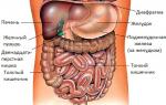
Diagram of the location of the abdominal organs
 So, if you look into the section of the anterior abdominal wall under the human diaphragm, you can see the following organs immediately below it:
- 1. The abdominal part of the esophagus is a small section 1–3 cm long, which immediately passes into the stomach.
- 2. Stomach (gaster) - a muscular sac with a capacity of about 3 liters.
- 3. Liver (hepar) - the largest digestive gland, located on the right under the diaphragm;
- 4. The gallbladder (vesica fellea) is a hollow organ that stores bile. It is located under the liver in the fossa of the gallbladder.
- 5. The pancreas is the second largest digestive gland after the liver. It lies behind the stomach in the retroperitoneal space on the left.
- 6. Spleen (lien) - located behind the stomach in the upper part of the abdominal cavity on the left.
- 7. Small intestine (intestinum tenue) - located between the stomach and large intestine and includes three sections that lie sequentially one after another: duodenum, jejunum, ileum.
- 8. Large intestine (intestinum crassum) - starts from the small intestine and ends with the anus. It also consists of several sections: the cecum, colon (which consists of the ascending, transverse, descending, sigmoid colon), rectum.
- 9. Kidneys (ren) - paired organs located in the retroperitoneal space.
- 10. Adrenal glands (glandulae suprarenale) - paired glands lying above the kidneys, lie in the retroperitoneal space.
- 11. Ureters (ureter) - paired tubes connecting the kidneys to the bladder and also lying in the retroperitoneal space.
- 12. The bladder (vesica urinaria) is a hollow organ lying in the pelvis.
- 13. Uterus (uterus), vagina (vagina), ovaries (ovarium) - female genital organs lying in the pelvis, related to the abdominal organs.
- 14. Seminal vesicles (vesiculæ seminales) and prostate gland (prostata) - male genital organs of the small pelvis.

Anatomical structure of the digestive system organs
The structure of organs related to the gastrointestinal tract is the same for both men and women.
Stomach
The stomach is a muscular cavity that lies between the esophagus and the duodenum. Serves for food accumulation, mixing and digestion, as well as partial absorption of substances.
In the anatomical structure of the stomach, the anterior and posterior walls are distinguished. Their connection above forms the lesser curvature of the stomach, and below - the greater curvature. The junction of the esophagus with the stomach is the cardiac foramen (at the level of the 11th thoracic vertebra), and the junction of the stomach with the duodenum is the pyloric foramen (pyloric foramen) - at the level of the 1st lumbar vertebra. The fundus is also distinguished from the stomach - the part of the stomach located to the left of the cardial opening, in which gases accumulate. The body of the stomach is the larger part that lies between the two openings. The approximate volume of the stomach is 3 liters.
The wall of the stomach includes the mucous membrane, muscular and serous:

Liver
 The liver is the largest digestive gland in the human body. A parenchymal organ that serves for the secretion of bile, neutralization of poisons and toxins, hematopoiesis in the fetus during pregnancy and participation in various metabolic processes.
The liver has 2 surfaces: diaphragmatic, facing the diaphragm, and visceral, bordering other organs of the abdominal cavity. Also, the liver has 2 large lobes: right and left, with the right one being the largest. Another important formation of the liver is the gate of the liver, which includes the portal vein, hepatic artery and nerves, and exits the common hepatic duct and lymphatic vessels. The organ itself consists of tiny hepatocyte cells that participate in the production of bile.
Gallbladder
 The gallbladder is a hollow organ , which is involved in the accumulation of bile. It lies under the liver in the fossa of the gallbladder.
This organ has a bottom that protrudes from under the lower edge of the liver; the neck is the narrow end heading towards the gate of the liver and the body of the bladder is the expansion lying between the bottom and the neck. The cystic duct departs from the neck, which, connecting with the common hepatic duct, forms the common bile duct. It, in turn, opens into the duodenum.
The wall of the gallbladder consists of mucous, submucosal, muscular and serous membranes:
Pancreas
 The pancreas is the second largest after liver iron gastrointestinal tract. It is located behind the stomach in the retroperitoneal space.
In the anatomical structure of the pancreas, it has a head, body and tail. The head of the gland lies to the right, near the pancreas, and the tail is directed to the left, approaching the gate of the spleen. The pancreas produces pancreatic juice, rich in enzymes necessary for digestion, as well as the hormone insulin, which regulates blood glucose levels.
Spleen
 The spleen is a parenchymal lymphoid organ. Located on the left side of the upper abdomen, just below the diaphragm, behind the stomach.
This organ has 2 surfaces: diaphragmatic and visceral and 2 poles: posterior and anterior. The outside of the spleen is covered with a capsule, and inside there is a pulp, which is divided into red and white. The spleen performs the function of a blood depot, immune function and hematopoietic function of the fetus.
Small intestine
The small intestine is the longest organ of the digestive system (in men - 7 m, in women - 5 m).
The small intestine consists of 3 sections: duodenum, jejunum and ileum.
The duodenum is about 30 cm long and lies between the stomach and jejunum. It has 4 parts: upper, descending, horizontal, ascending.

The jejunum and ileum form the mesenteric part of the small intestine, as they have a mesentery. They occupy most of the hypogastrium. The loops of the jejunum lie in the upper left, and the ileum - in the lower right part of the abdominal cavity.
The wall of the small intestine consists of mucous, submucosal, muscular and serous membranes:

Colon
Large intestine - located from the small intestine to the anus.
It consists of several sections: the cecum; colon (it includes the ascending, transverse, descending, sigmoid colon); rectum. The total length is about 1.5 m.
The colon has ribbons - longitudinal muscle fibers; haustra - small protrusions in the form of bags between the ribbons and omental processes - protrusion of the serous membrane with adipose tissue inside.
The vermiform appendix extends from the cecum by 2–20 cm.
At the junction of the ileum and the cecum there is an ileointestinal opening.
When the ascending colon passes into the transverse colon, a right bend of the colon is formed, and when the transverse colon passes into the descending colon, a left bend is formed.
The wall of the cecum and colon includes the mucous, submucosal, muscular and serous membrane.
The sigmoid colon begins from the descending colon and continues into the rectum, where it ends at the anus.
The length of the rectum is 15 cm, it accumulates and removes feces. At the level of the sacrum, it forms an extension - an ampulla (accumulation occurs in it), after which there is an anal canal, which opens with the anus.
The wall of the rectum consists of mucous, submucosal, muscular and serous membranes.
Kidneys
 Kidneys are paired parenchymal organs.
They are located in the retroperitoneal space. The right kidney is located slightly lower than the left, as it borders the liver. They are shaped like beans. On the outside, each kidney is covered with a fibrous capsule, and the parenchyma consists of the cortex and medulla. The structure of these organs determines their function. Inside each kidney there is a system of small renal calyces, which pass into large renal calyces, and these, in turn, open into the renal pelvis, from which the ureter departs to remove accumulated urine. The structural and functional unit of the kidney is the nephron.
Adrenal glands
 The adrenal glands are paired glands located above the kidneys.
They consist of cortex and medulla. There are 3 zones in the cortex: glomerular, fascicular and reticular. The main function of the adrenal glands is endocrine.
Ureters
The ureters are paired tubes that extend from the kidneys and connect them to the bladder.
The wall of the organ is represented by mucous, muscular and connective tissue membranes.

Bladder
 The bladder is a hollow organ that stores urine in the human body.
The size of the organ may vary depending on the amount of contents in it. From below, the organ narrows somewhat, passing into the neck of the bladder, which ends in the urethra. The bladder also has a body - its larger part and a bottom - its lower part. On the back surface, two ureters flow into the bladder, which deliver urine from the kidneys. At the bottom of the bladder, a vesical triangle is distinguished, the base of which is the openings of the ureters, and the apex is the opening of the urethra. This triangle contains the internal sphincter, which prevents involuntary urination.
Female genital organs related to the abdominal cavity
 The uterus is a muscular organ in which the development of the fetus occurs during pregnancy. It consists of several parts: bottom, body and neck. The lower part of the cervix transitions into the vagina. The uterus also has 2 surfaces: the anterior one, facing the bladder, and the posterior one, facing the rectum.
The wall of the organ has a special structure: perimeter (serous membrane), myometrium (muscular), endometrium (mucous membrane).
The vagina is a muscular organ about 10 cm long. The vaginal wall consists of 3 layers: mucous, muscle and connective tissue. The lower part of the vagina opens into the vestibule. The walls of the vagina are studded with glands that produce mucus.
The ovary is a paired organ of the female reproductive system that performs a reproductive function. They consist of connective tissue and cortex with follicles at different stages of development.
Normally, the ovaries on ultrasound look like this:

Genital organs in men related to the abdominal cavity
 The seminal vesicles are paired organs of the male reproductive system. The tissue of this organ has a structure in the form of cells.
Prostate gland (prostate) is the male gland. It surrounds the neck of the bladder.
In the abdominal cavity of the human body, in both men and women, there is a complex of internal organs of two important systems: digestive and genitourinary. Each organ has its own location, anatomical structure and its own characteristics. Basic knowledge of human anatomy leads to a better understanding of the structure and functioning of the human body.
The abdominal cavity is limited in front and on the sides by the abdominal walls, behind by the lumbar region, and above by the diaphragm; from below it passes into the pelvic cavity. It contains the abdominal cavity and abdominal organs.
Abdomen(cavum peritoneale) is represented by a space surrounded by a serous membrane - the peritoneum (peritoneum). It includes all organs covered by the peritoneum (Fig. 133). The serous layer covering the walls of the abdomen from the inside is called parietal, or parietal, and the layer adjacent to the organs is called splanchnic, or visceral. Both sheets are one whole; they directly transform into one another. Between the layers of the peritoneum there is a small amount of serous fluid - up to 30 ml.
Rice. 133. Sinuses and canals of the abdominal cavity.
I - hepatic bursa; II - pregastric bursa; III - right mesenteric sinus; IV - left mesenteric sinus; V - right channel; VI - left channel, 1 - diaphragm; 2 - coronary ligament of the liver; 3 - liver; 4 - stomach; 5 - spleen; 6 - transverse colon: 7 - duodenal-small intestinal bend; 8 - descending colon: 9 - sigmoid colon; 10 - bladder; 11 - terminal ileum; 12 - cecum with vermiform appendix; 13 - root of the mesentery of the small intestine; 14 - ascending colon; 15 - duodenum; 16 - gallbladder.
Most organs (stomach, small intestine, cecum, transverse colon and sigmoid colon, spleen) are enveloped by the peritoneum on all sides, i.e. they lie intraperitoneally, or intraperitoneally. They are supported by the mesentery or ligaments formed by the layers of the peritoneum. Other organs (liver, gall bladder, ascending and descending colon, part of the duodenum, pancreas, rectum) are covered by the peritoneum on three sides, with the exception of the posterior one, i.e. they are located mesoperitoneally. A small number of organs (duodenum, pancreas, kidneys, ureters, large blood vessels) lie behind the peritoneum - they occupy a retroperitoneal position.
Using the position of the transverse colon with its mesentery, the abdominal cavity is divided into upper and lower floors, which approximately corresponds to the plane passing through the ends of the X ribs. In the upper floor there are three sacs (or bursae): hepatic, pregastric and omental. The hepatic bursa (bursa hepatica) is located between the diaphragm, the anterior wall of the abdomen and the right lobe of the liver. The pregastric bursa (bursa pregastrica) is located in front of the stomach with its ligaments and is adjacent to the left lobe of the liver and spleen. These bags are separated from each other by the falciform ligament of the liver. The omental bursa (bursa omentalis) is represented by a slit-like space limited in front by the stomach with its ligaments, below by the left part of the transverse colon with its mesentery, on the left by the spleen with its ligaments and behind by the peritoneum of the posterior abdominal wall covering the pancreas, left kidney with the adrenal glands, aorta and inferior vena cava; on top, the omental bursa adjoins the caudate lobe of the liver (Fig. 134). This bag communicates with the common cavity through the omental foramen of Winslowi (for. epiploicum Winslowi), bounded by the peritoneum-covered right kidney with the adjacent inferior vena cava behind, the initial part of the duodenum below, the caudate lobe of the liver above and the hepatoduodenal ligament in front.

Rice. 134. The course of the peritoneum on a sagittal section of the abdomen (semi-schematic). The abdominal aorta is slightly displaced to the right and left undissected. 1 - diaphragm; 2 - small oil seal; 3 - gland hole; 4 - truncus coeliacus; 5 - a. mesenterica superior; 6 - pancreas; 7 - a. renalis; 8 - cisterna chyli and a. testicularis; 9 - duodenum; 10 - a. mesenterica inf.; 11 - latero- and retroaortic lymph nodes; 12 - mesenterium; 13 - vasa iliaca communia; 14 - greater omentum: 15 - colon transversum; 16 - mesocolon transversum; 17 - stomach; 18 - liver.
In the lower floor of the abdominal cavity, the right and left mesenteric sinuses and lateral canals are distinguished. The right sinus (sinus mesentericus dexter) is bounded above by the mesentery of the transverse colon, on the right by the ascending colon, on the left and below by the mesentery of the small intestine and in front by the greater omentum. The inflammatory processes occurring here are to a certain extent confined within the sinus. The left mesenteric sinus (sinus mesentericus sinister) is bounded above by the mesentery of the transverse colon, on the right by the mesentery of the small intestines, on the left by the descending colon and in front by the greater omentum. At the bottom, the sinus is open into the pelvic cavity, which makes it possible for pus or blood to spread here. Both mesenteric sinuses communicate through a gap limited by the initial part of the small intestine and the mesentery of the transverse colon. The right lateral canal (canalis lateralis dexter) is limited by the side wall of the abdomen and the ascending colon, the left (canalis lateralis dexter) is limited by the side wall of the abdomen and the descending colon. Both canals at the top communicate with the upper floor of the abdominal cavity, but on the left this communication is limited due to the existence of lig. phrenicocolicum. Inflammatory processes can spread through these channels.