Examples of description of external injuries (from the point of view of a forensic expert). Examples of descriptions of external injuries (from the point of view of a forensic expert) Do not neglect the ambulance
Headband - cap ">
Headband - "cap".
Sling-like bandage on the forehead.
Soft tissue injuries to the scalp are always dangerous. They can be accompanied by heavy bleeding, bone damage, brain contusion (concussion) or cerebral hemorrhage (hematoma), the occurrence of cerebral edema and inflammation of the meninges (meningitis, encephalitis). Signs of damage to the brain and bones of the skull, the development of inflammatory complications are headache, nausea, impaired vision and sensitivity of the skin of the extremities or weakness in them, an increase in body temperature, clouding of consciousness up to its loss.
Help: 1. Clean and wash the wound. A wound contaminated with soil or any other foreign object must be cleaned using tweezers or by hand. Then the wound is thoroughly washed with hydrogen peroxide or a weak solution of potassium permanganate (2-3 grains per glass, preferably boiled, water). You can wash the wound with tap water. With severe bleeding, first of all, it is necessary to stop the bleeding.
2. Treat the skin around the wound. Before treating the skin, it is necessary to cut the hair at a distance of two centimeters around the wound. Then gently smear the edges of the wound with a solution of iodine, brilliant green (brilliant green), a saturated solution of potassium permanganate or alcohol. In this case, alcohol is strictly not allowed to enter the wound.
3. Stop bleeding. When bleeding from a wound of the scalp, it is most effective to pack it with a sterile napkin or a sterile bandage. You can use gauze, cotton wool or any clean cloth. The swab is tightly pressed to the edges and bottom of the wound for 10-15 minutes. If the bleeding does not stop, then a pressure bandage is applied to the tampon inserted into the wound.
4. Apply a bandage (preferably sterile). Applying a bandage on the wound of the scalp is carried out as follows: tear off a piece (tie) about 1 m in size from the bandage, put it on the top of the head, the ends are lowered vertically down in front of the ears; the patient himself or one of the assistants keeps them taut. The tour of the bandage starts from the left side at the level of the forehead, goes to the right side back to the back of the head, thus making two rounds with the obligatory fixation of the first round. The third round of the bandage is wrapped around the string either on the left or on the right, so that it overlaps the previous round of the bandage by 1/2 or 2/3. Each subsequent tour leads higher and higher until the entire scalp is bandaged. The last round of the bandage is tied to the remaining vertical part of the tie from either side. The vertical ends of the tie are fixed under the chin.
5. Apply cold. Cold is applied to the bandage in the wound area. Cooling the injured area reduces bleeding, pain, and swelling. You can apply an ice pack, ice wrapped in a plastic bag, a heating pad filled with cold water, or a cloth soaked in cold water. As it warms up, the ice is changed. As a rule, it is enough to keep the cold at the site of injury for 2 hours, proceeding as follows: 15-20 minutes the cold is kept at the site of injury, then it is removed for 5 minutes, and a new portion of ice is applied again for 15-20 minutes, etc.
6. Consult a doctor. External signs of a head injury do not always reflect the condition of the victim. Invisible internal damage is fraught with danger to the life of the victim. You can not delay in contacting a doctor. In all cases of head injury, seek medical attention without delay.
 After proper treatment of an open wound, it is left alone for 2 days, then healing ointments can be used.
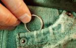
Everyone has been traumatized as a child. Often these are cuts.
The victim may not pay attention to the incised wound.
Use search
Is there any problem? Enter in the form "Symptom" or "Name of the disease" press Enter and you will find out all the treatment of this problem or disease.Adults are injured in everyday life, cut with knives, razors.
Damage with pus
 Every person in his life faced with purulent wounds. Treat such wounds 2 times a day.
Rinse with an antiseptic. The most suitable substances are chlorhexidine and peroxide.
It is good to use these 2 substances at the same time. You can use a weak solution of potassium permanganate. After you can process green.
A person consists in laying an ointment, for example:
- Levomikol.
- Levosin.
This procedure is preferably carried out in the morning and evening. This will get rid of pus - will promote rapid healing.
 It would seem, what can a cut on the skin lead to? The consequences of neglecting your body can be costly.
A cut can damage a vessel or nerve.
If a fresh wound is not treated, then germs will get there, and this leads to inflammation or even gangrene, followed by amputation.
With purulent - consult a doctor.
We treat at home
 We treat the wound at home:
- With household cuts and - it is important to remove dirt. This can be done under a stream of cold water, gently using soap.
- Disinfection. Often use hydrogen peroxide, brilliant green. If it happens that there are no medicines at hand, then you can use a saline solution.
- Cover the area with a band-aid or bandage. If the injury is severe and deep, see a doctor.
 Without a medical education, you can help a person with an open wound.
If the wound is small and clean, then after proper treatment, you will not need to see a doctor.
First, stop the bleeding. It's not always possible to stop bleeding. If the open wound is not deep, then it is enough to press this place.
But if the blood cannot be stopped and it has a rich scarlet color, then contact the medical staff. Just before that, you need to apply a tourniquet. Do not overtighten the tourniquet, it can harm - further interfere with processing.
If an artery is injured, then a tourniquet is applied above the site of injury by a centimeter, and if a vein, then lower.
Once the bleeding has stopped, disinfect the area. Everything is done with clean and processed hands. Hydrogen peroxide is a cleaner and disinfectant.

After peroxide treatment, you can treat the area around the damage with alcohol or brilliant green. Then you should apply a bandage. If there are no sterile bandages on hand, then any clean cloth will do.
A small wound needs to be looked after. If necessary, the first few days can be treated with saline.
Video
Disinfection after surgery
 Surgery is a major procedure that may involve the removal of non-viable tissue or foreign bodies to prevent infection.
The operation helps scarring - the speedy healing of tissues. After the operation, the wound is sutured. The wound after the operation is completely sterile - this is the key to rapid healing.
Clean postoperative wounds are treated with antiseptics, they include peroxide, chlorhexidine or furacilin solution.
Dressings are done daily until the stitches are removed. After treatment with an antiseptic, the edges of the damage are smeared with a solution of 70% alcohol or iodine. After treatment, you can lubricate the seam with ointment for speedy healing. When the procedures are completed, apply a bandage.
Make sure the dressing is dry and not wet. If the dressing gets wet, it should be changed. The postoperative wound must be monitored especially carefully to prevent infection.
head trauma
 There is a certain set of rules that will minimize the risk of infection.
Any damage to the soft tissues of the head must be washed and cleaned of visible dirt.
Foreign objects must be removed. Treat with hydrogen peroxide. If you're bleeding, then stop the bleeding.
Bleeding can be stopped by tamponing it with a clean bandage or using cotton.
Press the swab for ten minutes. If the blood does not stop, then the swab is pressed with a bandage for a while. In addition to cleaning the damage itself, it is important to treat the area around. It is necessary to shave off the hair and lubricate the edges with brilliant green or alcohol.

When the cleansing procedures are completed, apply a sterile dressing. If the damaged area hurts a lot, then it is permissible to apply cold to the bandage. This will relieve pain, swelling.
After any, it is advisable to consult a doctor, because the injury is much more dangerous than it seems visually.
shallow cut
 A cut is a very common household injury. After proper treatment, a shallow cut will soon stop bothering the victim.
How to properly treat a wound:
- Remove contamination.
- As visible dirt and objects have been removed, the place must be treated with peroxide or potassium permanganate solution. Can be treated with brilliant green or chlorhexidine. The use of any aggressive means is prohibited.
- Use a band-aid or bandage to cover the injury. If the injury is not large, then this can be limited.
Post-burn treatment
 A burn is an unpleasant injury that worries especially the first days. Rapid healing will depend on first aid for the burn.
The damaged area after the burn must be cooled. The first time after a burn, do not apply ointments to the damaged area of \u200b\u200bthe skin.
Cleanse the skin with ether, alcohol. If everything is done quickly, then the skin can quickly regenerate.
At first, it is permissible to apply lotions with antiseptic agents.
When time passes, you can apply ointments that have a healing effect.
These ointments include:
- Solcoseryl.
- "Rescuer".
They help tissue heal quickly, dry the damaged area so that it heals faster, and provide the skin with the building material for rapid regeneration.
Damage heals faster if it is properly monitored and treated properly. The body will fight itself, it is important to simply help it with the healing process.
When to go to the doctor
Minor abrasions, scratches and cuts can be treated independently at home, using the right tools for this and carrying out the necessary treatments in time.
You should consult a doctor in the presence of minor injuries only if, despite all the treatments, an inflammatory process has begun in the wound, and suppuration has appeared.
You can treat yourself without contacting a doctor only for shallow cuts, the length of which does not exceed 2 cm.
If you get a larger cut after the initial treatment, you should immediately consult a doctor, as suturing may be required.
If you receive serious and large wounds, you should contact the doctor immediately, it is important to provide the victim with the correct first aid before the ambulance arrives.
Possible consequences
Injury contamination is dangerous due to the penetration of anaerobic microbes. They do not need air, and they multiply quickly, causing dangerous complications. The danger is not exaggerated - gangrene will become a consequence of suppuration.
Traumatic (hemorrhagic) shock is a serious pathological condition that is life-threatening. It develops at the time of injury, without proper assistance will cause loss of consciousness and even death of the victim.
A seroma is a collection of purulent fluid due to inflammation. Exudate accumulates immediately, causing suppuration. It is necessary to pump out using a puncture or by making an additional incision.
A hematoma is a collection of blood clots under the skin. Appears if the bleeding was not stopped immediately. A comfortable environment for the accumulation of microbes additionally puts pressure on the tissues, infringing them.
Blood must be removed from the tissues, for this an additional incision is made or the blood is pumped out using a puncture.
Necrosis - appears due to damage to the work of blood vessels. Formed on the tissues around the cut. 2 types: wet and dry. Wet necrosis is removed immediately due to the accumulation of pus in deep tissues, dry necrosis does not need to be touched, it protects the skin from infection.
4.9 / 5 ( 10 votes)
1. BURNED WOUND
Description. In the right half of the frontal region, on the border of the scalp, there is a “P”-shaped (when the edges are brought together) wound, with a side length of 2.9 cm, 2.4 cm and 2.7 cm. In the center of the wound, the skin is exfoliated in the form flap in the area of 2.4 x 1.9 cm. The edges of the wound are uneven, up to 0.3 cm wide, bruised. The ends of the wound are blunt. Breaks 0.3 cm and 0.7 cm long extend from the upper corners, penetrating to the subcutaneous base. At the base of the flap there is a strip-like abrasion, 0.7x2.5 cm in size. Taking into account this abrasion, the entire damage has a rectangular shape, 2.9x2.4 cm in size. The right and upper walls of the wound are beveled, and the left one is undermined. Between the edges of the damage in the depth of the wound, tissue bridges are visible. The surrounding skin is not changed. In the subcutaneous base around the wound, there is a hemorrhage of dark red color, irregular oval shape, 5.6x5 cm in size and 0.4 cm thick.
DIAGNOSIS
Bruised wound of the right half of the frontal region.
2. BURNED WOUND
Description. In the right parietal-temporal part, 174 cm from the plantar surface and 9 cm from the anterior midline, in the area of 15x10 cm, there are three wounds (conventionally marked 1,2,3).
Wound 1. spindle-shaped, 6.5 x 0.8 x 0.7 cm in size. When the edges are brought together, the wound acquires a rectilinear shape, 7 cm long. The ends of the wound are rounded, oriented to 3 and 9 of the conventional clock face.
The upper edge of the wound is set to a width of up to 0.1-0.2 cm. The upper wall of the wound is bevelled, the lower one is undermined. The wound in the middle part penetrates to the bone.
Wound 2, located 5 cm down and 2 cm posterior to wound No. 1, has a star shape, with three rays oriented to 1. 6 and 10 of the conventional watch dial, 1.5 cm long, 1.7 cm and 0, 5 cm, respectively. The overall dimensions of the wound are 3.5x2 cm. The edges of the wound are set to the maximum width in the region of the anterior edge - up to 0.1 cm, the posterior edge - up to 1 cm. The ends of the wound are sharp. The front wall is undermined, the back is bevelled.
Wound 3 is similar in shape to wound No. 2 and is located 7 cm above and 3 cm anterior to wound No. 1. The length of the rays is 0.6, 0.9 and 1.5 cm. The total dimensions of the wound are 3x1.8 cm. Edges the wounds are planted to the maximum width in the region of the anterior margin - up to 0.2 cm, the posterior margin - up to 0.4 cm.
All wounds have uneven, raw, crushed, bruised edges, and tissue bridges at the ends. The outer boundaries of sedimentation are clear. The walls of the wounds are uneven, bruised, crushed, with intact hair follicles. The greatest depth of wounds is in the center, up to 0.7 cm at wound No. 1 and up to 0.5 cm at wounds No. 2 and 3. The bottom of wounds No. 2 and 3 is represented by crushed soft tissues. In the subcutaneous base around the wounds of hemorrhage, irregular oval shape, 7x3 cm in size at wounds N 1 and 4 x 2.5 cm at wounds N 2 and 3. The skin around the wounds (outside the sedimentation of the edges) is not changed.
DIAGNOSIS
Three bruised wounds of the right parietotemporal part of the head.
3. laceration
Description. On the right half of the forehead, 165 cm from the level of the plantar surface of the feet and 2 cm from the midline, there is a wound of irregular fusiform shape, 10.0 x 4.5 cm in size, with a maximum depth of 0.4 cm in the center. The length of the damage is located respectively 9-3 of the conventional clock face. When comparing the edges, the wound acquires an almost rectilinear shape, without a tissue defect, 11 cm long. The ends of the wound are sharp, the edges are uneven, without sedimentation. The skin along the edges of the wound is unevenly exfoliated from the underlying tissues to a width of up to: 0.3 cm - along the upper edge; 2 cm - along the bottom edge. In the formed "pocket" a flat dark red blood clot is determined. Hair along the edges of the wound and their bulbs are not damaged. The walls of the wound are sheer, uneven, with small focal hemorrhages. Between the edges of the wound in the region of its ends there are tissue bridges. The bottom of the wound is the partially exposed surface of the scales of the frontal bone. The length of the wound at the level of its bottom is 11.4 cm. Parallel to the length of the wound, a finely serrated edge of a fragment of the frontal bone protrudes 0.5 cm into its lumen, on which there are small focal hemorrhages. Around the wound on the skin and in the underlying tissues, no damage was found.
DIAGNOSIS
Rupture on the right side of the forehead.
4. BITED SKIN DAMAGE
Description. On the anteroexternal surface of the upper third of the left shoulder in the region of the shoulder joint, there is an unevenly pronounced red-brown annular sedimentation of an irregular oval shape measuring 4x3.5 cm, consisting of two arcuate fragments: upper and lower.
The upper fragment of the exudation ring has dimensions of 3x2.2 cm and a radius of curvature of 2.5-3 cm. It consists of 6 banded unevenly pronounced abrasions ranging in size from 1.2x0.9 cm to 0.4x0.3 cm, partially connected to each other. The maximum dimensions are located in the centrally located abrasions, the minimum - along the periphery of the sedimentation, especially at its upper end. The length of the abrasions is directed mainly from top to bottom (from the outer to the inner border of the semi-oval). The outer edge of the sedimentation is well pronounced, has the form of a broken line (step-shaped), the inner edge is sinuous, indistinct. The ends of the subsidence are U-shaped, the bottom is dense (due to drying), with an uneven striped relief (in the form of ridges and furrows running from the outer border of the semi-oval to the inner one). Precipitation has a greater depth (up to 0.1 cm) at the upper edge.
The lower fragment of the ring has dimensions of 2.5x1 cm and a radius of curvature of 1.5-2 cm. Its width is from 0.3 cm to 0.5 cm. on its left side. Here, the inner edge of sedimentation has a sheer or somewhat undermined character. The ends of the upsetting are U-shaped. The bottom is dense, grooved, deepest at the left end of the sedimentation. The bottom relief is uneven, there are 6 sinking sections located in a chain along the course of the abrasion, irregularly rectangular in size from 0.5 x 0.4 cm to 0.4 x 0.3 cm and up to 0.1-0.2 cm deep.
The distance between the inner boundaries of the upper and lower fragments of the “ring” of sedimentation is: on the right - 1.3 cm; in the center - 2 cm; on the left - 5 cm. The axes of symmetry of both semirings coincide with each other and correspond to the long axis of the limb. In the central zone of annular sedimentation, a blue bruise of an irregular oval shape, 2 x 1.3 cm in size, with fuzzy contours, is determined.
DIAGNOSIS
Abrasions and bruising on the anteroexternal surface of the upper third of the left shoulder.
5. CUT WOUND
Description. On the flexor surface of the lower third of the left forearm, 5 cm from the wrist joint, there is a wound (conventionally designated N 1) of an irregular fusiform shape, 6.5 x 0.8 cm in size, with the edges brought together - 6.9 cm long. From the outer (left) of the end of the wound, parallel to its length, there are 2 incisions, 0.8 cm long and 1 cm long with smooth edges ending in sharp ends. At 0.4 cm from the lower edge of the wound No. 2, parallel to its length, there is a superficial intermittent incision 8 cm long. The bottom of the wound at its inner (right) end has the greatest steepness and a depth of up to 0.5 cm.
2 cm down from the first wound there is a similar wound No. 2), 7x1.2 cm in size. The length of the wound is oriented horizontally. When the edges are reduced, the wound acquires a rectilinear shape, 7.5 cm long. Its edges are wavy, without sedimentation and crushing. The walls are relatively smooth, the ends are sharp. At the inner (right) end of the wound, parallel to the length, there are 6 skin incisions from 0.8 to 2.5 cm long, at the outer end - 4 incisions, from 0.8 to 3 cm long. The bottom is represented by dissected soft tissues and has the greatest steepness and the depth at the outer (left) end of the wound is up to 0.8 cm. In the depth of the wound, a vein is visible, on the outer wall of which there is a through damage of a spindle-shaped shape, 0.3x0.2 cm in size.
In the tissues surrounding both wounds, in an oval-shaped area measuring 7.5x5 cm, there are multiple dark red hemorrhages merging with each other, of irregular oval shape, ranging in size from 1x0.5 cm to 2x1.5 cm with uneven fuzzy contours.
DIAGNOSIS
Two incised wounds of the lower third of the left forearm.
6. STICK WOUND
Description.
On the left half of the back, 135 cm from the plantar surface of the feet, there is an irregular spindle-shaped wound measuring 2.3 x 0.5 cm. After closing the edges, the wound has a rectilinear shape 2.5 cm long. The edges of the wound are even, without sedimentation and bruising. The right end is U-shaped, 0.1 cm wide, the left end is in the form of an acute angle. The skin around the wound is free of damage and contamination.
On the posterior surface of the lower lobe of the left lung, 2.5 from its upper edge, a slit-like lesion is located horizontally. When the edges are brought together, it acquires a rectilinear shape, 3.5 cm long. The edges of the damage are even, the ends are sharp. The lower wall of the damage is bevelled, the upper one is undermined. On the inner surface of the upper lobe of the lung at the root, 0.5 cm of the damage described above, there is another (slit-like shape with smooth edges and sharp ends). There are hemorrhages along the wound channel.
Both injuries are connected by a straight single wound channel, having a direction from back to front and from bottom to top (provided that the body is in the correct vertical position). The total length of the wound channel (from the wound on the back to the damage to the upper lobe of the lung) is 22 cm.
DIAGNOSIS
Stab-cut blind wound of the left half of the chest, penetrating into the left pleural cavity, with penetrating damage to the lung.
7. CHOPPED WOUND
Description. On the anterior-internal surface of the lower third of the right thigh, 70 cm from the plantar surface of the feet, there is a gaping wound of irregular fusiform shape, 7.5x1 cm in size. After closing the edges, the wound takes a rectilinear shape, 8 cm long. smooth. One end of the wound is U-shaped, 0.4 cm wide, the other is in the form of an acute angle. The wound channel has a wedge-shaped shape and the greatest depth of up to 2.5 cm at its U-shaped end, ends in the muscles of the thigh. The direction of the wound channel is from front to back, from top to bottom and from left to right (subject to the correct vertical position of the body). The walls of the wound channel are even and relatively smooth. In the muscles around the wound channel, hemorrhage of an irregular oval shape, 6x2.5x2 cm in size.
On the anterior surface of the internal condyle of the right femur, a wedge-shaped lesion is 4x0.4 cm in size and up to 1 cm deep; The upper end of the damage is U-shaped, 0.2 cm wide, the lower end is sharp. The edges of the damage are even, the walls are smooth.
DIAGNOSIS
Chopped wound of the right thigh with an incision in the medial condyle of the femur.
8. FIRE BURN
Description. On the left half of the chest there is a red-brown wound surface, of an irregular oval shape, measuring 36 x 20 cm. The area of the burn surface, determined according to the “palms” rule, is 2% of the entire surface of the victim’s body. The wound is covered in places with a brownish scab, dense to the touch. The edges of the wound are uneven, coarse and finely wavy, somewhat elevated above the level of the surrounding skin and wound surface. The greatest depth of the lesion is in the center, the smallest - along the periphery. Most of the burn surface is represented by the exposed subcutaneous base, which has a wet, shiny appearance. In places, red small-focal hemorrhages are determined, oval in shape, ranging in size from 0.3 x 0.2 cm to 0.2 x 0.1 cm, as well as small thrombosed vessels. In the central part of the burn wound, there are separate areas covered with greenish-yellow purulent deposits, which alternate with pinkish-red areas of young granulation tissue. Soot deposits are determined in places on the wound surface. The vellus hairs in the wound area are shorter, their ends are swollen in a “flask-like” manner. When dissecting a burn wound in the underlying soft tissues, a pronounced edema is determined in the form of a gelatinous yellowish-gray mass, up to 3 cm thick in the center.
DIAGNOSIS
Thermal burn (by flame) of the left half of the chest, III degree, 2% of the body surface.
9. HOT WATER BURN
Description. On the anterior surface of the right thigh there is a burn wound of an irregular oval shape, 15x12 cm in size. The main part of the burn surface is represented by a group of confluent blisters containing a cloudy yellowish-gray liquid. The bottom of the blisters is a uniform pink-red surface of the deep layers of the skin. Around the zone of blisters there are areas of skin with a soft, moist, pinkish-reddish surface, on the border of which there are zones of peeling of the epidermis with its membranous exfoliation up to 0.5 cm wide. The edges of the burn wound are coarse and finely wavy, somewhat raised above the level of the surrounding skin, with "linguistic" protrusions, especially downwards (provided that the thigh is in the correct vertical position). Vellus hair in the area of the wound is not changed. When dissecting a burn wound in the underlying soft tissues, a pronounced edema is determined in the form of a gelatinous yellowish-grayish mass, up to 2 cm thick in the center.
DIAGNOSIS
Thermal burn with a hot liquid of the anterior surface of the right thigh II degree 1% of the body surface.
10. THERMAL FIRE BURN IV DEGREE
In the area of the chest, abdomen, buttocks, external genitalia and thighs, there is a continuous burn wound of irregular shape with wavy uneven edges. Borders of the wound: on the chest on the left - subclavian region; on the chest on the right - costal arch; on the back on the left - the upper part of the scapular region; on the back on the right - the lumbar region; on the legs - the right knee and the middle third of the left thigh. The wound surface is dense, red-brown, sometimes black. On the border with intact skin, there is a strip-like redness up to 2 cm wide. Vellus hair in the wound area is completely singed. On cuts in the underlying soft tissues, there is a pronounced gelatinous yellow-gray edema up to 3 cm thick.
11. LIGHTNING BURN
In the occipital region in the center there is a round dense light gray scar 4 cm in diameter with thinning of the skin, soldered to the bone. The borders of the scar are even, rise like a roller in the transition to intact skin. There is no hair in the scar area. Internal examination: The thickness of the scar is 2-3 mm. There is a round defect of the outer bone plate and spongy substance 5 cm in diameter with a flat, relatively flat and smooth, similar to a "polished" surface. The thickness of the bones of the cranial vault at the cut level is 0.4-0.7 cm, in the area of the defect the thickness of the occipital bone is 2 mm, the internal bone plate is not changed.
Penetrating injuries, wounds penetrating into cavities
12. STICK WOUND
Description. On the left half of the chest, along the midclavicular line in the IV intercostal space, there is a longitudinally located wound, of an irregular fusiform shape, measuring 2.9x0.4 cm. The upper part of the wound is of a rectilinear shape, 2.4 cm long; the lower one is arc-shaped, 0.6 cm long. The edges of the wound are even and smooth. The upper end of the wound is U-shaped, 0.1 cm wide, the lower end is sharp.
The wound penetrates the pleural cavity with damage to the left lung. The total length of the wound channel is 7 cm, its direction is from front to back and somewhat from top to bottom (with
condition of the correct vertical position of the body). There are hemorrhages along the wound channel.
DIAGNOSIS
Stab-cut wound of the left half of the chest, penetrating into the left pleural cavity with damage to the lung.
13. GUN SHORT THROUGH BULLET WOUND
On the chest, 129 cm from the level of the soles, 11 cm below and 3 cm to the left of the sternal notch, there is a wound of a rounded shape 1.9 cm with a tissue defect in the center and a circular belt of sediment along the edge, up to 0.3 cm wide. The edges of the wound uneven, scalloped, the lower wall is slightly beveled, the upper is undermined. In the bottom of the wound, the organs of the chest cavity are visible. On the lower semicircle of the wound, the imposition of soot in a semilunar area, up to 1.5 cm wide. On the back, 134 cm from the level of the soles, in the region of the 3rd left rib, 2.5 cm from the line of the spinous processes of the vertebrae, there is a wound forms (without a defect in the fabric) 1.5 cm long with uneven, finely patchwork edges, turned inside out and rounded ends. A white plastic fragment of the cartridge container will protrude from the bottom of the wound.
Examples of fracture fracture descriptions:
14. BROKEN RIB
On the 5th rib on the right between the angle and the tubercle, 5 cm from the articular head, there is an incomplete fracture. On the inner surface, the fracture line is transverse, with even, well-matched edges, without damage to the adjacent compact substance; the fracture zone is slightly gaping (signs of sprain). Near the edges of the rib, this line bifurcates (in the region of the upper edge at an angle of about 100 degrees, near the lower edge at an angle of about 110 degrees). The resulting branches pass to the outer surface of the rib and gradually, thinning, are interrupted near the edges. The edges of these lines are finely serrated and not densely comparable, the fracture walls are slightly sloping in this place (signs of compression.)
15. MULTIPLE RIB FRACTURES
The ribs 2-9 were broken along the left mid-axillary line. Fractures are of the same type: on the outer surface, the lines of fractures are transverse, the edges are even, tightly comparable, without damage to the adjacent compact (signs of stretching). On the inner surface, the lines of fractures are oblique-transverse, with coarsely serrated edges and small flakes and visor-shaped bendings of the adjacent compact substance (signs of compression). From the zone of the main fracture along the edge of the ribs, there are longitudinal linear splittings of the compact layer, which become hairy and disappear. 3-8 ribs are broken along the scapular line on the left with the same signs of compression on the outer and stretching on the inner surfaces as described above.
We process the wound on the head correctly.
Wounds on the head are usually divided into cut, stab, torn. They can appear as a result of injury from a blow, fall, bruise. The victim needs to be given first aid and brought to the traumatology department.
How to properly treat a head wound
A wound is a violation of the integrity of the skin or mucous membranes. It can be superficial or deep, cut or torn. Regardless of the severity of the lesion, the wound must be carefully treated.
What you need to treat a wound
- alcohol;
- brilliant green or iodine;
- chlorhexidine;
- hydrogen peroxide;
- potassium permanganate;
- package;
- heating pad;
- sterile gauze;
- bandage.
Preparation for the procedure
Before giving first aid, thoroughly wash your hands and treat them with rubbing alcohol or any other alcohol-containing liquid to prevent infection from entering the wound. It is necessary to clean the wound on the head with a sterile gauze swab. You should not use cotton wool, its particles can remain in the wound, which will provoke additional complications. When the scalp is damaged, you need to cut the hair around at a distance of two centimeters, wash the damaged area with chlorhexidine, 3% hydrogen peroxide or a weak solution of potassium permanganate.
Wound treatment
Around the wound, you need to generously lubricate the skin with alcohol, brilliant green, iodine, a saturated solution of potassium permanganate. It is important to ensure that drugs do not get into the damaged area, as they can cause tissue burns, which will seriously complicate the process of further healing.
When the bleeding doesn't stop
If the blood flow is plentiful, you need to independently attach a gauze sterile swab to the wound site. Then apply a pressure bandage. To reduce swelling, pain, stop bleeding, an ice pack or a heating pad filled with cold water should be applied to the bandage. As the water starts to warm up, change the heating pad. This is especially true for the warm season, when the path to the traumatology department takes a long time.
What to do with foreign objects in the wound
Such objects that are deep in the wound do not need to be removed by yourself. Doing this is very dangerous, as bleeding can increase. Only a qualified traumatologist or surgeon can carry out manipulations to cure foreign objects.
Don't neglect the emergency
Regardless of the degree of damage to the head, immediately call an ambulance or take the victim to the nearest traumatology department. In the case of a deep wound, there is a danger that the membranes of the brain will become inflamed, which sometimes leads to death, so even a slight delay in providing specialized medical care can cost the patient his life. See also: Cuts: how to act so as not to harm
Advice 1: How to treat a wound on the head.
Wounds on the head are divided into stab, cut, torn. They can appear as a result of injury when falling, hitting, bruising. The victim must be given first aid and taken to the traumatology department.

- How to treat a head wound
- How to treat a wound with iodine
- How to handle stitches after surgery
- - alcohol;
- - iodine;
- - greenery;
- - hydrogen peroxide;
- - chlorhexidine;
- - potassium permanganate;
- - ice;
- - package;
- - warmer;
- - sterile gauze;
- - bandage.
- head wound treatment
Tip 2: How to treat lacerations.

- how to treat lips
Tip 3: How to treat a wound to a cat.

- - sterile bandage or gauze;
- - scissors;
- - cotton swab;
- - greenery;
- - iodine;
- - vodka or alcohol;
- - tweezers;
- - 3% hydrogen peroxide solution;
- - streptocide tablet;
- - vaseline;
- Injuries in cats. How to treat wounds in a cat.
- how to treat a cat
Tip 4: How to treat a cut wound.

- - soap, alcohol-containing liquid;
- - potassium permanganate or hydrogen peroxide;
- - alcohol, iodine or brilliant green;
- - antiseptic ointment;
- - sodium chloride, furatsilin or antibiotics;
- - gauze swab, bandage, sterile gauze.
- Treatment and treatment of incised wounds
- how to treat a wound after a cut
Advice 5: How to treat a wound after castration of a cat.

The cat must be properly prepared for castration. During the operation, the bladder and digestive tract of the animal must be empty, therefore, 12 hours before castration, the cat cannot be fed, and even watered an hour before.
Wound treatment
If the doctor after the operation treated the wound with Terramycin or Alumazol spray, they remain on the skin for some time, in this case it is not necessary to treat the wound. If there was no such treatment, the wound must be washed with 3% hydrogen peroxide or furatsilin solution, dissolving one tablet in a glass of water. It is not recommended to treat the wound with brilliant green or an alcohol solution of iodine, they can dry out the skin.
So that the cat does not disturb the wound by licking it, he needs to put on a special collar around his neck, which will prevent him from reaching the back of the body. The collar is taken off only when eating. It is necessary to ensure that the cat does not rub against the floor with the back of the body.
The tray filler used at this time should be soft so as not to disturb the wound. It is better if it is white or at least a light shade, in which case the owners will be able to immediately notice the bleeding that has begun.
Possible Complications
The owners should be alerted by the increase in body temperature of the animal. The normal temperature for a cat is 38-39°C. In the first three days, it will inevitably be elevated, but if the temperature has not decreased on the fourth day, this is an occasion for an urgent appeal to the veterinarian. Moreover, you need to show the animal to the doctor if the wound began to fester. In this case, the veterinarian will prescribe an antibiotic.
On the first day after the operation, a decrease in temperature (less than 37 degrees) can also be observed, while the animal is sleeping. The cat needs to be warmed up by applying a heating pad and rubbing its paws. If this does not help, the cat still does not move and does not wake up, it is urgent to call a veterinarian or take the cat to the clinic.
It is also necessary to take the cat to the clinic if the suture begins to bleed.
After castration, the cat may suffer from constipation. Stool retention is inevitable during the first two to three days after anesthesia, but if the cat has no stool for more than four days, it is necessary to start giving him a laxative. Of course, this cannot be done without first consulting with the veterinarian, only he can choose the appropriate drug, taking into account the state of health and the characteristics of the body of a particular animal.
Injuries and wounds of the soft tissues of the head, symptoms and treatment.
Damage to the soft integument of the skull are closed and open. Bruises are closed, wounds (wounds) are open. Bruises occur as a result of hitting the head against hard objects, hitting the head with a hard object, when falling, etc.
As a result of the impact, the skin and subcutaneous tissue are damaged. From damaged blood vessels, blood flows into the subcutaneous tissue. When galea aponeurotica is intact, the outflowing blood forms a limited hematoma in the form of a protruding swelling (bump).
With more extensive damage to the soft tissues, accompanied by a rupture of the galea aponeurotica, the blood that has poured out of the damaged vessels forms a diffuse swelling. These extensive hemorrhages (hematomas) are soft in the middle and sometimes give a feeling of unsteadiness (fluctuation). These hematomas are characterized by a dense shaft around the hemorrhage. When feeling a dense shaft along the circumference of the hemorrhage, it can be mistaken for a skull fracture with pressure. A thorough examination, as well as an x-ray, makes it possible to correctly recognize the damage.
Wounds of the soft tissues of the head are observed as a result of injury from both sharp and blunt instruments (blunt violence). Injury to the soft integument of the skull is dangerous because local infection can spread to the contents of the skull and lead to meningitis, encephalitis and brain abscess, despite the integrity of the bone, due to the connection between the superficial veins and the veins inside the skull. The infection can also spread through the lymphatic vessels. Simultaneously with the injury of soft tissues, the bones of the skull and the brain can be damaged.
Symptoms. Symptoms depend on the nature of the injury. Cut and chopped wounds bleed heavily and gape. Stab wounds bleed a little. In the absence of infection complications, the course of wounds is favorable. If the wound was treated in the first hours, it may heal by first intention.
Symptoms of bruised wounds correspond to the nature of the wound. The edges of the bruised wound are uneven, with traces of a bruise (crush), soaked in blood, in some cases they are detached from the bone or underlying tissues. Bleeding is less abundant due to thrombosis of crushed and ruptured vessels. Contusion wounds can penetrate to the bone or be limited to soft tissue damage. A characteristic sign of lacerations is a significant detachment from the underlying bones and the formation of flaps.
A special type of damage to the scalp is the so-called scalping, in which a larger or smaller part of the scalp is torn off.
Treatment. In most cases, after careful pre-treatment of the wound itself and adjacent areas, it is sufficient to apply sutures to the wound, and for small wounds, a pressure bandage. In case of severe bleeding, the bleeding vessels should be tied off. Only a fresh, uncontaminated wound can be sutured. If the wound is contaminated, the objects that have fallen into the wound are removed with tweezers, the edges of the wound are lubricated with a solution of iodine tincture, the edges of the wound are refreshed (the primary treatment of the wound is performed), a solution of penicillin is poured into the wound (50,000-100,000 IU in a 0.5% solution of novocaine) or infiltrated with a solution penicillin wound edges, after which the wound is completely or partially sutured. In the latter case, the graduate is injected under the skin. After the inflammatory process subsides, a secondary suture can be applied to the wound. In some cases, intramuscular injection of a solution of penicillin is prescribed. If the wound is completely sewn up, and signs of inflammation appear in the following days, the sutures should be removed and the wound opened.
For the purpose of prophylaxis, anti-tetanus serum is administered to all wounded, and in case of severe wounds, especially those contaminated with earth, anti-gangrenous serum.
Care. Hair on the head contributes to pollution and makes it difficult to treat the skin and wound, and therefore it should be shaved as much as possible around the wound. When shaving, care must be taken not to introduce infection into the wound - it should be covered with a sterile napkin. Shaving is performed from the wound, not to the wound.
wound healing is a complex process consisting of several intersecting phases: inflammation, proliferation and remodeling. Each phase has its specific role and its specific features at the molecular and tissue levels. Healing can occur by primary, secondary and tertiary intention. Each type of healing has its advantages and disadvantages, the choice of healing method depends on the wound and on the characteristics of the process in each individual patient.
a) Epidemiology. Wounds can occur due to a variety of reasons, the most common of which are trauma and surgery. It is not possible to calculate the exact ratio of the causes of wounds.
b) Terminology. The wound healing process consists of three overlapping phases. The initial phase of wound healing is the inflammatory phase, which begins immediately after tissue damage. It is characterized by gradual wound closure and migration of inflammatory components of the immune system. In the proliferation phase, a stable wound matrix is formed, and granulation tissue is formed in the healing wound. In the remodeling phase, which lasts up to two years, the scar matures and strengthens.
Granulation tissue is new emerging tissue consisting of fibroblasts and developing blood vessels. Healing by primary intention occurs when primary sutures are applied, as a result of which the “dead space” is eliminated, and the wound surface quickly re-epithelializes. If the wound heals on its own, without any surgical intervention, the process is called healing by secondary intention. In infected wounds, secondary sutures are applied and the wound heals by tertiary intention. Infected wounds require daily care, and when the infection is resolved, the edges of the wound can be brought together surgically.
Wounds can capture all layers of tissue. Soft tissues include the skin and subcutaneous tissues (adipose tissue, muscles, nerves, blood vessels). More complex injuries are combined with damage to the cartilage and bones of the facial skeleton.
in) The course of wound healing:
1. Etiology. In the vast majority of cases, wounds occur as a result of trauma and surgical interventions.
2. Pathogenesis. In the absence of proper care, the outcome of the healing of open wounds can be unfavorable. Open wounds can become infected, causing tissue destruction and delaying the healing process. Also, wounds that are contaminated and covered with dry crusts heal worse, since in these cases the migration of the epithelium to the edges of the wound is disturbed. Unfavorable wound healing can lead not only to the formation of a rough scar, but also to functional impairment, for example, eyelid retraction or difficulty in nasal breathing if the wound is located near the eye or nose, respectively.
3. natural course of the process. During the inflammatory phase, a clot formed from the bleeding tissue closes the wound. This process is accompanied by primary vasoconstriction, which is then replaced by controlled vasodilation, during which platelets and fibrin migrate to the wound. The clot also protects the wound from the environment and contamination. Inflammatory cells that have migrated into the wound release a number of cytokines and immune factors that further regulate the healing process. These include fibroblast growth factor (FGF), platelet-derived growth factor (PDGF), transforming growth factors (TGFs).
Gradually formed fibronectin matrix on which proteins and cell complexes are subsequently deposited. The immune cells entering the wound bed, neutrophils and monocytes, are involved in phagocytosis. On the periphery of the wound, the migration of epithelial cells begins already 12 hours after the injury. This process is accompanied by flattening of epithelial cells and the formation of pseudopodia. In sutured wounds, the re-epithelialization process can be completed within 48 hours. Depending on the size and degree of contamination of the wound, the inflammatory phase lasts 5-15 days. Clinically, the processes described above are manifested by edema and inflammation.
During proliferative phase there is a regeneration of cellular structures inside the wound. At this time, there is an active proliferation of fibroblasts, accompanied by the deposition of collagen, and the formation of granulation tissue, consisting of inflammatory cells and new blood vessels. The clinically yellowish fibrinous plaque is gradually replaced by a clear red granulation tissue.
Remodeling phase starts after a few weeks. This is the longest phase, taking up to two years after the injury. The deposition of collagen continues, its fibers intersect, become thicker. Type III collagen is gradually replaced by type I collagen, which ensures the formation of a stronger scar. The cellular composition also undergoes changes that provide long-term maintenance of tissue integrity. For example, fibroblasts differentiate into myofibroblasts, contributing to wound contraction. Blood vessels slowly regress; clinically, this process is accompanied by the disappearance of hyperemia and the appearance of a mature scar of typically white color.
4. Possible Complications. If left untreated, the wound can become infected, resulting in healing resulting in the formation of a cosmetically unsatisfactory scar. If the large vessels of the face and neck are damaged, serious bleeding can occur. Unrecognized trauma to the facial nerve can lead to permanent paralysis. Damage to the parenchyma or duct of the parotid salivary gland may result in the formation of a salivary-cutaneous fistula or sialocele.
1. Complaints. If the wound is in the healing stage, patients usually complain of pain and discomfort. Deeper wounds to the face and neck may also be accompanied by dysfunction of the nerves or salivary glands. Sometimes patients do not attach importance to them, so the doctor must be careful to detect them. Damage to the bones of the facial skeleton can lead to additional complaints, such as diplopia in explosive orbital fractures, or malocclusion in fractures of the mandible or midface.
2. Survey. In most patients with soft tissue wounds, additional examination methods are not required. Penetrating head and neck injuries should alert the clinician to major vessel injury requiring CT angiography. In case of any bone injuries, it is necessary to perform CT scan. If surgical suturing of the wound is necessary, the main blood parameters (hemoglobin, electrolytes, indicators of the coagulation system) are determined.
3. Differential Diagnosis. The cause of an injury can often be identified at the patient's initial presentation. It is essential that when managing a patient with soft tissue injuries, the physician can formulate a “reconstructive algorithm”, which is a concept for the treatment of patients with soft tissue injuries. The algorithm starts with the simplest methods, and then gradually moves to the most complex ones.
 Areas of the face where wound healing is optimal by secondary intention.
As the complexity increases, reconstructive algorithm includes the following steps:
1. Wound healing without surgery (secondary intention)
2. Wound healing with delayed suturing (tertiary tension)
3. Simple wound closure (primary intention)
4. Complex wound closure with plasty with local tissues (primary intention)
5. Skin grafts
6. Comprehensive treatment using distant tissues (regional or free flaps).
e) Healing prognosis of head and neck wounds. The correct analysis of the existing wound and the choice of an appropriate method of treatment usually reduce the risk of a rough scar. Some wounds may require repeat surgery to achieve optimal results. First of all, the prognosis is influenced by the desire of both the patient and the surgeon to make every effort to promote favorable wound healing.