Chondroprotectors evidence-based medicine. Joint restoration - are chondroprotectors and myths about chondroitin needed? A bag that is always with you
V.V. KOSAREV, Doctor of Medical Sciences, Professor, S.A. BABANOV, MD SBEE HPE "Samara State Medical University" of the Ministry of Health of Russia
EFFICIENCY OF MODERN CHONDROPROTECTORS
IN OSTEOARTHRITIS
Keywords: osteoarthritis, articular cartilage, chondrocytes, delayed-acting symptom-modifying drugs, chondroitin sulfate, glucosamine
Osteoarthritis (OA) is a group of joint diseases of various etiologies that cause the destruction of cartilage and gradually lead to its loss.
Osteoarthritis occupies a leading position among diseases of the musculoskeletal system. According to WHO, more than 4% of the world's population suffers from osteoarthritis, in 10% of cases it is the cause of disability, worsening the quality of life and social adaptation of patients. Among people aged 65 years, the frequency of OA is 50%, over 75 years old - reaches 80%. The incidence of osteoarthritis in the Russian Federation is 580 per 100 thousand population. OA occupies up to 70% in the structure of rheumatic diseases. The prevalence of osteoarthritis is predicted to double in 10 years.
Osteoarthritis is currently regarded as a chronic progressive disease of the synovial
joints, which develops as a result of a complex set of biomechanical, biochemical and / or genetic factors. Degenerative dystrophic changes in OA are based on primary cartilage damage followed by an inflammatory response. The pathological process in OA involves not only the articular cartilage, but also the subchondral bone, ligaments, joint capsule, synovium, and periarticular muscles. OA is always associated with bone deformity, which is why it is also called deforming arthrosis.
The main clinical symptoms of OA are pain and deformity of the joints, which lead to their functional insufficiency. There are localized (with damage to one joint) and generalized forms of osteoarthritis (polyosteoarthrosis).
If the cause of the development of the disease is not established, osteoarthritis is called primary, or idiopathic.
OA can occur as a result of the interaction of various internal (age, female sex, menopause in women, developmental defects, hereditary predisposition) and external factors (trauma, excessive loads), which leads to damage to the articular cartilage and / or underlying bone tissue. The disease can be the result of both a single severe intra-articular injury and prolonged microtrauma to the joint. Excess weight occupies a special place among the risk factors for developing OA. So, in women with obesity, osteoarthritis of the knee joints develops 4 times more often compared to women with normal weight. It has been established that overweight is not only a risk factor for OA, but also contributes to its more rapid progression, up to disability. The occurrence of secondary osteoarthritis always has a specific cause.
ETIOLOGY AND RISK FACTORS OF OSTEOARTHRITIS
Risk factors for primary osteoarthritis ■ Older age ■ Female gender ■ Physical activity ■ Overweight ■ Past trauma ■ Hormone replacement therapy ■ Vitamin D deficiency ■ Smoking ■ Deformation of the articular surfaces ■ Weakness of the quadriceps femoris muscle, intense physical activity Causes of secondary osteoarthritis ■ Post-traumatic ■ Congenital , acquired diseases or endemic diseases: Perthes' disease, hypermobility syndrome, etc. ■ Metabolic diseases: ochronosis, hemochromatosis, Wilson's disease, Gaucher's disease ■ Endocrinopathy: acromegaly, hyperparathyroidism, diabetes mellitus, hypothyroidism ■ Calcium deposition disease ■ Nephropathy (Charcot's disease)
Articular cartilage is a highly specialized tissue consisting of a matrix containing two main macromolecules - glycosaminoglycans (proteoglycans) and collagen, and chondrocytes immersed in the matrix. Normally, the synthesis and degradation of cartilage in the joint are in a balanced state. However, under the influence of various risk factors, this balance is disturbed, as a result of which the processes of cartilage tissue destruction begin to go faster than the recovery processes. Further, a vicious circle is launched: the destruction of cartilage leads to inflammation, which, in turn, enhances the destructive processes in the joint. Currently, it is believed that the initiating role in the development of OA belongs to the subchondral bone: microfractures of the trabeculae and the production of cytokines trigger cartilage damage.
The high concentration of proteoglycans in the matrix provides an even distribution of the load that affects the cartilage and restoration of the shape after the load has ceased. With a decrease in the amount of glycosaminoglycans, the resistance of the cartilage matrix to the effects of physical activity decreases and the cartilage surface becomes susceptible to damage.
PATHOGENESIS OF OSTEOARTHROSIS
Etiological factors (genetic, mechanical, etc.)
Dysfunction of chondrocytes
Hyperproduction of cytokines (K-1p, TNF-a), overexpression of COX-2, transcription factor NF-kB, impaired synthesis of TGF-p
Activation of synovial fluid enzymes
Degradation of proteoglycans and collagen
Education
antibodies to collagen and proteoglycans
Subchondral bone remodeling
Osteoarthritis
Impaired function
The main clinical manifestations of osteoarthritis are pain and deformity of the joints. The disease begins with the appearance of a slight pain in the joints experiencing the greatest load (knee, hip, metatarsophalangeal joint of the first toe). First, one joint is affected, then others join. With the progression of the pathological process, pain appears not only after physical exertion, but also at night, the so-called pain may occur. meteosensitivity - a change in the intensity of pain depending on temperature, air humidity and atmospheric pressure, which affects the pressure in the joint cavity. As OA progresses, pain may occur (or increase) when changing body position, getting up from a chair, going down stairs. The appearance of pain at night indicates active
CLINICAL PICTURE AND STAGES OF OSTEOARTHRITIS
Joint pain
Crepitus in the affected joint
Clinical picture of OA
Joint deformity
Limited mobility in the affected joint
visualization of inflammation in the joint. The pain is often associated with morning stiffness, and there may be crepitus in the affected joint. The development of synovitis is accompanied by swelling of the joint, an increase in skin temperature above it. With the appearance of a reflex muscle spasm, there may be a restriction of movements in the affected joint up to the formation of tendon-muscle contractures. With a long course of the disease, Heberden's nodules appear on the distal interphalangeal joints, Bouchard's nodules appear on the proximal interphalangeal, as well as on the I metatarsophalangeal, knee and other joints.
Stages of osteoarthritis (according to J. Kellgren and J. Lawrence, 1952)
0 - no radiological signs
1 - doubtful radiological signs
II - minimal changes (slight narrowing of the articular
gaps, single osteophytes)
III - moderate manifestations (moderate narrowing of the articular
fissures, multiple osteophytes)
IV - pronounced changes (the articular space is almost not traced
alive, gross osteophytes are detected)
The course of osteoarthritis is variable. In some cases, despite the progression of radiological manifestations of the disease, the condition of patients remains stable for many years. Pain intensifies gradually against the background of the development of deformity and stiffness of the joints. Limitation of movements in the joints for a long time remains not so significant. Periodically, under the influence of provoking factors (cooling, respiratory infection), reactive synovitis occurs, the relapses of which become more frequent with the duration of the course of the disease. With severe osteoarthritis, “blockades” of the joints may occur.
Allocate osteoarthritis with rapid generalization of the process and osteoarticular destruction (erosive arthrosis). This course is more often observed in polyosteoarthrosis with the presence of Heberden's nodules and hereditary predisposition, as well as in women during menopause.
In men with a more powerful ligamentous-muscular apparatus, there is a milder course of osteoarthritis. They have episodic polyarthralgia with minor and slowly progressive radiological changes.
OA VARIANTS AND LOCALIZATION OF JOINT LESIONS
OPTIONS FOR THE COURSE OF OSTEOARTHRITIS
Localized OA:
■ Hand joints
■ Foot joints
■ Knee joints
■ Hip joints
■ Spine
■ Other joints
Generalized OA (3 groups of joints or more):
■ with damage to the distal and proximal interphalangeal joints
■ with damage to large joints
■ erosive
The prevalence of various localizations of OA in the population *
I | Knee joint (gonarthrosis) I I Hip joint (coxarthrosis) I I Hand and wrist joints I I Ankle joint I I Other joints** □ No OA
*U.K. residents aged 45 years and older seeking help for osteoarthritis
**Including OA of two or more joints of different localizations
DIAGNOSIS OF OSTEOARTHROSIS
The diagnosis of osteoarthritis can be established on the basis of the presence of characteristic clinical symptoms, radiological signs (narrowing of the joint space, flattening of the joint head and articular cavity, the presence of subchondral osteosclerosis, the appearance of bone growths along the edges of the articular surfaces - marginal osteophytes). Informative methods for diagnosing diseases
niya are also arthroscopy, computed tomography, magnetic resonance imaging. It should be borne in mind that clinical symptoms do not always correlate with X-ray data of the joints, magnetic resonance imaging, ultrasound methods, as well as with macro- and microscopic parameters obtained during arthroscopy or biopsy of the synovial membrane.
Many X-ray positive patients do not have clinical symptoms of OA, and vice versa: with a pronounced clinical picture of the disease, X-ray negativity can be observed.
Unlike arthritis, complete blood count is normal, however, with synovitis, ESR can rise up to 25 mm/h, fibrinogen is slightly increased. The synovial fluid is transparent, the number of cells is less than 2,000 per mm3.
Neurologist's consultation
R-graphy of the spine
Dispensary observation
No joint pathology
No clinical signs of OA, but there are neurological symptoms (pain)
No radiographic evidence of joint OA
No sonographic features
No biochemical
changes characteristic of OA
No immune changes characteristic of OA
Examination and examination by an orthopedist
R-graphy of the hip and knee joints
hip and knee joints
Biochemical study of urine and blood
Immunological study
Identified synovitis, pain during movements in extreme amplitude positions
Sclerosis, osteoporosis, subchondral bone cysts, osteophytes
Synovitis, capsule thickening, induration, and annular cysts
In the blood - deficiency of GAGs, in the urine - an increase in fragments of GAGs and collagen
Increased levels of CD3-CD16+CD56+, CD3+ CD25+, CD3+HLA-DR+, CEC, TNF-a
CLINICAL AND INSTRUMENTAL DIAGNOSTICS OF OSTEOARTHRITIS
Clinical symptoms Objective signs
■ Joint pain
■ Increased pain during exercise and decreased at rest
■ Morning stiffness (up to 30 minutes)
■ Pain on palpation of the joint, passive and active movements
■ Cracking in the joints during movement
■ Limitation of the range of motion of the joints
■ Swelling of the joints
■ Defiguration / deformity of the joints (including the presence of Heberden's, Bouchard's nodules)
X-ray signs
■ Joint space narrowing
■ Osteophytes
■ Subchondral sclerosis
It is recommended to reduce the load on the affected joints: do not recommend long walking, carrying weights. The use of a cane and a height-adjustable support crutch when walking significantly reduces the load on the hip joint.
When overweight, diet therapy is recommended. The goal is to achieve a body mass index of 18.5 to 25 kg/m2. You should reduce the amount of fat in food, increase the consumption of fish, fresh vegetables and fruits. The menu should include foods high in fiber and foods containing sulfur.
Physical exercises should be carried out without static loads (sitting, lying down, in the pool), slowly, smoothly, with a gradual increase in load, for 10-15 minutes several times during the day (30-40 minutes per day).
In osteoarthritis of the knee joints, the main exercises are those that help strengthen the muscles of the thigh (for example, raise the straightened leg in the supine position and hold it for several seconds); exercises aimed at increasing the range of motion (“bicycle”);
NON-PHARMACOLOGICAL METHODS OF OSTEOARTHRITIS THERAPY
Reduced stress on affected joints
■ Diet therapy (weight loss reduces stress on affected joints)
■ Weight restriction
■ Using a cane or crutch
Therapeutic and preventive effects
■ Therapeutic exercise
■ Dosed physical activity
Methods of surgical correction
■ Arthroplasty
■ Joint arthroplasty
exercises to improve the general aerobic condition of the body (walking on flat terrain in moderate
tempo). The duration of walking should be increased gradually up to 30-60 minutes 5-7 days a week.
Drug therapy for osteoarthritis is aimed at suppressing pain and normalizing the function of the affected joints.
Drugs used in OA are divided into two main groups: fast-acting and slow-acting symptom-modifying drugs. The first group includes non-steroidal anti-inflammatory drugs (NSAIDs), analgesics (simple and opioid), muscle relaxants, and glucocorticoids. Paracetamol, indomethacin, diclofenac, meloxicam, ibuprofen ointments, tramadol are used to reduce pain, joint swelling and stiffness.
Among the drugs of the second group (simptom-modifying drugs of delayed action), the primary role belongs to the natural components of the cartilage matrix - chondroitin sulfate and glucosamine, which are the most studied among the drugs of this group.
MEDICAL THERAPY OF OA
Drug groups Drugs Side effects
NSAIDs Indomethacin, diclofenac, meloxicam Risk of gastropathy, cardiovascular complications
Muscle relaxants Succinylcholine Bradycardia, hypotension, fasciculations, increased ocular pressure, malignant hyperthermia
Glucocorticosteroids Prednisolone, dexamethasone, methylprednisolone Ulcerogenic effect, steroid diabetes, Itsenko-Cushing's syndrome, osteoporosis, etc.
Delayed-acting symptom-modifying drugs Chondroitin sulfate, glucosamine Not expressed
These drugs slow down the progression of OA, prevent cartilage degradation, prevent the development of OA in intact joints,
which allows us to consider them as drugs of pathogenetic action in the treatment of osteoarthritis.
Chondroitin sulfate (CS) - one of the most important basic components of connective tissue, is part of the bone, cartilage, tendons, ligaments, largely provides the mechanical function of the joint, in particular resistance to compression. Osteoarthritis is associated with local deficiency of certain substances, including chondroitin sulfate, so its use in OA is justified. CS has an anti-inflammatory effect, stimulates the synthesis of proteoglycans, collagen and hyaluronic acid, reduces the catabolic activity of chondrocytes, affects the metabolism of subchondral bone. It has been proven that biological cholesterol activity is carried out by acting on NF-kB (one of the main regulators of the inflammatory response), reducing the expression of IL-ip by chondrocytes and synoviocytes, reducing the concentration of pro-inflammatory molecules (CRP, IL-6), and inhibiting the expression of COX-2.
Clinical pharmacokinetics of cholesterol
When taken per os, the drug is rapidly adsorbed from the gastrointestinal tract, enter the systemic circulation
CHONDROITIN SULPHATE IN OA THERAPY
Evidence base for chondroitin sulfate
Study Results
Analysis by Schneider H. et al. (2012) using the databases MEDLINE, Cochrane Register and EMBASE Analyzed 3 studies with adequate design (5 points on a scale of 1a (1af). The studies included 588 patients with knee OA, 291 of them were taking cholesterol and 297 were taking placebo. The results of the studies confirmed that that cholesterol at a dose of 1 g/day statistically significantly reduces the intensity of pain and improves the functional state of the joints.
A study by Michel et al. (2005) Observation of 300 patients with gonarthrosis for 2 years revealed a significant stabilizing effect of cholesterol on the width of the joint space, slowing down the progression of OA.
STOPP study (fahan A. et al.) (2009) The study included 622 patients with gonarthrosis. In the main group, a less pronounced narrowing of the joint space was registered compared to the control group (-0.07 and 0.31 mm, respectively, p< 0,0005) и меньшее число больных с рентгенологическим прогрессированием ^ 0,25 мм по сравнению с плацебо (28 против 41%; р < 0,0005).
predominantly low molecular weight derivatives up to 90% of the dose taken and 10% of native molecules. The bioavailability of cholesterol averages from 10 to 20%. The maximum concentration of cholesterol in the blood is reached 3-4 hours after ingestion, and in the synovial fluid - after 4-5 hours. Cholesterol is excreted mainly by the kidneys.
Clinical and pharmacological effects of cholesterol
■ Improves bone tissue trophism.
■ Reduces bone resorption.
■ Improves the elasticity of intra-articular cartilage.
Glucosamine is a monosaccharide and a natural component of glycosaminoglycans in the articular matrix and synovial fluid. There are several salts of glucosamine, glucosamine sulfate and glucosamine hydrochloride are used as medicines. The evidence base has glucosamine sulfate.
Clinical pharmacokinetics of glucosamine
The bioavailability of glucosamine when taken orally is 25%. When taking glucosamine sulfate in therapeutic doses, glucosamine enters both the plasma and the synovial fluid, while the concentration of the drug in the synovial fluid is 3.22-18.1 µmol/dL. The half-life of glucosamine is about 15 hours.
Clinical and pharmacological effects of glucosamine
■ Stimulates the synthesis by chondrocytes of a complete extracellular matrix (proteoglycans and hyaluronic acid)
GLUCOZAMINE IN OA THERAPY
Evidence base for glucosamine sulfate
Study Results
Towheed T.E. et al. (2005), Cochrane Database of Systematic Reviews The effectiveness of glucosamine sulfate (GS) is significantly higher than placebo in reducing the intensity of joint pain, improving the functional state of the joints, as assessed by the Lequesne index. Among patients taking GS, there was a higher percentage of patients who responded to therapy
Reginster J.Y. et al. (2001) 212 patients with gonarthrosis were randomized into two groups: glucosamine sulfate or placebo for 3 years. The width of the joint space in the main group increased by the end of the study by 0.12 mm, in the placebo group it decreased by 0.24 mm
Pavelka K. et al. (2002) 202 patients with knee OA (mean joint space width of about 4 mm, Lequesne score less than 9 points) were randomized to receive GS and placebo. After 3 years of observation, in patients in the placebo group, the width of the joint space decreased by 0.19 mm, in the GS group it increased by 0.04 mm
■ Significantly reduces the activity of catabolic enzymes in the cartilage, including matrix MMPs, suppresses syn-
tez nitric oxide, stimulates the synthesis of chondroitinsulfuric acid.
Teraflex (Bayer) is one of the most well-known combination drugs with proven chondroprotective activity. Teraflex contains 500 mg of glucosamine hydrochloride and 400 mg of chondroitin sulfate. The combination of two main chondroprotectors provides potentiation of the positive effect of each of them, since chondroitin sulfate and glucosamine are synergists, complementing and enhancing each other's action. In an experiment on a model of OA in rabbits, it was shown that combination drugs increase the synthesis of glycosaminoglycans by chondrocytes by 96.6%, and against the background of monotherapy with delayed-acting symptom-modifying drugs - only by 32%.
It is prescribed 3 capsules per day for the first 4 weeks, then 2 capsules per day. Duration of admission should be at least 6 months. The effectiveness of the drug increases with its long-term (many months and many years) intake. Given the aftereffect of the drug, it can be prescribed by repeated 6-month courses followed by a break of 3-6 months.
Teraflex has anti-inflammatory, analgesic and chondroprotective effects, as evidenced by the results of numerous studies.
COMBINED DRUG TERAFLEX IN THE THERAPY OF OSTEOARTHRITIS
Clinical and pharmacological effects of Teraflex:
■ Has anti-inflammatory and analgesic effect
■ Stimulates the formation of hyaluronon, the synthesis of proteoglycans and type II collagen
■ Suppresses the activity of enzymes that contribute to the degradation of cartilage
■ Stimulates the regeneration of cartilage tissue
■ Slows down the progression of osteoarthritis
Indications for Teraflex
Degenerative-dystrophic diseases of the joints and spine:
■ osteoarthritis 1-111 stage
■ osteochondrosis
Evidence base for the combination of chondroitin and glucosamine (including Teraflex)
Study
Analysis by McAlindon et al. (2000)
Das A. Jr. et al. GAIT (Glucosamine/Chondroitin Arthritis Intervention Trial) study (2000)
Research Institute of Rheumatology RAMS (2008) .
results
A meta-analysis of 15 double-blind, placebo-controlled studies of the efficacy of glucosamine and chondroitin sulfate was performed. The effectiveness of glucosamine and chondroitin sulfate as symptomatic agents has been proven
(pain reduction and improvement of functional status) in the treatment of osteoarthritis of the knee and hip joints
The efficacy of glucosamine, chondroitin sulfate and their combination was studied in 1,583 patients with knee OA. It was found that in patients with intense pain syndrome, the effectiveness of combination therapy (chondroitin sulfate and glucosamine hydrochloride) was significantly higher compared with placebo and monotherapy with these drugs.
The effectiveness of Teraflex was evaluated in 100 patients with gonarthrosis: 50 patients took Teraflex daily for 9 months. and 50 patients in an intermittent regimen (3 months on, 3 months off, 3 months on). After a 9-month course of therapy, the aftereffect of the drug was evaluated for 3 months. The analysis of the results showed that intermittent therapy with Teraflex is equally effective with continuous use of the drug in terms of the effect on pain, joint function and duration of aftereffect.
CONCLUSION
■ Osteoarthritis occupies a leading position among diseases of the musculoskeletal system. OA is considered as a chronic progressive disease of the synovial joints, which develops as a result of a complex set of biomechanical, biochemical and/or genetic factors. Degenerative dystrophic changes in osteoarthritis are based on primary cartilage damage followed by an inflammatory response.
■ The main basic chondro-protective drugs in the treatment of osteoarthritis are glucosamine sulfate / hydrochloride and chondroitin sulfate, due to high efficacy proven in clinical studies and an optimal safety profile. Chondroitin sulfate and glucosamine sulfate / hydrochloride have anti-inflammatory and analgesic effects, with prolonged use they slow down the progression of osteoarthritis.
■ Teraflex is a combined chondroprotective drug that contains 500 mg of glucosamine hydrochloride and 400 mg of chondroitin sulfate. The components of Teraflex are synergists, enhance and complement the pharmacological action of each other. The results of research and application experience allow us to consider Teraflex not only as a symptom-modifying and structure-modifying drug, but also as a means of pathogenetic therapy for osteoarthritis.
LITERATURE
1. Kotelnikov G.P., Lartsev Yu.V. Osteoarthritis: M.: Geotar-Media, 2009.
2. N. N. Kryukov, M. A. Kachkovsky, S. A. Babanov, and A. F. Verbovoi, J. Appl. Therapist's Handbook. Rostov n/a: Phoenix, 2013.
3. Svetlova M.S. Early stage gonarthrosis: clinical, instrumental, laboratory characteristics and disease-modifying therapy. Abstract diss. Dr. med. Sciences. Yaroslavl, 2009.
4. Alekseeva L.I., Sharapova E.P. Combined symptomatic slow-acting drugs in the treatment of osteoarthritis. RMJ, 2009, 17(4).
5. Alekseeva L.I. Combined drugs in the treatment of osteoarthritis. Medical Council, 2012, 8.
6. Nasonova V.A. Osteoarthritis is a problem of polymorbidity. Consilium medicum, 2009, 1:5-8.
7. Kovalenko V.N., Bortkevich O.P. Osteoarthritis. Practical guide. 2nd ed., revised. and additional K.: Morion, 2005.
8. Volpi N. Chondroitin sulfate for the treatment of osteoarthritis? Curr. Med. Chem., 2005, 4:221-34.
9. Chichasova N.V. Chondroitin sulfate (Structum) in the treatment of osteoarthritis: pathogenetic action and clinical efficacy. RMJ, 2009, 17(3).
10. HDchberg M. Structure effects of chondroitin sulfate in knee osteoarthritis: an updated meta-analysis of randomized placebo-controlled trial of 2-year duration. Osteoarthritis Cartilage, 2010, 18:28-31.
11. Zhang W, Nuki G, Moskowitz RW, et al. OARSI recommendations for the management of hip and knee osteoarthritis. Part III: changes in evidence following systematic cumulative update of research published through January 2009. Osteoarthritis Cartilage, 2010, 18: 476-99.
12. Schneider H., Maheu E., Cucherat M. Symptom-Modifying Effect of Chondroitin Sulfate in Knee Osteoarthritis: A Meta-Analysis of Randomized Placebo-Controlled Trials Performed with Structum®. Open Rheumatol. J., 2012, 6:183-189.
13. Han A. et al. Long-term effects of chondroitins 4 and 6 sulfate on knee osteoarthritis: The study on osteoarthritis progression prevention, a two-year, randomized, double-blind, placebo-controlled trial. Arthritis Rheum., 2009, 60 (2): 524-533.
14. Towheed TE, Maxwell L. Anastassiades TP et al. Glucosamine therapy for treating osteoarthritis. Cochrane Database of Systematic Reviews, 2005.
15. Pavelka K, Gatterova J, Olejarova M et al. Glucosamine sulfate use and delay of progression of knee osteoarthritis: a 3-year, randomized, placebo-controlled, double-blind study. Arch Intern Med, 2002, 162(18): 2113-23.
16 Reginster JY, Deroisy R, Rovati LC, et al. Long-term effects of glucosamine sulphate on osteoarthritis progression: a randomised, placebo-controlled clinical trial. Lancet, 2001, 357 (9252): 251-6.
17. Lippielo L, Woodword J, Karpman D et al. Beneficial effect of cartilage structure modyifing agents tested in chondrocyte and rabbit instability model osteoartrosis. Arthr. Rheum, 1999, suppl. 42:256.
18. McAlindon TE, LaValley MP, Gulin JP, Felson DT. Glucosamine and chondroitin for treatment of osteoarthritis: a systematic quality assessment and metaanalysis. JAMA, 2000, 283(11): 1469-1475.
19. Das A. Jr., Hammad T.A. Efficacy of a combination of FCHG49 glucosamine hydrochloride, TRH122 low molecular weight sodium chondroitin sulfate and manganese ascorbate in the management of knee osteoarthritis. Osteoarthritis Cartilage, 2000, Sep., 8(5): 343-350.
20. Alekseeva L.I., Kashevarova N.G., Sharapova E.P., Zaitseva E.M., Severinova M.V. Comparison of permanent and intermittent treatment of patients with osteoarthritis of the knee joints with the combined preparation "Teraflex". Scientific and practical rheumatology, 2008, 3: 68-72.
21. Arthritis Research UK 2013, Osteoarthritis in general practice.
For citation: Peshekhonova L.K., Peshekhonov D.V., Kuzovkina T.N. Clinical efficacy of chondroprotectors in the complex therapy of osteoarthritis of the knee joints // RMJ. 2009. No. 21. S. 1486
Currently, chondroprotectors are an obligatory component of the complex therapy of osteoarthritis (OA), since the pathogenetic basis of this disease is an imbalance in the processes of synthesis and resorption of articular tissues, mainly hyaline cartilage and subchondral bone. Cartilage destruction is more often caused by a person's professional activity, joint overload, sports injuries, metabolic disorders, rather than age-related wear of the joints. Thus, OA is a heterogeneous group of diseases of various etiologies with similar biological, morphological, clinical manifestations and outcome, which are based on damage to all components of the joint, primarily cartilage, as well as subchondral bone, synovial membrane, ligaments, capsule, periarticular muscles.
OA has a significant prevalence, especially in older age groups, where the frequency of its occurrence exceeds 50%. According to epidemiological data, 20 million adults in the United States have OA diagnosed by doctors. The most significant in our time is gonarthrosis, the occurrence of which is 2 times more common than coxarthrosis. No less important is disability, both temporary and permanent: in terms of the number of days of disability, OA is comparable to cardiovascular pathology. A significant percentage of disability in OA of the knee is due to the growing frequency of arthroplasty.
According to modern concepts, OA is based on many endogenous and exogenous factors. The first respectively include age, gender, developmental defects, hereditary predisposition, and the second - injuries, professional activities, sports activity and overweight. Because many risk factors are not modifiable, OA tends to be progressive disease.
Currently, there are 2 main forms of OA: primary (localized or generalized) and secondary (post-traumatic, caused by congenital, metabolic, endocrine and a number of other diseases).
Thus, modern OA therapy is a complex complex task, including load correction, the use of additional support, weight loss, muscle strength increase, which in general is a physical rehabilitation program, and drug therapy aimed at reducing pain and increasing joint mobility. Of course, the methods of treating OA of the knee joints are more complex, which served as the basis for the formulation of international recommendations of the special Commission of the Standing Committee of the European Antirheumatic League (EULAR) on international clinical trials in 2003. In accordance with these recommendations, the presence of risk factors must be taken into account in the treatment of OA of the knee joints ( obesity, undesirable mechanical factors, increased physical activity), the presence of common risk factors (age, comorbidities, taking medications of various groups), the severity of pain and functional insufficiency of the joints, the presence of signs of inflammation, including synovitis, localization and degree of structural damage .
According to evidence-based medicine and the opinion of EULAR international experts, complex therapy for OA should include slow-acting drugs (glucosamine sulfate, chondroitin sulfate, avocado/soybean unsaponifiable compounds, diacerein and hyaluronic acid). The essence of this fundamentally new approach to the treatment of OA is due to the effect of this group of drugs on the metabolic processes of cartilage tissue and the regeneration of the reparative capabilities of chondrocytes. The use of chondroitin, glucosamine and hyaluronan (HL) has been carried out in clinical practice since the early 1980s. At present, a significant scientific base has been accumulated, represented by dozens of controlled studies with a high level of evidence 1A-1B, according to the American College of Rheumatology (ACR) 2000, 2005.
In the complex therapy of OA, as mentioned above, it is necessary to include non-pharmacological treatment with regular educational programs, physical exercises, supporting means (knee joint fixators, orthoses, sticks). Symptomatic drugs that can eliminate pain and reduce the degree of functional insufficiency include paracetamol, non-steroidal anti-inflammatory drugs (NSAIDs), opioid analgesics, topical applications using NSAIDs and capsaicin. With exacerbations of pain in the knee joint, especially in the presence of concomitant synovitis, intra-articular injections of long-acting corticosteroids are indicated.
In OA therapy, one of the leading tasks is a complex effect on degenerating cartilage that loses its cushioning properties (Fig. 1). The pathogenesis of OA determines a significant increase in the load on the subchondral bone with a violation of bone remodeling, the development of osteoid and the appearance of osteophytes.
Treatment with chondroitin sulfate (CS) has a significant impact on the metabolic processes of various structures of the joint, affecting almost all the main pathogenetic mechanisms for the development of OA. First of all, the hyperexpression of pro-inflammatory mediators decreases, which contribute to inflammatory-degenerative processes in the joint and apoptosis of chondrocytes, replenishing the synthesis of glycosaminoglycans. The ability of cholesterol to influence cartilage metabolism was demonstrated in a one-year placebo-controlled study in patients with knee OA, in which there was a significant decrease in cartilage degradation markers (keratan sulfate) and bone tissue destruction markers (pyridinoline and deoxypyridinoline) . No less significant is the symptom-modifying effect of cholesterol, registered as a decrease in pain compared with NSAID monotherapy and an improvement in the functional ability of the joints.
A special place among chondroprotectors is occupied by preparations of hyaluronic acid - hyaluronan (GL), since these preparations are injected into the affected joints. The high frequency of damage to the knee joints in OA determines the need for a differentiated approach to the appointment of chondroprotectors: in case of polyosteoarthrosis - systemic administration of chondroitin and glucosamine, and in local forms - GL.
Hyaluronan in combination with collagen is the main component of proteoglycan, i.e. the connective tissue biopolymer (Fig. 2).
Proteoglycan has the ability to form a solid environment of hyaline cartilage. GL has unique viscoelastic properties, being present on the surface of the articular cartilage and synovium, it acts as a lubricant and absorber of mechanical shocks (Fig. 2).
Hyaluronan, in addition to its lubricant and damping properties, is used by chondrocytes in the process of synthesis of hyaline cartilage proteoglycans. Thus, Figure 3 shows the functional and metabolic relationships of synovial fluid and articular cartilage, chondrocytes and cartilage matrix, where the leading role belongs to hyaluronan. In osteoarthritis, the concentration of GL decreases, the molecules of synovial hyaluronic acid are shortened, which reduces the viscosity of the synovial fluid (Fig. 3).
Thus, the indication for the introduction of hyaluronan preparations into the affected joint is obvious.
In our study, in patients with gonarthrosis, we studied the clinical efficacy and tolerability of complex therapy for OA with cholesterol (as a systemic chondroprotector) and the intraarticularly administered drug Sinocrom (hyaluronan preparation) for 6 months. 60 outpatients (15 men and 45 women) aged 42-68 years were included in an open clinical study after a pre-signed informed consent (Table 1). The patients were divided into 2 groups. Patients of the 1st group received cholesterol 500 mg 2 times / day. during the 1st month and 250 mg 2 times / day. over the next 5 months. Patients of the 2nd group, who took cholesterol according to the same scheme, were additionally injected intraarticularly three times with an interval of 1 week. According to the indications, patients of both groups took NSAIDs in individually selected dosages (diclofenac 100 mg/day in the absence of gastroenterological pathology, meloxicam 15 mg/day in the presence of an ulcer history).
OA was diagnosed in accordance with the ACR 1987 criteria. In both observation groups, 2 non-modifiable risk factors prevailed - a history of trauma and age factor, as well as overweight as a modifiable factor (Fig. 4).
In the examined patients, pain was assessed according to VAS (100 mm visual analog scale) at rest and when walking, morning stiffness in minutes, functional insufficiency of the joints was determined (according to the WOMAC questionnaire), the therapeutic effect was assessed, according to the patient and the doctor, as well as adverse events during therapy. Clinical indicators were observed before the start of therapy, after 3 and 6 months.
As a result of the study, against the background of taking systemic chondroprotective therapy for cholesterol, there was a positive trend in assessing pain according to VAS both at rest and when walking. However, additional intra-articular administration of the drug Synocrom after 3 months. therapy showed a statistically significant difference from the results of the dynamics of pain in the 1st group. Over the next 3 months. the analgesic effect lasted longer and also statistically significantly differed from the result of patients in the first group (Fig. 5, 6).
The dynamics of the duration of morning stiffness observed by us in the examined patients in the observation groups did not differ statistically significantly (Table 2).
To assess the functional activity of patients, we studied the dynamics of the WOMAC index, which was positive in patients of the first observation group who received systemic cholesterol therapy. At the same time, in patients with OA of the 2nd group, stabilization of the state and expansion of functional capabilities were observed from the 3rd month of combination therapy, which was provided by a pronounced analgesic and anti-inflammatory effect of complex therapy, significantly and steadily optimizing functional activity (Fig. 7).
The overall assessment of the effectiveness of treatment by the patient and the doctor based on the results of 6 months. therapy is presented in table 3, the results of which reflect the improvement in the functional state of the knee joints against the background of pharmacotherapy with chondroprotectors, especially with the intra-articular administration of Synocrom, while there is no significant difference in the assessment of the effect by the patient and the doctor.
Assessing the tolerability of complex therapy in the observation groups, it should be noted that it was good and was canceled due to side effects in both observation groups. In 2 patients of the 1st group and in one patient of the 2nd group, dyspeptic symptoms were noted, which were stopped by diet correction, no medications were prescribed.
According to the results of the therapy, 6 patients (20%) of the 1st group and 10 patients (33.33%) of the 2nd group were able to completely abandon NSAIDs.
Thus, patients with OA should include chondroprotectors of the chondroitin sulfate group in complex therapy and, in case of localized lesions, additionally administer Synocrom, since these medications provide an effective effect on the critical links of pathogenesis, pain syndrome and the severity of functional deficiency.


Literature
1. Alekseeva L.I., Tsvetkova E.S. Osteoarthritis: from the past to the future // Scientific and Practical Rheumatology. No. 2. 2009, application. pp. 31-37.
2. Badokin V.V., Godzenko A.A., Korsakova Yu.L. Local therapy of osteoarthritis // The attending physician. No. 10. 2007. S. 2-4.
3. Belenky A.G. Hyaluronan preparations in the treatment of osteoarthritis of the knee and hip joints Textbook GOU DPO RMAPO Roszdrav of 23.04.2007.
4. Berglezov M.A., Andreeva T.M. Osteoarthritis (etiology, pathogenesis) // Bulletin of Traumatology and Orthopedics. N.N. Priorova.- 2006.- No. 4.- S. 79-86.
5. Vezikova N.N. Evaluation of the effectiveness of disease-modifying drugs and local therapies in osteoarthritis of the knee. Abstract diss. doc. honey. Sciences. Yaroslavl, 2005. 30 p.
6. Vertkin A.L., Alekseeva L.I., Naumov A.V. etc. Osteoarthritis in the practice of a general practitioner // RMJ. 2008. V.16. No. 7. pp. 478-480.
7. Goryachev D.V. The place of chondroitin sulfate preparations in the arsenal of drugs for the treatment of osteoarthritis // BC. T.16. No. 10, 2008. S. 3-7.
8. Clinical rheumatology. SPb., 2005. S. 386-388.
9. Clinical recommendations. Osteoarthritis. Diagnosis and management of patients with osteoarthritis of the knee and hip joints / Ed. O.M. Lesnyak.- M.: GEOTAR-Media, 2006. 176 p.
10. Nasonova V.A., Alekseeva L.I. Arkhangelskaya G.S. et al. Results of a multicenter clinical trial of Structum in Russia. New opportunities in the treatment of osteoarthritis and osteochondrosis. M., 2006. S. 5-7.
11. Nasonova V.A., Erdes Sh.F. About the World Decade of Osteoarticular Diseases 2000-2010 // Scientific and Practical Rheumatology. 2004. No. 4. pp. 14-16.
12. National leadership. Rheumato-logia / Ed. E.L. Nasonova, V.A. Nasonova.- M.: GEOTAR-Media, 2008. S. 573-588.
13. Pavlova V.N., Kapyeva T.N., Slutsky L.I., Pavlov G.G. Cartilage. Moscow: Medicine, 1998. 320 p.
14. Protsenko G.A. Chondroprotectors in the complex treatment of deforming arthrosis. Abstracts of the III Congress of Rheumatologists. Scientific and practical rheumatology, 2001. No. 3. P.98.
15. Rational pharmacotherapy of rheumatic diseases. M., 2003. V.3. pp. 143-149.
16. Guide for doctors. Health School. Osteoarthritis / Ed. O.M. Lesnyak.- M.: GEOTAR-Media, 2008.- P.81-89.
17. Salikhov I.G., Volkova E.R., Yakupova S.P. Periarticular use of chondroprotectors in patients with gonarthrosis with signs of damage to the tendon-ligamentous apparatus // Consislium medicum. 2006. V.8. No. 2. pp.59-61.
18. Conditions and requirements for intra-articular and periarticular administration of GCS preparations (method. instructions of the Ministry of Health of the Russian Federation No. 2001/25). M., 2001.
19. Tsurko V.V. Local treatment of articular syndrome in osteoarthritis: a rational choice of drug and dosage form // Handbook of a polyclinic doctor. No. 8. 2006. S. 3-8.
20. Tsurko V.V. Osteoarthritis: a geriatric problem // BC. T.13. No. 24. 2005. S. 1627-1631.
21. Chichasova N.V. The place of slow-acting drugs in the rational therapy of deforming osteoarthrosis // Consislium medicum. 2005. V.7. No. 8. pp. 634-638.
22. Chichasova N.V. Chondroitin sulfate (Structum) in the treatment of osteoarthritis: pathogenetic action and clinical efficacy // RMJ. T.17. Number 3. 2009. S. 3-7.
Your browser does not support JavaScript
To fully display the site, please enable JavaScript in your browser settings!
In the current age of the Internet and skillfully presented information, any literate patient can tell you what is needed in order to treat joints: you need to restore articular cartilage and you need to replenish the “lubrication” of the joint, i.e. intraarticular fluid. This goal, as if, is the best way to meet the appointment of drugs from the group of chondroprotectors.
The high cost of these drugs rolls over, the effect of their use is expected to be fabulous. But in reality, as a rule, regular intake of chondroprotectors leads to disappointment of patients. What are these drugs? And are they really as effective as the promotional materials promise us?
(chondros-cartilage, protector-protector) - literally: substances that protect cartilage (in particular, articular cartilage). This group of drugs appeared recently. The second generation of drugs has existed for a little over 10 years.
Depending on the time of development, 3 generations of chonroprotective drugs are distinguished:
I generation(of natural origin):
- animal origin (based on bone marrow and cartilaginous tissue of animals and fish) - Alflutop, Rumalon
- mucopolysaccharides - Arteparon (Mukartrin)
II generation(synthetic monopreparations)
- based on chondroitin sulfate - Mucosat, Chondroxide, Structum, Chonsurid, Chondrolon, Chondrogard, Chondrosat, Artradol, Artiflex, Artrox, Artrida, "Toad Stone" balm (BAA)
- based on glucosamine - Dona, Artron flex, Sustilak, Elbona, Sinarta
III generation(complex sinter preparations combining chondroitin sulfate with glucosamine) - Teraflex, Artra, Artron Complex, Chondroitin-complex, Formula-C, KONDROnova, Movex, Chondro-power, Protecon, Inoltra (BAA)
Now chondroprotectors combined with non-steroidal anti-inflammatory drugs are being developed and produced. For example, Teraflex advance (a combination of ibuprofen, glucosamine sulfate and chondroitin sulfate). Some researchers refer combinations of chondroprotectors with drugs from other groups to a new one, IV generation.
As a marketing move, the name "colloids" is sometimes used for some dietary supplements of the chondroprotective type, produced in the form of solutions. Although the name colloid does not characterize this particular group of drugs, but only one of the types of all existing solutions.
Separately, it should be noted substitutes (prostheses) for intra-articular fluid- preparations for intra-articular administration based on hyaluronic acid or polymers. These are Ostenil, Synvisc, Orthovisk, Hyalgan, Hyalual, Giastat, Hyalur, Giruan, Sinocrom, Suparts, Fermatron, Durolan, Coxartrum, ViscoPlus, Go-on, Euflexa, Hyalubrix, Adant, Noltrex.
Abroad, a group of chondroprotectors is called SYSADOA(Symptomatic Slow Acting Drags of Osteoarthritis - symptomatic slow acting drugs against osteoarthritis)
What do manufacturers of chondroprotectors say?
As indicated by the manufacturers, due to the fact that chondroprotectors contain components of articular cartilage, which, when ingested, are included in the composition of damaged cartilage and contribute to its renewal, they:
- contribute to the restoration of articular cartilage tissue, and also protect it from further destruction.
- they also contribute to the normalization of both the quantity and quality of the intra-articular (synovial) fluid.
In addition, the constituent substances of chondroprotectors have a moderate anti-inflammatory effect as an additional effect.
As a result of this effect, the patient's pain decreases, the function of the joint is restored and the articular cartilage is strengthened.
The therapeutic effect develops after prolonged use of drugs. Usually not less than 3 weeks, and most often after 3-6 months.
It should be noted that, guided by such logic (to replenish the diseased components of the joints with sources from the outside), people who have suffered from arthrosis for many years often act in this way. They try to eat jelly, gelatin, just a decoction of seeds. And bringing the comparison to the level of the grotesque, one can recall the North American Indians eating the heart of a defeated, but during the life of a former brave enemy, in order to gain his courage.
Yes, and chondroprotectors should act not only on articular cartilage, but on all cartilage of the body (including ear, nasal cartilage, etc.).
But the moderate anti-inflammatory effect of chondroprotectors needs to be looked at more carefully.
Long-term practice demonstrates the controversial effectiveness of chondroprotectors
But long-term practice demonstrates controversial effectiveness group of drugs - chondroprotectors. We regularly encounter patients who have been taking chondroprotectors daily for several years and do not notice any therapeutic effect from them. They continue to take these drugs, apparently, due to inertia and the ongoing intense impact of advertising information. Despite this, they continue to be widely prescribed by doctors and used by patients with joint and spinal problems.
The originally declared effect of the restoration of articular cartilage has not yet found a single confirmation. To determine whether the restoration of the articular cartilage has taken place, it is enough to compare the usual x-rays of the diseased joint taken before and after treatment with chondroprotectors. The height of the joint space indirectly indicates the thickness of the articular cartilage layer. Thus, after cartilage repair, the height of the joint space should increase. Unfortunately, this has never happened before. The height of the joint space always remains unchanged, and even decreases.
At best, only the effect of protecting the articular cartilage from further destruction during the period of taking these drugs is observed. But he is also doubtful.
It also raises many questions bioavailability various forms of drugs. What is the real amount of the active substance that enters the affected joint if the drug is not administered directly to the joint, but is taken orally (in the form of tablets, powders), in the form of intramuscular injections, and even more so in the form of ointments. Apparently, with such methods of administration, only a symbolic amount of the drug enters the required area.
Why is a positive effect sometimes noted when taking chondroprotectors?
Despite all the above circumstances, a relatively small part of patients sometimes develop a positive effect after taking chondroprotectors.
In the vast majority of cases, these are patients in the mild stages of the disease. In advanced cases of diseases, the effectiveness of chondroprotectors is close to zero.
What can be associated with this improvement in the condition in some patients, if the use of chondroprotectors does not cause the restoration of articular cartilage? And if there is no clear evidence of an improvement in the condition of the cartilage due to the use of chondroprotectors? The positive effect in these cases can be explained not chondroprotective action, but anti-inflammatory. The mechanism of action of chondroprotectors is associated with stimulation of the function of chondrocytes, a decrease in the activity of lysosomal enzymes (metalloproteinases), and an increase in the resistance of chondrocytes to the effects of pro-inflammatory cytokines. Those. chondroprotectors have the same effect that non-steroidal anti-inflammatory drugs (NSAIDs) have - Diclofenac, Movalis, Ortofen and many others. True, the anti-inflammatory effect of chondroprotectors is weaker than that of NSAIDs and develops for a long period of time.
Yes, long-term use of NSAIDs can cause various side effects (primarily affecting the gastric mucosa). But chondroprotectors with long-term use (and they are recommended to be taken for many months) can also initiate side effects. A natural question arises: in this case, is it not better to use non-steroidal anti-inflammatory drugs?
Let's even express such a "rambling" thought: maybe there is no drug group of chondroprotectors (in the sense that there are no drugs that "protect" articular cartilage)? Maybe the so-called chondroprotectors should be attributed to the group of anti-inflammatory drugs with moderate activity? Then non-steroidal anti-inflammatory drugs (NSAIDs) are more active anti-inflammatory drugs. And the most active are hormonal anti-inflammatory drugs (glucocorticoids). In this case, everything falls into place.
It is not for nothing that they try to include non-steroidal anti-inflammatory drugs (for example, Teraflex advance) in the composition of the most modern chondroprotectors. The efforts of pharmacists are directed not to protect and "restore" cartilage, but to strengthen the fight against inflammation.
Also, do not forget about the banal self-hypnosis effect caused by an aggressive advertising campaign.
It should also be remembered that, according to the recommendations of the developers of chondroprotective drugs themselves, it makes sense to prescribe them only in the initial and middle stages of arthrosis (I and II). In advanced stages (III and IV), there are no indications for the appointment of chondroprotectors, even according to the official version.
Unbiased Research
Here are just some of the data from unbiased foreign studies published in authoritative publications in the US and the UK:
- The New England Journal of Medicine (2006): conclusion of a meta-study conducted on a group of 1583 patients: chondroitin sulfate, glucosamine and any combination thereof did not surpass the effect obtained with conventional placebo.
- American College of Physicians (2007): Large-scale studies indicate that the symptomatic benefit of chondroitin is minimal or non-existent. The use of chondroitin in routine clinical practice must therefore be rethought.
- The BMJ: leading general medical journal (2010): conclusions based on 10 studies conducted on a group of 3803 patients: compared with placebo, chondroitin and glucosamine and their combinations did not show any benefit.
A fundamentally different approach to the treatment of joints in ACTA ® Arthroclinic
AT ACTA ® Arthroclinic have long abandoned the use of chondroprotectors. For the treatment of joints, a fundamentally different approach is used, which has been demonstrating its high efficiency over many years of practice.
Your browser does not support the video tag.
Many degenerative diseases of the supporting apparatus are classified as cartilage damage, which subsequently leads to the formation of severe pain and difficulty in mobility. In this case, doctors often prescribe chondroprotectors for the joints to their patients. However, it is worth noting that the drugs are effective at the initial stage of the disease, at a late stage they will not have any result.
What are chondroprotectors? Chondroprotectors are drugs that act on the area where the problem is located. The active ingredients help reduce the amount of effusion in the joint bag.
It is worth noting that chondroprotectors are names that connect a diverse group of medications and biological additives. These drugs contribute to the dynamic restoration and preservation of the integrity of the cartilage. Of course, the treatment takes a lot of time, you need a course of at least 2 months. The constituent substances of chondroprotectors are chondroitin sulfate, glucosamine. Tablets also have auxiliary components: antioxidants, vitamins, minerals.
Are chondroprotectors effective? Taking drugs helps to reduce inflammation, normalizes the overall structure of porous cartilage tissue. As a result, the pain begins to subside. A feature of these funds is that they do not contribute to the development of new tissues, but to the regeneration of old cartilage. But, an effective result will be if there is at least a small layer of cartilage in the damaged joint.
Medicines can be used together with analgesics. With changing pathologies of the musculoskeletal system, these tablets will have an effective result only when the disease is in the initial stage of development.
Classification of drugs
 The classification of chondroprotectors is divided by composition, generation, method of application.
- The first classification divides these funds according to the time they were introduced into medicine, consists of 3 generations:
- I generation (Alflutop, Rumalon, Mukartrin, Arteparon) - products of natural origin, consist of plant extracts, animal cartilage;
- II generation - the composition includes hyaluronic acid, chondroitin sulfate, glucosamine; very good drugs are produced by the pharmaceutical company Evalar;
- III generation - a combined remedy - chondroitin sulfate + hydrochloride.
- Another chondroprotectors, their classification is divided into groups, depending on their composition:
- drugs, the main substance of which is chondroitin (Chondrolon, Chondrex, Mucosat, Structum);
- mucopolysaccharides (Arteparon);
- preparations consisting of a natural extract of animal cartilage (Alflutop, Rumalon);
- drugs with glucosamine (Don, Artron flex);
- the best chondroprotectors of complex action (Teraflex, Artron complex, Formula-C).
- There is also a classification, in the essence of which their release form is located:
- injection chondroprotectors (Elbon, Chondrolon, Moltrex, Adgelon), any of these injections are more effective than capsules, tablets, since they begin their action immediately; intramuscular injection is used; course of treatment - 10-20 days for 1 injection, then treatment with tablets continues;
- capsules, tablets (Dona, Structum, Artra, Teraflex), their characteristic feature is that they begin to act only after 2-3 months, but after half a year an excellent result is observed; despite the fact that these drugs are used for a long time, they are normally tolerated by the body and have practically no side effects;
- substitutes for the fluid present in the joint (Fermatron, Sinocrom, Ostenil, Synvisc), they are used by direct injection into the joint; the course of treatment is usually 3-5 injections, but it happens that the desired result is already noticeable after the first injection; if you need the need for re-treatment, then this is possible only after six months.
The list of chondroprotectors is quite diverse, so you do not need to select them yourself. You should first visit a doctor, he will prescribe the right medicine, because in each situation it is selected individually for each person.
Indications and contraindications
 So, chondroprotectors can be used for the prevention and treatment of such diseases:
- cervical, thoracic, lumbar osteochondrosis;
- periodontal disease;
- traumatic joint disorders;
- arthrosis (gonarthrosis, coxarthrosis);
- periarthritis, arthritis;
- postoperative period;
- dystrophic lesions in the cartilage.
The use of these drugs is not always possible. There are the following contraindications:
- pregnancy, during lactation;
- an allergic reaction to the components of the drug;
- the last stage of dystrophic, degenerative diseases of the skeletal system;
- children under 12 years of age.
With forethought, use natural chondroprotectors in violation of the digestive system.
 Any drug should be used only as prescribed by a doctor. In order for chondroprotectors to have a favorable result from the joints, they must be used at an early stage in the development of the disease. The patient must follow the following guidelines:
- no need to load the damaged joint very much;
- a person should not be too full, with a decrease in body weight, joint pain also decreases;
- do not make movements with a load on the damaged joint;
- do not overcool the lower limbs;
- conduct physical therapy;
- do not forget about rest;
- good for hiking.
Diseases for which they are used
 These remedies can treat the following pathologies:
- Osteochondrosis. For the treatment of the disease, chondroprotectors are used for oral administration (Don, Honda Evalar, Teraflex, Artra, etc.). They restore damaged cartilage tissue, relieve pain. In combination with other means, their effectiveness increases.
- Arthritis. They use drugs (Chondroxide, Dona, Structum) along with anti-inflammatory, painkillers. Systematic treatment helps to reduce swelling, pain, stiffness of the joints. In case of damage to large joints (knees), intra-articular injections are used.
- Arthrosis. Effective chondroprotectors for the treatment of arthrosis (Artron flex, Dona, Honda Evalar, Alflutop), stimulate the production of intra-articular fluid, normalize its lubricating effects.
- Coxarthrosis. It is better to choose drugs that contain glucosamine, chondroitin sulfate (Teraflex, Chondroxide), they activate the renewal of cartilage, improve metabolism.
List of the most effective
 What chondroprotectors can have an effective effect and how to choose? You can select a list of drugs of the best drugs for therapy and restoration of joints:

How to use?
You can see the positive effect of the use of these funds only when the therapeutic course is long (about six months at least).
You also need to know that, together with these drugs, you need to use anti-inflammatory drugs, do massage, physiotherapy, follow a diet, and monitor your weight.
Numerous studies have confirmed the high safety of chondroprotectors in case of consumption of the recommended dose. They have no side effects, except for possible allergic reactions. Drugs are excreted through the kidneys, regardless of the route of administration.
Dear friends, hello!
After a short break, we return to the conversation about drugs, and today's conversation will be devoted to a group that causes a lot of controversy. We will talk about chondroprotectors.
Throughout the past week, I have been studying this issue and came to the conclusion that modern chondroprotective drugs are still a “dark horse”.
But one thing is clear: the whole people is divided in relation to this group into 2 camps. And they all share:
- Doctors. Some consider chondroprotectors to be the main pathogenetic treatment for arthrosis. Others say that this is pure profanity. The latter, in particular, include your beloved Elena Malysheva, who from the big podium, or rather, directly from the TV, said that chondroprotectors are drugs with unproven effectiveness.
- Pharmacy staff. Some, having read publications and clinical studies, think the same way as a TV star. Others argue that chondroprotectors really work. This, firstly, is said by grateful customers, secondly, “I took it myself, it became easier”, thirdly, “I gave it to my mother, there is an effect.”
- Sufferers who know what it is, firsthand. Some write reviews like: “drank, it’s no use. Just wasted money." Others retort them: “But it helped me!”
After studying and comprehending the videos, clinical studies and the opinions of doctors, I formed my own opinion.
CHONDROPROTECTOR DRUGS WORK, unless…
Although no, we will not run ahead of the locomotive.
I feel now how happy the supporters of this group were, and how their opponents frowned, dreaming of throwing rotten tomatoes at me.
Do not order to execute, order to say a word!
Moreover, it is in your own interests to fall in love with this group of funds: otherwise, how are you going to sell them?
We will now consider the following questions:
- Why don't chondroprotectors always help?
- How do they share?
- Why do they have side effects?
- Which is better: a single drug or a combination drug?
- What are the features and "chips" of popular chondroprotectors?
But first, as usual, let's remember how the joint is arranged in our body, and due to what it works.
How is the joint arranged?

So, the joint is a connection of articular surfaces of bones, each of which is covered with cartilage.
The joint is enclosed in a joint bag, or capsule, which is attached to the articulating bones. It provides tightness of the joint and protects it from damage.
The cartilage of the joint is a kind of gasket that is necessary for the smooth sliding of the heads of the bones relative to each other and for absorbing the loads that the joint experiences during movement.
Between the heads of the bones is a slit-like space - the joint cavity.
The inner lining of the joint capsule is called synovial and produces synovial fluid into the joint cavity.
Synovial fluid is needed to lubricate the articular surfaces of the bones, so that the cartilage does not dry out, and for all the functions of the ship to work properly.
Cartilage resembles a sponge in its structure: when the joint cavity is loaded, synovial fluid is released from the cartilage, and as soon as the compression stops, the fluid returns back to the cartilage.
What is articular cartilage made of?

Cartilage is made up of collagen fibers that run in different directions to form a network. The mesh cells contain proteoglycan molecules that hold water in the joint. Therefore, cartilage is approximately 70-80% water.
Proteoglycans are made up of protein and glycosaminoglycans.
Glycosaminoglycans are carbohydrates, which include hyaluronic acid and chondroitin sulfate, among others. Look at the picture above: chondroitin is the brush hairs in proteoglycans.
Both require glucosamine to produce. It is formed by cartilage tissue cells, chondrocytes, from substances that enter the body with food.
In other words, glucosamine is the building block for chondroitin. And chondroitin is needed for the synthesis of hyaluronic acid.
What is synovial fluid?
It is a blood plasma filtrate, which contains hyaluronic acid, obsolete joint cells, electrolytes, proteolytic enzymes that destroy old proteins.
Hyaluronic acid binds and retains water in the joint cavity, due to which the synovial fluid moisturizes the articular surfaces of the bones, and they move relative to each other like clockwork.
And one more important point. The fluid in the joint cavity is not worth it, as in a swamp.
She circulates. Old cells die, new ones are born, the blood plasma filtrate is renewed, and for this process, like air, movement is necessary.
How is the joint fed?
The nutrition of the joint leaves much to be desired.
It has no independent blood supply.
Its "nurse" is the synovial fluid, from where the cartilage, through osmosis, that is, leakage, takes the nutrients it needs. And they enter the synovial fluid from the blood vessels passing near the joint.
But even here it is not so simple.
The cartilage absorbs the synovial fluid only when moving: the leg was bent, the synovial fluid came out of the cartilage into the joint cavity, straightened it went back into the cartilage, delivering the necessary “food” to it.
When moving, the muscles that attach to the elements of the joint contract, and due to this, blood is pumped through their vessels, delivering more nutrients to the cartilage.
More about chondrocytes
Chondrocytes are involved in the restoration and production of substances necessary for cartilage. But the whole problem lies in the fact that there are very few of them: only 5%, and everything else (95%) is cartilage matrix (collagen fibers).
In addition, among chondrocytes there are young, mature and aged cells. The parade is commanded, of course, by mature people. Others either STILL do not have enough strength to synthesize the substances necessary for cartilage, or ALREADY do not have enough.
But with adequate loads and normal nutrition of the joint, this is enough.
conclusions
Thus, for the normal operation of the joint, you need:
- Mature chondrocytes receiving adequate nutrition.
- Normal blood supply to the joint.
- Adequate work of the muscles surrounding the joint.
Why does arthritis develop?
It most often develops as a result of one of four problems:
- Or they overloaded the joint (excess weight or sports loads that exceed the ability of the cartilage to extinguish them).
- Or they didn’t load it (physical inactivity, as a result of which the blood supply to the joint is disturbed, the cartilage does not receive adequate nutrition and begins to collapse).
- Or all together (+ hypodynamia).
- Or a serious injury in which the metabolism in the joint and its nutrition are disturbed.
What happens in the joint under the influence of these factors?
- Chondrocytes do not have time (with OVERLOAD) or cannot (with UNDERload) form a sufficient amount of glucosamine.
- If there is no glucosamine, chondroitin is not formed.
- If chondroitin is not formed, hyaluronic acid is not formed.
- If hyaluronic acid is not formed, fluid is not retained in the joint.
- If there is little fluid in the joint, the articular heads of the bones are not moistened.
And then this is what happens:
Stages of arthrosis

Stage 1 arthrosis:
- Cartilage loses water, i.e. dries up.
- Collagen fibers are torn or completely destroyed.
- The cartilage becomes dry, rough and cracks.
- Instead of sliding freely, the cartilages of the articulating bones "cling" to each other.
Stage 2 arthrosis:
- The pressure on the bone increases.
- The heads of the bones begin to gradually flatten out.
- The cartilage thins out.
- The joint space is reduced.
- The joint capsule and synovial membrane "wrinkle".
- Bone outgrowths - osteophytes - appear along the edges of the bones.
Stage 3 arthrosis:
- The cartilage disappears completely in places.
- The bones begin to rub against each other.
- The deformity of the joint increases.
Stage 4 arthrosis:
- The cartilage is completely destroyed.
- The joint gap is practically absent.
- The articular surfaces are exposed.
- The deformity of the joint reaches its maximum.
- Movement is not possible.

As a result of these changes, inflammation develops in the joint. It becomes edematous, but intensifies.
Now let's move on to the drugs.
But first, a few basics.
When do chondroprotectors "work"?
First of all, let's clarify the following for ourselves:
- Chondroitin and glucosamine are effective on 1-2 stages of arthrosis, when there is no cartilage destruction yet, and chondrocytes are alive.
- Chondroitin sulfate is a large molecule, about 100 times larger than glucosamine, so its bioavailability is only 13%.
- The bioavailability of glucosamine is greater, but also not much, only 25%. This means that 25% of the dose taken will reach the joint directly.
- The optimal daily therapeutic dosages of chondroprotectors for oral administration, according to practitioners, are as follows:
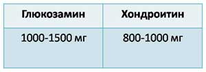
- To get real results, you need 2-3 courses of treatment with these drugs, which will take up to 1.5 years.
- Practitioners advise taking chondroprotectors continuously for 3-5 months and repeating the course every six months.
- Chondroprotectors should be taken regularly, in courses, and not on a case by case basis.
- It is pointless to take chondroprotective drugs if you continue to mock the joint with excessive loads. To achieve the effect, you need to reduce weight, and athletes abandon conventional training.
- You can take this group for a very long time and not see the result if you do not provide normal nutrition to the joint. This requires special (!) Exercises.
- For the production of chondroitin and glucosamine, cartilage of cattle, extracts from marine fish are used. It is difficult to achieve 100% purification, therefore, when taking these drugs allergic reactions occur and problems from the gastrointestinal tract (abdominal pain, diarrhea, constipation, etc.).
- Chondroitin sulfate reduces clotting blood, so it can not be used together with anticoagulants and with a tendency to bleeding. contraindicated pregnant and lactating women, children.
- Diabetics when taking these drugs need to carefully control their sugar levels. It can rise (carbohydrates after all).
How do chondroprotectors work?
What does glucosamine do?
- Stimulates the activity of chondrocytes.
- Necessary for the synthesis of chondroitin sulfate and hyaluronic acid.
- Prevents the destructive effect on the cartilage of NSAIDs and glucocorticosteroids.
What does chondroitin sulfate do?
- Necessary for the synthesis of hyaluronic acid.
- Normalizes the production of synovial fluid.
- Reduces the activity of enzymes that damage cartilage.
- It has an anti-inflammatory effect.
Types of chondroprotectors
Let's analyze how chondroprotectors are divided.
According to the way of taking exist:
- Preparations for oral administration (Struktum, Dona powders and tablets, Artra, etc.)
- Preparations for injections (Dona r / r, Alflutop, Rumalon, etc.)
- Preparations for external use (Chondroxide, Chondroitin, etc.).
With parenteral administration, the bioavailability of chondroprotectors is significantly higher, so they are prescribed when you need to quickly relieve an exacerbation, or when the patient prefers short courses of treatment, or when there are problems with the liver, so as not to burden it.
Preparations for external use are effective only in combination with other forms of release.
By composition, chondroprotectors are divided into:
- Monopreparations that contain only chondroitin sulfate (CS) or glucosamine (GA): Structum, Don.
- Combined products containing both one and the other component: Artra, Teraflex.
- Means that, in addition to cholesterol and GA, contain a non-steroidal (i.e., non-hormonal) anti-inflammatory agent: Teraflex Advance.
With the latter, everything is clear: if there are signs of inflammation (severe pain, swelling), at first we recommend a drug with NSAIDs. After 2-3 weeks, you can switch to a "clean" chondroprotector.
As for the first two, there is no unequivocal answer to the question “which is better”. Some doctors prefer single drugs, others combined, and still others prescribe both, depending on the situation.
But I noticed that glucosamine gives more side effects from the gastrointestinal tract.
Therefore, the combination of GA and CS seems to me the most optimal: it increases the bioavailability of the drug and reduces the frequency of adverse reactions.
Well, now let's go over the drugs.
I'll start with the "oldies":
RUMALON- solution for intramuscular injection.

Compound:
Glycosaminoglycan-peptide complex derived from cartilage and bone marrow of calves (powerful allergen due to animal proteins).
What is he doing:
It improves the synthesis of cholesterol, promotes the maturation of chondrocytes, stimulates the synthesis of collagen and proteoglycans. Moreover, the manufacturer writes that the drug is effective both in the early and late stages of arthrosis. The latter makes me doubt.
Application: administered according to the scheme for 5-6 weeks, 2 times a year.
Side effects: allergic reactions.
ALFLUTOP- injection.
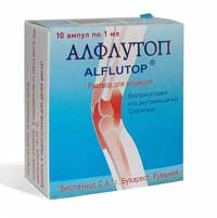
Ingredients: bioactive concentrate from small marine fish.
Contains amino acids useful for cartilage, mucopolysaccharides, trace elements: sodium, magnesium, zinc, iron, etc.
What it does: It inhibits the activity of hyaluronidase, an enzyme that breaks down hyaluronic acid. So the latter becomes larger, and the condition of the cartilage improves.
Application:
There are 2 ways to use it:
- Intramuscularly daily 1 ml for 20 days.
- Intra-articular 1 or 2 ml per joint every 3-4 days. Only 5-6 injections.
The course is repeated after six months.
Sometimes doctors start with intra-articular injections, then move on to intramuscular injections. It depends on the doctor. How many doctors, so many methods.
Contraindications: Allergy to seafood (sometimes very strong).
CHONDROLONE- lyophilisate (i.e. the active substance is in a dried state) for the preparation of a solution

Composition: contains chondroitin sulfate 100 mg per ampoule.
Since the bioavailability is high with this administration, this dosage is sufficient.
It is obtained from the cartilage of the trachea of cattle.
What it does: inhibits the activity of enzymes that cause cartilage destruction, stimulates the production of glycosaminoglycans by chondrocytes, normalizes the production of synovial fluid, and has an anti-inflammatory effect.
Application: in / m 1-2 ampoules every other day. Only 25-30 injections. The course is repeated after six months.
DONA- monopreparation.

Ingredients: contains glucosamine sulfate.
What it does: stimulates the synthesis of hyaluronic acid and other glycosaminoglycans, inhibits enzymes that cause cartilage destruction.
In one tablet 750 mg HA.
How to take: 1 t. 2 times a day with meals. Improvement occurs in 2-3 weeks. The minimum course is 4-6 weeks. Repeat the course after 2 months.
The powder contains 1500 mg of HA.
For whom is it optimal this form of release: powders are especially good for working citizens, who find it more convenient to take the drug only 1 time per day.
And also for those who have difficulty swallowing tablets.
Application: the powder is dissolved in a glass of water and taken 1 time per day (also better with meals). The course is 6 weeks, repeated after 2 months.
Solution for i / m administration: in 1 ampoule 400 mg of glucosamine. Bioavailability 95%. In addition to glucosamine, it contains lidocaine, therefore it has many contraindications: cardiovascular insufficiency, impaired liver and kidney function, epileptic seizures, etc. There are many side effects.
Medical prescription only!
Application: Enter 3 times a week for 4-6 weeks. And then as the doctor decides. Maybe he will switch to powders or tablets.
STRUKTUM- capsules.

Ingredients: contains chondroitin sulfate.
There are 250 mg and 500 mg. To be honest, I don’t know why the first form of release exists, since the manufacturer recommends taking 500 mg 2 times a day.
Judging by the presence in Moscow pharmacies, Structum 250 mg is leaving the shelves. Maybe I'm wrong.
What is he doing? Stimulates the synthesis of glycosaminoglycans, improves the metabolic process in the cartilage.
Application: take it 500 mg 2 times a day for 6 months.
The action after cancellation lasts 3-5 months, then you need to repeat the course.
- combined remedy.

Composition : contains very adequate dosages of chondroitin and glucosamine: 500 mg each
What it does: All the good things HA and CS do in the joint.
Application: take this drug 1 ton 2 times a day for the first 3 weeks, then 1 ton a day for a long time, but not less than 6 months.
TERAFLEX ADVANCE - another combination drug.
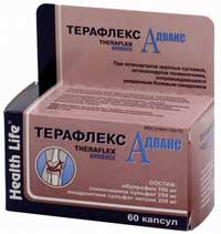
Composition: it contains: GA 250 mg, cholesterol 200 mg and ibuprofen 100 mg.
So, in addition to all the beneficial effects of the first two substances, it also has an anti-inflammatory and analgesic effect.
In addition to all the beneficial effects of the first two substances, it also has an anti-inflammatory and analgesic effect.
True, contraindications and side effects due to ibuprofen become several times larger.
Application: take it 2 capsules 3 times a day after meals for no more than 3 weeks. Then they switch to the usual Teraflex.
TERAFLEX
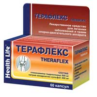
Composition: contains GA 500 mg, cholesterol 400 mg.
Application: take it for the first 3 weeks, 1 capsule 3 times a day, then 1 capsule 2 times a day for 3-6 months, preferably with meals. Then, as usual, the course is repeated.
External chondroprotectors
Here I will focus only on the most popular drug Chondroxide.
CHONDROXIDE

Composition: contains 1 g of 50 mg of chondroitin sulfate.
Release form: ointment and gel.
Application:
A large molecule of chondroitin cannot penetrate the skin on its own, therefore, in order to pass it through cell membranes, dimexide is added to the drug, which also has an anti-inflammatory and analgesic effect.
Do not apply to open wounds.
CHONDROXIDE FORTE – cream

Composition: contains cholesterol and the anti-inflammatory substance Meloxicam, that is, it reduces inflammation and pain.
Contraindications standard for NSAIDs.
Given this composition, it is better not to advise the elderly. For them, there is a gel for the period of exacerbation.
This is not just a cream, it is a transdermal glucosamine complex (glucosamine + triglycerides).

Compound . contains glucosamine, and not chondroitin, like the previous forms, and dimexide, so we recommend it when an allergic reaction to other external forms of chondroxide has been noted in the past.
And also when the buyer does not care about the high price. The main thing is that the effect is maximum.
The active substance is enclosed in a shell of lipids, which together form a micelle (nanoparticle), which delivers the active substance to the joint in a concentration comparable to an injection.
Application: apply it 2-3 times a day for 3-4 weeks. If necessary, the course is repeated.
I end with this.
You have a lot of chondroprotectors in your assortment: both drugs and dietary supplements.
But knowing the basic things that I talked about, you can now independently understand the composition of such a tool and its effectiveness.
I hope that now you can easily continue the phrase:
CHONDROPROTECTORS WORK, unless…
And as a homework, I suggest you think about:
What questions should a buyer ask when choosing a chondroprotector?
Having studied everything thoroughly, I understood why in some countries all chondroprotectors are considered additives: because their bioavailability is low (and manufacturers, by the way, do not hide this), and the therapeutic effect is greatly delayed in time.
And in conclusion, I will answer the most common question:
Why are there many bad results of using chondroprotectors?
- Because, as usual, people are hoping for a magic pill without putting in the effort to lose weight and work muscles.
- Because they want quick results, and not seeing them, they stop treatment.
- Because they begin to "drink Borjomi when the kidneys have failed", i.e. take chondroprotectors at 3-4 stages of arthrosis.
That's all.
How did you like this article, friends?
What do you think about chondroprotectors YOU?
Add, comment, share your experience, click on the social buttons. networks.
See you on the blog for hard workers!
With love to you, Marina Kuznetsova


