Cellular elements in secretions from the nipple of the mammary gland. Manifestations of the epithelial component of the mammary gland Varieties of benign changes
Examples of using RTM diagnostics in mammology
(examinations were carried out on a microwave mammograph RTM-01-RES) .
1. Patient K., 58 years old.
Mammography: fibrocystic mastopathy of a fibrous character. Cicatricial changes.
Clinic: both glands with symptoms of diffuse cystic mastopathy. The postoperative scar is compacted in places. Nodular formations are not defined. Regional lymph nodes are not enlarged.
RTM - conclusion: the thermogram is characteristic of the disease of the left breast in the upper inner quadrant.
Diagnostic puncture - erythrocytes, drops of fat.
The field of internal temperatures of the patient K., 58 years old
Conclusion of the oncologist: diffuse fibrocystic mastopathy.
Considering the conclusion of the RTM diagnostics, the patient was scheduled for a follow-up examination after 3 months.
After 4 months.
RTM - conclusion: the thermogram is characteristic of the disease of the left breast in the upper inner quadrant.
Clinic: the left mammary gland is cicatricial - changed. Under the areola, a nodular formation of a densely elastic consistency up to 1 cm in diameter with fairly clear contours. Regional lymph nodes are not enlarged.
Conclusion of the oncologist: nodular fibrocystic mastopathy, suspicion of cancer.
Diagnostic puncture: structureless masses, single leukocytes.
Mammography: fibrocystic mastopathy, on the left, the appearance of a nodular formation up to 1.5 cm in diameter, at the site of fibrosis, with an enlightenment rim.
Diagnostic puncture: cancer cytogram.
The final conclusion of the oncologist: cancer of the left breast.
2. Patient K., 53 years old.
Mammography: severe diffuse fibrocystic mastopathy, uneven tissue resorption.
Clinical examination: area of local fibrosis in the upper parts of the left breast.
Diagnostic puncture: erythrocytes, drops of fat.
Conclusion: fibrocystic mastopathy.
RTM - conclusion: the thermogram is typical for the disease of the left breast at the border of the upper quadrants.

The field of internal temperatures of the patient K., 53 years old.
After 4 months
Clinic: in the left mammary gland on the border of the upper quadrants, an area of doughy consistency with a rim of enlightenment is determined.
Conclusion: nodular fibrocystic mastopathy, suspicion of cancer of the left breast.
Mammography: on a fibrous background, a site of nodular mastopathy is determined in places with a stringy contour, up to 2.5 cm in diameter. Conclusion: suspicion of cancer of the left breast.
Diagnostic puncture: drops of fat.
In the city hospital No. 60, the patient underwent a sectoral resection of the left breast with an urgent histological examination.
Histological examination results: infiltrative lobular cancer, cancer metastases in one lymph node.
Postoperative opinion of the oncologist: cancer of the left breast IIb stage. A mastectomy was performed according to Pati on the left.
3. Patient L., 33 years old.
Clinic:
postoperative scar on the right. In the mammary glands, the phenomenon of moderately pronounced lobulation, heaviness, nodular formations are not determined. Conclusion: fibrocystic mastopathy.
RTM diagnostics: Thermogram characteristic of the disease left breast at the border of the lower quadrants.

Temperature field of patient L., 33 years old
After 1 year and 8 months.
Mammography: Moderately expressed diffuse fibrocystic mastopathy with a small cystic component on the left.
Clinic: fibrocystic mastopathy, on the left colostral discharge with an admixture of ichor.
Diagnostic puncture: drops of fat
After another 7 months.
Clinic: serous discharge on the left. In the mammary glands, the phenomenon of fibrocystic mastopathy.
After another 8 months.
Clinic: fibrocystic mastopathy with the presence of a nodular formation on the left in the lower quadrant near the areola. Cyst?
Mammography: remnants of the glandular triangle, a nodular formation on the left up to 2 cm in diameter, like a cyst.
Diagnostic puncture: cytological picture of cancer, possibly mucous.
Final diagnosis: cancer of the left breast T2N0M0 stage II.
4. Patient L., 59 years old.
Mammography
Clinic: Diffuse fibrocystic mastopathy.
RTM - conclusion: The thermogram is typical for the disease of the right mammary gland in the caudal region.
 
|
Internal temperature field and thermogram of patient L., 59 years old
.
Based on the results of RTM diagnostics, a diagnostic puncture was performed.
Result: cancer cytogram.
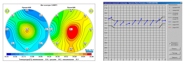
5. Patient B., 57 years old.
Clinic: In the left mammary gland, at the border of the lower quadrants, a nodular formation of 14 mm in size with clear boundaries (ultrasound) is determined. A diagnostic puncture yielded dense contents.
Mammography: Fibrocystic mastopathy. Nodular formation on the border of the lower quadrants of the cyst type.
Cytological examination: Drops of fat, erythrocytes.
RTM diagnostics: The thermogram is typical for the disease of the left breast at the border of the lower quadrants.

Internal temperature field and thermogram of patient B., 57 years old.

Temperature field of the skin and combined thermogram of patient B., 57 years old.
Histological examination: Infiltrating intraductal carcinoma.
6. Patient P., 58 years old.
Mammography: on the border of the outer quadrants of the left mammary gland, a nodular formation of irregular shape, about 3.5 cm in diameter, is determined. The structure of the mammary gland is diffusely changed. Skin, areola thickened.
Conclusion: edematous form of left breast cancer.
Clinic: complaints about a painful formation in the left mammary gland, which the patient noticed 2 days ago, the temperature rose to 38? Objectively, the left mammary gland is sharply hyperemic, edematous, the nipple is retracted. On the border of the outer quadrants, a large infiltrate is palpated, pain on palpation. An enlarged lymph node was palpated in the left axillary region.
Conclusion: acute mastitis.
RTM - conclusion: the thermogram is typical for the disease of the left breast.

The field of internal temperatures of the patient P., 58 years old (before treatment)
After 5 months the patient was called for a follow-up RTM examination
RTM - conclusion: the thermogram is characteristic of the disease of the left breast in the lower outer quadrant.

The field of internal temperatures of the patient P., 58 years old (after treatment).
Clinically, the patient showed positive dynamics in the treatment of acute mastitis, however, the focus of elevated temperature in the lower outer square of the left breast did not disappear.
A week later, the patient went to the doctor because of severe pain in the mammary gland, and soon she was operated on for recurrent mastitis.
Routine histological examination revealed intraductal carcinoma mammary gland
7. Patient B., 45 years old.
Clinic: against the background of fibrocystic mastopathy, on the border of the upper quadrants of the left mammary gland, a seal is indistinctly palpable in the form of a gross deformation of the tissues, not soldered to the skin.
Mammography: On the border of the upper quadrants of the left mammary gland, a nodular formation of an irregular shape measuring 1.5 x 0.7 cm with stringy contours is determined.
Conclusion: suspected left breast cancer.
Diagnostic puncture: structureless masses, drops of fat
Revision of the radiograph: no data for cancer.
RTM diagnostics: the thermogram is typical for the disease of the left breast at the border of the upper quadrants closer to the upper outer quadrant.

Temperature field and thermogram of patient B., 45 years old.
The patient underwent sectoral resection of the left breast.
The diagnosis was established after a planned histological examination.: invasive ductal carcinoma, 0.9 cm in diameter, no cancer metastases were found in any of the 11 lymph nodes.
8. Patient B., 50 years old.
Clinic: in the upper outer quadrant of the left breast there is a glandular component with a tendency to nodulation
Mammography: diffuse fibrocystic mastopathy more pronounced in the outer parts of the left mammary gland.
RTM diagnostics: the thermogram is typical for the disease of the left breast.

Temperature field and thermogram of patient B., 50 years old.
Diagnostic puncture: among erythrocytes, destructively altered cells with precancerous proliferation.
Review of mammograms(done 4 months before): an area of nodular mastopathy in the upper outer quadrant of the left mammary gland up to 1.5 cm in diameter, a hyperplastic lobe is determined in the inner quadrant of the left mammary gland.
The patient underwent a Patey radical mastectomy for left breast cancer.
9. Patient T., 61 years old.
The first RTM examination was carried out after the treatment of acute mastitis
RTM diagnostics: the thermogram is typical for a disease of the left breast with a focus in the upper outer quadrant.

Clinic: After the therapy, swelling and hyperemia of the left breast disappeared. An area of local fibrosis is preserved.
Conclusion: Residual signs of left-sided mastitis. Suspicion of cancer of the left breast.
Diagnostic puncture: drops of fat, erythrocytes.
After 3 months
Mammography: No significant dynamics. In the left mammary gland in the upper outer quadrant, there is an area of fibrosis of the same size with various calcifications.
Clinic: There are no signs of inflammation and swelling. The nipple in the left mammary gland is retracted (always). Seals and nodular formations are not determined.
RTM diagnostics: risk group, positive dynamics, repeated RTM examination is required.

Temperature field and thermogram of patient T., 61 years old.
After another 6 months.
After another 6 months.
Clinic: No complaints. There are no signs of inflammation or swelling. Limited fibrosis in the left mammary gland. Suspicion of cancer of the left breast.
Mammography: Against the background of involutive changes in the left mammary gland in the upper outer quadrant, there is a site of restructuring of the tissue structure up to 1.0 cm in size with stringy contours, of low intensity.
Conclusion: Suspicion of left breast cancer.
RTM diagnostics: The thermogram is typical for the disease of the left breast.

Temperature field and thermogram of patient T., 61 years old.
1 more month later
Clinic: After normalization, hyperemia and swelling of the outer sections of the left mammary gland are again noted. Conclusion: acute left-sided mastitis, suspicion of cancer of the left breast.
With a diagnosis of suspected cancer of the left breast (without cytological verification), hospitalization is recommended.
In the surgical department, the patient underwent a radical mastectomy of the left mammary gland according to Patey and then radiation therapy was performed.
10. Patient S., 40 years old.
Mammography: moderately expressed fibrocystic mastopathy. In the right mammary gland in the lower outer quadrant, a nodular formation of irregular shape with stringy contours, of medium intensity, up to 1.5 cm in diameter, is determined.
Conclusion
Clinic: mammary glands without features. There are no divisions. In the right mammary gland in the lower outer quadrant, a round, mobile, dense formation 1.5 cm in diameter is palpated. No other seals and nodular formations are detected.
Conclusion: suspected right breast cancer.
Diagnostic puncture: drops of fat.
: erythrocytes, drops of fat.
RTM - diagnostics: The thermogram is typical for the disease of the right mammary gland.

Temperature field and thermogram of patient S., 40 years old.
Given the clinical picture, mammography and RTM data, hospitalization is recommended with a diagnosis of suspected right breast cancer.
The patient underwent a sectoral resection of the right mammary gland with an urgent histological examination - nodular fibrocystic mastopathy.
Planned histological examination: medullary breast cancer, the diameter of the tumor node is 1.7 * 1.2 * 1.2 cm.
The final diagnosis was cancer of the right breast T 1 N 0 M 0 1 stage.
11. Patient P., 51 years old.
Of interest is the medical history of a patient who underwent radical sectoral resection and postoperative radiation therapy. As a result, the patient developed rough scars. 2 years after the treatment, the patient was examined in the MMD branch. Questionable data were obtained clinically and radiographically, which did not allow us to make a definite statement regarding the recurrence of cancer in the scar.
RTM data revealed a clear hyperthermia at the local point of the scar. Subsequent puncture examination confirmed the presence of cancer recurrence in the scar.

Temperature field of the skin and combined thermogram of patient P, 51 years old.
Clinic: in the outer parts of the right mammary gland, an area of local fibrosis is palpated. Swelling of the right arm after swimming - lymphostasis
Mammography: in the upper outer quadrant of the right mammary gland, an area of local fibrosis with fuzzy contours about 1.5 cm in size is determined. Negative dynamics.
Conclusion: suspected right breast cancer.
RTM - examination: the thermogram is typical for the disease of the right mammary gland in the areola. The appearance of thermoasymmetry according to the IR sensor at the border of the outer quadrants near the areola in the right mammary gland.
Diagnostic puncture: cancer cytogram
Histological conclusion: in the area of the postoperative scar, a nodule with a diameter of 0.4 cm is determined subcutaneously - a recurrence of infiltrating ductal cancer.
12. Patient K., 54 years old.
January 98.- Mammography: fibro-fatty involution with areas of fibrosis.
Clinic: complaints about the compaction on the right. Lipoma on the right chest wall.
Diagnostic puncture: drops of fat.

RTM - diagnostics: risk group. There is an increase in internal temperatures in both mammary glands, more on the left. Required RTM - examination.
February 98.- RTM - diagnostics: the thermogram is typical for the disease of the left breast.
Temperature field and thermogram of patient K., 54 years old.
Repeated diagnostic puncture: drops of fat.
December 98- No complaints.
Diagnostic puncture: fat drops.
October 99: diagnostic puncture: drops of fat.
2001- Mammography: fibro-fat involution.
Clinic: in the mammary glands, against the background of the phenomena of fatty involution, nodular formations are not determined. On the right, along the mid-axillary line, the formation of a dense consistency with a diameter of about 3 cm, such as a fibrolipoma, is palpated. No atypical cells were found in numerous diagnostic punctures. Surgical treatment is recommended.
2003 There are no complaints. In 2002 - resection of a node on the left lobe of the thyroid gland.
Mammography: On the left, in the upper outer quadrant, a nodular formation of irregular shape with fuzzy contours about 1.5 cm in size is determined. Suspicion of cancer of the left breast.
Ultrasound: On the left in the upper outer quadrant, a 0.9 * 1.05 cm area of an inhomogeneous structure without clear contours with a rupture of Cooper's ligaments is visualized. Suspicion of cancer of the left breast.
Clinic: On the left in the upper outer quadrant, a seal up to 1 cm is palpated.
Diagnostic puncture: cancer cytogram.
RTM diagnostics: the thermogram is characteristic of a disease of the left mammary gland in the upper outer quadrant in the periphery.


Temperature field and thermogram of patient K., 54 years old, after 4 years 6 months.
The final diagnosiscancer of the left breast T 1 N 0 M 0 1st stage.
Breast cytology is a laboratory study in which a specialist studies the structure and size of tissue cells. The survey is carried out all over the world, thanks to which thousands of lives have been saved.
According to statistics, every eighth woman in the world is faced with an oncological disease - breast cancer. It has been proven that breast self-examination is not an effective method for detecting tumors. Doctors insist on regular mammological examinations and annual screening with mammography. Cytology is another clinical method for examining the health of the breast.
Indications for cytology
Clinical indicators of breast cytology have accurate results. Their reliability is 90 - 97%. Doctors recommend screening under the following circumstances:
- The presence of tumor-like formations in the tissues of the breast:
- To determine the features of the neoplasm (benign or malignant);
- In order to determine the stage of maturity and spread of the tumor;
- Setting the nature of the tumor (how its shape, structure, density changes);
- Examination of new formations (polyps and granulomas, fixation of chronic inflammations);
- Prediction of the disease, options for tumor growth;
- The study of background changes, bacterial flora.
- With discharge from the nipple.
- With changes in skin color on the chest of a non-traumatic nature (the integrity of the skin is broken, peeling and irritation are revealed).
- The presence of chronic diseases of the genital organs.
- The mammary gland has been bruised, traumatized, or pain occurs.
- Women who are planning to become pregnant, or those who have not been able to get pregnant for a long time.
For analysis, you need to contact a cytologist, attending gynecologist, mammologist, ultrasound specialist.
The main criteria for passing the cytology of the separated breast
Carrying out cytology of the separated breast is an effective method for determining malignant tumors. The presence of secretions unrelated to the lactation period is a pathology. It is necessary to examine the state of health of the woman.
Important. Discharge from the nipples may be spontaneous or permanent. Liquid appears in case of pressure on the areola of the nipple. It has a color from milky yellow to red or brown.
The method of cytological examination of the discharged mammary gland is absolutely safe, and its reliability is at least 97%. The results of the examinations are compiled quickly, which allows you to timely determine the cause and nature of the disease, prescribe the appropriate treatment. The following recommendations should be followed before analysis:
- Seven days before the examination, it is forbidden to take aspirin and other antiagulants;
- On the day of the examination, do not use deodorant under the armpits, as well as any other flavoring substances;
- It is recommended to wear a bra during the procedure;
- Before taking the discharge, it is necessary to thoroughly wash the chest;
- Sedative medications are allowed.
Contraindications for breast cytology
The procedure is prohibited in case of suspected intraepithelial cancer with a limited lesion. Similar examination criteria are only being developed, the features of the examination method for this disease have not yet been studied.
General contraindications include:
- The presence of infection in the body, exacerbation of somatic diseases;
- Increased body temperature;
- Surgical intervention, which was carried out shortly before the examination;
- Impaired blood clotting;
- Pregnancy at any time;
- lactation period.
Breast Cytology Technique
Cytology of the breast is carried out in various ways. Depending on the results of the tests and the clinical state of health of the patient, the material for examinations are:
- scraping taken from breast tissue;
- punctate taken from the mammary gland;
- discharge from the nipple;
- biopsy print;
- material taken from erosive surfaces.
Taking a puncture
The algorithm for taking a puncture is standard. Small additions are possible, which are based on the diagnosis of the woman's state of health. The breast cytology technique consists of the following steps:
- The doctor selects a point on the chest for the injection. The site is the suspected formation of a cyst or tumor (determined by palpation).
- The area to be injected is treated with an antiseptic composition. If the breast is small, then the skin is completely processed.
- The injection is performed with an aspiration needle.
- The doctor collects the contents of the cyst. He performs two or three sharp suction movements to collect the required amount of material for research.
- The needle is then removed from the breast.
- The injection site is additionally treated with an antiseptic composition. A patch impregnated with bactericidal components is applied to the injection site.
The duration of oncocytology is 5-10 seconds. Doctors recommend performing the procedure between the 6th and 14th days of the menstrual cycle. The mammary glands during this period are characterized by softness and suppleness. They do not cause painful and unpleasant sensations, as is the case with the onset of menstruation. For women in menopause, the procedure is carried out on any day.
Performing a biopsy
The cytological smear is applied in a uniform layer on the disinfected glass. So that it does not dry out, it is additionally processed with a mixture of ethyl alcohol and ethers.
Operating material
Taking a biopsy obtained by the surgical method causes pain. The doctor, using a scalpel, makes an incision in the lymph node or the identified seal. Then a glass for examination is applied to the incision site. If the contents of the tumor are soft, then the imprint remains on the surface. If the contents are solid, then a scraping is performed from the seal incision.
Discharge from the mammary gland
A small amount of discharge is applied to the glass. To save the smear, special aerosols and mixtures of ethyl alcohol with ethers are used.
Smear-imprint from an eroded surface
Disinfected glass is applied to the lesion. Cells of the discharge remain on the surface. The resulting material is used for surveys.
Deciphering the cytology of the breast
The correct interpretation of the obtained test results allows the doctor to prescribe the appropriate treatment. Having received a document with a conclusion, the patient should contact the attending physician. To clarify the indications, below is a list of decoding the results of the survey:
- If the conclusion indicates an incomplete result, then it is necessary to perform an additional examination. In most cases, this problem occurs due to insufficient amount of collected material.
- The indicator norm indicates the normal state of health of the patient. The tissues taken for analysis have no pathologies; foreign and malignant bodies were not found.
- The presence of benign cells indicates the absence of signs that are characteristic of cancer cells.
- The presence of non-cancerous cells indicates that abnormal accumulations of cells and compounds are present in the test material. Although the formations are of non-tumor origin, they indicate the presence of cysts, mastitis, as well as other variants of inflammatory processes.
- Malignant neoplasms signal the presence of a cancerous tumor in the mammary glands. Information about the boundaries, structure, stage and localization of the tumor is additionally attached to the results of the analysis.
Important. It is not recommended to completely rely on the survey data, since even in such a procedure errors are possible. If the doctor doubts the results of the examination, an additional procedure should be performed or another method of examining the breast should be used.
After the procedure, scars and deformations do not remain on the body. In some cases, a hematoma forms, which disappears within a few days.
Fluid Cytology of the Breast
Liquid cytology of the breast refers to the morphological method of examination. This study option is the most accurate way to study tissue material. Preparations that are prepared on the basis of a cytocentrifuge have a single-layer structure. They are evenly distributed over the surface of medical glass. On the one hand, this allows you to save on the reagents used (the cost of the procedure is low), and on the other hand, the results are easier to decipher. Punctates from cysts and tumors, discharge from the nipples, prints are used as the material under study.
Cytology with a breast cyst
The cyst is the most common formation in the mammary gland. Pathology is found in women aged 35 to 50 years. The cause of the disease is stress and hormonal disruptions. In the presence of cysts, women complain of pain in the chest area, discharge from the nipples.
In this case, it is recommended to undergo an examination by a mammologist, perform an ultrasound examination and computed tomography. A puncture is used to collect secretions. As a result of examinations, cancer cells or other diseases that need to be monitored and treated are detected.
Cytology in breast fibroadenoma
Fibroadenoma is a tumor lesion of the breast. Swabs are taken for testing. If the diagnosis was not made on time, the fibroadenoma is transformed into a sarcoma. At this stage of the disease, no liquid is released from the nipples.
The following tumor variants are distinguished according to cytology:
- The presence of epithelial and connective cellular elements;
- The predominance of the epithelium and the minimum amount of connective tissue substance;
- The tumor is dominated by cellular elements that have many similarities with the cystic cavity.
Cytology in breast cancer
The detection of cancer in the breast has a number of characteristic features, which makes it possible to obtain test results with 90% accuracy:
- Colloidal cancer is a dense formation, because the cells in it are closely interconnected and held together by mucus in the cytoplasm.
- Papillary cancer is characterized by pronounced cell polymorphism. This means that the formation has uneven contours, it contains hyperchromic nuclei.
- Cancer, accompanied by a low degree of differentiation, has a monomorphic picture of cytology. Cells have a rounded shape, the nuclei are located in the central part of the cell. The disease has common features with the cytogram of malignant lymphoma.
- Paget's cancer is characterized by the presence of large, light-colored cells, which indicates the presence of cancer.
- Cancer with squamous metaplasia has polymorphic cells. They are scattered, characterized by abundant homogeneous cytoplasm, as well as hyperchromic nuclei.
Cytology of secretions from the mammary glands
Cytology of secretions from the mammary gland involves the study of the bacterial and cellular component of the fluid. The method is based on the study of a smear. Discharge from the nipples is the cause of various diseases and formations. Cytology is able to recognize the nature of the disease and identify its cause.
Breast cancer occurs not only in women, but also in men. Although the disease occurs 100 times less often in them and is detected in adulthood. Thanks to the use of the breast cytology method, it is possible to detect early forms of malignant and benign tumors. The method is characterized by a high degree of efficiency, so experts recommend their patients to use it in the examination process.
To date, the most common problem on the way to women's health are changes affecting the epithelium of the breast. Starting from adolescence, when there is a sharp increase in the mammary gland, together with the development of a pronounced nipple, and ending with the onset of menopause, which is characterized by a decrease in size and ducts. Accordingly, the breast tissue also changes.
The effect of age on the epithelium
In the normal state, the tissue component of the mammary glands, due to which the reproductive function becomes feasible, is a combination of stromal (adipose and fibrous connective tissue) and epithelial tissue. Branching ducts directly connected with the nipple and lobules act as epithelium, each formed at a certain age. Stratified squamous epithelium occurs in the area of the nipple, in particular the excretory ducts.
When only a girl is born, the epithelium consists of a small number of so-called rudimentary streams located deeper than the nipple and areola. Further, the prepubertal period is characterized by the slow growth of these ducts, while they branch, and the stromal component, in turn, increases. For the post-pubertal period, an increase in the volume of the gland is characteristic.
At the end of the gestation period, the glandular component reaches such a size that the entire mammary gland is overgrown with glandular tissue. At the end of feeding, the glandular tissue atrophies and the stroma comes to the fore. During menopause, glandular components also atrophy, accompanied by a decrease in the number of lobules, and sometimes their complete disappearance. All of the above is considered to be adequate changes in the glands regarding their functionality and structure.
Influence of hyperplasia
Changes in the epithelium of the mammary glands can be closely related to various diseases that a woman has. Such a fairly common disease is considered to be hyperplasia that occurs in the tissues of the breast. In order to identify the maximum number of symptoms, the diagnosis of the epithelium should be carried out, which ultimately will help to fix the type of this disease. It is worth understanding that for the prevention of the disease, it is necessary to periodically visit doctors such as a gynecologist and a mammologist.

There are the following types and hyperplasia:
Nodular - manifested by secretions in the form of blood, mucus and milk;
cystic - shows hardened nodes that can be easily felt, besides, they are motionless;
fibrous hyperplasia of the epithelium of the mammary gland - is distinguished by the presence of a cyst in the mammary gland, which causes severe pain when touched, and when pressed on the gland - they are simply unbearable.
Any detected neoplasms should be the reason for contacting a qualified specialist. With a typical course of the disease at an early stage, therapy is most effective, and when there is a precancerous condition, they resort to surgical intervention.
In most cases, benign changes in the mammary gland are associated with cellular hyperplasia. In this option, the doctor should consider such changes through the prism of the likely development of malignant tumors.
Varieties of benign changes
As scientifically confirmed, all benign changes are divided into certain groups, depending on the risk of developing malignant tumors. The first group includes non-proliferative processes, which include the following changes:
- apocrine metaplasia - a process that affects the epithelium of the mammary gland, when cuboidal cells turn into cylindrical ones;
- - includes benign epithelial and stromal elements, while the tumor is clearly delimited from other tissues.
The second group is represented by proliferative processes that occur without atypia and which include the following:
- severe (moderate) hyperplasia - filled epithelial cells of the lumen of the duct and its further expansion;
- intraductal papilloma, the lumen of which is framed by a formation consisting of papillae, which are covered with epithelial cells in two layers;
- sclerosing adenosis - display squeezing and changing the shape of the gland.
The third group includes the following atypical hyperplasias:
- ductal - has an epithelial nature of the structure, which has several signs of ductal cancer;
- lobular - absorbs the growth of cells that look small and the same.
Based on this, it follows that epithelial hyperplasia is a risk of degeneration of the epithelium into a malignant form, so a timely and comprehensive examination is required.
The hormonal component of the epithelium
The normal development of the breast is promoted by hormones such as estrogen and progesterone. Due to estrogen receptors, the milk ducts increase, as a result of which the fatty pad of the breast is formed. The presence of progesterone receptors promotes the growth of alveoli (milk production), milk lobules and lobes
Progesterone, as you know, causes cells to divide, that is, to act as a stimulant, or, conversely, to suppress them. The tissue component of the mammary gland absorbs well both progestins (steroids for stimulating pregnancy) and progesterone.
In normal non-dividing mammary glands, epithelial cells should not contain either progesterone or estrogen receptors. Outside of lactation in the normal state, the epithelium is directly dependent on periodic changes in hormones, which is why it is able to change during menstruation.
The ovulatory cycle is represented by a combination of estrogen and progesterone, respectively, the growth of breast tissue occurs precisely at such moments.
Corresponding changes in the mammary glands also occur under the influence of the following hormonal substances:
- epidermal growth factor - the activity of progesterone receptors in response to increasing estrogens;
- prolactin;
- thyroid hormones;
- insulin.
Thus, the epithelial cells in the mammary gland undergo changes throughout the life of a woman, the main thing is to find out with medical help what kind they are and to prevent the development of the disease in time.
Video
To identify various pathologies of the breast, many methods are used. Consider the methodology for conducting a cytological study, which is based on microscopic examination and evaluation of cellular material obtained from the focus of pathology. This analysis refers to oncomorphology, but it should not be opposed to histological.
Benefits of diagnostics:
- Harmlessness.
- Rapidity.
- Availability and simplicity.
- Possibility of repeated research.
- The use of a small amount of material for microscopic examination
The main goal is to make a correct diagnosis, which will avoid surgical intervention when performing a biopsy and will make it possible to draw up an effective treatment plan.
Research material can be:
- Scraping from breast tissue or tumor removed during surgery.
- Punctate mammary glands.
- Erosive surface material.
- Discharge from the nipple.
- Biopsy prints.
It is extremely important to obtain a complete material. It must be taken from the lesion, not the surrounding tissues.
- Puncture
It is carried out in a clinical laboratory or treatment room. It is performed under X-ray control, ultrasound or CT. This is necessary to control the position of the needle. Before the puncture, the area used is well palpated to determine mobility, communication with surrounding tissues, and to select the optimal fixation. The tissues are fixed with fingers and an aspiration needle is inserted. Upon reaching the focus of pathology, using a syringe, a couple of sharp suction movements are made to take the material.
The contents of the needle are blown onto a glass slide or into a container with a solution. If liquid appears during the puncture, then a test tube is placed under the needle and collected. After removing the fluid, the gland tissues are carefully palpated to exclude residual masses, which may be cystic contents.
- Biopsy
Preparations for cytology are allowed to be made from tissues obtained using this method. The imprint is performed by moving the biopsy with a needle on the glass, while avoiding injury to the tissues taken.
- Operating material
With the help of a scalpel, an incision is made in the lymph node, tumor or induration. The material is obtained by applying a piece of glass to the incision. If the consistency of the tissue is dense, which makes it impossible to make an imprint, then a scraping is made from the surface of the tumor incision.
- Discharge from the mammary gland
A drop of discharge is applied to the glass and a smear is prepared. If the discharge is small, then to obtain a smear with the help of decanting movements, they press on the area of the peripapillary zone.
- Smears-imprints from eroded surfaces
I apply glass to the lesion, on which the cellular elements of the discharge remain. You can also use a cotton swab. All material obtained is sent to the laboratory immediately after sampling.
Deciphering the cytology of the breast
Diagnostic testing is essential in making a diagnosis and developing a treatment plan. Its effectiveness largely depends on the method of implementation and decoding. Breast cytology is one of the most popular and truthful methods for detecting pathologies. After receiving the results, patients should understand that the final conclusion can only be made by a doctor who operates on symptoms, test results, images and other data.
Interpreting cytology results is a complex process. Consider the main transcripts of the analysis:
- Incomplete result - this conclusion indicates the need for additional research. Most likely, the difficulties arose due to the small volume of cellular material. With this conclusion, the doctor recommends a second procedure.
- Normal - tissues taken for analysis contain cells that do not have pathological signs. No additional bodies or inclusions were found.
- Benign cells - there are no signs characteristic of cancer cells.
- Non-cancerous cells - Abnormal accumulations of atypical cells and compounds were found in the examined tissues. But they are of non-tumor origin. Such results may indicate cysts, mastitis, or other variants of the inflammatory process.
- Malignant neoplasms - confirm the presence of a cancerous tumor in the mammary gland. The transcript should contain additional information about the stage, boundaries and localization of the tumor. Tumor features are evident and characteristic aggregations are present.
It is not recommended to fully rely on the information received, since errors are quite likely even in the cytological conclusion. If the doctor has doubts about the veracity of the results, then another sampling is foreseen for study.
Fluid Cytology of the Breast
One of the leading methods in determining pathological processes in the body is morphological. It is based on the study of cytological and histological material. Liquid breast cytology is considered the best way to process tissue material. Preparations prepared on a cytocentrifuge have a single-layer structure and are evenly distributed on a certain surface. This allows you to save expensive reagents when conducting immunocytochemical studies. And the results of such diagnostics are easy to interpret.
The cytologist examines the material, taking into account clinical and anamnestic data, the results of ultrasound, CT and mammography. For study, punctates of tumor formations, discharge from the nipples, prints of foci of pathology are suitable. In addition to liquid cytology, fixation and staining of materials are used.
Cytology with a breast cyst
One of the most common diseases of the breast is a cyst. Most often, pathology appears in patients 35-50 years old. The cause of the disease is hormonal disorders. Cysts can be unilateral and bilateral, single and multiple. Diagnostics is resorted to with appropriate clinical manifestations. The tissues of the glands are compacted and rough, pains appear, discharge from the nipples. On palpation, a small formation of a densely elastic consistency is determined.
Cytology with a breast cyst is carried out with appropriate indications, which are obtained using mammography, ultrasound and CT. Particular attention is paid to the differential diagnosis with cancer and fibroadenoma. A puncture is used to collect material. This is because the cyst is a fluid-filled sac. During the study, it is pierced with a special thin needle, and the liquid contents are sent for cytological examination.
The main task of the analysis is to identify atypical, that is, cancerous cells. If there are no conditions for safe sampling of material, manipulation may affect further treatment, or other diagnostic procedures have established the presence of metastasis, then puncture cytology is not performed.
Cytology in breast fibroadenoma
One type of breast cancer is fibroadenoma. This neoplasm belongs to leaf-shaped tumors. Smears used for cytology in breast fibroadenoma are represented by cuboidal epithelium and connective tissue elements of the stroma. Fibroadenoma is quite common, but leaf-shaped tumors do not exceed 2% of all fibroadenomas.
Such a tumor has the potential to transform into a sarcoma due to malignant changes in the stroma. And the presence of an epithelial component may indicate the development of carcinoma. Most often, the neoplasm is localized in the upper and central squares of the gland. There is no discharge from the nipples or metastases in the lymph nodes.
There are such variants of a leaf-shaped tumor according to cytology:
- With the presence of epithelial and connective tissue cellular elements.
- With a predominance of epithelial components and a meager amount of the connective tissue component.
- With a predominance of cellular elements similar in content to the cystic cavity.
- With scant epithelial or stromal component.
An accurate cytological result of fibroadenoma, that is, a benign form of a leaf-shaped tumor, is possible only with the first option.
Cytology in breast cancer
Breast cancer is characterized by cellular and nuclear polymorphism, which makes the cytological diagnosis 90% reliable. Consider the features of cytology in breast cancer and the types of cancerous lesions:
- Colloidal cancer - has densely packed cells in clusters and mucus production in the cytoplasm or in the form of non-filtered colored masses, that is, extracellularly.
- Papillary cancer - has a pronounced polymorphism of cellular elements, rough with uneven contours and hyperchromic nuclei.
- Cancer with a low degree of differentiation - cytology is characterized by a monomorphic picture. The cells are round in shape, and the nuclei occupy the central part of the cell. Sometimes the picture is similar to the cytogram of malignant lymphoma.
- Paget's cancer - most cells do not differ from low-grade or moderately differentiated forms of cancer. There are large light cells.
- Cancer with squamous metaplasia - polymorphic cells are present, which are scattered with abundant homogeneous cytoplasm and hyperchromic nuclei.
For the study, punctates of tumor formations, punctates of regional lymph nodes, discharges and scrapings from the nipple and erosive surfaces, the contents of cystic cavities, prints of the tumor or lymph nodes are used.
The main principles of cytological diagnostics are:
- The difference in cellular composition in pathology and normal.
- Assessment of the population of cells.
- Application of the pathoanatomical basis.
Each study should end with a detailed conclusion. Diagnostic criteria are based on the morphology of the nucleus and cell, let's consider them in more detail:
- Cell
It has an enlarged or gigantic size, which significantly complicates cytology. Similar is observed in lobular, mastitis-like and tubular cancer. There is a change in polymorphism and shape of cell elements. The state of the nucleus and cytoplasm are disturbed.
Mastopathy is a benign disease in which there are significant changes in the mammary glands in the form of hyperplasia and proliferation.
The main factors contributing to the appearance of this disease are:
- hereditary predisposition.
- Diseases of the biliary tract and liver.
- Endocrine disorders: hypothyroidism, diabetes mellitus.
- Gynecological diseases.
- abortion.
- stressful situations.
There are fibrous, nodular, lobular and cystic forms of mastopathy.
Symptoms of mastopathy
For mastopathy, the most characteristic signs of the disease are:
- the presence of slight discharge from the nipples;
- enlarged lymph nodes;
- sensation of engorgement of the glands;
- severe soreness.
Types of tumors and their diagnosis
On examination, the mammologist evaluates the condition of the glands, notes the shape and size of the seals. Superficial palpation of the lymph nodes and glands is performed.
The main methods of objective analysis of the state of the breast are mammography and ultrasound.
When nodular formations are detected, a biopsy of the mammary gland is performed. The resulting gland tissue cells are transferred to the laboratory for cytological examination. Discharge from the nipples is also subject to examination.
In accordance with the classification developed by the Problematic Commission on Tumor Morphology of the USSR Academy of Medical Sciences, taking into account also the histological classification of breast tumors proposed by WHO (1968-1969), dyshormonal hyperplasia and benign breast tumors can be distributed as follows:
A. Benign dysplasia(dyshormonal hyperplasia, mastopathy, fibroadenomatosis, gynecomastia in men):
a) non-proliferative (lobular, ductal, fibrous, cystic);
b) proliferative: epithelial (solid, papillary, cribriform); fibroepithelial (cystoadenopapillomatosis); myoepithelial (adenosis, sclerosing adenosis).
B. Benign tumors:
1. Fibroadenoma:
a) pericanalicular;
b) intracanalicular;
c) leaf-shaped (cellular intracanalicular fibroadenoma).
3. Breast adenoma.
4. Intraductal papilloma (papilloma of the main ducts).
5. Benign soft tissue tumors.
Cytological examination, taking into account the morphology of epithelial cells and the structures that they form, makes it possible to judge the degree of proliferation of the epithelium (moderate, pronounced, precancerous) and the nature of metaplasia (flattened, apocrine epithelium). However, differential cytological diagnosis of lobular (glandular) mastopathy, fibroadenomas and adenomas, especially with moderate epithelial proliferation, is often very difficult or impossible due to the similarity of cytograms in these processes.
The appearance in smears of a flattened, apocrine epithelium, papillary growths against a background characteristic of a cavity formed in an organ, or signs of a sharp secretory function of cells, makes it possible to cytologically differentiate cystic mastopathy and intraductal papilloma from fibroadenoma and adenoma.
The systematization of cytological pictures in mastopathy and fibroadenomas shows the possibilities of the cytological method in establishing a diagnosis.
Table of systematization of cytological pictures in mastopathy, fibroadenomas and sarcomas of the mammary gland
| Histological characteristics of the process | Cytological conclusion |
| Non-proliferative glandular, glandular-fibrous, glandular-cystic, cystic mastopathy, gynecomastia. Fibroadenoma. Adenoma (nipple, glands). Intraductal papilloma | Mastopathy or fibroadenoma (with moderate proliferation of the epithelium). Cystic mastopathy (with moderate proliferation, apocrinization of the epithelium). Papillary proliferation of the epithelium. Adenoma. Intraductal papilloma |
| Mastopathy or gynecomastia with solid, papillary, cribriform proliferation of the epithelium (including metaplastic). Fibroadenoma with solid proliferation of the epithelium. Adenosis. Sclerosing adenosis. Epitheliosis. Adenoma (nipple, glands) with solidification of structures. Intraductal papilloma | Mastopathy or fibroadenoma with precancerous proliferation of the epithelium. Cystic mastopathy with precancerous proliferation of the epithelium. Adenoma with precancerous proliferation of the epithelium. Intraductal papilloma with precancerous epithelial proliferation |
| Classical leaf-shaped tumor - intracanalicular fibroadenoma with proliferation of stroma elements | Fibroadenoma with proliferation of stroma elements. Leaf tumor |
| Intracanalicular fibroadenoma with proliferation of stromal elements and foci of its dedifferentiation - a leaf-shaped tumor with presarcomatous changes in the stroma | Fibroadenoma with atypical proliferation of stroma elements. Leaf-shaped tumor with atypical proliferation of stromal elements |
| Leaf-shaped tumor with sarcomatous transformation of the stroma | Sarcoma |
| Sarcoma (various forms, including the so-called "malignant mesenchymoma") | Sarcoma (various forms) |
Learn more about the types of breast cancer
Mastopathy and fibroadenomas with moderate proliferation of the epithelium
Glandular (lobular) mastopathy and fibroadenomas (pericanalicular, intracanalicular and mixed). In the preparations, relatively small epithelial cells (diameter 10–20 μm) are visible, similar in structure to the cells of the epithelium of the alveoli and glandular tubes of the unchanged mammary gland. They can be located separately, in groups, in large clusters, sometimes in multilayered microscopically small tissue patches and multicellular rounded structures without a lumen.
A sign of proliferation can be either changes in the morphological properties of cells, or the appearance of multicellular epithelial structures unusual for an unchanged organ.
Cellular criteria for proliferation include an increase in the size of cells and their nuclei. Rounded or slightly oval, as if swollen nuclei predominate with clearly defined, gently looped, intensely colored chromatin. In individual nuclei, single, relatively small, brightly colored nucleoli can be found. The cytoplasm of most cells is colored in intense dove tones, sometimes lilac or blue. Cell complexes characterizing proliferation have the form of rounded formations similar to primary glandular vesicles (acini), but lacking a lumen. There are also papillary-like complexes with a dense arrangement of cells and multilayer layers.
If fibrosis predominates in the pathological focus (fibrous mastopathy and fibroadenomas), then a very small number of small, monomorphic hyperchromic cells with homogeneous rounded hyperchromic nuclei and a barely noticeable narrow rim of intensely stained cytoplasm are detected in the preparations. From the elements of scirrhous cancer, these cells differ precisely in the monomorphism of the nuclei.
Cystic mastopathy
Cystic (glandular-cystic, cystic-fibrous) mastopathy is recognized due to the appearance in the cytograms of flattened epithelial cells of the walls of cystic cavities.

These are large cells, often polygonal, characterized by a centrally located small, usually rounded nucleus and abundant homogeneous or fine-grained cytoplasm, stained unequally intensely.
The number of such cells in smears is different; their arrangement in the form of layers is typical. During proliferation, they can acquire a bizarre shape, and the layers become multilayered.
Dystrophic changes in the cells of the cyst lining are often accompanied by rarefaction of chromatin, which gives the impression of its coarse heaviness and a significant increase in the size of the nucleoli. The background of cytograms is usually represented by unstructured masses, among which phagocytes, histiocytes, and elements of altered blood are present in varying numbers. Sometimes, along with flattened cells, cubic and prismatic elements are found, both scattered and in the form of clusters, papillary and rounded complexes, and tissue shreds.
In cystic mastopathy with apocrinization of the epithelium, punctate or discharge from the nipple may contain cells in a state of apocrine secretion, which, against the general background of the drug, are distinguished by their large size and the peculiar structure of the cytoplasm. They can be of various shapes, but high prismatic and irregularly rounded cells predominate. Their nuclei are rounded, with a clear finely looped chromatin pattern, sometimes with enlarged nucleoli.
The cytoplasm of all cells is abundant and, as a rule, unevenly stained with the identification of two zones (basal and apical). The apical part of the cell often contains dust-like secretion granules, and sometimes granularity is also detected in the basal zone. If there are many cells in the preparation, it is possible to trace the various stages of apocrine secretion up to the detachment of the apical part of the cell. The background of the drug is made up of fragments of the cytoplasm.
Mastopathy and fibroadenomas with precancerous proliferation of the epithelium
For the cytological diagnosis of precancerous proliferation of the epithelium against the background of mastopathy or fibroadenoma, the presence in the preparation of individual, sharply enlarged epithelial cells with signs of ahypicity and their intimate relationship with the cells of the proliferating epithelium described above is necessary. The size of large atypical cells can be quite significant (up to 20.3+2.8 µm cubic and prismatic cells and up to 44.2 ± 6.7 µm flattened and apocrine cells). Such cells are distinguished by the intensity of the staining of the nuclei and cytoplasm. At the same time, the contours of the nuclei remain even, distinct, sometimes wavy, with small bay-like impressions. The chromatin of the nuclei is uniform, small-clumped or looped, compact, intensely colored. A frequent sign of atypicality of the described cells is a significant increase in the nuclear cytoplasmic ratio.
Precancerous proliferation of the epithelium of the cystic cavity wall may be accompanied by the appearance of such atypical cell forms that it becomes necessary to differentiate such cytograms from cancer cytograms. The absence of clumpy chromatin, uneven contours of the nuclei, and a sharp increase in nucleoli in flattened cells indicate precancer. The intimate connection of large cells with the total mass of proliferating epithelium without signs of atypia also indicates a precancerous pathology.
Precancerous proliferation of the epithelium is characterized by the appearance of multicellular structures in the form of massive rounded complexes or papillary structures with a disorderly arrangement of cells of cubic and prismatic epithelium, multilayer layers of flattened epithelium with an abundance of binuclear cells.
Nipple adenomas and breast adenomas
Adenoma is characterized by a cellular composition of cubic, prismatic or cylindrical cells, which are scattered, in groups, clusters or glandular structures. Usually there is a different intensity of staining of the nuclei and a significant increase in their size in individual cells.

Differential diagnosis by cytograms in panchromic color is usually impossible due to their similarity with cytograms of glandular mastopathy and fibroadenomas.
In the preparations of discharge from the nipple, there are altered and unchanged erythrocytes, macrophages with hemosiderin in the cytoplasm, a different number of histiocytic elements, and proliferating cuboidal epithelium in the form of round and papillary complexes, often branching and bizarre. The cells of such complexes undergo fatty and vacuolar degeneration. Sometimes cells such as colostrums and elements of a flattened epithelium are found.

In smears, one can see scattered even more rounded epithelial cells, differently stained with signs of dystrophy.
Sometimes there are very few epithelial cells in the discharge from the nipple with intraductal papilloma and they are difficult to detect when viewing many preparations. In rare cases, they are completely absent and then only altered erythrocytes, phagocytes and histiocytes are visible in the preparation. Such drugs suggest the possibility of intraductal papilloma.
Precancerous proliferation with intraductal papilloma is accompanied by the appearance in the cytogram of large cells with signs of atypia. which are in intimate connection with the bulk of the cells of the proliferating epithelium and form papillae or rounded complexes.
Treatment
Conservative treatment consists in the use of oral contraceptives, vitamin therapy, sedatives and anti-inflammatory drugs. Homeopathic remedies are prescribed. Phytotherapy is widely used for the treatment of mastopathy. A good effect is obtained with the use of iodine preparations. Eleutherococcus and Schizandra tincture are prescribed to strengthen the immune system.
With nodular forms, surgery is mainly used. Sectoral resection of the gland consists in the complete excision of the pathological focus of the disease.
From physiotherapeutic procedures, electrophoresis and laser exposure are used.
Prevention
- For any form of mastopathy, it is recommended to exclude visiting the bath. Artificial tan is contraindicated;
- Periodic observation by a mammologist is considered one of the important points in preventing the disease;
- Self-examination of the glands should also become a mandatory preventive procedure;
- It is useful to constantly use the intake of natural antioxidants: selenium, zinc, beta-carotene, vitamins C and E;
- It is necessary to control weight, avoid stressful overloads and emotional breakdowns;
- It is advisable to change the diet and include seafood in the menu, with an increased content of iodine;
- Termination of pregnancy should be abandoned;
- Giving up bad habits (smoking and alcohol) also serves as a good prophylaxis to prevent the development of mastopathy.


