Normal heart ecg. Deciphering the ECG in adults: what do the indicators mean
An electrocardiogram is a diagnostic method that allows you to determine the functional state of the most important organ of the human body - the heart. Most people at least once in their lives have dealt with a similar procedure. But having received the ECG result in hand, not every person, unless he has a medical education, will be able to understand the terminology used in cardiograms.
What is cardiography
The essence of cardiography is the study of electrical currents that occur during the work of the heart muscle. The advantage of this method is its relative simplicity and availability. Strictly speaking, it is customary to call a cardiogram the result of measuring the electrical parameters of the heart, displayed in the form of a time graph.
The creation of electrocardiography in its modern form is associated with the name of the Dutch physiologist of the early 20th century, Willem Einthoven, who developed the basic methods of ECG and the terminology used by doctors to this day.
Thanks to the cardiogram, it is possible to obtain the following information about the heart muscle:
- Heart rate,
- Physical state of the heart
- The presence of arrhythmias
- The presence of acute or chronic damage to the myocardium,
- The presence of metabolic disorders in the heart muscle,
- The presence of violations of electrical conductivity,
- The position of the electrical axis of the heart.
Also, the electrocardiogram of the heart can be used to obtain information about some vascular diseases that are not related to the heart.
An ECG is usually performed in the following cases:
- Feeling an abnormal heartbeat;
- Attacks of shortness of breath, sudden weakness, fainting;
- Heartache;
- Murmurs in the heart;
- Deterioration of the condition of patients with cardiovascular diseases;
- Passage of medical examinations;
- Medical examination of people over 45 years old;
- Inspection before the operation.
- pregnancy;
- Endocrine pathologies;
- Nervous diseases;
- Changes in blood counts, especially with an increase in cholesterol;
- Over 40 years of age (once a year).
Where can I get a cardiogram?
If you suspect that your heart is not all right, then you can contact a general practitioner or cardiologist to give you a referral for an ECG. Also, on a paid basis, a cardiogram can be done at any clinic or hospital.
Procedure procedure
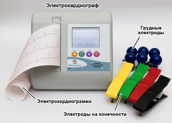
The ECG recording is usually performed in the supine position. To take a cardiogram, a stationary or portable device is used - an electrocardiograph. Stationary devices are installed in medical institutions, and portable ones are used by emergency teams. The device receives information about the electrical potentials on the surface of the skin. For this, electrodes are used, attached to the chest and limbs.
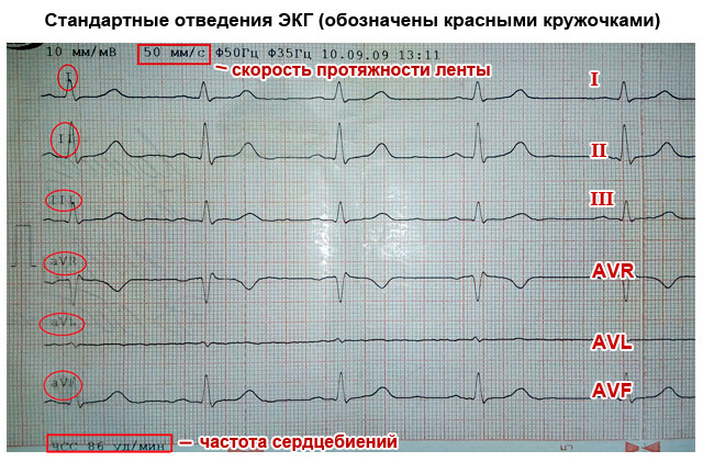
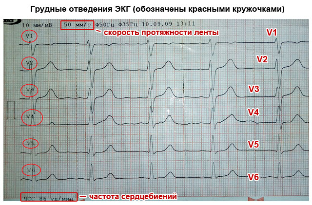
These electrodes are called leads. On the chest and limbs, 6 leads are usually installed. The chest leads are designated V1-V6, the leads on the limbs are called the main (I, II, III) and enhanced (aVL, aVR, aVF). All leads give a slightly different pattern of fluctuations, however, summing up the information from all the electrodes, you can find out the details of the work of the heart as a whole. Sometimes additional leads (D, A, I) are used.
Usually, the cardiogram is displayed as a graph on paper containing millimeter markings. Each lead-electrode has its own schedule. The standard belt speed is 5 cm/s, other speeds can be applied. In the cardiogram displayed on the tape, the main parameters, norm indicators and the conclusion generated automatically can also be indicated. Also, data can be recorded in memory and on electronic media.
After the procedure, it is usually required to decipher the cardiogram by an experienced cardiologist.
Holter monitoring
In addition to stationary devices, there are also portable devices for daily (Holter) monitoring. They are attached to the patient's body along with electrodes and record all the information received over a long period of time (usually within a day). This method gives much more complete information about the processes in the heart compared to a conventional cardiogram. So, for example, when taking a cardiogram in stationary conditions, the patient must be at rest. Meanwhile, some deviations from the norm can manifest themselves during physical exertion, in sleep, etc. Holter monitoring provides information about such phenomena.

Other types of procedures
There are several other methods of carrying out the procedure. For example, this is monitoring with physical activity. Abnormalities are usually more pronounced on the exercise ECG. The most common way to provide the body with the necessary physical activity is a treadmill. This method is useful in cases where pathologies can manifest themselves only in case of increased work of the heart, for example, if coronary disease is suspected.
With phonocardiography, not only the electrical potentials of the heart are recorded, but also the sounds that arise in the heart. The procedure is prescribed when it is necessary to clarify the occurrence of heart murmurs. This method is often used for suspected heart defects.
The patient must be calm during the procedure. Between physical activity and the procedure must pass a certain period of time. It is also not recommended to undergo the procedure after eating, drinking alcohol, caffeinated drinks or cigarettes.
Causes that can affect the ECG:
- Times of Day,
- electromagnetic background,
- Physical exercise,
- food intake,
- The position of the electrodes.
Prong types
First, let's talk a little about how the heart works. It has 4 chambers - two atria and two ventricles (left and right). The electrical impulse, due to which it is reduced, is formed, as a rule, in the upper part of the myocardium - in the sinus pacemaker - the nerve sinoatrial (sinus) node. The impulse propagates down the heart, first affecting the atria and causing them to contract, then it passes through the atrioventricular ganglion and another ganglion - the bundle of His, and reaches the ventricles. The main burden of pumping blood is taken by the ventricles, especially the left one, which is involved in the systemic circulation. This stage is called contraction of the heart or systole.
After the contraction of all parts of the heart, the time comes for their relaxation - diastole. Then the cycle repeats again and again - this process is called the heartbeat.
The state of the heart, in which there is no change in the propagation of impulses, is reflected on the ECG in the form of a straight horizontal line, called the isoline. The deviation of the graph from the isoline is called a tooth.
One heartbeat on an ECG contains six teeth: P, Q, R, S, T, U. The teeth can be directed both up and down. In the first case, they are considered positive, in the second - negative. The Q and S waves are always positive, and the R wave is always negative.

The teeth reflect the different phases of the contraction of the heart. P reflects the moment of contraction and relaxation of the atria, R - excitation of the ventricles, T - relaxation of the ventricles. Special designations are also used for segments (gaps between adjacent teeth) and intervals (sections of the graph, including segments and teeth), for example, PQ, QRST.
Correspondence of the stages of heart contraction and some elements of cardiograms:
- P - atrial contraction;
- PQ - horizontal line, the transition of the discharge from the atria through the atrioventricular node to the ventricles. The Q wave may be absent normally;
- QRS - ventricular complex, the element most often used in diagnostics;
- R - excitation of the ventricles;
- S - relaxation of the myocardium;
- T - relaxation of the ventricles;
- ST - horizontal line, myocardial recovery;
- U - may be absent in the norm. The reasons for the appearance of a tooth have not been clearly elucidated, however, the tooth is valuable for diagnosing certain diseases.
Below are some ECG abnormalities and their possible explanations. This information, of course, does not negate the fact that it is more expedient to entrust the decoding to a professional cardiologist who knows better all the nuances of deviations from the norms and related pathologies.
Major abnormalities and diagnosis
| Description | Diagnosis |
| The distance between the R teeth is not the same | atrial fibrillation, heart block, weakness of the sinus node, extrasystole |
| P wave too high (more than 5 mm), too wide (more than 5 mm), consists of two halves | atrial thickening |
| P wave absent in all leads except V1 | the rhythm does not come from the sinus node |
| PQ interval lengthened | atrioventricular block |
| QRS extension | ventricular hypertrophy, bundle branch block |
| No gaps between QRS | paroxysmal tachycardia, ventricular fibrillation |
| QRS in the form of a flag | heart attack |
| Deep and wide Q | heart attack |
| Wide R (more than 15 mm) in leads I, V5, V6 | left ventricular hypertrophy, bundle branch block |
| Deep S in III, V1, V2 | left ventricular hypertrophy |
| S-T above or below the isoline by more than 2 mm | ischemia or infarction |
| Tall, double-humped, pointed T | cardiac overload, ischemia |
| T merging with R | acute infarction |
Table of cardiogram parameters in adults
The norm of the duration of the elements of the cardiogram in children
The norms indicated in the table may also depend on age.
Rhythm of contractions
Violation of the rhythm of contractions is called. Rhythm irregularity in arrhythmia is measured as a percentage. An irregular rhythm is indicated by a deviation of the distance between similar teeth by more than 10%. Sinus arrhythmia, that is, arrhythmia associated with sinus rhythm, may be a variant of the norm for adolescents and young adults, but in most cases indicates the onset of a pathological process.
A kind of arrhythmia is extrasystole. He is told to her in the case when extraordinary reductions are observed. Single extrasystoles (no more than 200 per day with Holter monitoring) can also be observed in healthy people. Frequent extrasystoles that appear on the cardiogram in the amount of several pieces may indicate ischemia, myocarditis, heart defects.
Heart rate
This option is the most simple and clear. It determines the number of contractions in one minute. The number of contractions may be higher than normal (tachycardia) or lower than normal (bradycardia). Normal heart rate in adults can range from 60 to 80 beats. However, the norm in this case is a relative concept, so bradycardia and tachycardia may not always be evidence of pathology. Bradycardia can occur during sleep or in trained people, and tachycardia can occur during stress, after physical exertion, or at elevated temperature.
Heart rate norms for children of different ages

Photo: Africa Studio/Shutterstock.com
Types of heart rate
There are several types of heart rhythm, depending on where the nerve impulse begins to spread, leading to the contraction of the heart:
- sinus,
- atrial,
- atrioventricular,
- Ventricular.
Normally, the rhythm is always sinus. In this case, sinus rhythm can be combined with both a heart rate above normal and a heart rate below normal. All other types of rhythms are evidence of problems with the heart muscle.
atrial rhythm
Atrial rhythm also often appears on the cardiogram. Is the atrial rhythm normal or is it a kind of pathology? In most cases, the atrial rhythm on the ECG is not normal. However, this is a relatively mild degree of cardiac arrhythmias. It occurs in case of oppression or disruption of the sinus node. Possible causes are ischemia, hypertension, sick sinus syndrome, endocrine disorders. However, individual episodes of atrial contractions can be observed in healthy people. This type of rhythm can take on both the nature of bradycardia and the nature of tachycardia.
atrioventricular rhythm
Rhythm emanating from the atrioventricular node. In atrioventricular rhythm, the pulse rate usually drops to less than 60 beats per minute. Causes - weakness of the sinus node, atrioventricular blockade, taking certain drugs. Atrioventricular rhythm, combined with tachycardia, can occur during heart surgery, rheumatism, heart attack.
Ventricular rhythm
In ventricular rhythm, contractile impulses propagate from the ventricles. The contraction rate drops below 40 beats per minute. The most severe form of rhythm disturbance. Occurs in acute myocardial infarction, heart defects, cardiosclerosis, cardiac circulatory failure, in the preagonal state.
Electrical axis of the heart
Another important parameter is the electrical axis of the heart. It is measured in degrees and reflects the direction of propagation of electrical impulses. Normally, it should be somewhat inclined to the vertical and be 30-69º. At an angle of 0-30º, they talk about the horizontal location of the axis, at an angle of 70-90º - about the vertical. Axial deviation in one direction or another may indicate a disease, for example, hypertension or intracardiac blockades.
What do the conclusions on cardiograms mean?
Consider some of the terms that an ECG decoding may contain. They do not always indicate serious pathologies, however, in any case, they require a visit to a doctor for advice, and sometimes additional examinations.

Photo: Have a nice day Photo/Shutterstock.com
Atrioventricular block
It is reflected on the graph as an increase in the duration of the P-Q interval. 1 degree of the disease is reflected in the form of a simple prolongation of the interval. Grade 2 is accompanied by a deviation of the QRS parameters (loss of this complex). At grade 3, there is no connection between P and the ventricular complex, which means that the ventricles and atria work each in their own rhythm. The syndrome in stages 1 and 2 is not life-threatening, but requires treatment, as it can go into an extremely dangerous stage 3, in which the risk of cardiac arrest is high.
Ectopic rhythm
Any heart rhythm other than sinus. May indicate the presence of blockades, coronary heart disease, or be a variant of the norm. It can also appear as a result of an overdose of glycosides, neurocirculatory dystonia, hypertension.
Sinus bradycardia or tachycardia
Sinus rhythm on the ECG that is below (bradycardia) or above (tachycardia) the normal range. It can be both a variant of the norm and be a symptom of some pathologies. However, in the latter case, this symptom will most likely not be the only one indicated in the transcript of the cardiogram.
Nonspecific ST-T wave changes
What it is? This entry suggests that the reasons for the interval change are unclear and more research is needed. It may indicate a violation of metabolic processes in the body, for example, a change in the balance of potassium, magnesium, sodium ions, or endocrine disorders.
Conduction disorders within the ventricles
As a rule, they are associated with conduction disturbances inside the His nerve bundle. May affect the trunk of the beam or its legs. May lead to delayed contraction of one of the ventricles. Direct therapy for blockades of the His bundle is not carried out, only the disease that caused them is treated.
Incomplete blockade of the right leg of the bundle of His (RBBBB)
Widespread violation of ventricular conduction. In most cases, however, it does not lead to the development of pathologies and is not their consequence. If the patient does not have problems with the cardiovascular system, then this symptom does not require treatment.
Complete right bundle branch block (RBBBB)
This violation is more serious than an incomplete blockade. May indicate myocardial damage. It usually occurs in older and older people, it is rarely found in children and adolescents. Possible symptoms are shortness of breath, dizziness, general weakness and fatigue.
Blockade of the anterior branch of the left leg of the bundle of His (BPVLNPG)
Occurs in patients with hypertension who have had a heart attack. It may also indicate cardiomyopathy, cardiosclerosis, atrial septal defect, mitral valve insufficiency. Has no characteristic symptoms. It is observed mainly in the elderly (over 55 years).
Blockade of the posterior branch of the left leg of the bundle of His (B3VLNPG)
As a separate symptom, it is rare, as a rule, it is combined with blockade of the right bundle leg. May indicate a heart attack, cardiosclerosis, cardiomyopathy, calcification of the conduction system. The blockade is indicated by a deviation in the electrical axis of the heart to the right.
Metabolic changes
Reflect malnutrition of the heart muscle. First of all, it concerns the balance of potassium, magnesium, and sodium. The syndrome is not an independent disease, but indicates other pathologies. It can be observed with ischemia, cardiomyopathy, hypertension, rheumatism, cardiosclerosis.
Low voltage ECG
Electrodes installed on the patient's body capture currents of a certain voltage. If the voltage parameters are below normal, then they talk about low voltage. This indicates insufficient external electrical activity of the heart and may be the result of pericarditis or a number of other diseases.
Paroxysmal tachycardia
A rare condition that differs from normal (sinus) tachycardia, primarily in that it has a very high heart rate - more than 130 beats / s. In addition, the basis of paroxysmal tachycardia is the incorrect circulation of an electrical impulse in the heart.
Atrial fibrillation
At the heart of atrial fibrillation are atrial fibrillation or flutter. Arrhythmia caused by atrial fibrillation can also occur in the absence of heart pathologies, for example, with diabetes, intoxication, and also with smoking. Atrial flutter may be characteristic of cardiosclerosis, certain types of coronary disease, inflammatory processes of the myocardium.
Sinoatrial blockade
Difficulty in the output of the impulse from the sinus (sinoatrial) node. This syndrome is a type of sick sinus syndrome. It is rare, mostly in the elderly. Possible causes are rheumatism, cardiosclerosis, calcification, severe hypertension. May lead to severe bradycardia, syncope, convulsions, respiratory failure.
Hypertrophic conditions of the myocardium
They indicate an overload of certain parts of the heart. The body feels this situation and reacts to it by thickening the muscular walls of the corresponding department. In some cases, the causes of the condition may be hereditary.
Myocardial hypertrophy
General myocardial hypertrophy is a protective reaction, indicating an excessive load on the heart. May lead to arrhythmia or heart failure. Sometimes it is the result of a heart attack. A variation of the disease is hypertrophic cardiomyopathy - a hereditary disease that leads to an abnormal arrangement of heart fibers and carries a risk of sudden cardiac arrest.
Left ventricular hypertrophy
The most common symptom, which does not always indicate severe pathologies of the heart. May be characteristic of arterial hypertension, obesity, some heart defects. Sometimes it is also observed in trained people, people engaged in heavy physical labor.
Right ventricular hypertrophy
A rarer, but at the same time much more dangerous symptom than left ventricular hypertrophy. Indicates insufficiency of pulmonary circulation, severe pulmonary diseases, valvular defects or severe heart defects (tetralogy of Fallot, ventricular septal defect).
Left atrial hypertrophy
It is reflected in the form of a change in the P wave on the cardiogram. With this symptom, the tooth has a double top. It indicates mitral or aortic stenosis, hypertension, myocarditis, cardiomyopathies. Leads to chest pain, shortness of breath, increased fatigue, arrhythmias, fainting.
Right atrial hypertrophy
Less common than left atrial hypertrophy. It can have many causes - pulmonary pathologies, chronic bronchitis, arterial embolism, tricuspid valve defects. Sometimes seen during pregnancy. May lead to circulatory disorders, edema, shortness of breath.
Normocardia
Normocardia or normosystole refers to a normal heart rate. However, the presence of normosystole in itself is not evidence that the ECG is normal and everything is in order with the heart, since it may not exclude other pathologies, such as arrhythmias, conduction disorders, etc.
Nonspecific T wave changes
This symptom is typical for about 1% of people. A similar conclusion is made if it cannot be unambiguously associated with any other disease. Thus, with non-specific changes in the T wave, additional studies are needed. The symptom may be characteristic of hypertension, ischemia, anemia and some other diseases, and may also occur in healthy people.
tachysystole
Also often called tachycardia. This is the general name for a number of syndromes in which there is an increased frequency of contractions of various parts of the heart. There are ventricular, atrial, supraventricular tachysystoles. Such types of arrhythmias as paroxysmal tachycardia, atrial fibrillation and flutter also belong to tachysystoles. In most cases, tachysystoles are a dangerous symptom and require serious treatment.
ST depression of the heart
ST segment depression is common in high-frequency tachycardias. Often it indicates a lack of oxygen supply to the heart muscle and may be characteristic of coronary atherosclerosis. At the same time, the appearance of depression in healthy people is also noted.
Borderline ECG
This conclusion often frightens some patients who have found it on their cardiograms and are inclined to think that "borderline" means almost "mortal". In fact, such a conclusion is never given by a doctor, but is generated by a program that analyzes the parameters of the cardiogram on an automatic basis. Its meaning is that a number of parameters go beyond the norm, but it is impossible to unequivocally conclude that there is any pathology. Thus, the cardiogram is on the border between normal and pathological. Therefore, upon receipt of such a conclusion, a doctor's consultation is required, and, perhaps, everything is not so scary.
Pathological ECG
What it is? This is a cardiogram, which clearly showed some serious deviations from the norm. These can be arrhythmias, conduction disorders or nutrition of the heart muscle. Pathological changes require immediate consultation with a cardiologist, who must indicate the treatment strategy.
Ischemic changes on the ECG
Ischemic disease is caused by impaired blood circulation in the coronary vessels of the heart and can lead to such serious consequences as myocardial infarction. Therefore, the identification of ischemic signs on the ECG is a very important task. Ischemia at an early stage can be diagnosed by changes in the T wave (rise or fall). At a later stage, ST segment changes are observed, and at an acute stage, Q wave changes are observed.
Deciphering the ECG in children
In most cases, deciphering the cardiogram in children is simple. But the parameters of the norm and the nature of the violations may differ compared to those in adults. So, children normally have a much more frequent heartbeat. In addition, the sizes of teeth, intervals and segments are somewhat different.
Article publication date: 03/02/2017
Article last updated: 12/18/2018
From this article you will learn about such a diagnostic method as an ECG of the heart - what it is and what it shows. How is the registration of the electrocardiogram, and who can most accurately decipher it. And also you will learn to independently determine the signs of a normal ECG and the main heart diseases that can be diagnosed by this method.
What is an ECG (electrocardiogram)? This is one of the simplest, most accessible and informative methods for diagnosing heart diseases. It is based on the registration of electrical impulses that occur in the heart, and their graphic recording in the form of teeth on a special paper film.
Based on these data, one can judge not only the electrical activity of the heart, but also the structure of the myocardium. This means that with the help of an ECG, many different heart diseases can be diagnosed. Therefore, an independent interpretation of the ECG by a person who does not have special medical knowledge is impossible.
All that a simple person can do is only to roughly assess the individual parameters of the electrocardiogram, whether they correspond to the norm and what kind of pathology they can talk about. But the final conclusions on the conclusion of the ECG can only be made by a qualified specialist - a cardiologist, as well as a general practitioner or family doctor.
Method principle
The contractile activity and functioning of the heart is possible due to the fact that spontaneous electrical impulses (discharges) regularly occur in it. Normally, their source is located in the uppermost part of the organ (in the sinus node, located near the right atrium). The purpose of each impulse is to pass along the conductive nerve pathways through all departments of the myocardium, inducing their contraction. When an impulse arises and passes through the myocardium of the atria, and then the ventricles, their alternate contraction occurs - systole. During the period when there are no impulses, the heart relaxes - diastole.
ECG diagnostics (electrocardiography) is based on the registration of electrical impulses that occur in the heart. For this, a special device is used - an electrocardiograph. The principle of its work is to capture on the surface of the body the difference in bioelectric potentials (discharges) that occur in different parts of the heart at the time of contraction (in systole) and relaxation (in diastole). All these processes are recorded on a special heat-sensitive paper in the form of a graph consisting of pointed or hemispherical teeth and horizontal lines in the form of gaps between them.
What else is important to know about electrocardiography
Electrical discharges of the heart pass not only through this organ. Since the body has good electrical conductivity, the strength of the excitatory heart impulses is sufficient to pass through all the tissues of the body. Best of all, they spread to the chest in the region of the heart, as well as to the upper and lower extremities. This feature underlies the ECG and explains what it is.
In order to register the electrical activity of the heart, it is necessary to fix one electrode of the electrocardiograph on the arms and legs, as well as on the anterolateral surface of the left half of the chest. This allows you to catch all directions of propagation of electrical impulses through the body. The paths of the discharges between the areas of contraction and relaxation of the myocardium are called cardiac leads and are indicated on the cardiogram as follows:
- Standard Leads:
- I - the first;
- II - second;
- Ш - the third;
- AVL (similar to the first);
- AVF (analogue of the third);
- AVR (mirror image of all leads).
The significance of the leads is that each of them registers the passage of an electrical impulse through a certain part of the heart. Thanks to this, you can get information about:
- How the heart is located in the chest (the electrical axis of the heart, which coincides with the anatomical axis).
- What is the structure, thickness and nature of the blood circulation of the myocardium of the atria and ventricles.
- How regularly impulses occur in the sinus node and whether there are any interruptions.
- Are all impulses conducted along the paths of the conducting system, and are there any obstacles in their path.
What is an electrocardiogram
If the heart had the same structure of all its departments, nerve impulses would pass through them at the same time. As a result, on the ECG, each electrical discharge would correspond to only one tooth, which reflects the contraction. The period between contractions (pulses) on the EGC has the form of a flat horizontal line, which is called an isoline.
The human heart consists of the right and left halves, in which the upper section is distinguished - the atria, and the lower - the ventricles. Since they have different sizes, thicknesses and are separated by partitions, the excitatory impulse passes through them at different speeds. Therefore, different teeth are recorded on the ECG, corresponding to a specific section of the heart.

What do the teeth mean
The sequence of propagation of systolic excitation of the heart is as follows:
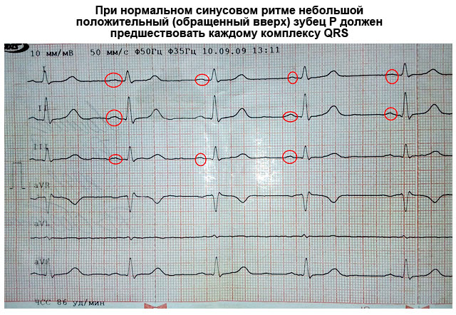
- The origin of electropulse discharges occurs in the sinus node. Since it is located close to the right atrium, it is this section that contracts first. With a slight delay, almost simultaneously, the left atrium contracts. On the ECG, such a moment is reflected by the P wave, which is why it is called atrial. It is facing up.
- From the atria, the discharge passes to the ventricles through the atrioventricular (atrioventricular) node (accumulation of modified myocardial nerve cells). They have good electrical conductivity, so there is normally no delay in the node. This is displayed on the ECG as a P-Q interval - a horizontal line between the corresponding teeth.
- Excitation of the ventricles. This part of the heart has the thickest myocardium, so the electrical wave passes through them longer than through the atria. As a result, the highest tooth appears on the ECG - R (ventricular), facing up. It may be preceded by a small Q wave that points in the opposite direction.
- After the completion of the ventricular systole, the myocardium begins to relax and restore energy potentials. On the ECG, it looks like an S wave (facing down) - a complete lack of excitability. After it comes a small T wave, facing up, preceded by a short horizontal line - the S-T segment. They say that the myocardium has fully recovered and is ready to make another contraction.


Since each electrode attached to the limbs and chest (lead) corresponds to a specific part of the heart, the same teeth look different in different leads - in some they are more pronounced, and in others they are less.
How to decipher a cardiogram
Sequential ECG decoding in both adults and children involves measuring the size, length of the teeth and intervals, assessing their shape and direction. Your actions with decryption should be as follows:
- Unfold the paper with the recorded ECG. It can be either narrow (about 10 cm) or wide (about 20 cm). You will see several jagged lines running horizontally, parallel to each other. After a short gap, in which there are no teeth, after the recording is interrupted (1–2 cm), a line with several complexes of teeth begins again. Each such graph displays a lead, so it is preceded by a designation of which lead it is (for example, I, II, III, AVL, V1, etc.).
- In one of the standard leads (I, II, or III) that has the highest R wave (usually the second), measure the distance between three consecutive R waves (R-R-R interval) and determine the average value of the indicator (divide number of millimeters by 2). This is necessary to calculate the heart rate in one minute. Remember that such and other measurements can be made with a ruler with a millimeter scale or counting the distance on the ECG tape. Each large cell on paper corresponds to 5 mm, and each dot or small cell inside it corresponds to 1 mm.
- Evaluate the gaps between the R waves: they are the same or different. This is necessary in order to determine the regularity of the heart rate.
- Sequentially evaluate and measure each wave and interval on the ECG. Determine their compliance with normal indicators (table below).
Important to remember! Always pay attention to the speed of the tape - 25 or 50 mm per second. This is fundamentally important for calculating the heart rate (HR). Modern devices indicate the heart rate on the tape, and the calculation does not need to be carried out.
How to calculate the heart rate
There are several ways to count the number of heartbeats per minute:
- Typically, an ECG is recorded at a speed of 50 mm/sec. In this case, you can calculate the heart rate (heart rate) using the following formulas:
HR=60/((R-R (in mm)*0.02))
When recording an ECG at a speed of 25mm/s:
HR=60/((R-R (in mm)*0.04)
- You can also calculate the heart rate on the cardiogram using the following formulas:
- When recording at 50 mm/sec: HR = 600/average number of large cells between R waves.
- When recording at 25 mm/sec: HR = 300/average number of large cells between R waves.

What does an ECG look like in normal and pathological conditions?
What a normal ECG and wave complexes should look like, what deviations are most common and what they indicate, is described in the table.
Important to remember!
- One small cell (1 mm) on an ECG film corresponds to 0.02 seconds at 50 mm/sec and 0.04 seconds at 25 mm/sec (for example, 5 cells - 5 mm - one large cell corresponds to 1 second).
- The AVR lead is not used for evaluation. Normally, it is a mirror image of standard leads.
- The first lead (I) duplicates the AVL, and the third (III) duplicates the AVF, so they look almost identical on the ECG.

| ECG parameters | Norm indicators | How to decipher deviations from the norm on the cardiogram, and what they indicate |
|---|---|---|
| Distance R-R-R | All spaces between R waves are the same | Different intervals can talk about atrial fibrillation, extrasystole, weakness of the sinus node, heart block |
| Heart rate | In the range from 60 to 90 bpm | Tachycardia - when the heart rate is more than 90 / min Bradycardia - less than 60/min |
| P wave (atrial contraction) | Turns upwards in arch type, about 2 mm high, precedes each R wave. May be absent in III, V1 and AVL | High (more than 3 mm), wide (more than 5 mm), in the form of two halves (two-humped) - thickening of the atrial myocardium |
| Not present at all in leads I, II, FVF, V2-V6 - rhythm does not come from the sinus node | ||
| Several small teeth in the form of a "saw" between the R waves - atrial fibrillation | ||
| P-Q interval | Horizontal line between P and Q waves 0.1–0.2 seconds | If it is elongated (more than 1 cm when recording 50 mm / s) - the heart |
| Shortening (less than 3 mm) - WPW syndrome | ||
| QRS complex | The duration is about 0.1 sec (5 mm), after each complex there is a T wave and there is a gap in the horizontal line | Expansion of the ventricular complex indicates hypertrophy of the ventricular myocardium, |
| If there are no gaps between the high complexes facing up (they go continuously), this indicates either ventricular fibrillation | ||
| Has the form of a "flag" - myocardial infarction | ||
| Q wave | Facing down, less than ¼ R deep, may be absent | A deep and wide Q wave in standard or chest leads indicates an acute or previous myocardial infarction |
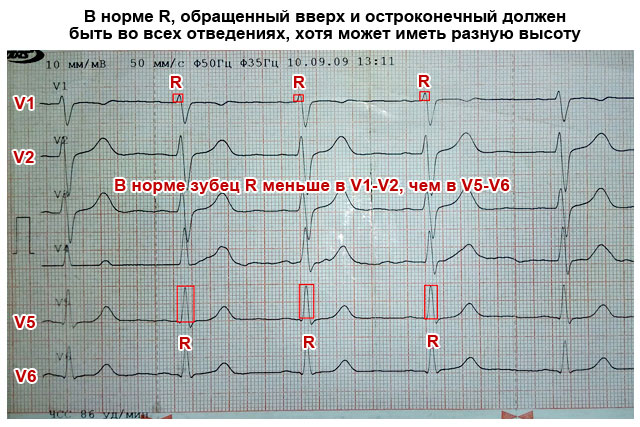
| R wave | Tallest, facing upwards (about 10–15 mm), spiky, present in all leads | It may have a different height in different leads, but if it is more than 15–20 mm in leads I, AVL, V5, V6, this may indicate. Serrated at the top R in the form of the letter M indicates the blockade of the legs of the bundle of His. |
| S wave | Present in all leads, downward facing, pointed, may vary in depth: 2–5 mm in standard leads | Normally, in the chest leads, its depth can be as many millimeters as the height R, but should not exceed 20 mm, and in leads V2–V4, the depth of S is the same as the height of R. Deep or serrated S in III, AVF, V1, V2 - left ventricular hypertrophy. |
| S-T segment | Corresponds to the horizontal line between the S and T waves | Deviation of the electrocardiographic line up or down from the horizontal plane by more than 2 mm indicates coronary disease, angina pectoris or myocardial infarction |
| T wave | Turned upwards in an arc less than ½ R high, in V1 may have the same height, but should not be higher | High, peaked, double-humped T in standard and chest leads indicates coronary disease and heart overload |
| The T wave merging with the S-T interval and the R wave in the form of an arcuate "flag" indicates an acute period of infarction |


Something else important
The characteristics of the ECG described in the table in normal and pathological conditions are only a simplified version of the interpretation. A full assessment of the results and a correct conclusion can only be made by a specialist (cardiologist) who knows the extended scheme and all the subtleties of the method. This is especially true when you need to decipher the ECG in children. The general principles and elements of the cardiogram are the same as in adults. But there are different rules for children of different ages. Therefore, only pediatric cardiologists can make a professional assessment in controversial and doubtful cases.
Pathology of the cardiovascular system is one of the most common problems affecting people of all ages. Timely treatment and diagnosis of the circulatory system can significantly reduce the risk of developing dangerous diseases.
To date, the most effective and easily accessible method of studying the work of the heart is an electrocardiogram.
When examining the results of the examination of the patient, doctors pay attention to such components of the ECG as:
- teeth;
- intervals;
- Segments.
Not only their presence or absence is assessed, but also their height, duration, location, direction and sequence.
There are strict normal parameters for each line on the ECG tape, the slightest deviation from which may indicate violations in the work of the heart.
ECG analysis
The entire set of ECG lines is examined and measured mathematically, after which the doctor can determine some parameters of the work of the heart muscle and its conduction system: heart rate, heart rate, pacemaker, conduction, electrical axis of the heart.
To date, all these indicators are investigated by high-precision electrocardiographs.
Sinus rhythm of the heart
This is a parameter that reflects the rhythm of heart contractions that occur under the influence of the sinus node (normal). It shows the coherence of the work of all parts of the heart, the sequence of processes of tension and relaxation of the heart muscle.
The rhythm is very easy to identify by the tallest R waves: if the distance between them is the same throughout the entire recording or deviates by no more than 10%, then the patient does not suffer from arrhythmia.
heart rate
The number of beats per minute can be determined not only by counting the pulse, but also by the ECG. To do this, you need to know the speed at which the ECG was recorded (usually 25, 50 or 100 mm / s), as well as the distance between the highest teeth (from one peak to another).
By multiplying the recording time of one mm by length of segment R-R you can get your heart rate. Normally, its performance ranges from 60 to 80 beats per minute.

Source of excitation
The autonomic nervous system of the heart is designed in such a way that the process of contraction depends on the accumulation of nerve cells in one of the zones of the heart. Normally, this is the sinus node, the impulses from which diverge throughout the nervous system of the heart.
In some cases, other nodes (atrial, ventricular, atrioventricular) can take on the role of the pacemaker. This can be determined by examining the P wave is inconspicuous, located just above the isoline.

You can read detailed and comprehensive information about the symptoms of cardiosclerosis of the heart.
Conductivity
This is a criterion showing the process of momentum transfer. Normally, impulses are transmitted sequentially from one pacemaker to another, without changing the order.
Electric axle
An indicator based on the process of excitation of the ventricles. Mathematical analysis of Q, R, S waves in leads I and III allows you to calculate a certain resulting vector of their excitation. This is necessary to establish the functioning of the branches of the His bundle.
The obtained angle of inclination of the axis of the heart is estimated by the value: 50-70° normal, 70-90° deviation to the right, 50-0° deviation to the left.
In cases where there is a tilt of more than 90° or more than -30°, there is a serious malfunction in the bundle of His.
Teeth, segments and intervals
Teeth - ECG sections lying above the isoline, their meaning is as follows:
- P- reflects the processes of contraction and relaxation of the atria.
- Q, S- reflect the processes of excitation of the interventricular septum.
- R- the process of excitation of the ventricles.
- T- the process of relaxation of the ventricles.
Intervals are sections of the ECG lying on the isoline.
- PQ- reflects the propagation time of the impulse from the atria to the ventricles.
Segments - sections of the ECG, including an interval and a wave.
- QRST- the duration of the contraction of the ventricles.
- ST- the time of complete excitation of the ventricles.
- TP is the time of electrical diastole of the heart.

Norm in men and women
The decoding of the ECG of the heart and the norms of indicators in adults are presented in this table:

Healthy Childhood Outcomes
Deciphering the results of ECG measurements in children and their norm in this table:

Dangerous diagnoses
What dangerous conditions can be determined by the ECG readings during decoding?
Extrasystole
This phenomenon characterized by irregular heartbeat. A person feels a temporary increase in the frequency of contractions, followed by a pause. It is associated with the activation of other pacemakers, sending along with the sinus node an additional burst of impulses, which leads to an extraordinary contraction.
If extrasystoles appear no more than 5 times per hour, then they cannot cause significant harm to health.
Arrhythmia
Characterized change in the frequency of sinus rhythm when pulses arrive at different frequencies. Only 30% of these arrhythmias require treatment, because can lead to more serious illnesses.
In other cases, this may be a manifestation of physical activity, a change in hormonal levels, the result of a fever and does not threaten health.

Bradycardia
It occurs when the sinus node is weakened, unable to generate impulses with the proper frequency, as a result of which the heart rate also slows down, up to 30-45 beats per minute.
Tachycardia
The opposite phenomenon, characterized by an increase in heart rate over 90 beats per minute. In some cases, temporary tachycardia occurs under the influence of strong physical exertion and emotional stress, as well as during illnesses associated with fever.

Conduction disorder
In addition to the sinus node, there are other underlying pacemakers of the second and third orders. Normally, they conduct impulses from the first-order pacemaker. But if their functions weaken, a person may feel weakness, dizziness caused by depression of the heart.
It is also possible to lower blood pressure, because. the ventricles will contract less frequently or arrhythmically.
Many factors can lead to disruption in the work of the heart muscle itself. Tumors develop, muscle nutrition is disrupted, and depolarization processes fail. Most of these pathologies require serious treatment.
Why there might be differences in performance
In some cases, when re-analyzing the ECG, deviations from previously obtained results are revealed. With what it can be connected?
- different time of day. Usually, an ECG is recommended to be done in the morning or afternoon, when the body has not yet had time to be influenced by stress factors.
- Loads. It is very important that the patient is calm while recording the ECG. The release of hormones can increase heart rate and distort performance. In addition, before the examination, it is also not recommended to engage in heavy physical labor.
- meal. Digestive processes affect blood circulation, and alcohol, tobacco and caffeine can affect heart rate and pressure.
- electrodes. Improper overlap or accidental shifting can seriously change the performance. Therefore, it is important not to move during the recording and degrease the skin in the area where the electrodes are applied (the use of creams and other skin products before the examination is highly undesirable).
- Background. Sometimes other devices can interfere with the operation of the electrocardiograph.

Additional examination methods
Halter
Method long-term study of the work of the heart, made possible by a portable compact tape recorder that is capable of recording results on magnetic tape. The method is especially good when it is necessary to investigate recurrent pathologies, their frequency and time of occurrence.

Treadmill
Unlike a conventional ECG recorded at rest, this method is based on the analysis of the results after exercise. Most often, this is used to assess the risk of possible pathologies not detected on a standard ECG, as well as when prescribing a course of rehabilitation for patients who have had a heart attack.
Phonocardiography
 Allows analyze heart sounds and murmurs. Their duration, frequency and time of occurrence correlate with the phases of cardiac activity, which makes it possible to assess the functioning of the valves, the risks of developing endocarditis and rheumatic heart disease.
A standard ECG is a graphic representation of the work of all parts of the heart. Its accuracy can be affected by many factors, so doctor's advice should be followed.
The examination reveals most of the pathologies of the cardiovascular system, however, additional tests may be required for an accurate diagnosis.
Finally, we suggest watching a video course on decoding “ECG for everyone”:
Formation of the conclusion on the electrocardiogram (ECG) is carried out by a doctor of functional diagnostics or a cardiologist. This is a difficult diagnostic process that requires special training and practice. A doctor describing an ECG should know the basics of cardiac electrophysiology, variants of a normal cardiogram, and be able to identify functional and morphological changes in the heart. He should be able to analyze the violation of the functions of automatism, conduction, excitability of the heart, evaluate the effect of medications and other external factors on the formation of ECG waves and intervals.
The description of the electrocardiogram includes several successive steps. First, the gender and age of the patient are evaluated, since different age groups may have their own ECG features, and the cardiogram is different for men and women. Then the duration and amplitude of the waves and intervals of the cardiogram are determined. After that, the rhythm is assessed, the features of the position of the heart in the chest, conduction disturbances, signs of focal changes in the myocardium and hypertrophy of the heart departments are analyzed. Then the final conclusion is formed. If possible, the ECG is compared with previously recorded films of the same patient (analysis in dynamics).

Analysis of the P wave involves measuring its amplitude, duration, determining polarity and shape. Determine the duration of the P-Q interval.
Analysis of the ventricular QRS complex is an assessment of the ratio of the teeth in all leads, measuring the amplitude and duration of these teeth.
To analyze the ST segment, it is necessary to determine its displacement up or down relative to the isoelectric line and evaluate the shape of this displacement.
When evaluating the T wave, you need to pay attention to its polarity, shape, amplitude.
Then the Q-T interval is measured and compared with the proper value, determined by a special table.
Normal ECG
Normally, the rhythm of the heart is regular, correct, its source is the sinus node. Sinus rhythm at rest has a rate of 60 to 100 beats per minute. The heart rate is determined by measuring the distance between adjacent R waves on the ECG (R-R interval).
The direction of the so-called electrical axis of the heart is determined, showing the position of the resulting electromotive force vector (angle alpha). It is indicated in degrees. The normal axis corresponds to an alpha value between 40 and 70 degrees.
The presence of turns of the heart around its axis is determined.
Heart rhythm disorder

A heart rhythm disorder, or arrhythmia, is diagnosed if the following ECG abnormalities are detected:
- an increase in the heart rate of more than 100 per minute or a decrease of less than 60 per minute;
- wrong rhythm;
- non-sinus rhythm;
- violation of the conduction of an electrical signal through the conduction system of the heart.
Arrhythmias are divided into the following main groups.
On the basis of a violation of the formation of an impulse:
- violations of automatism of the sinus node (sinus tachycardia, bradycardia, arrhythmia);
- ectopic (non-sinus) rhythms caused by the predominance of automatism of non-sinus centers (slip-out, accelerated ectopic rhythms, pacemaker migration);
- ectopic rhythms due to the re-entry mechanism (paroxysmal tachycardia, atrial and ventricular fibrillation and flutter).
On the basis of conduction disorders:
- blockade (sinoatrial, intraatrial, atrioventricular, intraventricular blockade, in particular);
- ventricular asystole;
- ventricular preexcitation syndromes, in particular.
The electrocardiographic features of these disorders are varied and complex.
Hypertrophy of the heart
Myocardial hypertrophy is an adaptive reaction of the body in response to an increase in load, manifested in an increase in the mass of the heart and the thickness of its walls.
Changes in hypertrophy of any parts of the heart are due to increased electrical activity of the corresponding chamber, slowing down the propagation of an electrical signal in its wall, as well as ischemic and dystrophic changes in the heart muscle.
Using the ECG, you can determine the signs of hypertrophy and, as well as their combinations.
Myocardial blood supply disorders
With the help of an ECG, it is possible in some cases to assess the blood supply to the heart muscle. This method has become especially important in the diagnosis of myocardial infarction: an acute disturbance of blood flow in the coronary vessels, accompanied by necrosis (necrosis) of a section of the heart muscle, followed by the formation of cicatricial changes in this zone.
ECG during the course of myocardial infarction has a natural dynamics, which allows you to monitor the development of the process, determine its prevalence and identify complications. With the help of an ECG, the localization of myocardial infarction is also determined.
Other ECG changes
By analyzing changes in the ventricular complex, the ST segment, and the T wave, many other pathological conditions can be diagnosed, such as pericarditis, myocarditis, electrolyte disturbances, and other processes.
Video course "ECG for everyone", lesson 1 - "Conducting system of the heart, electrodes"
Video course "ECG for everyone", lesson 2 - "Teeth, segments, intervals"
Video course “ECG is within the power of everyone”, lesson 3 - “ECG analysis algorithm”
Electrocardiography (ECG): fundamentals of theory, removal, analysis, detection of pathologies
Applied for practical purposes in the 70s of the 19th century by the Englishman A. Waller, an apparatus that records the electrical activity of the heart continues to faithfully serve humanity to this day. Of course, for almost 150 years it has undergone numerous changes and improvements, but the principle of its work, based on records of electrical impulses propagating in the heart muscle, remained the same.
Now almost every ambulance team is equipped with a portable, lightweight and mobile electrocardiograph, which allows you to quickly take an ECG, not lose precious minutes, diagnose and promptly deliver the patient to the hospital. For large-focal myocardial infarction, and other diseases that require emergency measures, minutes count, so an urgent electrocardiogram saves more than one life every day.
Deciphering the ECG for the doctor of the cardiology team is a common thing, and if it indicates the presence of an acute cardiovascular pathology, then the team immediately, turning on the siren, goes to the hospital, where, bypassing the emergency room, they will take the patient to the intensive care unit for urgent care. The diagnosis with the help of an ECG has already been made and no time has been lost.
Patients want to know...
Yes, patients want to know what the incomprehensible teeth on the tape left by the recorder mean, therefore, before going to the doctor, patients want to decipher the ECG themselves. However, everything is not so simple, and in order to understand the "tricky" record, you need to know what a human "motor" is.
The heart of mammals, which includes humans, consists of 4 chambers: two atria, endowed with auxiliary functions and having relatively thin walls, and two ventricles, which carry the main load. The left and right parts of the heart also differ from each other. Providing blood to the pulmonary circulation is less difficult for the right ventricle than pushing blood into the systemic circulation with the left. Therefore, the left ventricle is more developed, but also suffers more. However, regardless of the difference, both parts of the heart should work evenly and harmoniously.

The heart is heterogeneous in its structure and electrical activity, since contractile elements (myocardium) and irreducible elements (nerves, blood vessels, valves, fatty tissue) differ in varying degrees of electrical response.
Usually patients, especially older ones, are worried: are there any signs of myocardial infarction on the ECG, which is quite understandable. However, for this you need to learn more about the heart and the cardiogram. And we will try to provide this opportunity by talking about waves, intervals and leads and, of course, about some common heart diseases.
Ability of the heart
For the first time, we learn about the specific functions of the heart from school textbooks, so we imagine that the heart has:
- automatism, due to the spontaneous generation of impulses, which then cause its excitation;
- excitability or the ability of the heart to activate under the influence of exciting impulses;
- or the “ability” of the heart to ensure the conduction of impulses from their place of origin to contractile structures;
- Contractility, that is, the ability of the heart muscle to carry out contractions and relaxation under the control of impulses;
- tonicity, in which the heart in diastole does not lose its shape and provides continuous cyclic activity.
In general, the heart muscle in a calm state (static polarization) is electrically neutral, and biocurrents(electrical processes) in it are formed under the influence of exciting impulses.
Biocurrents in the heart can be recorded
Electrical processes in the heart are due to the movement of sodium ions (Na +), which are initially located outside the myocardial cell, inside it and the movement of potassium ions (K +), rushing from the inside of the cell to the outside. This movement creates conditions for changes in transmembrane potentials during the entire cardiac cycle and repeated depolarizations(excitation, then contraction) and repolarizations(transition to the original state). All myocardial cells have electrical activity, but slow spontaneous depolarization is characteristic only of the cells of the conduction system, which is why they are capable of automatism.
 Excitation propagated through conducting system, sequentially covers the departments of the heart. Starting in the sinoatrial (sinus) node (the wall of the right atrium), which has maximum automatism, the impulse passes through the atrial muscles, the atrioventricular node, the bundle of His with its legs and goes to the ventricles, while exciting the sections of the conduction system even before the manifestation of its own automatism .
The excitation that occurs on the outer surface of the myocardium leaves this part electronegative in relation to the areas that the excitation has not touched. However, due to the fact that the tissues of the body have electrical conductivity, biocurrents are projected onto the surface of the body and can be registered and recorded on a moving tape in the form of a curve - an electrocardiogram. The ECG consists of teeth that are repeated after each heartbeat, and through them shows those violations that are in the human heart.
How is an EKG taken?
Many people can probably answer this question. Making an ECG, if necessary, is also not difficult - there is an electrocardiograph in every clinic. EKG technique? It only seems at first glance that she is so familiar to everyone, but meanwhile, only health workers who have received special training in taking an electrocardiogram know her. But it is hardly worthwhile for us to go into details, since no one will allow us to do such work without preparation anyway.

Patients need to know how to properly prepare: that is, it is advisable not to overeat, do not smoke, do not consume alcoholic beverages and drugs, do not get involved in heavy physical labor and do not drink coffee before the procedure, otherwise you can deceive the ECG. It will certainly be provided, if not something else.
So, a completely calm patient undresses to the waist, frees his legs and lies down on the couch, and the nurse will lubricate the necessary places (leads) with a special solution, apply electrodes, from which wires of different colors go to the device, and take a cardiogram.
The doctor will then decipher it, but if you are interested, you can try to figure out your own teeth and intervals on your own.
Teeth, leads, intervals
Perhaps this section will not be of interest to everyone, then it can be skipped, but for those who are trying to figure out their ECG on their own, it may be useful.
The teeth in the ECG are indicated using Latin letters: P, Q, R, S, T, U, where each of them reflects the state of different parts of the heart:
- P - atrial depolarization;
- QRS complex - depolarization of the ventricles;
- T - repolarization of the ventricles;
- A small U wave may indicate repolarization of the distal ventricular conduction system.

To record an ECG, as a rule, 12 leads are used:
- 3 standard - I, II, III;
- 3 reinforced unipolar limb leads (according to Goldberger);
- 6 reinforced unipolar chest (according to Wilson).
In some cases (arrhythmias, abnormal location of the heart), it becomes necessary to use additional unipolar chest and bipolar leads and according to Nebu (D, A, I).

When deciphering the results of the ECG, the duration of the intervals between its components is measured. This calculation is necessary to assess the frequency of the rhythm, where the shape and size of the teeth in different leads will be an indicator of the nature of the rhythm, the electrical phenomena occurring in the heart and (to some extent) the electrical activity of individual sections of the myocardium, that is, the electrocardiogram shows how our heart works in that or other period.
Video: lesson on ECG waves, segments and intervals
ECG analysis
A more rigorous interpretation of the ECG is carried out by analyzing and calculating the area of \u200b\u200bthe teeth using special leads (vector theory), however, in practice, they generally manage with such an indicator as electrical axis direction, which is the total QRS vector. It is clear that each chest is arranged in its own way and the heart does not have such a strict location, the weight ratio of the ventricles and the conductivity inside them are also different for everyone, therefore, when decoding, the horizontal or vertical direction of this vector is indicated.
Doctors analyze the ECG in sequential order, determining the norm and violations:
- Assess the heart rate and measure the heart rate (with a normal ECG - sinus rhythm, heart rate - from 60 to 80 beats per minute);
- Intervals (QT, normal - 390-450 ms) are calculated, characterizing the duration of the contraction phase (systole) using a special formula (more often I use the Bazett formula). If this interval is lengthened, then the doctor has the right to suspect,. And hypercalcemia, on the contrary, leads to a shortening of the QT interval. The pulse conductivity reflected by the intervals is calculated using a computer program, which significantly increases the reliability of the results;
- they begin to count from the isoline along the height of the teeth (normally R is always higher than S) and if S exceeds R, and the axis deviates to the right, then they think about violations of the right ventricle, if vice versa - to the left, and at the same time the height of S is greater than R in II and III leads - suspect left ventricular hypertrophy;
- The QRS complex is studied, which is formed during the conduction of electrical impulses to the ventricular muscle and determines the activity of the latter (the norm is the absence of a pathological Q wave, the width of the complex is not more than 120 ms). If this interval is displaced, then they speak of blockades (full and partial) of the legs of the His bundle or conduction disturbance. Moreover, incomplete blockade of the right leg of the His bundle is an electrocardiographic criterion for right ventricular hypertrophy, and incomplete blockade of the left leg of the His bundle may indicate left hypertrophy;
- The ST segments are described, which reflect the period of recovery of the initial state of the heart muscle after its complete depolarization (normally located on the isoline) and the T wave, which characterizes the process of repolarization of both ventricles, which is directed upwards, is asymmetric, its amplitude is below the wave in duration, it is longer than the QRS complex.
Only a doctor carries out the decoding work, however, some ambulance paramedics perfectly recognize a common pathology, which is very important in emergency cases. But first you still need to know the ECG norm.

This is how a cardiogram of a healthy person looks like, whose heart works rhythmically and correctly, but not everyone knows what this record means, which can change under various physiological conditions, such as pregnancy. In pregnant women, the heart occupies a different position in the chest, so the electrical axis shifts. In addition, depending on the period, the load on the heart is added. An ECG during pregnancy will reflect these changes.
The indicators of the cardiogram are also excellent in children, they will “grow” with the baby, therefore they will change according to age, only after 12 years the child’s electrocardiogram begins to approach the ECG of an adult.
Worst Diagnosis: Heart Attack

The most serious diagnosis on the ECG, of course, is, in the recognition of which the cardiogram plays the main role, because it is she (the first!) Finds the zones of necrosis, determines the localization and depth of the lesion, and can distinguish an acute infarction from the scars of the past.
The classic signs of myocardial infarction on the ECG are the registration of a deep Q wave (OS), segment elevationST, which deforms R, smoothing it, and the subsequent appearance of a negative pointed isosceles T wave. Such an elevation of the ST segment visually resembles a cat's back ("cat"). However, myocardial infarction is distinguished with and without a Q wave.

Video: signs of a heart attack on the ECG
When there's something wrong with the heart
Often in the conclusions of the ECG you can find the expression: "". As a rule, people whose heart has carried an additional load for a long time, for example, with obesity, have such a cardiogram. It is clear that the left ventricle in such situations is not easy. Then the electric axis deviates to the left, and S becomes greater than R.

hypertrophy of the left (left) and right (right) ventricles of the heart on the ECG
Video: cardiac hypertrophy on the ECG
related materials:
In questions about the interpretation of the ECG, be sure to indicate the gender, age, clinical data, diagnoses and complaints of the patient


