Where is the right atrium located in humans? General circulation. Development mechanism and types of right atrial hypertrophy
This defect forms 2% of the total number of congenital heart defects in children of the first year of life; the pulmonary veins, along with this defect, do not communicate with the left atrium. They flow directly into the right atrium or are connected to it through one or a pair of veins of a huge circle, such as the right superior vena cava, azygos vein, left brachiocephalic trunk, coronary sinus and ductus venosus. Along with this, the pulmonary veins almost at any time merge into a single collector, which passes behind the left atrium, but does not connect with it. This is the key to successful surgical correction.
The defect appears due to the fact that in embryogenesis the unspecialized pulmonary vein does not connect to the left atrium; as a result, the pulmonary venous plexus connects with one of the adjacent veins of a huge circle. Three main anatomical types of this defect are outlined: supracardial, intracardial and subcardial (or subdiaphragmatic); in addition, possibly a mixed type. In about a quarter of patients, blood from the lungs returns to the heart through a non-specialized collector located directly behind the left atrium and flows through a vertical vein into the left brachiocephalic vein, which already flows, as normal, into the superior vena cava. In another quarter of patients, the nonspecialized pulmonary vein collector descends below the diaphragm, joins the ductus venosus, and returns blood to the heart through the inferior vena cava. In the intracardiac type, the pulmonary veins drain directly into the right atrium or coronary sinus.
Characteristic hemodynamic disturbances and clinical manifestations appear with obstruction of the pulmonary venous return in children with complete anomalous pulmonary venous return. In the subcardiac type, severe obstruction is not uncommon at almost any time. It can appear due to the length and narrowness of the non-specialized pulmonary vein collector, its compression in the esophageal opening of the diaphragm, and much more often due to narrowing of the venous duct (which is the norm); blood from the pulmonary veins in this case is forced to pass through the portal system of the liver. With supracardial confluence of the pulmonary veins, obstruction appears much less. Its circumstance may be a limited narrowing of the lumen of the vessel, but more often it appears when the vertical vein is compressed between the left pulmonary artery in front and the left main bronchus behind. This occurs if the vertical vein passes not in front of, but behind the left pulmonary artery. With intracardiac and mixed types of anomalous confluence of the pulmonary veins, obstruction also occurs.
Concomitant heart defects occur in 30% of patients with complete anomalous pulmonary venous return. These are in most cases complex defects, such as non-specialized AV canal, transposition of the main arteries. single ventricle and situs ambiguus (right and left atrial isomerism).
Hemodynamics
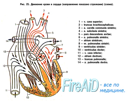
All blood from the pulmonary veins returns to the right atrium, where it mixes with venous blood. Some of this blood then flows through the dilated foramen ovale into the left atrium, left ventricle, and aorta; the rest enters through the right side of the heart into the vessels of the lungs. As a result of obligate shunting of blood from right to left, SaO 2 falls at the level of the atria; in rare cases, blood is discharged through the oval window mainly from the pulmonary veins, then SaO 2 practically does not decrease. The decrease in SaO 2 depends on the ratio of pulmonary and systemic blood flow and is rarely so pronounced that oxygen supply to tissues is disturbed. Because the blood from the pulmonary veins mixes with venous blood at the level of the right atrium, or moreover proximal, the oxygen content in the blood of all four chambers of the heart and in the main arteries in most cases is not uncommon.

In the supracardial and intracardiac type of malformation, obstruction of the pulmonary venous return is usually absent or small, therefore, pulmonary blood flow is increased, pulmonary hypertension may be expressed to varying degrees, and pulmonary vascular resistance is relatively low. Along with this, children survive the first weeks or months of life, but in the absence of correction of the defect, they can die from severe heart failure in the first year of life. At almost any time in the subcardial type of malformation, and in about a third of cases of the supracardial type, there is severe obstruction of the pulmonary venous return, leading to severe pulmonary hypertension, pulmonary blood flow restriction, pulmonary congestion, and interstitial pulmonary edema. The pressure in the pulmonary artery quite often exceeds the systemic pressure. Hell, without correction of the defect, death occurs in the first weeks of life. With all types of defect, the pressure in the right atrium is invariably higher than in the left; at times, resistance to pulmonary venous return appears due to restriction of shunting through the foramen ovale.
Clinical manifestations
8090% of patients have tachypnea, heart failure and developmental delay. Cyanosis may be minimal if there is no obstruction of the pulmonary venous return, but it becomes more pronounced as heart failure progresses. In newborns, the pulse is quickened, a cardiac impulse is determined, but the heart sounds are of a simple volume; murmurs are rarely heard. In the future, the auscultatory picture changes. A four-beat gallop rhythm often appears. A soft mesosystolic murmur is heard along the left edge of the sternum, and a rumbling mesodiastolic murmur is heard from below at the left edge of the sternum and at the top. From time to time, a constant vascular murmur from the pulmonary veins may be heard.

With obstruction of the pulmonary venous return, severe shortness of breath appears very early. It progresses rapidly, congestion develops in a small circle, cyanosis and right ventricular failure. The second heart sound is loud, slightly split, a gallop rhythm may be heard. There are no loud noises, but from below, at the left edge of the sternum, it is possible to hear a soft blowing systolic murmur of mitral regurgitation.
Chest x-ray
In the absence of obstruction of the pulmonary venous return, the cardiac shadow is enlarged, the vessels of the lungs are full-blooded. In children older than a year, a shadow in the form of a figure eight or a snowwoman is possible; it is formed by dilated vertical, left brachiocephalic and superior vena cava, sitting astride an enlarged heart.

With obstruction of the pulmonary venous return, the radiographic picture is very characteristic. The cardiac shadow is normal or slightly dilated, and the lung fields are covered with a thick reticulate haze. Such transformations of the lung fields with normal sizes of the heart shadow and the absence of pulmonary sounds often suggest the idea of a primary lung pathology (hyaline membrane disease, interstitial lung inflammation). For differential diagnosis, especially if the transformations on the radiograph are not resolved, early echocardiography is required.
ECG
On the ECG there are indicators of right ventricular hypertrophy, and quite often also right atrial hypertrophy. Hypertrophy quite often exceeds the physiological hypertrophy of the right ventricle in newborns, which is manifested by a qR complex in the right chest leads, a weak left ventricular depolarization vector, and the absence of T-wave inversion in the first days of life.

echocardiography
This method allows with high sensitivity and specificity to diagnose and establish the type of defect, especially in situs solitus, the confluence of the pulmonary veins through a non-specialized collector and in the absence of other major heart defects. Based on this, all newborns with severe respiratory disorders and cyanosis, especially if they are planned to be transferred to extracorporeal membrane oxygenation. an echocardiogram should be done.
Cardiac catheterization
Catheterization and radiopaque study of the heart are not very informative, while in addition, if carried out carefully, they are still fraught with an enormous aggravation of the condition. These studies should be carried out only with complex heart defects, at a time when there is an abnormal confluence of the pulmonary veins of a mixed type and it is not possible to clarify the situation with the help of EchoCG.

Treatment
In severe obstruction of the pulmonary venous return, early surgical correction is the only way to save the child. They begin intensive treatment of hypoxemia and metabolic acidosis, prescribe diuretics, oxygen, independent breathing under constant positive pressure, at one moment preparing the patient for surgery in conditions of unnatural circulation.
When the pulmonary veins flow through a non-specialized collector, the goal of the operation is to return the non-specialized pulmonary vein to the left atrium. Impose a wide anastomosis between the anterior wall of the pulmonary vein collector and the posterior wall of the left atrium. With small sizes of the left atrium, the interatrial septum is displaced to the right. The abnormal communication of the pulmonary vein collector is ligated and dissected. With an intracardiac type of defect, it is possible that the displacement of the interatrial septum is sufficient to the right of the confluence of the pulmonary vein collector into the right atrium.
In the absence of obstruction of the pulmonary venous return, radical surgery restores normal hemodynamics, while mortality is very moderate at 5% or less. With obstruction of the pulmonary venous return, especially in the subcardiac type of defect, mortality is higher, despite the fact that early diagnosis, intensive treatment of metabolic acidosis and urgent correction of the defect allowed to reduce mortality and take good long-term results. But, it should be emphasized that in approximately 10% of patients in the early stages after the end of the operation (24 months), obstruction of the pulmonary veins begins. Its circumstance may be a stricture of the anastomosis or multiple stenoses of the pulmonary veins in the region of the orifices or lobar branches. In the case of multiple stenoses, the prognosis is very negative.
Anatomical features
The right atrium is located in front and to the right of the left. Outside, it is covered with an epicardium, under which there is a thin layer of the myocardium and an inner layer - the endocardium. From the inside of the atrium, the surface is smooth, except for the inner surface of the auricle and part of the anterior wall, where ribbing is noticeable. This ribbing is due to the presence of pectinate muscles, which are delimited by a border crest from the rest of the inner surface. The right ear is an additional cavity in the shape of a pyramid.
The auricle functions as a blood reservoir and decompression chamber during ventricular systole. The ear also has a receptor zone, which allows it to take part in the regulation of heart contractions. Not far from the ear, on the anterior wall, there is an atrioventricular opening, through which communication occurs with the ventricle. The medial wall of the atrium plays the role of the interatrial septum. It has an oval fossa, which is closed by a thin connective tissue membrane.

Before birth and during the neonatal period, in its place is an oval hole, which takes part in the fetal circulation. After birth, the function of the foramen ovale is lost and it closes, leaving a fossa. In a quarter of the population, the opening does not close and an atrial septal defect, called the foramen ovale, develops.
In most cases, the defect does not cause any problems, but over time, with a large size of the foramen ovale, there is a risk of paradoxical embolism and infarcts. The oval window also ensures the discharge of blood from the left to the right atrium, which causes mixing of arterial and venous blood and a decrease in cardiac output.
2 Inflowing vessels

The superior and inferior vena cava are the two largest veins in the body, to which blood flows from all organs and tissues. Along with the vena cava, the smallest veins of the heart and the coronary sinus flow into the right atrium. The smallest veins of the heart open into the atrium over its entire surface. The coronary sinus is a collector of the veins of the heart, which, with the help of the mouth, opens into the atrial cavity between the opening of the inferior vena cava and the atrioventricular opening. The veins that empty into the coronary sinus represent the main pathway for the outflow of venous blood from the heart. After passing through the atrium, it goes to the ventricle.
3 Beginning of the conduction system of the heart
Between the mouth of the superior vena cava and the right ear is the sinoatrial node. It coordinates the work of different parts of the heart, ensuring normal cardiac activity. The sinoatrial node generates impulses and is the pacemaker of the first order (70 per minute). From it, the right and left branches of the sinoatrial node go to the myocardium.
4 Physiology and significance in the cardiac cycle

It is the anatomical features of the structure of the atrium that ensure the continuity and constancy of blood flow even during ventricular contraction. Constant venous inflow is promoted by a number of factors, one of which is thin walls. Thin walls cause the atrium to stretch, as a result of which it does not have time to overflow with blood. Due to the thin muscle layer, the right atrium does not fully contract during systole, which ensures the transient blood flow from the veins through the atrium to the ventricle.
Since the contractions are rather weak, they do not cause a significant increase in pressure that would impede venous flow or encourage backflow of blood into the veins. Another factor that ensures continuous circulation is the absence of inlet valves of the mouth of the vena cava, which would require an increase in venous pressure to open. In addition, the presence of atrial volume receptors plays a significant role in maintaining blood flow.
These are low pressure baroreceptors that send signals to the hypothalamus when pressure is reduced. A decrease in pressure indicates a decrease in blood volume. The hypothalamus responds to this by releasing vasopressin. Summarizing the above, we can conclude that without the right atrium, due to the periodic increase in pressure during ventricular contraction, the blood flow to the heart would be jerky, which would affect the overall rate of blood circulation in the direction of its decrease.
atrium are blood-receiving chambers, the ventricles, on the contrary, eject blood from the heart into the arteries. The right and left atria are separated from each other by a septum, as are the right and left ventricles. On the contrary, between the right atrium and the right ventricle there is a message in the form right atrioventricular orifice, ostium atrioventriculare dextrum; between left atrium and left ventricle - ostium atrioventriculare sinistrum.
Through these openings, blood during atrial systole is directed from the cavities of the latter into the cavities of the ventricles.
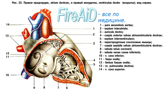
Right atrium, atrium dextrum, has the shape of a cube. From behind they pour into it at the top v. cava superior and below v. cava inferior, anteriorly, the atrium continues into a hollow process - the right ear, auricula dextra. The right and left ears cover the base of the aorta and pulmonary trunk. Partition between the atria, septum interatriale, set obliquely, from the anterior wall it goes back and to the right, so that the right atrium is located on the right and in front, and the left one is on the left and behind. The inner surface of the right atrium is smooth, with the exception of a small area in front and the inner surface of the ear, where a number of vertical ridges from those located here are visible. comb muscles, musculi pectinati. At the top musculi pectinati end scallop, crista terminalis, which on the outer surface of the atrium corresponds to sulcus terminalis. This groove indicates the junction of the primary sinus venosus with the atrium of the fetus. On the septum separating the right atrium from the left, there is an oval-shaped depression - Fossa ovalis, which is bounded at the top and front by an edge - limbus fossae ovalis. This recess is the remnant of a hole - foramen ovale through which the atria communicate with each other during the prenatal period. In! / C cases, foramen ovale persists for life, as a result of which periodic displacement of arterial and venous blood is possible if the contraction of the atrial septum does not close it. Between the openings of the superior and inferior vena cava on the posterior wall, there is a noticeable slight elevation, tuberculum intervenosum, behind the top section Fossae ovalis. It is believed that it directs blood flow in the embryo from the superior vena cava to ostium atrioventriculare dextrum.
From the bottom edge of the hole v. cava inferior to limbus fossae ovalis a crescent-shaped fold stretches, variable in size, - valvula venae cavae inferioris.
It is of great importance in the embryo, directing blood from the inferior vena cava through the foramen ovale into the left atrium. Below this damper, between the holes v. cava inferior and ostium atrioventriculare dextrum, flows into the right atrium sinus coronarius cordis collecting blood from the veins of the heart; in addition, small veins of the heart, independently flow into the right atrium. Their little holes foramina vendrum minimorum, scattered over the surface of the walls of the atrium. Near the opening of the venous sinus there is a small endocardial fold, valvula sinus corondrii. In the lower anterior part of the atrium, a wide right atrioventricular orifice, ostium atrioventriculare dextrum, leads to the cavity of the right ventricle.
Left atrium, atrium sinistrum, adjacent to the back of the descending aorta and esophagus. On each side, two pulmonary veins flow into it; left ear, auricula sinistra, protrudes anteriorly, bending around the left side of the aortic trunk and pulmonary trunk. In the ear there are muscle pectinati. In the lower anterior left atrioventricular orifice, ostium atrioventriculare sinistrum, oval shape leads into the cavity of the left ventricle.
Every educated person knows that the heart consists of four sections, each of which performs a specific function. Currently, there are a large number of negative factors that contribute to the development of pathologies and an increase in the size of the heart.
One such disease is right atrial hypertrophy. From the school anatomy course, many remember that blood from the atria enters the ventricles, and then spreads throughout the body. Hypertrophy slows down this process, so many health problems arise.
Diseases of the cardiovascular system are very serious and in no case should you self-medicate. Most likely, you will only harm your body and exacerbate the problem. In this article, we will try to describe in more detail what right atrial hypertrophy is, what symptoms you should pay attention to, what diagnostic and treatment methods are used in modern medicine.
Hypertrophy of the right atrium - a characteristic of the disease

Right atrial hypertrophy
The heart pumps blood around the body. From the atria through the openings, blood enters the ventricles, and then is pushed into the vessels. The right atrium is able to contain a certain volume of blood, if this volume exceeds the allowable one for some reason, the muscle tissue of the heart begins to work more actively.
In order to expel this extra volume, protective mechanisms are launched and the muscle tissue grows - hypertrophies, the walls of the atrium thicken - so it is easier for them to cope with the load. This condition is right atrial hypertrophy. All the causes leading to hypertrophy can be divided into two large groups: heart disease and lung disease.
Let's take a closer look at these reasons:
- Chronic lung diseases: chronic obstructive pulmonary disease, bronchial asthma, pulmonary emphysema.
- Chest deformity: kyphosis, severe scoliosis;
- Changes in the tricuspid valve: narrowing (stenosis) or insufficiency.
- Myocarditis;
- Endocarditis;
- Congenital heart defects: atrial septal defect, Ebstein anomaly, tetralogy of Fallot.
With lung pathology, there is an increase in pressure in the pulmonary artery system, pressure in the right ventricle increases, and then in the right atrium, hypertrophy of the right heart occurs;
In the case of a narrowing of the opening connecting the right ventricle and the right atrium, the blood cannot flow into the ventricle in full, the right atrium overflows, thickens, and subsequently expands, blood stagnates in the atrium and in the system of vena cava.
In case of valve insufficiency, on the contrary, blood flows abundantly into the atrium, with the contraction of the ventricle, which also leads to thickening and hypertrophy;
Myocardial cells (cardiomyocytes) are quite highly specialized and are not able to multiply by simple division, therefore, myocardial hypertrophy occurs due to an increase in the number of intracellular structures and cytoplasm volume, as a result of which the size of cardiomyocytes changes and myocardial mass increases.
Cardiac hypertrophy is an adaptive process, that is, it occurs in response to various disorders that prevent its normal functioning.Under such conditions, the myocardium is forced to contract with an increased load, which entails an increase in metabolic processes in it, an increase in cell mass and tissue volume.
At the initial stages of its development, hypertrophy is adaptive in nature, and the heart is able to maintain normal blood flow in the organs due to an increase in its mass. However, over time, the functionality of the myocardium is depleted, and hypertrophy is replaced by atrophy - the opposite phenomenon, characterized by a decrease in cell size.
Depending on the structural changes in the heart, it is customary to distinguish two types of hypertrophy:
- Concentric - when the size of the heart increases, its walls thicken, and the cavities of the ventricles or atria decrease in volume;
- Eccentric - the heart is enlarged, but its cavities are expanded.
It is known that hypertrophy can develop not only with some disease, but also in a healthy person with increased load. So, in athletes or people engaged in heavy physical labor, hypertrophy of both skeletal muscles and heart muscles occurs.
There are many examples of such changes, and sometimes they have a very sad outcome up to the development of acute heart failure. Excessive physical activity at work, the pursuit of pronounced muscles among bodybuilders, increased heart function, say, among hockey players, are fraught with such dangerous consequences, therefore, when doing such sports, you need to carefully monitor the state of the myocardium.
Thus, given the causes of myocardial hypertrophy, there are:
- Working (myofibrillar) hypertrophy, which occurs as a result of an excessive load on the organ under physiological conditions, that is, in a healthy body;
- Substitutive, which is the result of the body's adaptation to functioning in various diseases.
It is worth mentioning such a type of this pathology of the myocardium as regenerative hypertrophy. Its essence lies in the fact that when a connective tissue scar is formed at the site of the infarction (since the heart muscle cells are not able to multiply and make up for the defect that has appeared), the surrounding cardiomyocytes increase (hypertrophy) and partially take over the functions of the lost area.
In order to understand the essence of such changes in the structure of the heart, it is necessary to mention the main causes of hypertrophy in its various departments in pathological conditions.

Congenital malformations such as tetralogy of Fallot can lead to right atrial hypertrophy. Pressure overload of the right atrium is characteristic of tricuspid valve stenosis. This is an acquired heart disease in which the area of the opening between the atrium and the ventricle decreases. Stenosis of the tricuspid valve may be due to endocarditis.
With another acquired heart disease - tricuspid valve insufficiency - the right atrium experiences volume overload. In this condition, the blood from the right ventricle during its contraction enters not only into the pulmonary artery, but also back into the right atrium, forcing it to work with overload.
The right atrium is enlarged in some congenital heart defects. For example, with a significant atrial septal defect, blood from the left atrium enters not only the left ventricle, but also through the defect into the right atrium, causing its overload.
Congenital heart defects, accompanied by the development of HPP in children - Ebstein's anomaly, Fallot's tetralogy, transposition of the great vessels and others. Overload of the right atrium can occur quickly and appear mainly on the electrocardiogram.
This condition can occur during an attack of bronchial asthma, pneumonia, myocardial infarction, pulmonary embolism. In the future, with recovery, the symptoms of HPP gradually disappear.
Sometimes electrocardiographic signs of GPP appear with an increase in heart rate, for example, against the background of hyperthyroidism. In thin people, electrocardiographic signs of GPP may be normal.It is important to consider some other factors:
- The right atrium is overloaded with tricuspid valve stenosis.
- The right atrium is overloaded with volume due to tricuspid valve insufficiency, which is also an acquired heart disease.
- Some heart defects with which children are already born are also important.
This heart defect is acquired under the influence of various factors. If it is, then the area of the opening between the ventricle and the atrium becomes smaller. This defect is sometimes a consequence of endocarditis.
In this case, the blood from the right ventricle in the process of its contraction penetrates not only into the pulmonary artery, but even back, that is, into the right atrium. For this reason, it functions with overload.
For example, if there is a defect in the septum located between the atria, then blood from the left atrium enters both the left ventricle and the right atrium, which is why it is overloaded. Congenital heart defects that cause the development of hypertrophy in children include Tetralogy of Fallot, Ebstein's anomaly, and some others.
Overload of the right atrium can develop quite quickly. This shows up well on the ECG. Other causes include pneumonia, myocardial infarction, and pulmonary embolism. When recovery occurs, the symptoms of HPP disappear, but this does not happen immediately, but gradually.
Sometimes signs of hypertrophy on the ECG are observed with an increase in the frequency of heart contractions, and hyperthyroidism can serve as a background for this. If the patient has a lean body build, signs of ECG hypertrophy may be considered normal.The listed reasons due to which right atrial hypertrophy develops differ from hypertrophy of other parts of the heart, for example, the left ventricle. In this case, the causes are constant high blood pressure, too much exercise, hypertrophic cardiomyopathy, and so on.
Left atrial hypertrophy may develop due to general obesity. This condition is very dangerous if it manifests itself in children and at a young age. Of course, some reasons may be similar, but there is still a difference.
The main reason for the development of right atrial hypertrophy is pressure overload of blood flow in the pulmonary artery. In addition, the following conditions may be the cause of the development of this pathology:
- Respiratory pathology. They can cause an increase in blood pressure in the pulmonary artery.
- Pulmonary embolism. The formation of a blood clot interferes with the free flow of blood flow, resulting in an increased workload on the heart.
- Narrowing of the lumen in the tricuspid valve. The septum, located between the ventricle and the right atrium, contributes to the normal circulation of blood. If the lumen in it narrows, then, accordingly, the amount of blood flowing through it decreases. As a result, blood stasis occurs, and in order to cope with it, the load on the right atrium increases.
- Congenital heart defects. Any pathology in the structure of a vital organ causes disturbances in blood flow in it.
- Hypertrophy of the right ventricle.
- Stenosis of the tricuspid valve. Reducing the size of the opening between the ventricle and the atrium causes a violation of the outflow of blood, which contributes to the development of hypertrophy.
In addition, there are certain prerequisites, the presence of which in a patient provokes the development of this pathology. These include the following:
- significant overweight;
- rib deformity;
- stress;
- smoking and alcohol abuse.
Depending on the cause of the disease, 3 types of hypertrophy are distinguished: myofibrillar, replacement and regenerative:
- Myofibrillar hypertrophy occurs in a healthy person against the background of constant increased loads.
- Replacement is the result of the adaptation of the heart to a normal mode of operation in the presence of other pathologies.
- Regenerative hypertrophy develops as a result of a heart attack.


With defects in the tricuspid valve (this is the tricuspid septum between the right atrium and the ventricle), the hole through which blood normally flows freely from the atrium to the ventricle is severely narrowed or does not close enough.
This disrupts intracardiac blood flow:
- after filling the ventricle at the time of diastole (relaxation), an extra portion of blood remains in the atrium;
- it presses on the walls of the myocardium more than with normal filling and provokes their thickening.
With pathology in the pulmonary circulation (with pulmonary diseases), the blood pressure in the pulmonary vessels and in the right ventricle increases (from it the small or pulmonary circulation begins). This process prevents the free flow of the required volume of blood from the atrium to the ventricle, part of it remains in the chamber, increases pressure on the walls of the atrium and provokes the growth of the muscle layer of the myocardium.
Most often, right atrial hypertrophy develops against the background of cardiovascular disorders, but sometimes it becomes the result of regular physical exertion or myocardial necrosis.
Depending on the factor under the influence of which the thickening of the chamber walls appeared, there are:
- Regenerative hypertrophy due to scarring at the site of the focus of necrosis (after a heart attack). The atrial myocardium grows around the scar, trying to restore cell function (conduction and contraction).
- Replacement as a way for the heart muscle to compensate for circulatory deficiencies under the influence of various pathologies and negative factors.
- Working - a form that develops under the influence of regular physical activity (professional training), as a protective mechanism in case of increased heart rate, hyperventilation of the lungs, an increase in the pumped blood volume, etc.
Signs of right atrial hypertrophy
Hypertrophy of the right atrium is expressed by pain in the chest, respiratory disorders, and fatigue. Often, adverse symptoms are preceded by: pneumonia, exacerbation of bronchial asthma, pulmonary embolism, etc.
After treatment of the underlying disease, anxiety symptoms may subside and even disappear completely. In addition to the clinical manifestations of pulmonary problems, signs of venous stasis are possible with hypertrophy. Alarming signs of right atrial hypertrophy are characterized by:
- cough, shortness of breath, deterioration in respiratory function;
- puffiness;
- blanching of the skin, cyanosis;
- dullness of attention;
- slight tingling, discomfort in the region of the heart;
- pathology of the heart rhythm.
In most cases, hypertrophy is asymptomatic, and the manifestation of clinical symptoms is noted already in the advanced stage. Consult your doctor immediately if you notice - palpitations, dizziness (loss of consciousness), swelling of the lower extremities.


HPP by itself does not cause any symptoms. Everything rests on the signs that are associated with the main disease. For example, when chronic cor pulmonale is formed, the symptoms may be as follows:
- shortness of breath at rest and with little exertion;
- night cough;
- coughing up blood.
When the right atrium is no longer able to cope with a large load, signs of insufficiency of blood circulation in the main circle begin to appear, which are associated with venous blood stagnation.
Clinical signs:
- heaviness in the hypochondrium on the right;
- change in the size of the abdomen in a big way;
- the appearance of enlarged veins on the abdomen;
- swelling of the lower extremities and some other symptoms.
During pregnancy, there is a high probability of developing this pathology, because for the entire period of pregnancy there is a huge amount of hormonal changes in the body, pressure surges caused by weight gain.
Also, the pressure rises due to difficulty breathing, high physical activity. A pregnant woman is at risk of developing right atrial hypertrophy.If a doctor diagnoses a pathology, it is prescribed to place a pregnant woman in a hospital in order to conduct a thorough study of the problem, select treatment methods and a method of delivery. After all, with serious problems with the heart during childbirth, the mother may die.


Diagnosis of GPPA is carried out in several stages, depending on the stage of development of the pathology. For example, if hypertrophy has developed significantly and characteristic symptoms have begun to appear, it is advisable to interview the patient with a doctor, followed by a visual examination.
During the survey, the doctor finds out what symptoms the patient observed in the period from the onset of the development of the pathology to the present. If the symptoms converge with GPPA, then the doctor refers the patient to additional procedures that will confirm the diagnosis:
- Ultrasound of the heart.
- Chest x-ray with contrast. Allows you to see the boundaries of the heart, an increase in its departments and the state of the vessels. Important! As you can see, right atrial hypertrophy on an ECG is easier to recognize than on any other device, since electrocardiographic data are based not on one, but on several indicators at once, indicating the presence of a pathology in the body.
On the cardiogram, hypertrophy is expressed by a sharp deviation of the electrical axis to the right side with a slight shift forward and down. According to the R and S wave, the doctor determines the condition of the patient's atrium and ventricle at the time of the examination
If the R wave is pointed, the amplitude is increased, then the diagnosis of GPPA is confirmed with almost one hundred percent probability. Right atrial hypertrophy according to the results of the ECG is determined on the basis of a combination of several signs at once, indicated by the R wave, therefore it is almost impossible to make an erroneous diagnosis after this study.
During this procedure, the doctor examines the heart, its chambers and valves in detail for visual abnormalities. If during the examination on the screen a significant increase in PP, thickening of the walls is noticeable, then the doctor can diagnose the patient with right atrial hypertrophy.
Doppler study shows hemodynamics in the heart, overload of the PP due to problems with the valve on it is clearly visible.
However, it is still recommended to carry out the ECG procedure in conjunction with other studies so that the diagnosis is as accurate as possible. After all, right atrial hypertrophy is a very serious pathology that negatively affects the heart and cardiovascular system.
If it is not properly treated, it can easily provoke a heart attack, leading to death. Therefore, immediately after identifying the cause of the disease, the patient is recommended to immediately begin a course of therapy.
Additional diagnostics
If the ECG shows signs of atrial enlargement, the patient is recommended additional examinations to confirm hypertrophy and find out its causes. The simplest diagnostic methods - percussion (tapping), palpation (feeling) and auscultation (listening) - will be used already during the examination in the cardiologist's office.
Of the hardware studies, most likely, echocardiography (EchoCG - ultrasound of the heart) will be prescribed: it is safe for all groups of patients, including the elderly, young children and pregnant women, and is suitable for multiple examinations over time.
Modern echocardiographs use special software for 3D visualization of the structure of the heart and its valves; at the same time, it is possible to measure both functional and physical parameters (in particular, the volume of parts of the heart, wall thickness, etc.).
Together with EchoCG in cardiology, Dopplerography and color DS (Doppler scanning) are used: these examinations supplement the EchoCG result with information on hemodynamic characteristics and a color image of blood flow. In rare cases, a situation is possible when the result of echocardiography does not correspond to clinical manifestations.
The fact is that the picture that we see on the monitor of the EchoCG machine is in fact only a model built by the program based on calculations. Programs, like people, make mistakes. So, if ultrasound does not help determine the diagnosis, a contrast radiography or computed tomography is prescribed.
Both of these x-ray methods provide a reliable image of the heart against the background of other anatomical structures, which is very important in GLP caused by pulmonary diseases.Naturally, X-ray diagnostics has its own contraindications, and arterial catheterization during X-rays and the introduction of a contrast agent into the bloodstream also increase the trauma of the procedure for the patient.
Ecg - signs

With hypertrophy of the right atrium, the EMF created by it increases, while the excitation of the left atrium occurs normally.
The top figure shows normal P wave formation:
- excitation of the right atrium begins somewhat earlier and ends earlier (blue curve);
- excitation of the left atrium begins somewhat later and ends later (red curve);
- the total EMF vector of excitation of both atria draws a positive smoothed P wave, the leading edge of which forms the beginning of excitation of the right atrium, and the rear edge forms the end of excitation of the left atrium.
With hypertrophy of the right atrium, the vector of its excitation increases, which leads to an increase in the amplitude and duration of the first part of the P wave (lower figure), due to excitation of the right atrium.
With hypertrophy of the right atrium, its excitation ends simultaneously with the excitation of the left atrium or even somewhat later. As a result, a high peaked P wave is formed - a characteristic sign of right atrial hypertrophy:
- The height of the pathological P wave exceeds 2-2.5 mm (cells);
- The width of the pathological P wave is not increased; less often - increased to 0.11-0.12 s (5.5-6 cells);
- As a rule, the top of the pathological P wave is symmetrical;
- An abnormal high P wave is recorded in standard leads II, III and enhanced lead aVF.
Characteristic signs of a pathological P wave in right atrial hypertrophy in various leads:
- In standard lead I, the P wave is often negative or flattened (rarely, a high, pointed P wave is observed in leads I, aVL);
- In lead aVR, the presence of a deep pointed negative P wave is characteristic (its usual width is not increased);
- In the chest leads V1, V2, the P wave becomes high pointed or biphasic with a sharp predominance of the first positive phase (normally, the P wave in these leads is biphasic smoothed);
- Occasionally, the P wave in lead V1 is weakly positive, weakly negative, or smoothed, but in leads V2, V3, a tall peaked P wave is recorded;
- The greater the hypertrophy of the right atrium, the greater the number of chest leads marked high pointed positive P wave (in leads V5, V6 P wave is usually reduced in amplitude).
The activation time of the right atrium is measured in III or aVF or V1 leads. Hypertrophy of the right atrium is characterized by a prolongation of its activation time in these leads (exceeds 0.04 s or 2 cells).
With hypertrophy of the right atrium, the Macruse index (the ratio of the duration of the P wave to the duration of the PQ segment) is often less than the lower acceptable limit - 1.1.
An indirect sign of right atrial hypertrophy is an increase in the amplitude of the P waves in leads II, III, aVF, while the pathological P wave in each of the leads is larger in amplitude than the following T wave (normally PII, III, aVF).


Since right atrial hypertrophy is a secondary problem, there is one treatment feature. To make the size normal, to improve the supply of oxygen to the body through good cardiac functioning, is possible only with the help of treating the root cause.
Doctors carry out medical correction of the patient's condition. But the patient himself must also make some changes. He needs to adjust his lifestyle. The efforts of specialists can be useless if you treat your body incorrectly.
It is necessary to give up bad addictions, improve nutrition, normalize body weight and lead an active lifestyle, playing sports. Thanks to such measures, the recovery process will be quick and effective, and the risk of relapse will also decrease.
If a cor pulmonale is detected, which is the result of problems with the lungs, the actions of doctors are aimed at compensating for lung function. Measures are taken to prevent inflammation, bronchodilators and other medications are prescribed.
Cardiac glycosides are prescribed to eliminate the symptoms of diseases of the heart muscle. If valve defects are detected, surgery is performed. To eliminate the symptoms of diseases of the heart muscle, antiarrhythmic therapy is prescribed, which includes cardiac glycosides.
An important role is played by drugs that stimulate the exchange of muscle structures. It is the modern hypertrophy detected with the help of ECG that makes it possible to prescribe treatment in time, which increases the possibility of complete recovery and a long, fulfilling life.In no case should you prescribe treatment yourself, you can cause serious damage to your health. Preventive measures of hypertrophy are aimed at the implementation of a healthy lifestyle, a balanced diet, and the right regimen.
You do not need to exhaust yourself with physical exercises, but they certainly must be present in a person's life. In addition, it is important to carry out timely treatment of diseases, and those associated with the heart, blood vessels and other body systems.
To obtain the desired positive effect from complex treatment, the following doctor's recommendations must be followed:
- complete cessation of smoking and alcohol;
- gradual weight loss;
- regular performance of a complex of physiotherapy exercises;
- normalization of the diet under the supervision of a dietitian.
An effective therapeutic tactic implies the mandatory treatment of the underlying disease. If there are indications (congenital or acquired defects, thromboembolism), surgical intervention is performed.
In other cases, drug treatment will be optimal, aimed at normalizing blood flow through the pulmonary artery, correcting diseases of the lungs and bronchi, normalizing blood pressure and preventing myocardial infarction.Hypertrophy of the right heart is almost always secondary changes, therefore, with timely treatment of the primary pathology, there will be no serious problems in the atrium and ventricle.


Drug treatment of right ventricular hypertrophy consists in taking the following groups of drugs:
- Regular intake of diuretics;
- Beta-blockers (drugs of this pharmacological group are incompatible with alcoholic beverages and smoking);
- Calcium channel antagonists;
- Anticoagulants;
- Magnesium and potassium preparations;
- The use of cardiac glycosides is acceptable in the minimum dosage;
- Medicines that lower blood pressure.
Concomitant appointments are possible to normalize the functioning of the lungs and eliminate stenosis of the pulmonary valve. In some cases, it may be necessary to take some of the above medicines throughout life. If there is no positive dynamics or any improvement, the patient may be scheduled for surgery.
Patients should be aware of the dangers of self-medication and not try to pick up drugs on their own. People suffering from overweight, as well as systematically subjected to physical activity, are recommended to be regularly examined by a cardiologist.


In the treatment of right ventricular hypertrophy, surgery is usually performed at an early age. The efforts of the surgeon can be directed to prosthetic valves or removal of pathological openings and vessels. However, sometimes the causes of such changes are associated with an incurable congenital pathology of the respiratory system, which can be dealt with only by transplanting the entire heart-lung complex or only the lungs.
Operative tactics in most cases slows down the increase in the mass of muscle cells of the ventricles and helps to eliminate the cause of the disease. Perform several types of surgery:
- Aortic valve replacement only. The operation can be performed in the traditional way with the opening of the chest or in a minimally invasive way, when the valve is delivered to a predetermined position in the folded state through a puncture in the femoral artery.
- Valve prosthesis together with part of the aorta. This intervention is more traumatic and requires a lot of experience of the surgeon. The prostheses themselves can be artificial or biological, made from processed pig tissues.
Before performing such an operation, it is necessary to perform a large number of compatibility tests, and after the intervention, drugs should be taken to prevent the development of a rejection reaction. Since only a doctor can develop an effective treatment strategy, it is necessary to trust a competent specialist.


Before using any folk recipes, you need to coordinate this with the doctor. After analyzing the stage of development of the disease, he will determine whether it is possible to use folk remedies.
The most effective traditional medicine recipes:
- St. John's wort has a calming effect and will be useful for the heart muscle.
- Drops from lily of the valley flowers.
- A decoction of cornflowers is effective for headaches, in addition, it cleanses the blood.
- Hypertrophy can be treated with an infusion of spring adonis, but this is a poisonous plant, so it is important to follow the recommended dose exactly.
- If you are worried about severe shortness of breath, fresh nettles will help.
- Rosemary decoction helps to support the work of the heart.
- Decoction from young shoots of blueberry bushes. To prepare it, you need to pour 1 tablespoon of raw materials with a glass of water and boil for 10 minutes. It should be taken one sip in the morning at lunchtime and in the evening.
- A very useful cranberry. Grind fresh berries with sugar and store in the cold. Take one tablespoon after meals.
- Soothes and improves health herbal tea.
For cooking, it is necessary to pour 100 grams of dry raw materials into an enamel bowl, add 2 liters of clean water and boil for 10 minutes. After that, the pan should be wrapped in a towel and insisted for at least an hour.
Strain the infused broth through gauze and add 200 milliliters of May honey. The mixture should be poured into a glass container, closed with a lid and stored in the refrigerator. Take a decoction three times a day, three tablespoons 30 minutes before meals for 1 month.
You will need a half-liter jar of dark glass, it must be filled with fresh flowers and poured with alcohol. Close all this with a lid and place in a place where the sun's rays do not fall for two weeks.
After this time, pass through gauze and drink 15 drops, previously dissolved in a small amount of water, three times a day before meals. The course of treatment is 2 months.For cooking, you need 1 tablespoon of dry cornflowers, which should be placed in a ceramic pan, add 250 milliliters of boiled water and put in a water bath for a quarter of an hour. Then, the cooled broth should be filtered and taken 100 milliliters three times a day 20 minutes before meals. The course of treatment is 2 weeks.
You will need 1 teaspoon of herbs, which you need to pour 200 milliliters of boiling water and insist under a closed lid for half an hour. Strain the infusion and take 1 tablespoon before meals three times a day.
Fresh leaves and stems need to be chopped, 5 tablespoons separated into a jar, add the same amount of honey there and put in a place where daylight does not fall. Every day, for two weeks, the medicine must be shaken.
Then heat it in a water bath to a liquid state and strain. Take 1 tablespoon three times daily before meals. You need to store it in the refrigerator.
To get it, you need to mix 3 tablespoons of motherwort, 2 tablespoons of wild rosemary, 2 tablespoons of cudweed and 1 tablespoon of kidney tea. The ingredients must be placed in an airtight container.
Then separate 1 tablespoon of the collection and pour 300 milliliters of boiling water. Boil for three minutes and infuse the decoction for 4 hours. After that, pass through gauze and drink warm 100 milliliters three times a day half an hour before meals.To prepare, you will need 1 teaspoon of hawthorn, fragrant rue and valerian flowers. Add 500 milliliters of boiling water to the ingredients and leave for half an hour. Then filter, divide into three servings and drink throughout the day for three months.
Possible Complications
Dilatation of the cavity of the right atrium with thickening of the myocardial wall in the absence of adequate therapy can cause the following dangerous complications:
- chronic heart failure;
- progressive cor pulmonale;
- cardiac arrhythmia and conduction disturbance by the type of blockade;
- complete thromboembolism of the pulmonary artery;
- myocardial infarction;
- sudden cardiac death.


Since the condition in question is a consequence of another disease, the prognosis will be based on the effectiveness of the treatment of the root cause of this pathology. An important role in this is played by the presence of irreversible changes in the tissue and functioning of the heart muscle, as well as the severity of such changes.
For example, if the cause of hypertrophy of the right side of the heart was a defect, then the presence of concomitant pathologies, the general condition of the patient's body and the peculiarities of hemodynamics matter. It is believed that this disease does not pose a serious threat to the life of the patient, if it was diagnosed in time, and the patient accepts and complies with all the recommendations and prescriptions of the doctor.
In order to prevent the development of this pathology, it is necessary to lead a healthy lifestyle, eat right and observe the daily routine. You should not load yourself with significant physical exertion if the type of activity is associated with one or another kind of professional sports.
It is enough to carry out daily walks, go swimming, ride a bike. Many studies confirm that excessive stress on the heart increases the pressure in the cardiac circulation and leads to disruption of the functioning of a vital organ.
The procedure for the prevention of this disease is very well known to everyone. The first thing is a healthy lifestyle. Thanks to normal sleep, proper nutrition, constant moderate physical activity, they can easily prevent the appearance of pathologies with the heart.
A prerequisite is a moderate load on the body. Do not think that the heart of a bodybuilder who carries heavy barbells is always healthy. This is where the secret lies, since a person makes exorbitant loads on the body, which significantly increases the pressure in the entire circulatory system.This causes non-pathological hypertrophy. For this reason, you should try not to overload yourself. Movement is life, especially if this procedure is carried out in the form of a game. It is also health care. It is recommended to regularly walk outside, ride a bike, do light jogging. People who do this every day have 10 times fewer heart problems.
Well, and, of course, for prevention, you need to try to be less nervous. It's better to laugh and be happy. This is what doctors recommend. It is also necessary to timely treat diseases that can create complications and spread to the cardiovascular system.
History: "doctor-cardiologist.ru; cardio-life.ru; vashflebolog.ru; diabet-gipertonia.ru; zabserdce.ru; tonometra.net; iserdce.ru; ritmserdca.ru; oserdce.com; esthetology.com.ua ;ocardio.com"
called blood circulation. Through blood circulation, blood communicates
all organs of the human body, there is a supply of nutrients and
oxygen, excretion of metabolic products, humoral regulation, etc.
Blood moves through the blood vessels. They represent
elastic tubes of different diameters. The main circulatory system is
The heart is a hollow muscular organ that performs rhythmic contractions.
Thanks to its contractions, the movement of blood in the body occurs. The doctrine of
regulation of blood circulation was developed by I.P. Pavlov.
There are 3 types of blood vessels: arteries, capillaries and veins.
arteries Vessels that carry blood from the heart to the organs. They have
thick walls, from 3 layers:
outer layer ( adventitia) - connective tissue;
- medium ( media) - consists of smooth muscle tissue and contains
connective elastic fibers. Reducing this shell
accompanied by a decrease in the lumen of the vessels;
Internal ( intima) - formed by connective tissue and from the side
the lumen of the vessel is expelled by a layer of flat endothelial cells.
The arteries are located deep under the muscle layer and are reliably protected from
damage. As they move away from the heart, the arteries branch into smaller vessels,
and then to the capillaries.
Depending on the blood supply to organs and tissues, arteries are divided into:
1. Parietal ( parietal) - blood supply walls of the body.
2. Visceral ( visceral) - blood supply to internal organs.
Before an artery enters an organ, it is called an organ artery, having entered the organ -
intraorgan. Depending on the development of different layers of the artery wall
divided into vessels:
- muscular type- they have a well-developed middle shell, fibers
are arranged spirally according to the type of spring;
Mixed ( muscular-elastic) type - in the walls approximately equal
the number of elastic and muscle fibers (carotid, subclavian);
- elastic type in which the outer shell is thinner than the inner.
These are the aorta and the pulmonary trunk, into which blood enters under high pressure.
In children, the diameter of the arteries is larger than in adults. In newborns, the arteries
predominantly of the elastic type, the arteries of the muscular type are not yet developed.
capillaries are the smallest blood vessels
lumen from 2 to 20 µm. The length of each capillary does not exceed 0.3 mm. Them
the number is very large, so for 1 mm2 of fabric there are several hundred
capillaries. The total lumen of the capillaries of the whole body is 500 times greater than the lumen of the aorta.
In the resting state of the organ, most of the capillaries do not function and the current
their blood stops. The capillary wall consists of one layer
endothelial cells. Cell surface facing the capillary lumen
uneven, folds form on it. Exchange of substances between blood and tissues
occurs only in capillaries. Arterial blood throughout the capillaries
turns into a venous, which is collected first in postcapillaries, and then in
Distinguish capillaries:
1. Nourishing- provide the body with nutrients and O2, and
2. Specific- enable the organ to perform its function
(gas exchange in the lungs, excretion in the kidneys).
Vienna are the vessels that carry blood from the organs to the heart. They are,
like arteries, they have three-layer walls, but contain less elastic and
muscle fibers, so they are less elastic and fall off easily. Veins have
valves that open with blood flow. This promotes the movement of blood into
one direction. The movement of blood in one direction in the veins is facilitated by
not only the semilunar valves, but also the pressure difference in the vessels and contractions
muscular layer of veins.
Each area or organ receives its blood supply from several vessels.
Distinguish:
1. Main vessel- the biggest.
2. Additional ( collateral) is a lateral vessel that carries out
roundabout blood flow.
3. Anastomosis is the third vessel that connects the other 2. Otherwise
called connecting vessels.
There are also anastomoses between the veins. Termination of current in one vessel
leads to increased blood flow through collateral vessels and anastomoses.
CIRCULATION SCHEME
Blood circulation is necessary to nourish the tissues where the exchange takes place.
substances through the walls of the capillaries. Capillaries are the main part
microcirculatory bed, in which blood microcirculation occurs and
microcirculation is the movement of blood and lymph in a microscopic
parts of the vascular bed. The microcirculatory bed according to V.V. Kupriyanov includes
5 links:
1. Arterioles- the smallest parts of the arterial system.
2.Precapillaries- an intermediate link between arterioles and true
capillaries.
3. Capillaries.
4. Postcapillaries.
5. Venules.
All blood vessels in the human body make up 2 circles of blood circulation:
small and big.
Lecture 9. LYMPHATIC SYSTEM
It is represented by lymph nodes and lymphatic vessels, in
which lymph circulates.
Lymph in its composition resembles blood plasma, in which
lymphocytes. In the body there is a constant formation of lymph and its outflow through
lymphatic vessels into veins. The process of lymph formation is associated with the exchange of substances between
blood and tissues.
As blood flows through the blood capillaries, some of its plasma
tissue and constitutes tissue fluid. Tissue fluid bathes the cells
there is a constant exchange of substances between the fluid and the cells: in
cells receive nutrients and oxygen, and back - metabolic products.
Tissue fluid containing metabolic products partially re-enters
blood through the walls of blood vessels. At the same time, another part of the tissue
fluid does not enter the blood, but into the lymphatic vessels and makes up the lymph. So
Thus, the lymphatic system is an additional outflow system,
supplementing the function of the venous system.
Lymph- translucent yellowish liquid
tissue fluid. In its composition, it is close to blood plasma, but the proteins in it
less. Lymph contains many leukocytes that enter it from
intercellular spaces and lymph nodes. Lymph flowing from various
organs, has a different composition. It enters the lymphatic vessels
circulatory system (about 2 liters per day). Lymph nodes perform protective
empties into the right venous angle. Lymph flows into it from the right half
chest, right upper limb, right half of the head, face and neck.
Through the lymphatic vessels, along with the lymph, can spread
pathogenic microbes and particles of malignant tumors.
On the way of lymphatic vessels in some places there are lymph nodes. By
bringing lymph flows to the nodes through the vessels relevant- flows away from them.
Lymph nodes are small round or oblong
bodies. Each node consists of a connective tissue sheath, from which
crossbars come off. The backbone of the lymph nodes consists of reticular tissue. Between
the crossbars of the nodules are the follicles in which reproduction occurs
lymphocytes.
Functions lymph nodes:
They are hematopoietic organs
Perform a protective function (delay pathogenic microbes);
in such cases, the nodes increase in size, become dense and may
be felt.
Lymph nodes are located in groups. Lymph from each organ or area
premature puberty.
THYMUS
Thymus located in the upper part of the anterior mediastinum
directly behind the breastbone. It consists of two (right and left) lobes , upper
the ends of which can exit through the upper opening of the chest, and the lower
often extend to the pericardium and occupy the upper interpleural
triangle. The size of the gland during a person's life is not the same: its mass in
a newborn on average 12 g, at 14-15 years old - about 40, at 25 years old - 25 and at 60 years old -
close 15g . In other words, the thymus gland, having reached the greatest development
time of onset of puberty, subsequently gradually reduced.
The thymus gland is of great importance in immune processes, its hormones up to
the onset of puberty inhibit the function of the gonads, regulate __________ growth
bones (osteosynthesis), etc.
ADRENAL GLAND
adrenal gland(glandiila suprarenalis) steam room, refers to so
called the adrenal system. Located in the retroperitoneum
directly on the upper pole of the kidney. This gland is shaped like a three-
faceted pyramid facing the top to the diaphragm, and the base to the kidney.
Its dimensions in an adult: height 3-6 cm , base diameter about 3 cm
and the width is close 4-6mm , weight - 20 g . On the anterior surface of the gland are
gate - the place of entry and exit of blood vessels and nerves. iron covered
connective tissue capsule, which is part of the renal fascia. From-
the sprouts of the capsule penetrate into it through the gate and form, as it were, an organ stroma.
In cross section, the adrenal gland consists of an outer cortical
substance and internal medulla.
The adrenal medulla secretes a group of adrenaline-
series that stimulate the function of the sympathetic nervous system: constriction
vayut blood vessels, excite the process of splitting glycogen in the liver and
other Hormones secreted by the adrenal cortex, or
choline-like substances, regulate water-salt metabolism and affect the function
sex glands.
Lecture 11
DEVELOPMENT OF THE NERVOUS SYSTEM
Stage 1 - net nervous system. At this stage (intestinal)
The nervous system is made up of nerve cells, numerous processes of which
connect with each other in different directions, forming a network. Reflection of this
stage in humans is the network-like structure of the nervous system of the digestive
Stage 2 - nodal _________nervous system. At this stage, the (invertebrate) nervous
cells converge into separate clusters or groups, and from clusters
cell bodies, nerve nodes are obtained - centers, and from clusters of processes -
nerves. With a segmental structure, nerve impulses that occur at any point
bodies, do not spread throughout the body, but spread along transverse trunks in
within this segment. A reflection of this stage is the preservation in humans
primitive features in the structure of the autonomic nervous system.
Stage 3 - tubular nervous system. Such a nervous system (NS) in chordates
(lancelet) arose in the form of a neural tube with segmental
nerves to all segments of the body, including the apparatus of movement - the trunk brain. At
vertebrates and humans, the trunk brain becomes the spinal cord. Phylogeny of NS
determines the embryogenesis of human NS. NS is laid in the human embryo on
second or third week of intrauterine development. It comes from the outside
germ layer - ectoderm, which forms the brain plate. This
plate deepens, turning into a brain tube. brain tube
represents the rudiment of the central part of the NS. The rear end of the tube forms
rudimentary spinal cord. Anterior extended end by constriction
is divided into 3 primary cerebral vesicles, from which the head
144
The neural plate initially consists of a single layer of epithelial
cells. During its closure in the brain tube, the number of cells increases
and there are 3 layers:
Internal, from which the epithelial lining of the brain stems
cavities;
The middle one, from which the gray matter of the brain develops (embryonic
nerve cells);
External, developing into white matter (processes of nerve cells). At
separation of the brain tube from the ectoderm ganglionic plate. From her
spinal nodes develop in the region of the spinal cord, and in the region of the brain
brain - peripheral nerve nodes. Part of the ganglionic neural plate is
on the formation of ganglion nodes) of the autonomous NS, located in the body on
different distances from the central nervous system (CNS).
The walls of the neural tube and the ganglionic plate are composed of cells:
Neuroblasts from which neurons develop (functional unit
nervous system);
Neuroglial cells are divided into macroglial and microglial cells.
Macroglial cells develop like neurons, but are unable to conduct
excitation. They perform protective functions, the function of nutrition and contact.
between neurons.
Microglial cells originate from mesenchyme (connective tissue). Cells
together with blood vessels enter the brain tissue and are phagocytes.
THE SIGNIFICANCE OF THE NERVOUS SYSTEM
1. The National Assembly regulates the activities of various organs, organ systems and all
organism.
2. Carries out the connection of the whole organism with the external environment. All annoyances from
the external environment are perceived by the NS with the help of the sense organs.
3. The National Assembly carries out communications between different bodies and systems and
coordinates the activities of all organs and systems, determining the integrity
organism.
4. The human brain is the material basis of thinking and
speech associated with it.
CLASSIFICATION OF THE NERVOUS SYSTEM
The NS is divided into two closely related parts.


